Diastolic Function Assessment New Guideline Update Practical Approach
|
|
|
- Clarissa Anderson
- 6 years ago
- Views:
Transcription


1 Mayo Clinic Department of Cardiovascular Diseases Mayo Clinic Echocardiography Review Course for Boards and Recertification Diastolic Function Assessment New Guideline Update Practical Approach Jae K. Oh, MD Echo Hawaii 2017 January 26 th, MFMER slide-1 Learning Objectives for Diastology After this talk, you should be able to Apply the new 2016 ASE/EACVI Guideline Classify and grade diastolic function Estimate filling pressure in most patients 2016 MFMER slide-2 1


2 Evaluation of Diastolic Function 2017 Update New 2016 ASE/EACVI Recommendation Typical cases : Gr. 1, 2, and 3 Specific and challenging situations Discordant Echo Parameters Hypertrophic CM Mitral Annulus Calcification Mitral Regurgitation Atrial Fibrillation My recommendation Summary 2016 MFMER slide-3 Normal Diastole Adequate LV filling without increased filling pressure at rest and exertion Med e 7 cm/s Lat e 10 cm/s TR 2.8 m/sec LAVI 34 ml/m 2 PV S = PV D 2016 MFMER slide-4 2


3 Abnormal diastole Delayed relaxation, stiffness, and increased filling pressure e = 5 cm/s DT 140 msec E/e = MFMER slide-5 Evaluation of Diastolic Function Mitral Inflow and Annulus Velocity Normal Abn Relax Pseudo Restrictive Grade 1 Grade 2 Grade 3 Mitral flow Mitral annulus velocity Preload dependent Preload independent Sohn et al: JACC, 1997 CP MFMER slide-6 3
, 10 (lat) cm/s 2. E/e 14 (Av), 15(Med) 3. TR velocity 2.8 m/sec 4.")
4 LV filling pressure 1/18/2017 Estimation of LV Filling Pressures EF >50% EF <50% E/E <8 E/E 8-15 E/E >15 Ommen SR et al: Circulation 102:1788, MFMER slide-7 Four Major Parameters in Diastology Normal values Four Major Diagnostic Parameters Normal Values 1. E velocity 7(med), 10 (lat) cm/s 2. E/e 14 (Av), 15(Med) 3. TR velocity 2.8 m/sec 4. LAVI 34 ml/m 2 JASE and EJ CV Imaging April MFMER slide-8 4


5 New Criteria for Diastolic Function Assessment In pts with normal LVEF 50% 1 Septal e velocity 7 cm/s or lateral e velocity 10 cm/s 2 Average E/e 14, 15 (Med) 3 TR velocity 2.8 m/s 4 LA volume index 34 ml/m 2 3 Normal 2 and 2 3 Abnormal Normal diastolic function Indeterminate Diastolic dysfunction Normal Diastolic Filling Pressure Increased Diastolic Filling Pressure Criteria for diagnosis of LV diastolic dysfunction in patients with normal LVEF 2016 MFMER slide-9 Mitral inflow E/A E <50 cm/s E/A E >50 cm/s or E/A >0.8-<2 E/A 2 3 criteria to be evaluated* 2 of 3 negative 1 Average E/e >14 2 TR velocity >2.8 m/s 3 LA volume index >34 ml/m 2 2 of 3 or 3 of 3 positive When only 2 criteria are available 2 negative 1 positive and 1 negative 2 positive Normal LAP Grade I diastolic dysfunction Cannot determine LAP and diastolic dysfunction grade* LAP Grade II diastolic dysfunction LAP Grade III diastolic dysfunction If symptomatic consider CAD or proceed to diastolic stress test In patients with depressed LVEF or normal EF with diastolic dysfunction 2016 MFMER slide-10 5


6 67 yo man with ischemic CM and HF Gr. 1 dysfunction with normal filling pressure E= 45 cm/sec A= 90 cm/sec E/A = 0.5 Grade 1 Dysfunction E/A 0.8 E <50 cm/sec 2016 MFMER slide year old man with ischemic CM and HF E= 45 cm/s E/A = 0.5 E/e = 12 Medial e = 4 cm/s 2 months before E= 120 cm/sec E/A =0.9 E/e = 30 TR Vel = 3 m/sec 2016 MFMER slide-12 6

7 Ischemic Cardiomyopathy Echo Predictor STICH Trial (N=1511) Best survival with E/A Lin et al AHA 2016 MFMER slide year old man with ischemic CM and HF E= 120 cm/sec E/A =0.9 TR Vel = 3 m/sec e = 4 E/e = MFMER slide-14 7


8 After anterior STEMI : Grade 2 E= 120 A= 110 E/A = 1.1 Lat e = 6 Med e = MFMER slide-15 Cardiac Amyloidosis Grade 3 Dysfunction E/A = 6 E = 4 cm/sec E/e = 30 TR = 4 m/sec 2016 MFMER slide-16 8
 Delayed")


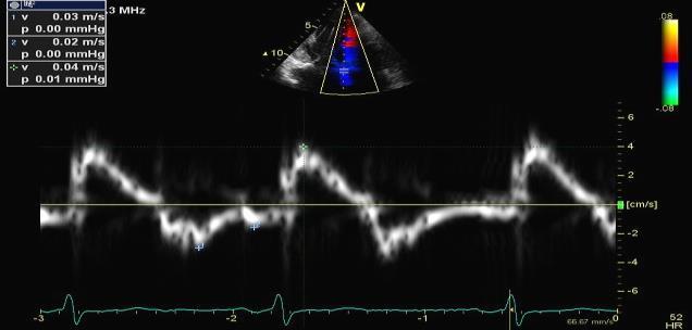
9 Mid-diastolic mitral flow (L) Delayed relaxation A. Fib Frommelt et al: J Am Soc Echocardiogr 16:176, 2003 CP MFMER slide-17 What to do in indeterminate cases? 2016 MFMER slide-18 9


10 Case #1 67 year old woman with HPT and SOB LAVI = 54 and TR= 2.8 m/s E= 90 A= 40 E/A= E +/- 2. E/e NL 3. TR LAVI ++ Lat e =8 E/e = 12 Med e = 7 E/e = MFMER slide-19 Indeterminate? E +/- E/e < 15 LA > 34 TR > 2.8 m/sec TR = 2.83 m/s 2016 MFMER slide-20 10


11 Valsalva Maneuver : E/A reduced > 0.5 Grade 2 Dysfunction E= 90 A= 40 E/A=2.3 E= 60 A= 60 E/A= MFMER slide-21 Case #2 23 YO with HCM Case # 6 1. Grade 1 2. Grade 2 3. Grade 3 4. Possibly normal E = 70 cm/s A= 30 cm/s E/A = MFMER slide-22 11


12 E = 70 cm/s 23 yo with HCM Medial e = 8 cm/sec Lateral e = 10 cm/sec E/e = 9 E/e = 7 Can he have a normal diastolic function? 2016 MFMER slide yo man with HCM LAVI 29 ml/m 2 TR = 2 m/sec IVRT = 120 msec 2016 MFMER slide-24 12


13 Valsalva in 23 yo HCM Normal filling pressure E/A= 2.3 E/A = MFMER slide-25 Mitral A duration is shorter than PV AR Increased LVEDP 2011 MFMER slide MFMER slide-26 13


14 Doppler Determination of LVEDP Mitral flow velocity EDP (mm Hg) r=0.68 P< LV pressure PVad-Ad (ms) mm Hg Rossvoll and Hatle: JACC, Pulmonary vein Ma=100 ms PVa=165 ms LVEDP can be increased with normal mean LV diastolic pressure CP MFMER slide-27 Mitral inflow E/A E <50 cm/s E/A E >50 cm/s or E/A >0.8-<2 E/A 2 3 criteria to be evaluated* 2 of 3 negative 1 Average E/e >14 2 TR velocity >2.8 m/s 3 LA volume index >34 ml/m 2 2 of 3 or 3 of 3 positive When only 2 criteria are available 2 negative 1 positive and 1 negative 2 positive Normal LAP Grade I diastolic dysfunction Cannot determine LAP and diastolic dysfunction grade* LAP Grade II diastolic dysfunction LAP Grade III diastolic dysfunction If symptomatic consider CAD or proceed to diastolic stress test In patients with depressed LVEF or normal EF with diastolic dysfunction reduced e 2016 MFMER slide-28 14


15 Case #3 72 yo woman with HCM Med e = 2 E/e = 40 E=80 A=95 E/A = 0.85 Lat e = 5 E/e = MFMER slide yo woman with apical HCM Grade 2 dysfunction with LAVI 37 ml/m 2 TR = 3 m/sec 2016 MFMER slide-30 15
16 Medial E-e ratio 1/18/2017 Medial E/e Ratio Versus Mean LAP Simultaneous studies <48 hours between studies All studies All = 0.45x r=0.44 P<0.001 Simultaneous studies y=0.28x r=0.28 P= Mean LAP (mmhg) Geske et al: Circulation; 116:2702, MFMER slide-31 Mean LAP vs Medial E-e ratio Hypertrophic CM Mean LAP (mmhg) Simultaneous studies <48 hours between studies < >15 Mean E-e Ratio Geske et al: Circulation; 116:2702, MFMER slide-32 16

17 Mitral annulus e velocity ASE/EACVI recommends average value E from one location is acceptable We need a caution in using e Primary pulmonary hypertension Pacemaker LBBB Wall motion abnormality Mitral annulus calcification Hypertrophic CM 2016 MFMER slide-33 Case # 4 Mitral annulus calcification and TAVR E= 100 cm/sec Lat e = 4 cm/s Med e = 3 cm/s 2016 MFMER slide-34 17


18 Mitral annulus calcification and TAVR E is reduced TR = 2.4 m/sec 2016 MFMER slide-35 Variable Mitral annulus e velocity vs MAC Mean age 73 years Group 1 n=79 no MAC Group 2 n=38 mild MAC Group 3 n=38 mod-severe MAC Agatston Score >119 P for trend Septal e 5.96± ± ± Lateral e 7.37± ± ± Average e 6.63±2 6.02± ± E/avg e ratio 13± ± ±8.26 <0.001 LV diastolic parameters are altered in the presence of MAC. This could be due to direct effects of MAC or might reflect truly reduced diastolic function. Interpretation of diastolic parameters in patients with MAC should be performed with caution. Codolosa et al: Am J Cardiol 2016;117: MFMER slide-36 18


19 71 year old woman with LAVI = 39 ml/m 2 Case #5 E/e NL < 14 E NL > 7 LAVI Enlarged TR NL < 2.8 Lateral e = 10 cm/sec Medial e = 9 cm/sec E/e = 9 E/e = MFMER slide year old woman with Scleroderma Normal Diastolic Function with large LA LVOT TVI = 26 cm 2016 MFMER slide-38 19
 Increased")

20 Reasons for LA enlargement Diastolic dysfunction Increased filling pressure (Not always) Increased volume Athlete s heart Measurement error 2016 MFMER slide-39 E=120 L wave E = 6 E/e =20 JASE MFMER slide-40 20
 DT < 130 msec")


21 Diastolic Function in A. Fib DT < 160 msec (with reduced EF) DT < 130 msec poor survival (Hurley, Oh) Other measurements E acceleration > 1900 cm/sec 2 IVRT 65 msec E/e 11 IVRT/ T E-e TR velocity 2016 MFMER slide year old man with dyspnea PFO velocity LAP- RAP = 36 mmhg 2016 MFMER slide-42 21


22 PFO Velocity for estimating LA pressure Baseline After Diuresis Peak V = 3.2 m/s Peak V = 1.6 m/s From Chandra, MD & Will Miranda, MD 2016 MFMER slide-43 Learning Points Earliest diastolic abnormality is delayed relaxation which can be detected by reduced e velocity by tissue Doppler Integrated approach with 4 parameters E velocity E/e ratio LAVI TR velocity If indeterminate with 4 parameters, use other parameters (PV, Valsalva, PFO, IVRT, T E-e ) 2011 MFME 2016 MFMER slide-44 22
Diastolic Function Assessment Practical Ways to Incorporate into Every Echo
 Diastolic Function Assessment Practical Ways to Incorporate into Every Echo Jae K. Oh, MD Echo Hawaii 2018 2018 MFMER 3712003-1 Learning Objectives My presentation will help you to Appreciate the importance
Choose the grading of diastolic function in 82 yo woman
 Question #1 Choose the grading of diastolic function in 82 yo woman E= 80 cm/s A= 70 cm/s LAVI < 34 ml/m 2 1= Grade 1 2= Grade 2 3= Grade 3 4= Normal 5= Indeterminate 2018 MFMER 3712003-1 Choose the grading
An Integrated Approach to Study LV Diastolic Function
 An Integrated Approach to Study LV Diastolic Function Assoc. Prof. Adriana Ilieşiu, FESC University of Medicine Carol Davila Bucharest, Romania LV Diastolic Dysfunction impaired relaxation (early diastole)
Constriction vs Restriction Case-based Discussion
 Mayo Clinic Department of Cardiovascular Diseases Mayo Clinic Echocardiography Review Course for Boards and Recertification Constriction vs Restriction Case-based Discussion Jae K. Oh, MD Samsung Professor
Diastology State of The Art Assessment
 Diastology State of The Art Assessment Dr. Mohammad AlGhamdi Assistant professor, KSAU-HS Consultant Cardiologist King AbdulAziz Cardiac Center Ministry of National Guard Health Affairs Diagnostic Clinical
Diastolic Heart Function: Applying the New Guidelines Case Studies
 Diastolic Heart Function: Applying the New Guidelines Case Studies Mitral Regurgitation The New ASE William Guidelines: A. Zoghbi Role MD, of FASE, 2D/3D MACCand CMR Professor and Chairman, Department
Left ventricular diastolic function and filling pressure in patients with dilated cardiomyopathy
 Left ventricular diastolic function and filling pressure in patients with dilated cardiomyopathy Bogdan A. Popescu University of Medicine and Pharmacy Bucharest, Romania My conflicts of interest: I have
Diastole is Not a Single Entity Four Components of Diastolic Dysfunction
 Physiology of Diastolic Function Made Easy James D. Thomas, MD, FACC, FASE Director, Center for Heart Valve Disease Bluhm Cardiovascular Institute Professor of Medicine, Feinberg School of Medicine, Northwestern
Constrictive Pericarditis
 Constrictive Pericarditis Never Confused with Anything Else Jae K. Oh, MD 2018 MFMER 3712003-1 ARS #1 CP Which of following patients has constrictive pericarditis? 1 2 3 Medial e 13 cm/s Medial e 3 cm/s
Evalua&on)of)Le-)Ventricular)Diastolic) Dysfunc&on)by)Echocardiography:) Role)of)Ejec&on)Frac&on)
of)Le-)Ventricular)Diastolic) Dysfunc&on)by)Echocardiography:) Role)of)Ejec&on)Frac&on)") Evalua&on)of)Le-)Ventricular)Diastolic) Dysfunc&on)by)Echocardiography:) Role)of)Ejec&on)Frac&on) N.Koutsogiannis) Department)of)Cardiology) University)Hospital)of)Patras)! I have no conflicts of interest
Evalua&on)of)Le-)Ventricular)Diastolic) Dysfunc&on)by)Echocardiography:) Role)of)Ejec&on)Frac&on) N.Koutsogiannis) Department)of)Cardiology) University)Hospital)of)Patras)! I have no conflicts of interest
DOPPLER HEMODYNAMICS (1) QUANTIFICATION OF PRESSURE GRADIENTS and INTRACARDIAC PRESSURES
 QUANTIFICATION OF PRESSURE GRADIENTS and INTRACARDIAC PRESSURES") THORAXCENTRE DOPPLER HEMODYNAMICS (1) QUANTIFICATION OF PRESSURE GRADIENTS and INTRACARDIAC PRESSURES J. Roelandt DOPPLER HEMODYNAMICS Intracardiac pressures and pressure gradients Volumetric measurement
THORAXCENTRE DOPPLER HEMODYNAMICS (1) QUANTIFICATION OF PRESSURE GRADIENTS and INTRACARDIAC PRESSURES J. Roelandt DOPPLER HEMODYNAMICS Intracardiac pressures and pressure gradients Volumetric measurement
OPTIMIZING ECHO ACQUISTION FOR STRAIN AND DIASTOLOGY
 OPTIMIZING ECHO ACQUISTION FOR STRAIN AND DIASTOLOGY October 8, 2017 Deborah Agler, ACS, RDCS, FASE Coordinator of Education and Training Cleveland Clinic General Principles Diastology Clinical Data Heart
The Patient with Atrial Fibrilation
 Assessment of Diastolic Function The Patient with Atrial Fibrilation Assoc. Prof. Adriana Ilieşiu, FESC University of Medicine Carol Davila Bucharest, Romania Associated Conditions with Atrial Fibrillation
Diastolic Function: What the Sonographer Needs to Know. Echocardiographic Assessment of Diastolic Function: Basic Concepts 2/8/2012
 Diastolic Function: What the Sonographer Needs to Know Pat Bailey, RDCS, FASE Technical Director Beaumont Health System Echocardiographic Assessment of Diastolic Function: Basic Concepts Practical Hints
10/1/2016. Constrictive Pericarditis Unique Hemodynamics. What s New in Pericardial Disease? Case-based Discussion
 Mayo Clinic Department of Cardiovascular Diseases Mayo Clinic Echocardiography Review Course for Boards and Recertification What s New in Pericardial Disease? Case-based Discussion Jae K. Oh, MD Samsung
Diastolic Function Overview
 Diastolic Function Overview Richard Palma BS, RDCS, RCS, APS, FASE Director and Clinical Coordinator The Hoffman Heart and Vascular Institute School of Cardiac Ultrasound None Disclosures Learning Objectives
Μαρία Μπόνου Διευθύντρια ΕΣΥ, ΓΝΑ Λαϊκό
 Μαρία Μπόνου Διευθύντρια ΕΣΥ, ΓΝΑ Λαϊκό Diastolic HF DD: Diastolic Dysfunction DHF: Diastolic HF HFpEF: HF with preserved EF DD Pathophysiologic condition: impaired relaxation, LV compliance, LV filling
P = 4V 2. IVC Dimensions 10/20/2014. Comprehensive Hemodynamic Evaluation by Doppler Echocardiography. The Simplified Bernoulli Equation
 Comprehensive Hemodynamic Evaluation by Doppler Echocardiography Itzhak Kronzon, MD North Shore LIJ/ Lenox Hill Hospital New York, NY Disclosure: Philips Healthcare St. Jude Medical The Simplified Bernoulli
Pericardial Disease: Case Examples. Echo Fiesta 2017
 Pericardial Disease: Case Examples Echo Fiesta 2017 2014 2014 MFMER MFMER 3346252-1 slide-1 Objectives Have a systematic approach to evaluation of constriction 2014 MFMER 3346252-2 CASE 1 2013 MFMER 3248567-3
Echo Doppler Assessment of Right and Left Ventricular Hemodynamics.
 Echo Doppler Assessment of Right and Left Ventricular Hemodynamics. Itzhak Kronzon, MD, FASE, FACC, FESC, FAHA, FACP, FCCP Northwell, Lenox Hill Hospital, New York Professor of Cardiology Hofstra University
GENERAL PRINCIPLES FOR ECHO ASSESSMENT OF DIASTOLIC FUNCTION (For full recommendation refer to the Left Ventricular Diastolic Function Guideline)
") 1 THE AMERICAN SOCIETY OF ECHOCARDIOGRAPHY RECOMMENDATIONS FOR THE EVALUATION OF LEFT VENTRICULAR DIASTOLIC FUNCTION BY ECHOCARDIOGRAPHY: A QUICK REFERENCE GUIDE FROM THE ASE WORKFLOW AND LAB MANAGEMENT
1 THE AMERICAN SOCIETY OF ECHOCARDIOGRAPHY RECOMMENDATIONS FOR THE EVALUATION OF LEFT VENTRICULAR DIASTOLIC FUNCTION BY ECHOCARDIOGRAPHY: A QUICK REFERENCE GUIDE FROM THE ASE WORKFLOW AND LAB MANAGEMENT
LV FUNCTION ASSESSMENT: WHAT IS BEYOND EJECTION FRACTION
 LV FUNCTION ASSESSMENT: WHAT IS BEYOND EJECTION FRACTION Jamilah S AlRahimi Assistant Professor, KSU-HS Consultant Noninvasive Cardiology KFCC, MNGHA-WR Introduction LV function assessment in Heart Failure:
Diastology Disclosures: None. Dias2011:1
 Diastology 2011 James D. Thomas, M.D., F.A.C.C. Cardiovascular Imaging Center Department of Cardiology Cleveland Clinic Foundation Cleveland, Ohio, USA Disclosures: None Dias2011:1 Is EVERYBODY a member!?!
Jong-Won Ha*, Jeong-Ah Ahn, Jae-Yun Moon, Hye-Sun Suh, Seok-Min Kang, Se-Joong Rim, Yangsoo Jang, Namsik Chung, Won-Heum Shim, Seung-Yun Cho
 Eur J Echocardiography (2006) 7, 16e21 CLINICAL/ORIGINAL PAPERS Triphasic mitral inflow velocity with mid-diastolic flow: The presence of mid-diastolic mitral annular velocity indicates advanced diastolic
Swan Song: Echocardiography as a Pulmonary Artery Catheter? Interdepartmental Division of Critical Care Medicine
 Swan Song: Echocardiography as a Pulmonary Artery Catheter? The swan is without spot, and it sings sweetly as it dies, that song ending its life Leonardo Da Vinci Curr Opin Anesthesiol 2016, 29:36 45 Circulation.
Echo-Doppler evaluation of left ventricular diastolic function. Michel Slama Amiens France
 Echo-Doppler evaluation of left ventricular diastolic function Michel Slama Amiens France Left ventricular pressure Pressure A wave [ LVEDP LVEDP préa Congestive cardiac failure with preserved systolic
Comprehensive Hemodynamics By Doppler Echocardiography. The Echocardiographic Swan-Ganz Catheter.
 Comprehensive Hemodynamics By Doppler Echocardiography. The Echocardiographic Swan-Ganz Catheter. Itzhak Kronzon, MD, FASE, FACC, FESC, FAHA, FACP, FCCP North Shore HS, LIJ/Lenox Hill Hospital, New York
Prognostic Value of Left Atrial Size and Function
 Prognostic Value of Left Atrial Size and Function James D. Thomas, M.D., F.A.C.C. Cardiovascular Imaging Center Department of Cardiology Cleveland Clinic Foundation Cleveland, Ohio, USA Conflicts: None
Pericardial Diseases/Tamponade Illustrative Cases
 Pericardial Diseases/Tamponade Illustrative Cases Jae K. Oh, MD Echo Hawaii 2017 2012 MFMER 3200268v3(2010)-1 Case #1 47 year old man Chest pain Not exertional Normal Examination 2016 MFMER slide-2 1 47
How to Assess Diastolic Dysfunction?
 How to Assess Diastolic Dysfunction? Fausto J Pinto, MD, PhD, FESC, FACC, FASE Lisbon University Dyastolic Dysfunction Impaired relaxation Elevated filling pressures Ischemic heart disease Cardiomyopathies
E/Ea is NOT an essential estimator of LV filling pressures
 Euroecho Kopenhagen Echo in Resynchronization in 2010 E/Ea is NOT an essential estimator of LV filling pressures Wilfried Mullens, MD, PhD December 10, 2010 Ziekenhuis Oost Limburg Genk University Hasselt
Dobutamine Stress testing In Low Flow, Low EF, Low Gradient Aortic Stenosis Case Studies
 Dobutamine Stress testing In Low Flow, Low EF, Low Gradient Aortic Stenosis Case Studies Mitral Regurgitation The New ASE Guidelines: Role of 2D/3D and CMR William A. Zoghbi MD, FASE, MACC Professor and
Constrictive Pericarditis Curable Paradoxical Diastolic Heart Failure ASE EBRC 2018
 Constrictive Pericarditis Curable Paradoxical Diastolic Heart Failure ASE EBRC 2018 May 8 th, 2018 Jae K. Oh, MD Samsung Professor of CV Diseases Director, Pericardial Disease Clinic Co-Director, Integrated
Conflicts of interest: GE, Abbott, Edwards (honoraria)
") Understanding Diastole and Its Contribution to Heart Failure: State of the Art in 2016 James D. Thomas, MD, FACC, FASE Director, Center for Heart Valve Disease Bluhm Cardiovascular Institute Professor
Understanding Diastole and Its Contribution to Heart Failure: State of the Art in 2016 James D. Thomas, MD, FACC, FASE Director, Center for Heart Valve Disease Bluhm Cardiovascular Institute Professor
Basic Approach to the Echocardiographic Evaluation of Ventricular Diastolic Function
 Basic Approach to the Echocardiographic Evaluation of Ventricular Diastolic Function J A F E R A L I, M D U N I V E R S I T Y H O S P I T A L S C A S E M E D I C A L C E N T E R S T A F F C A R D I O T
HFNEF. Heart Failure is
 HFNEF Bijoy K. Khandheria, MD. FASE, FACP, FACC FESC Professor of Medicine University of Wisconsin Director. Echocardiography Services Aurora Health Care No conflicts or off label use CP1173868-1 Heart
HFpEF. April 26, 2018
 HFpEF April 26, 2018 (J Am Coll Cardiol 2017;70:2476 86) HFpEF 50% or more (40-71%) of patients with CHF have preserved LV systolic function. HFpEF is an increasingly frequent hospital discharge. Outcomes
Diastolic Functions: Evaluation & Clinical Applications
 Special Articles Diastolic Functions: Evaluation & Clinical Applications Senior Consultant Cardiologist, Metro Heart Institute, Delhi Immediate Past President, Cardiological Society of India (Cardiovasc.
The importance of left atrium in LV diastolic function
 II Baltic Heart Failure Meeting and Congress of Latvian Society of Cardiology The importance of left atrium in LV diastolic function Dr. Artem Kalinin Eastern Clinical University Hospital Riga 30.09.2010.
Highlights from EuroEcho 2009 Echo in cardiomyopathies
 Highlights from EuroEcho 2009 Echo in cardiomyopathies Bogdan A. Popescu University of Medicine and Pharmacy, Bucharest, Romania ESC Congress 2010 Hypertrophic cardiomyopathy To determine the differences
Value of echocardiography in chronic dyspnea
 Value of echocardiography in chronic dyspnea Jahrestagung Schweizerische Gesellschaft für /Schweizerische Gesellschaft für Pneumologie B. Kaufmann 16.06.2016 Chronic dyspnea Shortness of breath lasting
Ref 1. Ref 2. Ref 3. Ref 4. See graph
 Ref 1 Ref 2 Ref 3 1. Ages 6-23 y/o 2. Significant LVM differences by gender 3. For males 95 th percentiles: a. LVM/BSA = 103 b. LVM/height = 100 4. For females 95 th percentiles: a. LVM/BSA = 84 b. LVM/height
ECHO HAWAII. Role of Stress Echo in Valvular Heart Disease. Not only ischemia! Cardiomyopathy. Prosthetic Valve. Diastolic Dysfunction
 Role of Stress Echo in Valvular Heart Disease ECHO HAWAII January 15 19, 2018 Kenya Kusunose, MD, PhD, FASE Tokushima University Hospital Japan Not only ischemia! Cardiomyopathy Prosthetic Valve Diastolic
Effect of Heart Rate on Tissue Doppler Measures of E/E
 Cardiology Department of Bangkok Metropolitan Administration Medical College and Vajira Hospital, Bangkok, Thailand Abstract Background: Our aim was to study the independent effect of heart rate (HR) on
Doppler Basic & Hemodynamic Calculations
 Doppler Basic & Hemodynamic Calculations August 19, 2017 Smonporn Boonyaratavej MD Division of Cardiology, Department of Medicine Chulalongkorn University Cardiac Center, King Chulalongkorn Memorial Hospital
Echocardiography: Guidelines for Valve Quantification
 Echocardiography: Guidelines for Echocardiography: Guidelines for Chamber Quantification British Society of Echocardiography Education Committee Richard Steeds (Chair), Gill Wharton (Lead Author), Jane
Diastolic Heart Failure
 Chronic Heart Failure Prevalence overall = 2-3 % Diastolic Heart Failure Patrick Wouters University Hospital Ghent Belgium (Heart Failure + Asymptomatic Ventricular Dysfunction) Prevalence > 70 y = 10-20
The Doppler Examination. Katie Twomley, MD Wake Forest Baptist Health - Lexington
 The Doppler Examination Katie Twomley, MD Wake Forest Baptist Health - Lexington OUTLINE Principles/Physics Use in valvular assessment Aortic stenosis (continuity equation) Aortic regurgitation (pressure
HEMODYNAMIC ASSESSMENT
 HEMODYNAMIC ASSESSMENT INTRODUCTION Conventionally hemodynamics were obtained by cardiac catheterization. It is possible to determine the same by echocardiography. Methods M-mode & 2D echo alone can provide
LV geometric and functional changes in VHD: How to assess? Mi-Seung Shin M.D., Ph.D. Gachon University Gil Hospital
 LV geometric and functional changes in VHD: How to assess? Mi-Seung Shin M.D., Ph.D. Gachon University Gil Hospital LV inflow across MV LV LV outflow across AV LV LV geometric changes Pressure overload
LA Function analysis Marcia Barbosa Vice Presidente - Brazilian Soc of Cardiology President-elect - Interamerican Soc of Cardiology
 LA Function analysis Marcia Barbosa Vice Presidente - Brazilian Soc of Cardiology President-elect - Interamerican Soc of Cardiology Belo Horizonte Brazil DECLARATION OF CONFLICT OF INTEREST Nothing to
Review of Cardiac Imaging Modalities in the Renal Patient. George Youssef
 Review of Cardiac Imaging Modalities in the Renal Patient George Youssef ECHO Left ventricular hypertrophy (LVH) assessment Diastolic dysfunction Stress ECHO Cardiac CT angiography Echocardiography - positives
Imaging in Heart Failure: A Multimodality Approach. Thomas Ryan, MD
 Imaging in Heart Failure: A Multimodality Approach Thomas Ryan, MD Heart Failure HFrEF HFpEF EF50% Lifetime risk 20% Prevalence 6M Americans Societal costs - $30B 50% 5-year survival 1 Systolic
Nancy Goldman Cutler, MD Beaumont Children s Hospital Royal Oak, Mi
 Nancy Goldman Cutler, MD Beaumont Children s Hospital Royal Oak, Mi Identify increased LV wall thickness (WT) Understand increased WT in athletes Understand hypertrophic cardiomyopathy (HCM) Enhance understanding
Strain/Untwisting/Diastolic Suction
 What Is Diastole and How to Assess It? Strain/Untwisting/Diastolic Suction James D. Thomas, M.D., F.A.C.C. Cardiovascular Imaging Center Department of Cardiology Cleveland Clinic Foundation Cleveland,
Valvular Regurgitation: Can We Do Better Than Colour Doppler?
 Valvular Regurgitation: Can We Do Better Than Colour Doppler? A/Prof David Prior St Vincent s Hospital Melbourne Sports Cardiology Valvular Regurgitation Valve regurgitation volume loads the ventricles
European Heart Journal - Cardiovascular Imaging Advance Access published July 18, 2016
 European Heart Journal - Cardiovascular Imaging Advance Access published July 18, 2016 European Heart Journal Cardiovascular Imaging doi:10.1093/ehjci/jew082 Recommendations for the Evaluation of Left
Echocardiographic Evaluation of the Cardiomyopathies. Stephanie Coulter, MD, FACC, FASE April, 2016
 Echocardiographic Evaluation of the Cardiomyopathies Stephanie Coulter, MD, FACC, FASE April, 2016 Cardiomyopathies (CMP) primary disease intrinsic to cardiac muscle Dilated CMP Hypertrophic CMP Infiltrative
Adel Hasanin Ahmed 1
 Adel Hasanin Ahmed 1 PERICARDIAL DISEASE The pericardial effusion ends anteriorly to the descending aorta and is best visualised in the PLAX. PSAX is actually very useful sometimes for looking at posterior
Hemodynamic Assessment. Assessment of Systolic Function Doppler Hemodynamics
 Hemodynamic Assessment Matt M. Umland, RDCS, FASE Aurora Medical Group Milwaukee, WI Assessment of Systolic Function Doppler Hemodynamics Stroke Volume Cardiac Output Cardiac Index Tei Index/Index of myocardial
Low Gradient Severe? AS
 Low Gradient Severe? AS Philippe Pibarot, DVM, PhD, FACC, FAHA, FESC, FASE Canada Research Chair in Valvular Heart Diseases Institut Universitaire de Cardiologie et de Pneumologie de Québec / Québec Heart
ΚΑΡΔΙΟΛΟΓΟΣ EUROPEAN ACCREDITATION IN TRANSTHORACIC AND TRANSESOPHAGEAL ECHOCARDIOGRAPHY
 1 ΚΑΡΔΙΟΛΟΓΟΣ EUROPEAN ACCREDITATION IN TRANSTHORACIC AND TRANSESOPHAGEAL ECHOCARDIOGRAPHY 2 Constrictive pericarditis (CP) is characterized by impaired ventricular filling due to a stiffened or noncompliant
Evaluation of Left Ventricular Diastolic Dysfunction by Doppler and 2D Speckle-tracking Imaging in Patients with Primary Pulmonary Hypertension
 ESC Congress 2011.No 85975 Evaluation of Left Ventricular Diastolic Dysfunction by Doppler and 2D Speckle-tracking Imaging in Patients with Primary Pulmonary Hypertension Second Department of Internal
Role of Stress Echo in Valvular Heart Disease. Satoshi Nakatani Osaka University Graduate School of Medicine Osaka, Japan
 Role of Stress Echo in Valvular Heart Disease Satoshi Nakatani Osaka University Graduate School of Medicine Osaka, Japan Exercise echocardiography Dobutamine echocardiography Usefulness of exercise echo
Δυναμική υπερηχοκαρδιογραφία στις μυοκαρδιοπάθειες : έχει θέση και ποια ;
 Ελληνική Καρδιολογική Εταιρεία Σεμινάρια ομάδων εργασίας Θεσσαλονίκη, 8-10 Φεβρουαρίου 2018 Ομάδα εργασίας Ηχωκαρδιολογίας Δυναμική υπερηχοκαρδιογραφία στις μυοκαρδιοπάθειες : έχει θέση και ποια ; ΑΓΑΘΗ-ΡΟΖΑ
Low Gradient Severe AS: Who Qualifies for TAVR? Andrzej Boguszewski MD, FACC, FSCAI Vice Chairman, Cardiology Mid-Michigan Health Associate Professor
 Low Gradient Severe AS: Who Qualifies for TAVR? Andrzej Boguszewski MD, FACC, FSCAI Vice Chairman, Cardiology Mid-Michigan Health Associate Professor Michigan State University, Central Michigan University
Marti McCulloch, BS, MBA, RDCS, FASE Houston, Texas
 Marti McCulloch, BS, MBA, RDCS, FASE Houston, Texas Mitral Regurgitation What to Expect Review Specific Signs of Severity Supportive Signs of Severity Qualitative Parameters Structural Doppler Quantitative
22 nd Annual Conference of the Saudi Heart Association Riyadh, Saudi Arabia
 22 nd Annual Conference of the Saudi Heart Association Riyadh, Saudi Arabia New Echocardiographic Modalities to Evaluate Ventricular Function in Congenital Heart Disease: Tissue Doppler & Strain Rate Imaging
Global left ventricular circumferential strain is a marker for both systolic and diastolic myocardial function
 Global left ventricular circumferential strain is a marker for both systolic and diastolic myocardial function Toshinari Onishi 1, Samir K. Saha 2, Daniel Ludwig 1, Erik B. Schelbert 1, David Schwartzman
Hypertensive heart disease and failure
 Hypertensive heart disease and failure Prof. Dr. Alan Fraser Cardiff University The heart in hypertension Pathophysiology of LV adaptation Regional development of hypertrophy Stress testing - inducible
«Paradoxical» low-flow, low-gradient AS with preserved LV function: A Silent Killer
 «Paradoxical» low-flow, low-gradient AS with preserved LV function: A Silent Killer Philippe Pibarot, DVM, PhD, FACC, FAHA, FESC, FASE Canada Research Chair in Valvular Heart Diseases Université LAVAL
How to assess ischaemic MR?
 ESC 2012 How to assess ischaemic MR? Luc A. Pierard, MD, PhD, FESC, FACC Professor of Medicine Head, Department of Cardiology University Hospital Sart Tilman, Liège ESC 2012 No conflict of interest Luc
Cardiac hypertrophy : differentiating disease from athlete
 Cardiac hypertrophy : differentiating disease from athlete Ario Soeryo Kuncoro, MD, Cardiologist Echocardiography Division, National Cardiovascular Centre Harapan Kita-Jakarta Departement of Cardiology
Aortic Stenosis: Spectrum of Disease, Low Flow/Low Gradient and Variants
 Aortic Stenosis: Spectrum of Disease, Low Flow/Low Gradient and Variants Martin G. Keane, MD, FASE Professor of Medicine Lewis Katz School of Medicine at Temple University Basic root structure Parasternal
Strategie di Prevenzione del Rischio CCV Globale. La diagnosi pre-clinica della disfunzione ventricolare sinistra
 Strategie di Prevenzione del Rischio CCV Globale 1^ edizione: 13 novembre 2010 ore 9.00-13.00 SALA CARAVAGGIO - FIERA DI BERGAMO Via Lunga BERGAMO Per i MAP dei distretti di: Seriate Grumello Valle Cavallina
Tissue Doppler Imaging
 Cronicon OPEN ACCESS Hesham Rashid* Tissue Doppler Imaging CARDIOLOGY Editorial Department of Cardiology, Benha University, Egypt *Corresponding Author: Hesham Rashid, Department of Cardiology, Benha University,
Role of echocardiography in the assessment of ischemic heart disease 분당서울대학교병원윤연이
 Role of echocardiography in the assessment of ischemic heart disease 분당서울대학교병원윤연이 Outline Evaluation of Chest pain Evaluation of MI complications Prediction of Outcomes Evaluation of Chest pain Evaluation
ΚΑΡΔΙΑΚΗ ΑΝΕΠΑΡΚΕΙΑ ΜΕ ΔΙΑΤΗΡΗΜΕΝΟ ΚΛΑΣΜΑ ΕΞΩΘΗΣΗΣ
 ΚΑΡΔΙΑΚΗ ΑΝΕΠΑΡΚΕΙΑ ΜΕ ΔΙΑΤΗΡΗΜΕΝΟ ΚΛΑΣΜΑ ΕΞΩΘΗΣΗΣ ΙΓΝΑΤΙΟΣ ΟΙΚΟΝΟΜΙΔΗΣ MD, PHD, FESC ΑΝΑΠΛΗΡΩΤΗΣ ΚΑΘΗΓΗΤΗΣ ΚΑΡΔΙΟΛΟΓΙΑΣ Β ΚΑΡΔΙΟΛΟΓΙΚΗ ΚΛΙΝΙΚΗ, ΕΘΝΙΚΌ ΚΑΙ ΚΑΠΟΔΙΣΤΡΙΑΚΌ ΠΑΝΕΠΙΣΤΗΜΙΟ ΑΘΗΝΩΝ ΝΟΣΟΚΟΜΕΙΟ
ECHOCARDIOGRAPHY DATA REPORT FORM
 Patient ID Patient Study ID AVM - - Date of form completion / / 20 Initials of person completing the form mm dd yyyy Study period Preoperative Postoperative Operative 6-month f/u 1-year f/u 2-year f/u
MAKING SENSE OF MODERATE GRADIENTS IN PATIENTS WITH SYMPTOMATIC AORTIC STENOSIS
 MAKING SENSE OF MODERATE GRADIENTS IN PATIENTS WITH SYMPTOMATIC AORTIC STENOSIS David A. Orsinelli, MD, FACC, FASE Professor, Internal Medicine Director, Structural Heart Imaging The Ohio State University
Rest and Exercise Echocardiography in Hypertrophic Cardiomyopathy: Determinants of Exercise Peak Gradient and Predictors of Outcome
 Rest and Exercise Echocardiography in Hypertrophic Cardiomyopathy: Determinants of Exercise Peak Gradient and Predictors of Outcome G. Deswarte, AS. Polge, N. Lamblin, A. Millaire, M. Richardson, C. Bauters,
Echo assessment of the failing heart
 Echo assessment of the failing heart Mark K. Friedberg, MD The Labatt Family Heart Center The Hospital for Sick Children Toronto, Ontario, Canada Cardiac function- definitions Cardiovascular function:
History of Stress Testing. Disclosure. Overview. Stress Echocardiography New Applications. and Comparison with Other Stress.
 Stress Echocardiography New Applications and Comparison with Other Stress Modalities Robert T. Palac, MD April 28, 2016 Overview Stress Testing Basics Stress Echocardiography and Stress Nuclear Perfusion
Pericardial Diseases. Smonporn Boonyaratavej, MD. Division of Cardiology, Department of Medicine Chulalongkorn University
 Pericardial Diseases Smonporn Boonyaratavej, MD Division of Cardiology, Department of Medicine Chulalongkorn University Cardiac Center, King Chulalongkorn Memorial Hospital 21 AUGUST 2016 Pericardial
Chamber Quantitation Guidelines: What is New?
 Chamber Quantitation Guidelines: What is New? Roberto M Lang, MD J AM Soc Echocardiogr 2005; 18:1440-1463 1 Approximately 10,000 citations iase in itune Cardiac Chamber Quantification: What is New? Database
Constrictive/Restrictive Cardiomyopathies: Diagnosis and Management Update; Radiation Induced Heart Disease. Alexander (Sandy) Dick, MD
 Dick, MD") Constrictive/Restrictive Cardiomyopathies: Diagnosis and Management Update; Radiation Induced Heart Disease Alexander (Sandy) Dick, MD Outline Pericardial Constriction Diagnosis: Imaging, Hemodynamics
Constrictive/Restrictive Cardiomyopathies: Diagnosis and Management Update; Radiation Induced Heart Disease Alexander (Sandy) Dick, MD Outline Pericardial Constriction Diagnosis: Imaging, Hemodynamics
ASCeXAM / ReASCE. Practice Board Exam Questions. Tuesday Morning
 ASCeXAM / ReASCE Practice Board Exam Questions Tuesday Morning Congenital Heart Disease in Adults Congenital Heart Disease Cases Diastolic Function Pericardial Disease Congenital Heart Disease Cases Michael
Evaluation of the Right Ventricle in Candidates for Right Ventricular Assist Device Implantation.
 Evaluation of the Right Ventricle in Candidates for Right Ventricular Assist Device Implantation. Evaluation of RVAD Function. Ioannis A Paraskevaidis Attikon University Hospital Historical Perspective
Stage of Valvular AS. Outline 10/14/16. Low-flow and Other Challenges to the Assessment of Aortic Stenosis. Severe AS
 Low-flow and Other Challenges to the Assessment of Aortic Stenosis Nithima Ratanasit, MD, FACC, FASE Siriraj Hospital, Mahidol University Bangkok, Thailand Outline Types of low-flow aortic stenosis Assessment
Managing the Low Output Low Gradient Aortic Stenosis Patient
 Managing the Low Output Low Gradient Aortic Stenosis Patient R A Nishimura MD Judd and Mary Leighton Professor of CV Mayo Clinic No disclosures Valvular Stenosis Severity of Aortic Stenosis Mean gradient
Indicator Mild Moderate Severe
 Indicator Mild Moderate Severe Jet velocity (m/s) 2.0-2.9 3.0-3.9 4.0 Mean gradient (mmhg) < 20 20-39 40 Valve area (cm 2 ) 1.0 Valve area index (cm 2 /m 2 ) 0.6 1 Abnormal AV with Reduced Systolic Opening
NEW GUIDELINES. A Guideline Protocol for the Echocardiographic assessment of Diastolic Dysfunction
 NEW GUIDELINES A Guideline Protocol for the Echocardiographic assessment of Diastolic Dysfunction Echocardiography plays a central role in the non-invasive evaluation of diastole and should be interpreted
Usually we DON T need to go beyond the gradient
 Aortic Stenosis Going Beyond the Gradient James D. Thomas, MD, FACC, FASE Director, Center for Heart Valve Disease Bluhm Cardiovascular Institute Professor of Medicine, Feinberg School of Medicine, Northwestern
M-Mode Echocardiography Is it still Alive? Itzhak Kronzon, MD,FASE. Sampling Rate M-Mode: 1800 / sec 2D: 30 / sec
 M-Mode Echocardiography Is it still Alive? Itzhak Kronzon, MD,FASE Honoraria: Philips Classical M-mode Echocardiography M-Mode offers better time and image resolution. Sampling Rate M-Mode: 1800 / sec
Dr.Fayez EL Shaer Consultant cardiologist Assistant professor of cardiology KKUH
 Pulmonary Hypertension in patients with Heart Failure with Preserved Ejection Fraction Dr.Fayez EL Shaer Consultant cardiologist Assistant professor of cardiology KKUH Recent evaluation of available data
Echocardiography. Guidelines for Valve and Chamber Quantification. In partnership with
 Echocardiography Guidelines for Valve and Chamber Quantification In partnership with Explanatory note & references These guidelines have been developed by the Education Committee of the British Society
Noninvasive assessment of left ventricular (LV)
") Comparative Value of Tissue Doppler Imaging and M-Mode Color Doppler Mitral Flow Propagation Velocity for the Evaluation of Left Ventricular Filling Pressure* Michal Kidawa, MD; Lisa Coignard, MD; Gérard
Comparative Value of Tissue Doppler Imaging and M-Mode Color Doppler Mitral Flow Propagation Velocity for the Evaluation of Left Ventricular Filling Pressure* Michal Kidawa, MD; Lisa Coignard, MD; Gérard
ASCeXAM / ReASCE. Practice Board Exam Questions. Monday Afternoon
 ASCeXAM / ReASCE Practice Board Exam Questions Monday Afternoon Aortic Valve Prostheses Aortic Regurgitation Stress Echocardiography Ischemic Heart Disease and Myocardial Infarction 3D Echocardiography
Echocardiographic and Doppler Assessment of Cardiac Functions in Patients of Non-Insulin Dependent Diabetes Mellitus
 ORIGINAL ARTICLE JIACM 2002; 3(2): 164-8 Echocardiographic and Doppler Assessment of Cardiac Functions in Patients of Non-Insulin Dependent Diabetes Mellitus Rajesh Rajput*, Jagdish**, SB Siwach***, A
Prof. JL Zamorano Hospital Universitario Ramón y Cajal
 Prof. JL Zamorano Hospital Universitario Ramón y Cajal Should we forget TR? Nath J et al. Impact of tricuspid regurgitation on long-term survival. J Am Coll Cardiol. 2004; 43:405-409 Why is it difficult