Tissue Doppler and Strain Imaging. Steven J. Lester MD, FRCP(C), FACC, FASE
|
|
|
- Arthur Bruce
- 6 years ago
- Views:
Transcription

1 Tissue Doppler and Strain Imaging Steven J. Lester MD, FRCP(C), FACC, FASE
 None Off")
2 Relevant Financial Relationship(s) None Off Label Usage None

3 a. Turn the wall filters on and turn down the receiver gain. b. Turn the wall filters off and turn up the receiver gain. c. Turn the wall filters off and turn down the receiver gain. d. Turn the wall filters on and turn up the receiver gain.


4 a. You measure strain along the axis of the ultrasound beam. b. Velocity and strain measurements are measured from standard gray-scale images. c. Myocardial velocity measurements are not influenced by translational or tethering motion as they are when obtained by pulsed wave tissue Doppler imaging. d. You can measure longitudinal but not circumferential or radial strain.


5 a. Higher b. Lower c. The same

6 Objective way with which to quantify the minor amplitude and temporal subtleties in motion

7


8 1. What is myocardial imaging? 2. Potential Clinical Applications 3. Impediments to widespread clinical adoption.
")
 / c C= average speed of sound in")
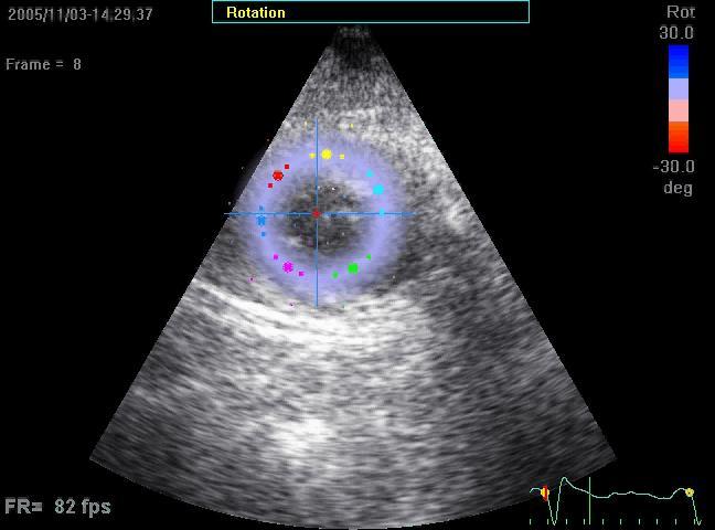
9 Christian Andreas Doppler Positive Frequency Shift Emitted Frequency (fo) Reflected Frequency (fr) (fr-fo) = 2fo v (cosθ) / c C= average speed of sound in tissue (1540m/sec)


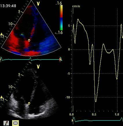
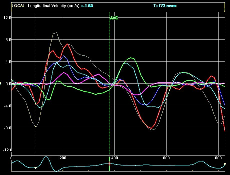
10 Doppler Tissue Imaging 1. Turn wall filters off 2. Turn down the gain


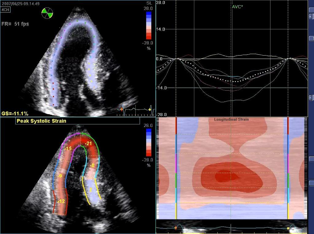
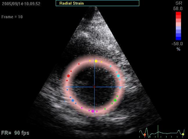
11 Septal Myocardial Velocity Traces S1 S2 e a


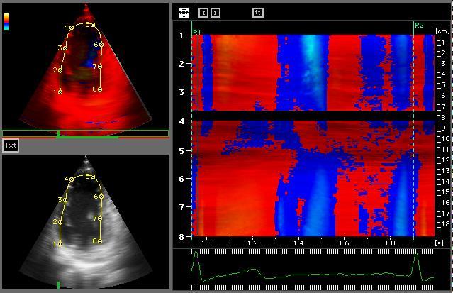
12 Normal


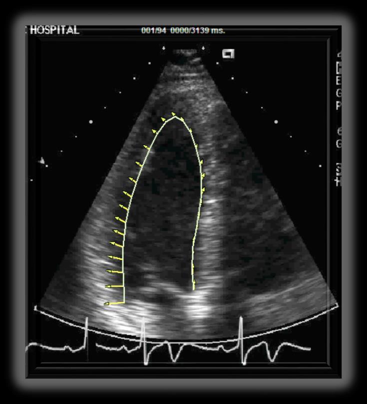
13 To Detect Regional Wall Motion


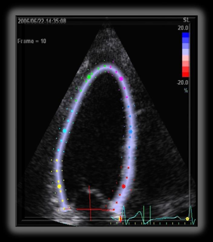
14 Peak Pulsed Velocities TD Mean Color Velocities TD 14 11


15 Translation and Tethering


16 Strain = deformation resulting from applied force Stress = force Courtesy of Ted Abraham
 Strain ( ) = L 1 -L 0 L 0 10 cm Strain rate 8 L 0 L 1-20% cm +20% 12 cm 0% 10")
17 Used to describe elastic properties of cardiac muscle (Mirsky and Parmley: Circ Res, 1973) Strain ( ) = L 1 -L 0 L 0 10 cm Strain rate 8 L 0 L 1-20% cm +20% 12 cm 0% 10 cm


18 Rate of deformation High strain rate Low strain rate Equal strain Courtesy of Andreas Heimdal


19 AoC Basal Mid wall Apical Basal Mid wall Apical


20 Movement of the myocardium relative to the sample volume fixed in space


21 Speckle Tracking Velocity is estimated as a shift of each object divided by time between successive frames (or multiplied by Frame Rate)--> 2D vector: (Vx, Vy) = (dx, dy) * FR Y dy 0 dx New location Old location X Courtesy Peter Lysysanksy


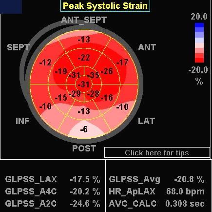
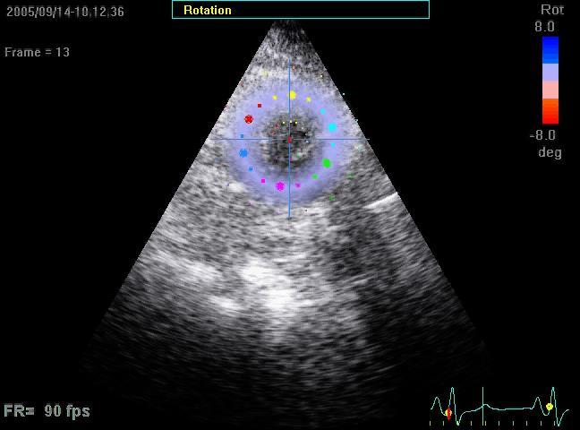
22 Potential Advantage? Signal noise Speckle tracking by principle is angle independent Gray scale (standard views) Monitor strain in two rather than one dimension Minimal user input Assessment of rotation: derived from circumferential strain at different levels in the heart (NO fixed sample volume)

23 Rotation/Twist/Torsion


24 Rotation and Torsion Rotation Basal Rotation Torsion Apex View from apex
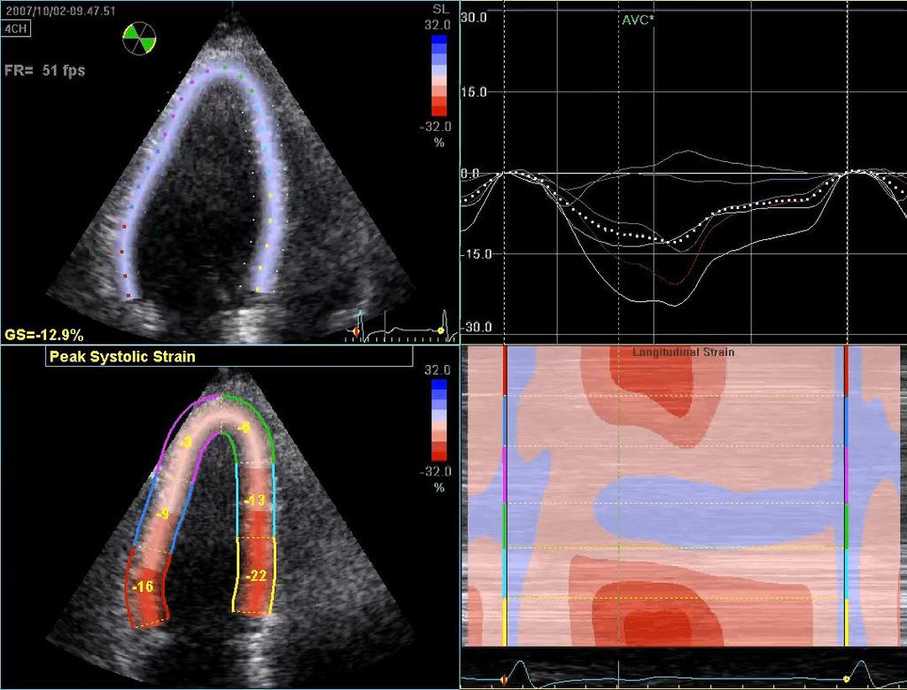
25 Normal Abnormal relaxation Pseudonormalization Restriction Mitral flow E A Tissue Doppler E A Apical rotation Basal rotation LV torsion Park et al: J Am Soc Echo Cardiogr 21:1129, 2008


26 Potential Clinical Applications

27 Impaired Systolic Function by Strain Imaging in Heart Failure With Preserved Ejection Fraction Strain Imaging detects impaired systolic function despite preserved global LVEF in HFpEF that may contribute to the pathophysiology of the HFpEF syndrome. J Am Coll Cardiol 2014;63:447-56
28 Strain (%) 0 Controls Hypertensive Heart Disease HFpEF Longitudinal Circumferential * * * * *p< compared to controls and between HHD and HFpEF J Am Coll Cardiol 2014;63:447-56


29 Longitudinal Strain NT-proBNP

30

31 Cardio-Oncology


32 76 year old male CMML/MDS with associated myeloid sarcoma skin lesions Experimental Chemotherapy ABT-348


33 Baseline 2 Months LVEF = 66% LVEF = 58%


34 Baseline 2 Months LVEF = 66% LVEF = 58% GLPSS Avg = -17.8% Troponin T = 0.02 GLPSS Avg = -14.3% Troponin T = 0.03

35 Global Longitudinal Peak Systolic Strain (GLS) Members of the in Chamber the range Quantification of -20% Writing Group are: Roberto M. Lang, MD, FASE, et al - Optimize image quality, maximize frame rate and minimize foreshortening. - When regional tracking is suboptimal in more than two J Am myocardial Soc Echocardiogr segments 2015;28:1-39 in a single view the calculation of GLS should be avoided.

36 Aortic Valve closure

37

38

39 Anthracyclines and Trastuzumab Can we predict a later (3 months) decline in LVEF? No decrease in GLS > 10% or elevated hstni have a 3% probability of a decrease in LVEF. If either a decrease in GLS or elevated hstni have a 9X increased risk for cardiotoxicity compared to those with no changes in either of these markers.

40 CRTCD if decrease in LVEF >10% to a value <53% - GLS -Reversible: is the optimal to within parameter 5 percentage of deformation points of for the baseline early detection of subclinical LV dysfunction. - In patients -Partially with reversible: available improved baseline by strain >10 percentage points measurements, from the nadir a but relative remaining percentage >5 percentage reduction points of below GLS baseline of <8% from baseline appears not to be meaningful, -Irreversible: and improved those >15% by <10 from percentage baseline are points very from likely the to nadir be and abnormal. remaining >5 percentage points below baseline


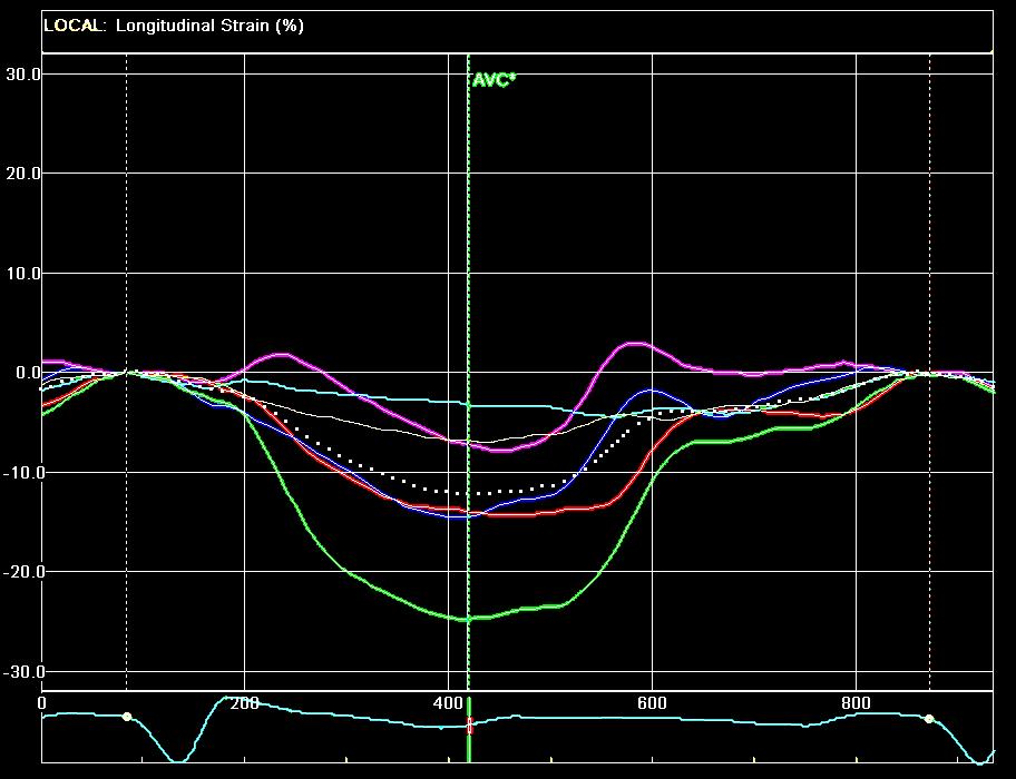
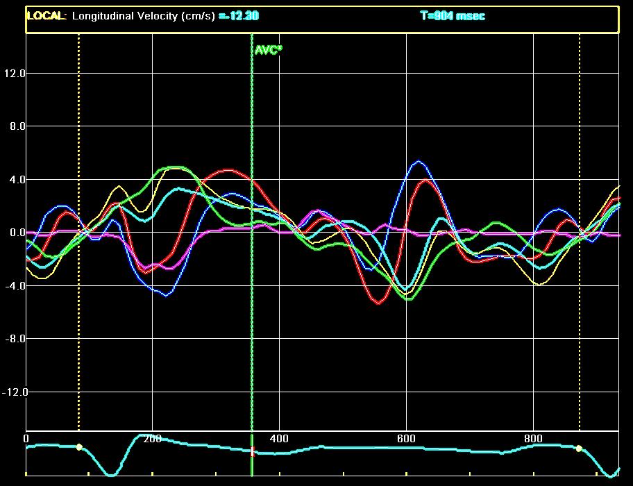
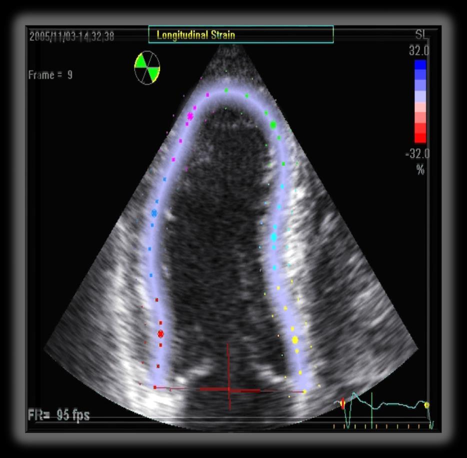
41 Athlete HTN HCM Infiltrative -amyloid Storage -Fabry


42 The Thinker Auguste Rodin


43

44 J Am Soc Echocardiogr 2013;26: Patients with HCM have significantly limited systolic function reserve and more dynamic dyssynchrony with exercise compared with those with HTN
45 Controls HTN HCM Rest Strain (%) * ** Exercise Strain (%) Rest TTP-SD (msec) Exercise TTP-SD (msec) Septal Thickness (mm) * ** ** * ** * **

46 Normal Abnormal relaxation Pseudonormalization Restriction Mitral flow E A Tissue Doppler E A Apical rotation Basal rotation LV torsion Park et al: J Am Soc Echo Cardiogr 21:1129, 2008
47 Controls HTN HCM Rest Circumferential Strain (%) Circumferential Strain Exercise (%) Septal Thickness (mm) * ** * ** * **


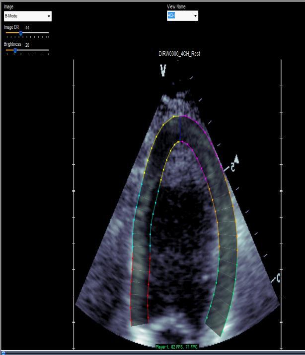
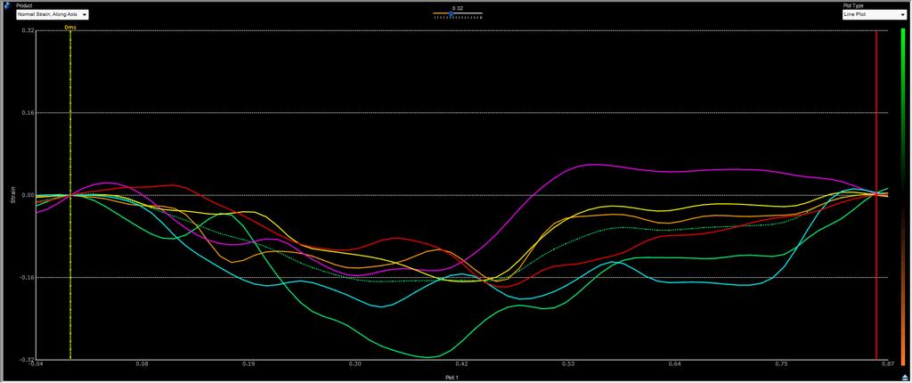
48 Apical HCM Septal HCM

49 Application of a Parametric Display of Two-Dimensional Speckle-Tracking Longitudinal Strain to Improve the Etiologic Diagnosis of Mild to Moderate Left Ventricular Hypertrophy J Am Soc Echocardiogr 2014;27:888-95


50 Cardiac Amyloidosis Hypertensive Heart Disease Hypertrophic Cardiomyopathy 14mm 14mm 13mm Mean Wall Left Ventricular Thickness

51


52 Longitudinal Strain (%) AVC Longitudinal Velocity cm/sec)
")
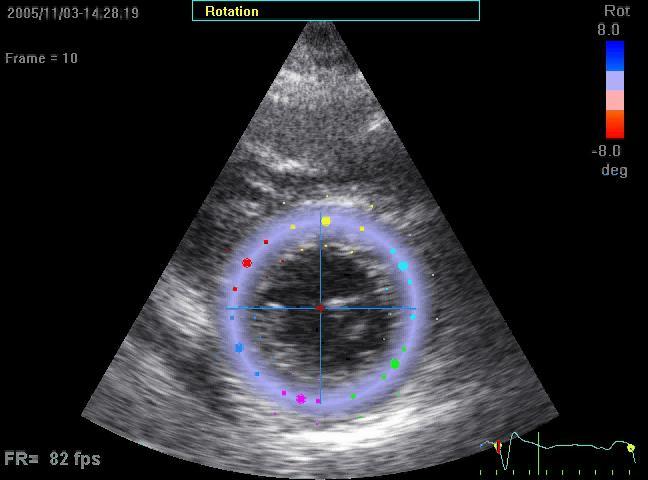
53 Torsion (degrees) 7º

54 Longitudinal Strain (%) AVC Longitudinal Velocity cm/sec)
")
55 Torsion (degrees) 16º


56 J Am Coll Cardiol Img 2014;7: LV dysfunction is frequently subclinical despite a normal ejection fraction. It may preceded the onset of symptoms an portend a poor outcome - The advent of novel tissue-tracking echo techniques has unleashed new opportunities for the clinical identification of early abnormalities in LV function.


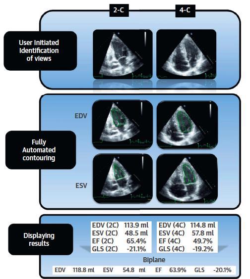
57 Impediments to Clinical Adoption? 1. Standardization 2. Workflow


58 Echocardiographic Measures of Myocardial Deformation by Speckle-Tracking Technologies: The Need for Standardization? J Am Soc Echocardiogr 2012:25:
")
59 (GE Vivid 7)


60 Image Area EchoInsight GLS % vs %; p= Movie GE
61 Need For Standardization Endocardium Endocardium/Epicardium Average of peaks Natural Lagrangian Natural Lagrangian Systole ± ± ± ±2.53 Systole/ diastole ± ±2.90* ± ±2.57 Peak of average vs ; p=0.02 Systole ± ± ± ±2.75 Systole/ diastole ± ± ± ±2.69 *Significant difference (P<.05) compared with Image-Arena GLS Significant difference (P<.001) compared with Image-Arena GLS J Am Soc Echocardiogr 2012:25:

62 vs ; p=0.02


63
64
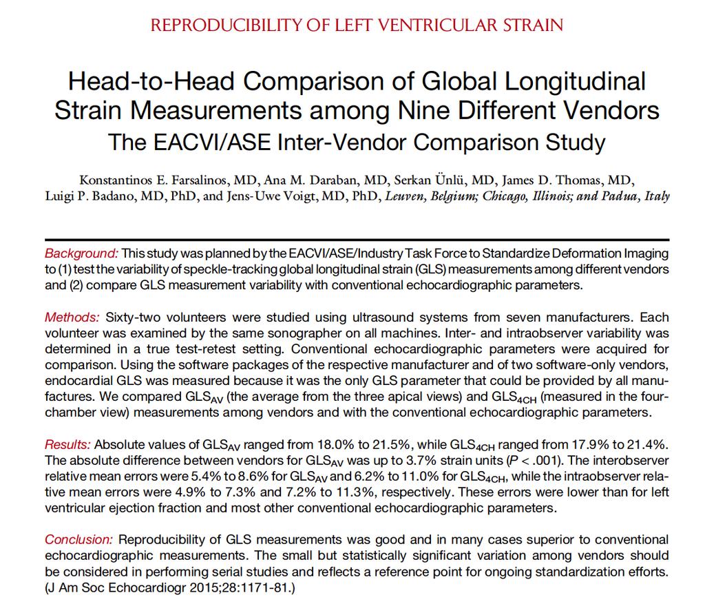
65 ± ± ± ± ± ± ± ± ± ± GLS AV, % Hitachi-A Esaote GE Philips Samsung Siemens Toshiba Epsilon Tomtec Mean of All Farsalinos et al. J Am Soc Echocardiogr 2015;28:
66 Mean Error, % GLS AV E E/A IVS LVEDD PW GLS AV Farsalinos et al. J Am Soc Echocardiogr 2015;28:
67 GLS AV, % Hitachi-A Esaote GE Philips Samsung Siemens Toshiba Epsilon Tomtec EF BI Farsalinos et al. J Am Soc Echocardiogr 2015;28:

68 Cross vendor variability in peak systolic global longitudinal strain may now be less than that of measures of left ventricular ejection fraction J Am Soc Echocardiogr 2015;28:183-93

69 Any innovation in imaging must be paralleled or exceeded by an innovation in workflow.
70 Fully Automated Versus Standard Tracking of Left Ventricular Ejection Fraction and Longitudinal Strain The Fast-EFs Multicenter Study J Am Coll Cardiol 2015;66:
71
72 1. AutoLV measurements were feasible in 98% of studies. 2. Average analysis time was 8+1 sec/patient. 3. Interobserver variability was higher for both visual and manual EF, but not different for LS.


73 Simultaneous measurement of Strain and Ejection Fraction Images courtesy of J. D Hooge et. al. Longitudinal Strain Regional Ejection Fraction Measures systolic shortening Sensitive measure of myocardium function Measures fractional change in volume Established, commonly used metric


74 Analysis October January EF Strain

75 Lower resolution (spatial and temporal)

76 a promising approach J Am Soc Echocardiogr 2012;25:68-79


77 What s Next Starts Soon Standardization Workflow Efficiency

78 It doesn t matter how slowly you go as long as you do Confucius not stop

79 1. DTI characterizes the low velocity, high intensity signals that come from the wall. 2. DTI is limited to movement relative to the sample volume fixed in space 3. Velocity: pitfalls of tethering and translational motion

80 4. Local parameters of deformation (strain and strain rate) are not influenced by tethering or translational motion 5. Feature or Speckle tracking can evaluate velocity, strain and strain rate from standard gray scale images 6. Feature tracking permits assessment of strain in the axis of movement rather than the axis of the ultrasound beam.
81 a. Turn the wall filters on and turn down the receiver gain. b. Turn the wall filters off and turn up the receiver gain. c. Turn the wall filters off and turn down the receiver gain. d. Turn the wall filters on and turn up the receiver gain.
82 a. You measure strain along the axis of the ultrasound beam. b. Velocity and strain measurements are measured from standard gray-scale images. c. Myocardial velocity measurements are not influenced by translational or tethering motion as they are when obtained by pulsed wave tissue Doppler imaging. d. You can measure longitudinal but not circumferential or radial strain.
83 a. Higher b. Lower c. The same
84
Tissue Doppler and Strain Imaging
 Tissue Doppler and Strain Imaging Steven J. Lester MD, FRCP(C), FACC, FASE Relevant Financial Relationship(s) None Off Label Usage None 1 Objective way with which to quantify the minor amplitude and temporal
Tissue Doppler and Strain Imaging
 Tissue Doppler and Strain Imaging Steven J. Lester MD, FRCP(C), FACC, FASE Relevant Financial Relationship(s) None Off Label Usage None 1 Objective way with which to quantify the minor amplitude and temporal
DISCLOSURE. Myocardial Mechanics. Relevant Financial Relationship(s) Off Label Usage
 Off Label Usage") 7th Annual Team Echocardiography: The Heart of Cardiovascular Medicine Tissue Doppler, Strain, Speckle: What? How? Christopher J Kramer RDCS Aurora Medical Group Advanced Cardiovascular Services, Aurora
7th Annual Team Echocardiography: The Heart of Cardiovascular Medicine Tissue Doppler, Strain, Speckle: What? How? Christopher J Kramer RDCS Aurora Medical Group Advanced Cardiovascular Services, Aurora
Strain and Strain Rate Imaging How, Why and When?
 Strain and Strain Rate Imaging How, Why and When? João L. Cavalcante, MD Advanced Cardiac Imaging Fellow Cleveland Clinic Foundation Disclosures: No conflicts of interest Movement vs Deformation Movement
2/2/2011. Strain and Strain Rate Imaging How, Why and When? Movement vs Deformation. Doppler Myocardial Velocities. Movement. Deformation.
 Strain and Strain Rate Imaging How, Why and When? João L. Cavalcante, MD Advanced Cardiac Imaging Fellow Cleveland Clinic Foundation Disclosures: No conflicts of interest Movement vs Deformation Movement
VECTORS OF CONTRACTION
 1/3/216 Strain, Strain Rate, and Torsion: Myocardial Mechanics Simplified and Applied VECTORS OF CONTRACTION John Gorcsan, MD University of Pittsburgh, Pittsburgh, PA Shortening Thickening Twisting No
Velocity, strain and strain rate: Doppler and Non-Doppler methods. Thoraxcentre, Erasmus MC,Rotterdam
 Velocity, strain and strain rate: Doppler and Non-Doppler methods J Roelandt J. Roelandt Thoraxcentre, Erasmus MC,Rotterdam Basics of tissue Doppler imaging Instantaneous annular velocity profiles IVCT
Global left ventricular circumferential strain is a marker for both systolic and diastolic myocardial function
 Global left ventricular circumferential strain is a marker for both systolic and diastolic myocardial function Toshinari Onishi 1, Samir K. Saha 2, Daniel Ludwig 1, Erik B. Schelbert 1, David Schwartzman
Nancy Goldman Cutler, MD Beaumont Children s Hospital Royal Oak, Mi
 Nancy Goldman Cutler, MD Beaumont Children s Hospital Royal Oak, Mi Identify increased LV wall thickness (WT) Understand increased WT in athletes Understand hypertrophic cardiomyopathy (HCM) Enhance understanding
Strain Imaging: Myocardial Mechanics Simplified and Applied
 9/28/217 Strain Imaging: Myocardial Mechanics Simplified and Applied John Gorcsan III, MD Professor of Medicine Director of Clinical Research Division of Cardiology VECTORS OF CONTRACTION Shortening Thickening
Tissue Doppler Imaging in Congenital Heart Disease
 Tissue Doppler Imaging in Congenital Heart Disease L. Youngmin Eun, M.D. Department of Pediatrics, Division of Pediatric Cardiology, Kwandong University College of Medicine The potential advantage of ultrasound
Alicia Armour, MA, BS, RDCS
 Alicia Armour, MA, BS, RDCS No disclosures Review 2D Speckle Strain (briefly) Discuss some various patient populations & disease pathways where Strain can be helpful Discuss how to acquire images for Strain
LV FUNCTION ASSESSMENT: WHAT IS BEYOND EJECTION FRACTION
 LV FUNCTION ASSESSMENT: WHAT IS BEYOND EJECTION FRACTION Jamilah S AlRahimi Assistant Professor, KSU-HS Consultant Noninvasive Cardiology KFCC, MNGHA-WR Introduction LV function assessment in Heart Failure:
How To Perform Strain Imaging; Step By Step Approach. Maryam Bo Khamseen Echotechnoligist II EACVI, ARDMS, RCS King Abdulaziz Cardiac Center- Riyadh
 How To Perform Strain Imaging; Step By Step Approach Maryam Bo Khamseen Echotechnoligist II EACVI, ARDMS, RCS King Abdulaziz Cardiac Center- Riyadh Outlines: Introduction Describe the basic of myocardium
Implementing New Technology
 Implementing New Technology PP16 Imaging Conference Bicol Hospital, Legaspi City, Philippines July 2016 David Adams, ACS, RCS, RDCS, FASE Duke University Medical Center Echo Evolution By Dr. Zoghbi Handheld/POC
New Approaches to Systolic Function: Strain Imaging
 New Approaches to Systolic Function: Strain Imaging James D. Thomas, MD, FACC, FASE Director, Center for Heart Valve Disease Bluhm Cardiovascular Institute Professor of Medicine, Feinberg School of Medicine,
Velocity Vector Imaging as a new approach for cardiac magnetic resonance: Comparison with echocardiography
 Velocity Vector Imaging as a new approach for cardiac magnetic resonance: Comparison with echocardiography Toshinari Onishi 1, Samir K. Saha 2, Daniel Ludwig 1, Erik B. Schelbert 1, David Schwartzman 1,
Three-dimensional Wall Motion Tracking:
 Three-dimensional Wall Motion Tracking: A Novel Echocardiographic Method for the Assessment of Ventricular Volumes, Strain and Dyssynchrony Jeffrey C. Hill, BS, RDCS, FASE Jennifer L. Kane, RCS Gerard
OPTIMIZING ECHO ACQUISTION FOR STRAIN AND DIASTOLOGY
 OPTIMIZING ECHO ACQUISTION FOR STRAIN AND DIASTOLOGY October 8, 2017 Deborah Agler, ACS, RDCS, FASE Coordinator of Education and Training Cleveland Clinic General Principles Diastology Clinical Data Heart
L ecocardiografia nello Scompenso Cardiaco Acuto e cronico: vecchi dogmi e nuovi trends.
 V SESSIONE SCOMPENSO CARDIACO 2015 Genova, 13-14 Novembre 2015 L ecocardiografia nello Scompenso Cardiaco Acuto e cronico: vecchi dogmi e nuovi trends. Gian Paolo Bezante, MD, FACC UOC Clinica di Malattie
Myocardial Strain Imaging in Cardiac Diseases and Cardiomyopathies.
 Myocardial Strain Imaging in Cardiac Diseases and Cardiomyopathies. Session: Cardiomyopathy Tarun Pandey MD, FRCR. Associate Professor University of Arkansas for Medical Sciences Disclosures No relevant
Measuring cardiac tissue motion and strain
 Ultrasound Measuring cardiac tissue motion and strain Automated Cardiac Motion Quantification A.I. (acmq A.I. ) David Prater, MS, Clinical Scientist, Philips Jane Vogel, MD, Senior Product Manager, Philips
Value of echocardiography in chronic dyspnea
 Value of echocardiography in chronic dyspnea Jahrestagung Schweizerische Gesellschaft für /Schweizerische Gesellschaft für Pneumologie B. Kaufmann 16.06.2016 Chronic dyspnea Shortness of breath lasting
22 nd Annual Conference of the Saudi Heart Association Riyadh, Saudi Arabia
 22 nd Annual Conference of the Saudi Heart Association Riyadh, Saudi Arabia New Echocardiographic Modalities to Evaluate Ventricular Function in Congenital Heart Disease: Tissue Doppler & Strain Rate Imaging
Strain/Untwisting/Diastolic Suction
 What Is Diastole and How to Assess It? Strain/Untwisting/Diastolic Suction James D. Thomas, M.D., F.A.C.C. Cardiovascular Imaging Center Department of Cardiology Cleveland Clinic Foundation Cleveland,
HYPERTROPHY: Behind the curtain. V. Yotova St. Radboud Medical University Center, Nijmegen
 HYPERTROPHY: Behind the curtain V. Yotova St. Radboud Medical University Center, Nijmegen Disclosure of interest: none Relative wall thickness (cm) M 0.22 0.42 0.43 0.47 0.48 0.52 0.53 F 0.24 0.42 0.43
10/7/2013. Systolic Function How to Measure, How Accurate is Echo, Role of Contrast. Thanks to our Course Director: Neil J.
 Systolic Function How to Measure, How Accurate is Echo, Role of Contrast Neil J. Weissman, MD MedStar Health Research Institute & Professor of Medicine Georgetown University Washington, D.C. No Disclosures
Advanced Echocardiography in the Evaluation of Chemotherapy Patients
 Advanced Echocardiography in the Evaluation of Chemotherapy Patients Juan Carlos Plana, MD, FACC, FASE Co-Director, Cardio-Oncology Center Section of Cardiovascular Imaging Department of Cardiovascular
Heart Failure in Women: Dr Goh Ping Ping Cardiologist Asian Heart & Vascular Centre
 Heart Failure in Women: More than EF? Dr Goh Ping Ping Cardiologist Asian Heart & Vascular Centre Overview Review pathophysiology as it relates to diagnosis and management Rational approach to workup:
Conflict of interest: none declared
 The value of left ventricular global longitudinal strain assessed by three-dimensional strain imaging in the early detection of anthracycline-mediated cardiotoxicity C. Mornoş, A. Ionac, D. Cozma, S. Pescariu,
Novel echocardiographic modalities: 3D echo, speckle tracking and strain rate imaging. Potential roles in sports cardiology. Stefano Caselli, MD, PhD
 Novel echocardiographic modalities: 3D echo, speckle tracking and strain rate imaging. Potential roles in sports cardiology. Stefano Caselli, MD, PhD Ospedale San Pietro Fatebenefratelli Rome, Italy Differential
RIGHT VENTRICULAR SIZE AND FUNCTION
 RIGHT VENTRICULAR SIZE AND FUNCTION Edwin S. Tucay, MD, FPCC, FPCC, FPSE Philippine Society of Echocardiography Quezon City, Philippines Echo Mission, BRTTH, Legaspi City, July 1-2, 2016 NO DISCLOSURE
Diastolic Function: What the Sonographer Needs to Know. Echocardiographic Assessment of Diastolic Function: Basic Concepts 2/8/2012
 Diastolic Function: What the Sonographer Needs to Know Pat Bailey, RDCS, FASE Technical Director Beaumont Health System Echocardiographic Assessment of Diastolic Function: Basic Concepts Practical Hints
Cardiac Chamber Quantification by Echocardiography
 Cardiac Chamber Quantification by Echocardiography Maryam Bokhamseen, RCS, RCDS, EACVI Echotechnologist ǁ, Non invasive Cardiac Laboratory King Abdulaziz Cardiac Center. Outline: Introduction. Background
Advanced Multi-Layer Speckle Strain Permits Transmural Myocardial Function Analysis in Health and Disease:
 Advanced Multi-Layer Speckle Strain Permits Transmural Myocardial Function Analysis in Health and Disease: Clinical Case Examples Jeffrey C. Hill, BS, RDCS Echocardiography Laboratory, University of Massachusetts
Right Ventricle Steven J. Lester MD, FACC, FRCP(C), FASE Mayo Clinic, Arizona
, FASE Mayo Clinic, Arizona") Right Ventricle Steven J. Lester MD, FACC, FRCP(C), FASE Mayo Clinic, Arizona 1. In which scenario will applying the simplified Bernoulli equation to the peak tricuspid regurgitation velocity and adding
Right Ventricle Steven J. Lester MD, FACC, FRCP(C), FASE Mayo Clinic, Arizona 1. In which scenario will applying the simplified Bernoulli equation to the peak tricuspid regurgitation velocity and adding
The Basics of Systolic Function
 The Basics of Systolic Function James D. Thomas, MD, FACC, FASE Director, Center for Heart Valve Disease Bluhm Cardiovascular Institute Professor of Medicine, Feinberg School of Medicine, Northwestern
Chamber Quantitation Guidelines: What is New?
 Chamber Quantitation Guidelines: What is New? Roberto M Lang, MD J AM Soc Echocardiogr 2005; 18:1440-1463 1 Approximately 10,000 citations iase in itune Cardiac Chamber Quantification: What is New? Database
Carlos Eduardo Suaide Silva, Luiz Darcy Cortez Ferreira, Luciana Braz Peixoto, Claudia Gianini Monaco, Manuel Adán Gil, Juarez Ortiz
 Silva et al Original Article Arq Bras Cardiol Study of the Myocardial Contraction and Relaxation Velocities through Doppler Tissue Imaging Echocardiography. A New Alternative in the Assessment of the Segmental
Restrictive Cardiomyopathy
 ESC Congress 2011, Paris Imaging Unusual Causes of Cardiomyopathy Restrictive Cardiomyopathy Kazuaki Tanabe, MD, PhD Professor of Medicine Chair, Division of Cardiology Izumo, Japan I Have No Disclosures
Mechanisms of False Positive Exercise Electrocardiography: Is False Positive Test Truly False?
 Mechanisms of False Positive Exercise Electrocardiography: Is False Positive Test Truly False? Masaki Izumo a, Kengo Suzuki b, Hidekazu Kikuchi b, Seisyo Kou b, Keisuke Kida b, Yu Eguchi b, Nobuyuki Azuma
Imaging in Heart Failure: A Multimodality Approach. Thomas Ryan, MD
 Imaging in Heart Failure: A Multimodality Approach Thomas Ryan, MD Heart Failure HFrEF HFpEF EF50% Lifetime risk 20% Prevalence 6M Americans Societal costs - $30B 50% 5-year survival 1 Systolic
Echocardiographic Assessment of the Left Ventricle
 Echocardiographic Assessment of the Left Ventricle Theodora Zaglavara, MD, PhD, BSCI/BSCCT Department of Cardiovascular Imaging INTERBALKAN EUROPEAN MEDICAL CENTER 2015 The quantification of cardiac chamber
Quantification of Cardiac Chamber Size
 2017 KSE 2017-11-25 Quantification of Cardiac Chamber Size Division of Cardiology Keimyung University Dongsan Medical Center In-Cheol Kim M.D., Ph.D. LV size and function Internal linear dimensions PLX
Dr. Dermot Phelan MB BCh BAO PhD European Society of Cardiology 2012
 Relative Apical Sparing of Longitudinal Strain Using 2- Dimensional Speckle-Tracking Echocardiography is Both Sensitive and Specific for the Diagnosis of Cardiac Amyloidosis. Dr. Dermot Phelan MB BCh BAO
Appendix II: ECHOCARDIOGRAPHY ANALYSIS
 Appendix II: ECHOCARDIOGRAPHY ANALYSIS Two-Dimensional (2D) imaging was performed using the Vivid 7 Advantage cardiovascular ultrasound system (GE Medical Systems, Milwaukee) with a frame rate of 400 frames
Affecting the elderly Requiring new approaches. Echocardiographic Evaluation of Hemodynamic Severity. Increasing prevalence Mostly degenerative
 Echocardiographic Evaluation of Hemodynamic Severity Steven J. Lester MD, FACC, FRCP(C), FASE Mayo Clinic, Arizona Relevant Financial Relationship(s) None Off Label Usage None A re-emerging public-health
Basic Approach to the Echocardiographic Evaluation of Ventricular Diastolic Function
 Basic Approach to the Echocardiographic Evaluation of Ventricular Diastolic Function J A F E R A L I, M D U N I V E R S I T Y H O S P I T A L S C A S E M E D I C A L C E N T E R S T A F F C A R D I O T
3D-stress echocardiography Bernard Cosyns, MD, PhD
 3D-stress echocardiography Bernard Cosyns, MD, PhD No Disclosure The Pro-Technology bias Sicari et al. Cardiovascular Ultrasound 2006, 4:11 Overview 2D stress echocardiography: main limitations 3D echocardiography:
Strain imaging in children: from Tissue Doppler to 3 D
 Strain imaging in children: from Tissue Doppler to 3 D Mark kk. Friedberg Fi Outline Deformation in the fetus and neonate Deformation in pediatric cardiomyopathy y (briefly!) Deformation in Congenital
Little is known about the degree and time course of
 Differential Changes in Regional Right Ventricular Function Before and After a Bilateral Lung Transplantation: An Ultrasonic Strain and Strain Rate Study Virginija Dambrauskaite, MD, Lieven Herbots, MD,
좌심실수축기능평가 Cardiac Function
 Basic Echo Review Course 좌심실수축기능평가 Cardiac Function Seonghoon Choi Cardiology Hallym university LV systolic function Systolic function 좌심실수축기능 - 심근의수축으로심실에서혈액을대동맥으로박출하는기능 실제임상에서 LV function 의의미 1Diagnosis
JACC: CARDIOVASCULAR IMAGING VOL. 8, NO. 2, 2015 ª 2015 BY THE AMERICAN COLLEGE OF CARDIOLOGY FOUNDATION ISSN X/$36.00
 JACC: CARDIOVASCULAR IMAGING VOL. 8, NO. 2, 15 ª 15 BY THE AMERICAN COLLEGE OF CARDIOLOGY FOUNDATION ISSN 1936-878X/$36. PUBLISHED BY ELSEVIER INC. http://dx.doi.org/1.116/j.jcmg.14.1.1 How to Define End-Diastole
Mechanisms of heart failure with normal EF Arterial stiffness and ventricular-arterial coupling. What is the pathophysiology at presentation?
 Mechanisms of heart failure with normal EF Arterial stiffness and ventricular-arterial coupling What is the pathophysiology at presentation? Ventricular-arterial coupling elastance Central arterial pressure
Global and Regional Myocardial Function Quantification by Two-Dimensional Strain Application in Hypertrophic Cardiomyopathy
 Journal of the American College of Cardiology Vol. 47, No. 6, 2006 2006 by the American College of Cardiology Foundation ISSN 0735-1097/06/$32.00 Published by Elsevier Inc. doi:10.1016/j.jacc.2005.10.061
Hypertensive heart disease and failure
 Hypertensive heart disease and failure Prof. Dr. Alan Fraser Cardiff University The heart in hypertension Pathophysiology of LV adaptation Regional development of hypertrophy Stress testing - inducible
Beginner s Guide to Strain: What should be in your lab in Disclosures
 Beginner s Guide to Strain: What should be in your lab in 2018 Bonita Anderson DMU (Cardiac), MApplSc (Med Ultrasound), ACS, AMS, FASE None Disclosures Calculation of Strain Strain can be Positive Strain
Assessment of cardiac function with 3D echocardiography. Đánh giá chức năng tim bằng siêu âm tim 3D
 Assessment of cardiac function with 3D echocardiography Đánh giá chức năng tim bằng siêu âm tim 3D TS. BS. Nguyễn Thị Thu Hoài Viện Tim Mạch Quốc Gia Việt Nam TỪ SIÊU ÂM M-mode ĐẾN SIÊU ÂM 3D TỪ SIÊU ÂM
Feasibility and limitations of 2D speckle tracking echocardiography
 ORIGINAL ARTICLE 204 A prospective study in daily clinical practice Feasibility and limitations of 2D speckle tracking echocardiography Lina Melzer, Anja Faeh-Gunz, Barbara Naegeli, Burkhardt Seifert*,
Diastology Disclosures: None. Dias2011:1
 Diastology 2011 James D. Thomas, M.D., F.A.C.C. Cardiovascular Imaging Center Department of Cardiology Cleveland Clinic Foundation Cleveland, Ohio, USA Disclosures: None Dias2011:1 Is EVERYBODY a member!?!
Chamber Quantitation Guidelines - Update II
 Chamber Quantitation Guidelines - Update II Right Heart Measurements Steven A. Goldstein MD FACC FASE Professor of Medicine Georgetown University Medical Center MedStar Heart Institute Washington Hospital
Tissue Doppler Imaging
 Cronicon OPEN ACCESS Hesham Rashid* Tissue Doppler Imaging CARDIOLOGY Editorial Department of Cardiology, Benha University, Egypt *Corresponding Author: Hesham Rashid, Department of Cardiology, Benha University,
Diastole is Not a Single Entity Four Components of Diastolic Dysfunction
 Physiology of Diastolic Function Made Easy James D. Thomas, MD, FACC, FASE Director, Center for Heart Valve Disease Bluhm Cardiovascular Institute Professor of Medicine, Feinberg School of Medicine, Northwestern
New Universal Strain Software Accurately Assesses Cardiac Systolic and Diastolic Function Using Speckle Tracking Echocardiography
 DOI: 10.1111/echo.12512 2014, Wiley Periodicals, Inc. Echocardiography New Universal Strain Software Accurately Assesses Cardiac Systolic and Diastolic Function Using Speckle Tracking Echocardiography
DECLARATION OF CONFLICT OF INTEREST. None
 DECLARATION OF CONFLICT OF INTEREST None Hot Topics in Echocardiography: The position of the EAE EAE / ASE recommendation about Echo Assessment of Cardiac Mechanics Jens-Uwe Voigt Dpt. of Cardiovascular
Quantitation of right ventricular dimensions and function
 SCCS Basics of cardiac assessment Quantitation of right ventricular dimensions and function Tomasz Kukulski, MD PhD Dept of Cardiology, Congenital Heart Disease and Electrotherapy Silesian Medical University
The rapid evolution of echocardiography during the past 25 years
 Evaluation of Myocardial Mechanics in the Fetus by Velocity Vector Imaging Adel K. Younoszai, MD, David E. Saudek, MD, Stephen P. Emery, MD, and James D. Thomas, MD, Denver, Colorado; Cleveland, Ohio;
The road to successful CRT implantation: The role of echo
 The road to successful CRT implantation: The role of echo Tae-Ho Park Dong-A University Hospital, Busan, Korea Terminology Cardiac Resynchronization Therapy (CRT) = Biventricular pacing (BiV) = Left ventricular
Diagnostic approach to heart disease
 Diagnostic approach to heart disease Initial work up History Physical exam Chest radiographs ECG Special studies Echocardiography Cardiac catheterization Echocardiography principles Technique of producing
Vevo 2100 System Cardio Measurements. Dieter Fuchs, PhD FUJIFILM VisualSonics, Inc.
 Vevo 2100 System Cardio Measurements Dieter Fuchs, PhD FUJIFILM VisualSonics, Inc. dfuchs@visualsonics.com Instructions This document is a guideline on how to assess cardiac function in rodents imaged
How NOT to miss Hypertrophic Cardiomyopathy? Adaya Weissler-Snir, MD University Health Network, University of Toronto
 How NOT to miss Hypertrophic Cardiomyopathy? Adaya Weissler-Snir, MD University Health Network, University of Toronto Introduction Hypertrophic cardiomyopathy is the most common genetic cardiomyopathy,
Left atrial function. Aliakbar Arvandi MD
 In the clinic Left atrial function Abstract The left atrium (LA) is a left posterior cardiac chamber which is located adjacent to the esophagus. It is separated from the right atrium by the inter-atrial
Coronary artery disease (CAD) risk factors
 risk factors") Background Coronary artery disease (CAD) risk factors CAD Risk factors Hypertension Insulin resistance /diabetes Dyslipidemia Smoking /Obesity Male gender/ Old age Atherosclerosis Arterial stiffness precedes
Background Coronary artery disease (CAD) risk factors CAD Risk factors Hypertension Insulin resistance /diabetes Dyslipidemia Smoking /Obesity Male gender/ Old age Atherosclerosis Arterial stiffness precedes
Evaluation of Left Ventricular Diastolic Dysfunction by Doppler and 2D Speckle-tracking Imaging in Patients with Primary Pulmonary Hypertension
 ESC Congress 2011.No 85975 Evaluation of Left Ventricular Diastolic Dysfunction by Doppler and 2D Speckle-tracking Imaging in Patients with Primary Pulmonary Hypertension Second Department of Internal
Μαρία Μπόνου Διευθύντρια ΕΣΥ, ΓΝΑ Λαϊκό
 Μαρία Μπόνου Διευθύντρια ΕΣΥ, ΓΝΑ Λαϊκό Diastolic HF DD: Diastolic Dysfunction DHF: Diastolic HF HFpEF: HF with preserved EF DD Pathophysiologic condition: impaired relaxation, LV compliance, LV filling
Acute impairment of basal left ventricular rotation but not twist and untwist are involved in the pathogenesis of acute hypertensive pulmonary oedema
 Acute impairment of basal left ventricular rotation but not twist and untwist are involved in the pathogenesis of acute hypertensive pulmonary oedema A.D. Margulescu 1,2, R.C. Sisu 1,2, M. Florescu 2,
E/Ea is NOT an essential estimator of LV filling pressures
 Euroecho Kopenhagen Echo in Resynchronization in 2010 E/Ea is NOT an essential estimator of LV filling pressures Wilfried Mullens, MD, PhD December 10, 2010 Ziekenhuis Oost Limburg Genk University Hasselt
Evalua&on)of)Le-)Ventricular)Diastolic) Dysfunc&on)by)Echocardiography:) Role)of)Ejec&on)Frac&on)
of)Le-)Ventricular)Diastolic) Dysfunc&on)by)Echocardiography:) Role)of)Ejec&on)Frac&on)") Evalua&on)of)Le-)Ventricular)Diastolic) Dysfunc&on)by)Echocardiography:) Role)of)Ejec&on)Frac&on) N.Koutsogiannis) Department)of)Cardiology) University)Hospital)of)Patras)! I have no conflicts of interest
Evalua&on)of)Le-)Ventricular)Diastolic) Dysfunc&on)by)Echocardiography:) Role)of)Ejec&on)Frac&on) N.Koutsogiannis) Department)of)Cardiology) University)Hospital)of)Patras)! I have no conflicts of interest
Fetal cardiac function: what to use and does it make a difference?
 17 th International Conference on Prenatal Diagnosis and Therapy Lisbon, June 2013 Fetal cardiac function: what to use and does it make a difference? Fàtima Crispi Department of Maternal-Fetal Medicine,
New aspects of Echocardiography in Hypertensive Heart Disease. Fausto J. Pinto, MD, PhD, FESC, FACC, FASE
 New aspects of Echocardiography in Hypertensive Heart Disease Fausto J. Pinto, MD, PhD, FESC, FACC, FASE Progressive increase in cardiovascular morbidity (left) and all-cause mortality (right) rates
Chamber Quantitation Guidelines II Right Heart Measurements
 Chamber Quantitation Guidelines II Right Heart Measurements Steven A. Goldstein MD FACC FASE Director, Noninvasive Cardiology MedStar Heart Institute Washington Hospital Center Sunday, October 9, 2016
Left Ventricular Dyssynchrony in Patients Showing Diastolic Dysfunction without Overt Symptoms of Heart Failure
 ORIGINAL ARTICLE DOI: 10.3904/kjim.2010.25.3.246 Left Ventricular Dyssynchrony in Patients Showing Diastolic Dysfunction without Overt Symptoms of Heart Failure Jae Hoon Kim, Hee Sang Jang, Byung Seok
Aortic valve Stenosis: Insights in the evaluation of LV function. Erwan DONAL Cardiologie CHU Rennes
 Aortic valve Stenosis: Insights in the evaluation of LV function Erwan DONAL Cardiologie CHU Rennes erwan.donal@chu-rennes.fr Preload Afterload Myocardial Fiber Shortening Circumferential Longitudinal
MYOCARDIAL DEFORMATION IMAGING ON EXERCISE IN CHRONIC PRIMARY MITRAL REGURGITATION
 MYOCARDIAL DEFORMATION IMAGING ON EXERCISE IN CHRONIC PRIMARY MITRAL REGURGITATION A thesis submitted to the University of Manchester for the degree of Doctor of Medicine in the Faculty of Medical and
The importance of left atrium in LV diastolic function
 II Baltic Heart Failure Meeting and Congress of Latvian Society of Cardiology The importance of left atrium in LV diastolic function Dr. Artem Kalinin Eastern Clinical University Hospital Riga 30.09.2010.
Echocardiographic Evaluation of the Cardiomyopathies. Stephanie Coulter, MD, FACC, FASE April, 2016
 Echocardiographic Evaluation of the Cardiomyopathies Stephanie Coulter, MD, FACC, FASE April, 2016 Cardiomyopathies (CMP) primary disease intrinsic to cardiac muscle Dilated CMP Hypertrophic CMP Infiltrative
Advanced imaging of the left atrium - strain, CT, 3D, MRI -
 Advanced imaging of the left atrium - strain, CT, 3D, MRI - Monica Rosca, MD Carol Davila University of Medicine and Pharmacy, Bucharest, Romania Declaration of interest: I have nothing to declare Case
Cardiac hypertrophy : differentiating disease from athlete
 Cardiac hypertrophy : differentiating disease from athlete Ario Soeryo Kuncoro, MD, Cardiologist Echocardiography Division, National Cardiovascular Centre Harapan Kita-Jakarta Departement of Cardiology
WHAT DO ELECTROPHYSIOLOGISTS WANT TO KNOW FROM ECHOCARDIOGRAPHERS BEFORE, DURING&AFTER CARDIAC RESYNCHRONIZATION THERAPY?
 WHAT DO ELECTROPHYSIOLOGISTS WANT TO KNOW FROM ECHOCARDIOGRAPHERS BEFORE, DURING&AFTER CARDIAC RESYNCHRONIZATION THERAPY? Mary Ong Go, MD, FPCP, FPCC, FACC OUTLINE What is CRT Who needs CRT What does the
Automated Volumetric Cardiac Ultrasound Analysis
 Whitepaper Automated Volumetric Cardiac Ultrasound Analysis ACUSON SC2000 Volume Imaging Ultrasound System Bogdan Georgescu, Ph.D. Siemens Corporate Research Princeton, New Jersey USA Answers for life.
PRESENTER DISCLOSURE INFORMATION. There are no potential conflicts of interest regarding current presentation
 PRESENTER DISCLOSURE INFORMATION There are no potential conflicts of interest regarding current presentation Better synchrony and diastolic function for septal versus apical right ventricular permanent
GENERAL PRINCIPLES FOR ECHO ASSESSMENT OF DIASTOLIC FUNCTION (For full recommendation refer to the Left Ventricular Diastolic Function Guideline)
") 1 THE AMERICAN SOCIETY OF ECHOCARDIOGRAPHY RECOMMENDATIONS FOR THE EVALUATION OF LEFT VENTRICULAR DIASTOLIC FUNCTION BY ECHOCARDIOGRAPHY: A QUICK REFERENCE GUIDE FROM THE ASE WORKFLOW AND LAB MANAGEMENT
1 THE AMERICAN SOCIETY OF ECHOCARDIOGRAPHY RECOMMENDATIONS FOR THE EVALUATION OF LEFT VENTRICULAR DIASTOLIC FUNCTION BY ECHOCARDIOGRAPHY: A QUICK REFERENCE GUIDE FROM THE ASE WORKFLOW AND LAB MANAGEMENT
Segmental Tissue Doppler Image-Derived Tei Index in Patients With Regional Wall Motion Abnormalities
 ORIGINAL ARTICLE DOI 10.4070 / kcj.2010.40.3.114 Print ISSN 1738-5520 / On-line ISSN 1738-5555 Copyright c 2010 The Korean Society of Cardiology Open Access Segmental Tissue Doppler Image-Derived Tei Index
Quantifying LV function how good are we?
 Quantifying LV function how good are we? Professor Alan G Fraser Wales Heart Research Institute Cardiff University, U.K. Support for research from Hitachi Aloka, & GE Ultrasound Visual assessment of synchronicity
We are IntechOpen, the world s leading publisher of Open Access books Built by scientists, for scientists. International authors and editors
 We are IntechOpen, the world s leading publisher of Open Access books Built by scientists, for scientists 4,000 116,000 120M Open access books available International authors and editors Downloads Our
Athlete s Heart vs. Cardiomyopathy
 Athlete s Heart vs. Cardiomyopathy Linda D. Gillam, MD, MPH, FASE Chair, Department of Cardiovascular Medicine Medical Director, Cardiovascular Service Line Former Team Cardiologist to the New York Jets
Utility of Echocardiography
 Hypertrophic Cardiomyopathy and Beyond- Echo Hawaii 2018 Lawrence Rudski MD FRCPC FACC FASE Professor of Medicine Director, Division of Cardiology and Azrieli Heart Center Jewish General Hospital, McGill
An Integrated Approach to Study LV Diastolic Function
 An Integrated Approach to Study LV Diastolic Function Assoc. Prof. Adriana Ilieşiu, FESC University of Medicine Carol Davila Bucharest, Romania LV Diastolic Dysfunction impaired relaxation (early diastole)
Conflict of Interests
 The Left Ventricle: How Should We Quantify Its Size and Function; Is It Time for 3D in Everyone? Roberto M Lang, MD Conflict of Interests Philips Medical Imaging Research Grants Speakers bureau Advisory
Assessment of left ventricular function by different speckle-tracking software
 European Journal of Echocardiography (2010) 11, 417 421 doi:10.1093/ejechocard/jep226 Assessment of left ventricular function by different speckle-tracking software Ana Manovel*, David Dawson, Benjamin