Diagnosis of Myocardial Infarction/Ischemia with Bundle Branch Blocks
|
|
|
- Cleopatra Hancock
- 6 years ago
- Views:
Transcription
1 Diagnosis of Myocardial Infarction/Ischemia with Bundle Branch Blocks Mark I. Langdorf, MD, MHPE, FACEP, FAAEM, RDMS Professor and Chair Associate Residency Director Department of Emergency Medicine University of California, Irvine
2 Objectives Interpretation of 12 lead ECGs with: Infarction or Ischemia In the presence of: Right bundle branch block Left bundle branch block Ventricular paced rhythms To understand and use decision rules
3 Take-home Messages: You can diagnose acute MI or ischemia in patients with bundle branch blocks or pacers Secondary ST-T wave changes are normal, and go in the opposite direction of the last part of the QRS complex. Primary ST-T wave changes mean ischemia or infarction, and go in the same direction as the last part of the QRS complex.
4 Take-home Messages: One half to two-thirds of patients with chest pain and LBBB that is new or not known to be old will have an AMI Reperfusion therapy is recommended. Serial ECGs may clarify the situation. Immediate angiogram is preferred.
5 Truth? The diagnosis of myocardial infarction in the presence of left bundle branch block is impossible.
6 Partially true True: Diagnosis of completed MI with LBBB is more difficult Q waves present in precordial leads without anterior infarction. ST segment elevation hides in repolarization changes. Do we care much about completed MI?
7 You Can Diagnose MI ongoing ischemia and injury can be detected in the presence of LBBB, and may be seen as often as they are in the presence of normal cardiac conduction. Fesmire, Annals EM, 26:69, 1995
8 Dr. Braunwald says: Some findings are highly specific and predictive (90-100%) for MI with left bundle branch block. Acute MI: Primary ST-T wave changes in two or more contiguous leads
9 ST-T Wave Changes with Bundle Branch Blocks: Changes are same with both right and left Secondary means normal, expected Primary means abnormal: ischemia or infarction
10 J point ST segment Last deflection of QRS
11 ST-T Wave Changes Even with bundle branch blocks ST segment elevation still means infarction ST segment depression/t wave inversion still means ischemia


12 No, no! I hate needles!
13 Secondary ST-T Wave Changes These are normal, expected Last deflection of QRS complex is key J point displaced away from the last portion of the QRS complex T wave oriented away from the last portion of the QRS complex
14 Primary ST-T Wave Changes Primary = abnormal, not a result of BBB ST segment displaced toward last portion of QRS complex T wave points toward last portion of QRS complex
15 Primary ST-T Wave Changes Must be in two contiguous leads Inferior: II, III and avf Septal: V 1 and V 2 Anterior: V 3 and V 4 Lateral: V 5, V 6, I and avl (high lateral) Don t call ischemia/infarction if only one lead in transition from positive to negative QRS
16 Concept of Dis/Concordance Refers to whether the last portion of the QRS complex goes in the same or different direction as the T wave Discordance=good=secondary Concordance=bad=primary
17 Right BBB in V 1 up down Secondary (normal, discordant) ST-T Wave changes
18 Right BBB in V 6 down up Secondary (normal, discordant) ST-T Wave Changes
19 Left BBB in V 1 down up Secondary (normal, discordant) ST-T Wave changes
20 Left BBB in V 6 up down Secondary (normal, discordant) ST-T Wave changes
21 Secondary=Discordant=Good Down up pattern Up down pattern
22 Right BBB in V 1 up up Primary Infarction (concordant) ST-T Wave changes
23 Right BBB in V 6 down down Primary Ischemic (concordant) ST-T Wave Changes
24 Left BBB in V 1 down down Primary Ischemic (concordant) ST-T Wave Changes
25 Left BBB in V 6 up up Primary Infarction (concordant) ST-T Wave Changes
26 Primary = Concordant = Bad Up up pattern Down down pattern
27 ECG of Evolving MI with Left BBB Review of 26,003 GUSTO patients (1993) Derivation set: 131 (0.5%) patients with LBBB Average time from onset of symptoms to ECG: 120 minutes Validation set: 45 patients from GUSTO-2A with AMI and LBBB Sgarbossa et al., NEJM, 334:481, 1996
28 ECG of Evolving MI with Left BBB Identified three predictive criteria: ST segment elevation > 1 mm concordant with QRS ST segment depression >1 mm concordant with QRS ST segment elevation > 5mm discordant with QRS
29 Left BBB in V mm > 5 mm normal too far up Primary Infarction ST-T Wave change Exaggerated ST Segment Elevation
30 ECG of Evolving MI with LBBB How did these factors perform on the validation set? ST elevation > 1 mm concordant (bad) with QRS Sensitivity: 73% Specificity: 92% Odds ratio: 25.2 (95% CI ) ST depression >1 mm concordant (bad) with QRS Sensitivity: 25% Specificity: 92% Odds ratio: 6.0 (95% CI )
31 ECG of Evolving MI with Left BBBB ST elevation > 5 mm discordant with QRS Sensitivity: 26% Specificity: 92% Odds ratio: 4.3 (95% CI ) Decision tree incorporates all three factors in order of predictive power
32 ECG of Evolving MI with LBBB Does the T wave go the wrong way up? Does the T wave go the wrong way down? Does the T wave/st segment go the right way, but too far? Three yes answers = 100% MI Three no answers = 16% MI


33 Wrong way up? elevation Wrong way down? depression Right way, but too far? Probability of MI %
34 Are These Criteria Valid? Poor performance 190 patients/13% with AMI Sensitivity: 0-16% Specificity: % Treat all LBBB not known to be old as acute MI Good performance 224 patients/45% with AMI Sensitivity: 73% (cardiologist) vs. 67% (EP) Specificity: 98% (both) Li, et al., Annals EM, 36:561, 2000 Sokolove, et al., Annals EM, 36:566, 2000


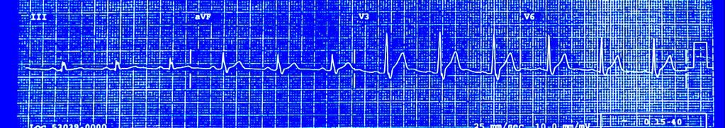
35 RBBB with Secondary ST-T Wave Changes

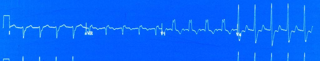
36 LBBB with Secondary ST-T Wave Changes
37 RBBB with Anteroseptal Infarction


38 LBBB with Exaggerated ST Elevation: Anteroseptal/ lateral Infarction > 5 mm
39 LBBB with Lateral Infarction

40 Left BBB with Anteroseptal Ischemia


41 Left BBB Primary ST T Wave depression Primary STT Wave elevation Exaggerated ST segment elevation
42 Should We Treat All LBBB Patients with Lytics? American Heart Association 2005: Treat new or presumably new LBBB Literature: not necessarily Yes! 35% with new LBBB and chest pain did not have an MI (Kontos) 48% with LBBB not known to be old did not have an MI (Edhouse) Kontos MC, et al., Ann Emerg Med. 37:431-8, 2001 Edhouse, et al., J Accid Emerg Med. 16:331-5, 1999.
43 Bottom Line Treating all LBBB as if AMI risks unnecessary thrombolytics in 1/3 to 1/2 Reasonable if low risk of complications Sgarbossa criteria are helpful If present, thrombolytics indicated If criteria absent serial ECGs for dynamic changes Transfer for cath if < 90 mins
44 Take-Home Messages: You can diagnose acute MI or ischemia with BBB Secondary (normal) ST-T changes are opposite the last part of the QRS Primary (abnormal) ST-T changes mean ischemic or infarction, and go in the same direction as the last part of the QRS
45 Take-Home Messages: One-half to two-thirds of patients with chest pain and new LBBB (or not known to be old) have AMI Reperfusion therapy is recommended Serial ECGs may clarify Immediate angiogram is preferred
46 Why, your fever s way down.
47
48 Next two slides good but no time in Buenos Aires
49 LBBB with Evolving MI 182 patients with LBBB and acute MI New LBBB: Sens = 46%, Spec = 65% Concordant ST-segment elevation or depression (Sgarbossa criteria) Specificity = 100% Positive predictive values = 100% Sensitivity for ST elevation = 8% Sensitivity for ST depression = 17% Kontos MC, et al., Ann Emerg Med. 37:431-8, 2001
50 ECG of Evolving MI with LBBB Of patients with acute MI and LBBB, the LBBB was NEW (from their MI) in only 46%. If we only treated NEW LBBB, we d miss treating 54% of patients who needed it. Conversely, only 65% of patients with NEW LBBB actually had MI. So, if we treated everyone with new LBBB and chest pain we d treat 35% unnecessarily.
51 ECG of LBBB without MI 124 patients with LBBB and no MI Only 1 had primary ST segment depression anteriorly Only 1 had primary ST segment elevation 9 had exaggerated ST segment elevation > 5 mm Sgarbossa criteria are sufficiently specific (few false positives) Madias JE, et al., Clin Cardiol. 24:652-5, 2001
52 ECG of LBBB with Evolving MI 414 LBBB patients with AMI vs. 85 without Sgarbossa criteria uncommon with AMI: Concordant ST-segment elevation: 6% Concordant ST-segment depression: 3% Discordant ST-segment elevation: 19% Concordant ST elevation (LR 5.3) and ST depression in V 1 -V 3 highly specific for AMI, but not sensitive Gula LJ et al., Coron Artery Dis. 14:387-93, 2003
53 17% sens, 94% spec International Journal of Cardiology 78 (2001) locate / ijcard ECG criteria in diagnosis of acute myocardial infarction in the presence of left bundle branch block Gunnar Gunnarsson*, Peter Eriksson, Mikael Dellborg Clinical Experimental Research Laboratory, Department of Medicine, Sahlgrenska University Hospital /O stra, Go teborg, Sweden Received 24 October 2000; received in revised form 30 December 2000; accepted 10 January 2001 Abstract Background: The diagnosis of acute myocardial infarction in the presence of left bundle branch block is difficult. Recently a diagnostic ECG scoring system was suggested, showing good diagnostic abilities. This scoring system has never been tested in a prospective manner; we have done so and investigated if it might bear prognostic information. Methods: A prospective multi-centre study. Consecutive patients with left bundle branch block and suspicion of acute myocardial infarction, admitted to 14 Swedish coronary care units. Recruitment from March 1996 to December ECG registered on admission and after h. Results: One hundred and fifty-eight patients were included, mean age 74.9 years. Seventy-six patients (48%) had an acute myocardial infarction. The proposed cut-off total score of $3 of the ECG scoring system for the diagnosis of acute myocardial infarction had a sensitivity of 17.1% (95% CI %) and specificity of 94.0% (95% CI %). Clinical judgement of acute myocardial infarction resulted in a sensitivity of 15.8% (95% CI %) and specificity of 96.0% (CI %). No difference was seen in 3-month or 1-year survival between those with total ECG score $3 versus total score,3. Conclusion: The diagnostic abilities of the proposed ECG criteria are low and not better than the clinical judgement. The criteria are therefore not suitable for screening patients with suspicion of acute myocardial infarction in the presence of left bundle branch block, nor do they seem to identify high risk patients. Ó 2001 Elsevier Science Ireland Ltd. All rights reserved.

54


55 ACCOUNTING CLERK 4 DRUG 3 REHAB POISON 2 CONTROL PUBLIC 5 WORKS SOCIAL 4 SERVICE Second Floor Please

56 RBBB with Anteroseptal Ischemia
57 One of these is a prescription and one is a receipt from a Chinese restaurant.
Goals: Widen Your Understanding of the Wide QRS!
 Goals: Widen Your Understanding of the Wide QRS! 1. Describe an approach to diagnosis of LBBB 2. Describe the predictive value of New LBBB 3. Describe the ST segment changes that are diagnostic of AMI
Marcin Dada, MD December 03, 2013
 STEMI Imposters Marcin Dada, MD December 03, 2013 Marcin Dada, MD Associate Director, Chest Pain Center Hartford Hospital, Hartford, CT Member, AHA Mission Lifeline Steering Committee Outline of Topics
Electrocardiography for Healthcare Professionals. Chapter 14 Basic 12-Lead ECG Interpretation
 Electrocardiography for Healthcare Professionals Chapter 14 Basic 12-Lead ECG Interpretation 2012 The Companies, Inc. All rights reserved. Learning Outcomes 14.1 Discuss the anatomic views seen on a 12-lead
Hot Topics in Cardiac Arrest. Should the patient go To the Cath Lab?
 Hot Topics in Cardiac Arrest Should the patient go To the Cath Lab? Tim Russert 1950-2008 Host of NBC s Meet the Press Sudden Cardiac Arrest : Autopsy showed plaque rupture in his LAD ( per LA Times,
Cardiac Ischemia ECG Workshop
 Cardiac Ischemia ECG Workshop Classic, Confusing, and Confounding Patterns Amal Mattu, MD, NE Professor and Vice Chair Department of Emergency Medicine University of Maryland School of Medicine amalmattu@comcast.net
Myocardial Infarction. Reading Assignment (p66-78 in Outline )
") Myocardial Infarction Reading Assignment (p66-78 in Outline ) Objectives 1. Why do ST segments go up or down in ischemia? 2. STEMI locations and culprit vessels 3. Why 15-lead ECGs? 4. What s up with avr?
Myocardial Infarction Reading Assignment (p66-78 in Outline ) Objectives 1. Why do ST segments go up or down in ischemia? 2. STEMI locations and culprit vessels 3. Why 15-lead ECGs? 4. What s up with avr?
A few new tools for better detection and understanding of STEMIs in the field.
 A few new tools for better detection and understanding of STEMIs in the field. Let s talk, prep and placement. Try to shoot for quality, consistency and no artifact! (looking sometimes for 1 or 2 mm changes)
Preface: Wang s Viewpoints
 AHA/ACCF/HRS Recommendations for the Standardization and Interpretation of the Electrocardiogram: Part IV, Ischemia and Infarction Presented by: WANG, TZONG LUEN, MD, PhD, JM, FACC, FESC, FCAPSC Professor,
All About STEMIs. Presented By: Brittney Urvand, RN, BSN, CCCC. Essentia Health Fargo Cardiovascular Program Manager.
 All About STEMIs Presented By: Brittney Urvand, RN, BSN, CCCC Essentia Health Fargo Cardiovascular Program Manager Updated 10/2/2018 None Disclosures Objectives Identify signs and symptoms of a heart attack
Section V. Objectives
 Section V Landscape of an MI Objectives At the conclusion of this presentation the participant will be able to Outline a systematic approach to 12 lead ECG interpretation Demonstrate the process for determining
The Fundamentals of 12 Lead EKG. ECG Recording. J Point. Reviewing the Cardiac Conductive System. Dr. E. Joe Sasin, MD Rusty Powers, NRP
 The Fundamentals of 12 Lead EKG Dr. E. Joe Sasin, MD Rusty Powers, NRP SA Node Intranodal Pathways AV Junction AV Fibers Bundle of His Septum Bundle Branches Purkinje System Reviewing the Cardiac Conductive
Understanding the 12-lead ECG, part II
 Bundle-branch blocks Understanding the 12-lead ECG, part II Most common electrocardiogram (ECG) abnormality Appears as a wider than normal S complex Occurs when one of the two bundle branches can t conduct
12 Lead Interpretation
 12 Lead Interpretation Objectives Ischemia, injury and infarction ECG complex review J point ST segment STEMI recognition Ischemia to Infarct Infarction is an evolving process As the infarct evolves ECG
Foundations EKG I - Unit 1 Summary
 Foundations EKG I - Unit 1 Summary The accurate diagnosis of ST elevation myocardial infarction (STEMI) is one of the most time critical duties in the practice of EM. Diagnosis is not always easy so guidelines
12 Lead ECG. Presented by Rebecca Sevigny BSN, RN Professional Practice & Development Dept.
 12 Lead ECG Presented by Rebecca Sevigny BSN, RN Professional Practice & Development Dept. Two Main Coronary Arteries RCA LCA which branches into Left Anterior Descending Circumflex Artery Two Main Coronary
INTERPRETAZIONE ECG NEL PAZIENTE CON SOSPETTO STEMI
 INTERPRETAZIONE ECG NEL PAZIENTE CON SOSPETTO STEMI Giacomo Veronese Scuola di Specializzazione Medicina d Emergenza e Urgenza Università Milano-Bicocca Siete d accordo se vi propongo per una relazione..
12 Lead EKG. The Basics
 12 Lead EKG The Basics Objectives Demonstrate proper 12 EKG lead placement Determine electrical axis Identify ST and T wave changes as they relate to myocardial ischemia Describe possible complications
Electrocardiography. Hilal Al Saffar College of Medicine,Baghdad University
 Electrocardiography Hilal Al Saffar College of Medicine,Baghdad University Which of the following is True 1. PR interval, represent the time taken for the impulse to travel from SA node to AV nose. 2.
Acute Coronary Syndromes Unstable Angina Non ST segment Elevation MI (NSTEMI) ST segment Elevation MI (STEMI)
 ST segment Elevation MI (STEMI)") Leanna R. Miller, RN, MN, CCRN-CSC, PCCN-CMC, CEN, CNRN, CMSRN, NP Education Specialist LRM Consulting Nashville, TN Objectives Evaluate common abnormalities that mimic myocardial infarction. Identify
Leanna R. Miller, RN, MN, CCRN-CSC, PCCN-CMC, CEN, CNRN, CMSRN, NP Education Specialist LRM Consulting Nashville, TN Objectives Evaluate common abnormalities that mimic myocardial infarction. Identify
12 LEAD EKG BASICS. By: Steven Jones, NREMT P CLEMC
 12 LEAD EKG BASICS By: Steven Jones, NREMT P CLEMC ECG Review Waves and Intervals P wave: the sequential activation (depolarization) of the right and left atria QRS complex: right and left ventricular
MICS OF MYOCARDIAL ISCHEMIA AND INFARCTION REVISED FOR LAS VEGAS
 ECG MIMICS OF MYOCARDIAL ISCHEMIA AND INFARCTION 102.06.05 Tzong-Luen Wang MD, PhD, JM, FESC, FACC Professor. Medical School, Fu-Jen Catholic University Chief, Emergency Department, Shin-Kong Wu Ho-Su
12 Lead Electrocardiogram (ECG) PFN: SOMACL17. Terminal Learning Objective. References
 PFN: SOMACL17. Terminal Learning Objective. References") 12 Lead Electrocardiogram (ECG) PFN: SOMACL17 Slide 1 Terminal Learning Objective Action: Communicate knowledge of 12 Lead Electrocardiogram (ECG) Condition: Given a lecture in a classroom environment
12 Lead Electrocardiogram (ECG) PFN: SOMACL17 Slide 1 Terminal Learning Objective Action: Communicate knowledge of 12 Lead Electrocardiogram (ECG) Condition: Given a lecture in a classroom environment
12 Lead ECG Interpretation: Color Coding for MI s
 12 Lead ECG Interpretation: Color Coding for MI s Anna E. Story, RN, MS Director, Continuing Professional Education Critical Care Nurse Online Instructional Designer 2004 Anna Story 1 Objectives review
Masqueraders of STEMI
 Masqueraders of STEMI Steven M. Costa, M.D. Assistant Professor Department of Medicine Division of Cardiology Scott & White Memorial Hospital and Clinic Texas A&M University Health Science Center Disclosures
12 Lead Acquisition and Interpretation APRIL 23 11:00 AM
 12 Lead Acquisition and Interpretation APRIL 23 11:00 AM Presented by : Jennifer Robson, Prehospital Care Specialist Dr. Don Eby, Local Medical Director Objectives Upon completion of this webinar, you
ECG Basics Sonia Samtani 7/2017 UCI Resident Lecture Series
 ECG Basics Sonia Samtani 7/2017 UCI Resident Lecture Series Agenda I. Introduction II.The Conduction System III.ECG Basics IV.Cardiac Emergencies V.Summary The Conduction System Lead Placement avf Precordial
Abnormalities Caused by Left Bundle Branch Block
 Marquette University e-publications@marquette Physician Assistant Studies Faculty Research and Publications Physician Assistant Studies, Department 12-17-2010 Abnormalities Caused by Left Bundle Branch
Disclosures. STEMI:To Call or Not to Call. Disclosures 9/18/2017. Alternate Title: Hey Doc, If you re not doing anything Saturday Night
 STEMI:To Call or Not to Call Disclosures No financial disclosures September, 2017 Frederick James Trip Meine III MD, FACC, FSCAI Cape Fear Heart Associates, Wilmington, NC Disclosures Alternate Title:
By the end of this lecture, you will be able to: Understand the 12 lead ECG in relation to the coronary circulation and myocardium Perform an ECG
 By the end of this lecture, you will be able to: Understand the 12 lead ECG in relation to the coronary circulation and myocardium Perform an ECG recording Identify the ECG changes that occur in the presence
Relax and Learn At the Farm 2012
 Relax and Learn At the Farm 2012 Session 2: 12 Lead ECG Fundamentals 101 Cynthia Webner DNP, RN, CCNS, CCRN-CMC, CHFN Though for Today Mastery is not something that strikes in an instant, like a thunderbolt,
ECG CONVENTIONS AND INTERVALS
 1 ECG Waveforms and Intervals ECG waveforms labeled alphabetically P wave== represents atrial depolarization QRS complex=ventricular depolarization ST-T-U complex (ST segment, T wave, and U wave)== V repolarization.
Family Medicine for English language students of Medical University of Lodz ECG. Jakub Dorożyński
 Family Medicine for English language students of Medical University of Lodz ECG Jakub Dorożyński Parts of an ECG The standard ECG has 12 leads: six of them are considered limb leads because they are placed
10 ECGs No Practitioner Can Afford to Miss. Objectives
 10 ECGs No Practitioner Can Afford to Miss Mary L. Dohrmann, MD Professor of Clinical Medicine Division of Cardiovascular Medicine University of Missouri School of Medicine No disclosures Objectives 1.
Left posterior hemiblock (LPH)/
/") ECG OF THE MONTH Left Postero-inferior Depolarization Delay Keywords Electrocardiography Intraventricular conduction delay, Inferoposterior hemiblock, Left posterior fascicular block, Left posterior hemiblock
ECG OF THE MONTH Left Postero-inferior Depolarization Delay Keywords Electrocardiography Intraventricular conduction delay, Inferoposterior hemiblock, Left posterior fascicular block, Left posterior hemiblock
Preface: Wang s Viewpoints
 AHA/ACCF/HRS Recommendations for the Standardization and Interpretation of the Electrocardiogram: Ischemia and Infarction 103.10.07 Presented by: WANG, TZONG LUEN, MD, PhD, JM, FACC, FESC, FCAPSC Professor,
Pennsylvania Academy of Family Physicians Foundation & UPMC 43rd Refresher Course in Family Medicine CME Conference March 10-13, 2016
 Pennsylvania Academy of Family Physicians Foundation & UPMC 43rd Refresher Course in Family Medicine CME Conference March 10-13, 2016 Disclosures: EKG Workshop Louis Mancano, MD Speaker has no disclosures
Disclosure. 3. ST depression indicative of ischemia is most commonly observed in leads: 1. V1-V2. 2. I and avl 3. V
 Interpreting Stress Induced Ischemia by ECG, Bundle Branch Block & Arrhythmias Disclosure Gregory S Thomas MD, MPH Medical Director, MemorialCare Heart & Vascular Institute, Long Beach Memorial Astellas
ACUTE CORONARY SYNDROME
 12 LEAD ECG INTERPRETATION in ACUTE CORONARY SYNDROME WAYNE W RUPPERT, CVT, CCCC, NREMT-P Cardiovascular Clinical Coordinator Bayfront Health Seven Rivers Crystal River, FL Education Specialist St. Joseph
ECG ABNORMALITIES D R. T AM A R A AL Q U D AH
 ECG ABNORMALITIES D R. T AM A R A AL Q U D AH When we interpret an ECG we compare it instantaneously with the normal ECG and normal variants stored in our memory; these memories are stored visually in
ECG pre-reading manual. Created for the North West Regional EMET training program
 ECG pre-reading manual Created for the North West Regional EMET training program Author:- Dr Juan Carlos Ascencio-Lane juan.ascencio-lane@ths.tas.gov.au 1 Disclaimer This handbook has been created for
STAT 12 Lead ECG Workshop: Basics & ACS
 STAT 12 Lead ECG Workshop: Basics & ACS Part 2: Acute Coronary Syndrome WAYNE W RUPPERT, CVT, CCCC, NREMT-P Cardiovascular Coordinator Bayfront Health Seven Rivers Crystal River, Florida Interventional
Medical Management of Acute Coronary Syndrome: The roles of a noncardiologist. Norbert Lingling D. Uy, MD Professor of Medicine UERMMMCI
 Medical Management of Acute Coronary Syndrome: The roles of a noncardiologist physician Norbert Lingling D. Uy, MD Professor of Medicine UERMMMCI Outcome objectives of the discussion: At the end of the
Bundle Branch & Fascicular Blocks. Reading Assignment (p53-58 in Outline )
") Bundle Branch & Fascicular Blocks Reading Assignment (p53-58 in Outline ) Objectives 1. QRS analysis of Right and Left BBB 2. Uncomplicated vs complicated BBB 3. Diagnosis of RBBB with LAFB and LPFB 4.
Bundle Branch & Fascicular Blocks Reading Assignment (p53-58 in Outline ) Objectives 1. QRS analysis of Right and Left BBB 2. Uncomplicated vs complicated BBB 3. Diagnosis of RBBB with LAFB and LPFB 4.
DR QAZI IMTIAZ RASOOL OBJECTIVES
 PRACTICAL ELECTROCARDIOGRAPHY DR QAZI IMTIAZ RASOOL OBJECTIVES Recording of electrical events in heart Established electrode pattern results in specific tracing pattern Health of heart i. e. Anatomical
THE emergency physician (EP), frequently the
, frequently the") 1256 ST-SEGMENT ELEVATION Brady et al. INTERPRETATION OF ST-SEGMENT ELEVATION Errors in Emergency Physician Interpretation of ST-segment Elevation in Emergency Department Chest Pain Patients WILLIAM J.
1256 ST-SEGMENT ELEVATION Brady et al. INTERPRETATION OF ST-SEGMENT ELEVATION Errors in Emergency Physician Interpretation of ST-segment Elevation in Emergency Department Chest Pain Patients WILLIAM J.
ECG Cases and Questions. Ashish Sadhu, MD, FHRS, FACC Electrophysiology/Cardiology
 ECG Cases and Questions Ashish Sadhu, MD, FHRS, FACC Electrophysiology/Cardiology 32 yo female Life Insurance Physical 56 yo male with chest pain Terminology Injury ST elevation Ischemia T wave inversion
Part One Objectives. Don t Worry About It. All done for you Paper Speed 25 mm/sec Calibration 1 mv charge over 20 ms = 10 mm tall Lincoln Hat
 12-lead and ACS Review North Lyon Refresher Part One Objectives 12 lead ECG Basics Anatomy and Physiology STEMI Diagnosis Types of MI ACS Review STEMI System and Interventional Cardiology Review The Value
TOPICS IN EMERGENCY MEDICINE SEMI-FINAL
 RISK ASSESSMENT IN PATIENTS WITH CHEST PAIN Nora Goldschlager, M.D. FACP, FACC, FAHA, FHRS Cardiology - San Francisco General Hospital UCSF Disclosures: None 1 CHEST PAIN NOT DUE TO MYOCARDIAL ISCHEMIA
What s New in IV Conduction? (Quadrafascicular, not Trifascicular)
") What s New in IV Conduction? (Quadrafascicular, not Trifascicular) Frank Yanowitz, MD Professor, University of Utah School of Medicine Medical Director, IHC ECG Services (Urban Central Region) http://ecg.utah.edu
What s New in IV Conduction? (Quadrafascicular, not Trifascicular) Frank Yanowitz, MD Professor, University of Utah School of Medicine Medical Director, IHC ECG Services (Urban Central Region) http://ecg.utah.edu
electrocardiograms Analysis of emergency department interpretation of
 Journal of Accident and Emergency Medicine 1994 11, 149-153 Correspondence: E.R. Snoey, Department of Emergency Medicine, Highland General Hospital 1411 East 31st Street, Oakland, California 94602, USA
Myocardial Infarction In Dr.Yahya Kiwan
 Myocardial Infarction In 2007 Dr.Yahya Kiwan New Definition Of Acute Myocardial Infarction The term of myocardial infarction should be used when there is evidence of myocardial necrosis in a clinical setting
ECG S: A CASE-BASED APPROACH December 6,
 ECG S: A CASE-BASED APPROACH December 6, 2018 1 Faculty Disclosure Faculty: Lorne Gula MD, FRCPC Professor, Western University Cardiologist, Hearth Rhythm Specialist Director, Electrophysiology Laboratory,
Different ECG patterns at presentation in ACS. D. Goldwasser F. Molina A. Bayes de Luna
 Different ECG patterns at presentation in ACS D. Goldwasser F. Molina A. Bayes de Luna Acute Coronary syndromes: The importance of the ECG There are two types of ACS: STE- ACS and Non STE-ACS The most
3/4/2018. March Martina Frost, PA C Desert Cardiology. Electricity moving towards/away from electrode create downward/upward directions of waves
 March 2018 Martina Frost, PA C Desert Cardiology Electricity moving towards/away from electrode create downward/upward directions of waves Frontal view Limb leads: I, II, III, avl, avf, (avr) Horizontal
Which Patients With Suspected Myocardial Ischemia and Left Bundle-Branch Block Should Receive Thrombolytic Agents?
 EDITORIAL: Which Patients With Suspected Myocardial Ischemia and Left Bundle-Branch Block Should Receive Thrombolytic Agents? From the Department of Emergency Medicine, Albert Einstein College of Medicine,
Acute Coronary Syndromes. Disclosures
 Acute Coronary Syndromes Disclosures I work for Virginia Garcia Memorial Health Center, Beaverton, OR. Jon Tardiff, BS, PA-C OHSU Clinical Assistant Professor And I am a medical editor for Jones & Bartlett
ECG Interpretation Cat Williams, DVM DACVIM (Cardiology)
") ECG Interpretation Cat Williams, DVM DACVIM (Cardiology) Providing the best quality care and service for the patient, the client, and the referring veterinarian. GOAL: Reduce Anxiety about ECGs Back to
ECG Interpretation Cat Williams, DVM DACVIM (Cardiology) Providing the best quality care and service for the patient, the client, and the referring veterinarian. GOAL: Reduce Anxiety about ECGs Back to
REtrive. REpeat. RElearn Design by. Test-Enhanced Learning based ECG practice E-book
 Test-Enhanced Learning Test-Enhanced Learning Test-Enhanced Learning Test-Enhanced Learning based ECG practice E-book REtrive REpeat RElearn Design by S I T T I N U N T H A N G J U I P E E R I Y A W A
Chapter 3 for 12 Lead Training -Precourse-
 ONTARIO BASE HOSPITAL GROUP Chapter 3 for 12 Lead Training -Precourse- Ontario Base Hospital Group Education Subcommittee 2008 TIME IS MUSCLE ONTARIO BASE HOSPITAL GROUP Introduction and Purpose Introduction
Conduction Problems / Arrhythmias. Conduction
 Conduction Problems / Arrhythmias Conduction Wolf-Parkinson White Syndrome (WPW) and Lown-Ganong-Levine (LGL): Atrial impulses bypass the AV node through an accessory pathway or bypass tract (bundle of
Stress ECG is still Viable in Suleiman M Kharabsheh, MD, FACC Consultant Invasive Cardiologist KFHI KFSHRC-Riyadh
 Stress ECG is still Viable in 2016 Suleiman M Kharabsheh, MD, FACC Consultant Invasive Cardiologist KFHI KFSHRC-Riyadh Stress ECG Do we still need stress ECG with all the advances we have in the CV field?
Solutions for Every Day Problems Cardiologists and the ECG: Are We Really That Good at It? Part II Daniel José Piñeiro Profesor Titular de Medicina,
 Solutions for Every Day Problems Cardiologists and the ECG: Are We Really That Good at It? Part II Daniel José Piñeiro Profesor Titular de Medicina, Universidad de Buenos Aires, Argentina Member, Membership
June 2009 CE. Site code # E-1209
 1 June 2009 CE Site code # 107200E-1209 Region X SOP Review ACS, Asystole/PEA, Bradycardia, VF/Pulseless VT, Conscious Sedation, Region X Field Triage Criteria Prepared by: Sharon Hopkins, RN, BSN, EMT-P
12 Lead ECG Interpretation
 12 Lead ECG Interpretation Julie Zimmerman, MSN, RN, CNS, CCRN Significant increase in mortality for every 15 minutes of delay! N Engl J Med 2007;357:1631-1638 Who should get a 12-lead ECG? Also include
The Efficient and Smart Methods for Diagnosis of SVT 대구파티마병원순환기내과정병천
 The Efficient and Smart Methods for Diagnosis of SVT 대구파티마병원순환기내과정병천 Differentiation Supraventricular Origin from Ventricular Origin on ECG. QRS-Complex Width. 1. Narrow QRS-Complex Tachycardia (
Name Authentication Date (Position or Committee) Quality & Patient Safety Steering. Meeting Minutes & 08/14 Committee
 Quality & Patient Safety Steering. Meeting Minutes & 08/14 Committee") Title: Document Number: Document Type: Affected Departments: Review Bodies: Revision/(Review) Dates: (Dates that included only a review, but no content 07/14 revision, are in parentheses) Effective Date:
Title: Document Number: Document Type: Affected Departments: Review Bodies: Revision/(Review) Dates: (Dates that included only a review, but no content 07/14 revision, are in parentheses) Effective Date:
VCU HEALTH SYSTEM EMERGENCY DEPARTMENT GUIDELINE
 VCU HEALTH SYSTEM EMERGENCY DEPARTMENT GUIDELINE SUBJECT: Care of the Chest Pain Patient in the Emergency Department FILE SECTION: VCUHS/ED Section: Please note: Clinical Practice Guideline Evidence-based
12-Lead ECG Interpretation. Kathy Kuznar, RN, ANP
 12-Lead ECG Interpretation Kathy Kuznar, RN, ANP The 12-Lead ECG Objectives Identify the normal morphology and features of the 12- lead ECG. Perform systematic analysis of the 12-lead ECG. Recognize abnormalities
12 Lead ECGs: Ischemia, Injury & Infarction. Kevin Handke NRP, FP-C, CCP, CMTE STEMI Coordinator Flight Paramedic
 12 Lead ECGs: Ischemia, Injury & Infarction Kevin Handke NRP, FP-C, CCP, CMTE STEMI Coordinator Flight Paramedic None Disclosures Objectives Upon completion of this program the learner will be able to
ECG Workshop. Nezar Amir
 ECG Workshop Nezar Amir Myocardial Ischemia ECG Infarct ECG in STEMI is dynamic & evolving Common causes of ST shift Infarct Localisation Left main artery occlusion: o diffuse ST-depression with ST elevation
UPDATE ON THE MANAGEMENTACUTE CORONARY SYNDROME. DR JULES KABAHIZI, Psc (Rwa) Lt Col CHIEF CONSULTANT RMH/KFH 28 JUNE18
 Lt Col CHIEF CONSULTANT RMH/KFH 28 JUNE18") UPDATE ON THE MANAGEMENTACUTE CORONARY SYNDROME DR JULES KABAHIZI, Psc (Rwa) Lt Col CHIEF CONSULTANT RMH/KFH 28 JUNE18 INTRODUCTION The clinical entities that comprise acute coronary syndromes (ACS)-ST-segment
UPDATE ON THE MANAGEMENTACUTE CORONARY SYNDROME DR JULES KABAHIZI, Psc (Rwa) Lt Col CHIEF CONSULTANT RMH/KFH 28 JUNE18 INTRODUCTION The clinical entities that comprise acute coronary syndromes (ACS)-ST-segment
Chapter 2 Practical Approach
 Chapter 2 Practical Approach There are beginners in electrocardiogram (ECG) analysis who are fascinated by a special pattern (e.g., a bundle-branch block or a striking Q wave) and thereby overlook other
12 Lead ECG Skills: Building Confidence for Clinical Practice. Presented By: Cynthia Webner, BSN, RN, CCRN-CMC. Karen Marzlin, BSN, RN,CCRN-CMC
 12 Lead ECG Skills: Building Confidence for Clinical Practice NTI 2009 Preconference Session 803 Presented By: Karen Marzlin, BSN, RN,CCRN-CMC 1 12 Lead ECG Fundamentals: The Starting Place for Linking
Acute Myocardial Infarction: Difference in the Treatment between Men and Women
 Quality Assurance in Hcahh Can, Vol. 5, No. 3, pp. 261-265,1993 Printed in Great Britain 1040-6166/93 $6.00 + 0.00 1993 Pergamon Press Ltd Acute Myocardial Infarction: Difference in the Treatment between
We are IntechOpen, the world s leading publisher of Open Access books Built by scientists, for scientists. International authors and editors
 We are IntechOpen, the world s leading publisher of Open Access books Built by scientists, for scientists 3,900 116,000 120M Open access books available International authors and editors Downloads Our
12 Lead ECG Interpretation: The Basics and Beyond
 12 Lead ECG Interpretation: The Basics and Beyond Cindy Weston, DNP, RN, CCRN, CNS-CC, FNP-BC Assistant Professor Texas A&M University College of Nursing cweston@tamhsc.edu Objectives Review the basics
Critical Review Form Therapy Objectives: Methods:
 Critical Review Form Therapy Clinical Trial Comparing Primary Coronary Angioplasty with Tissue-Plasminogen Activator for Acute Myocardial Infarction (GUSTO-IIb), NEJM 1997; 336: 1621-1628 Objectives: To
ECG in coronary artery disease. By Sura Boonrat Central Chest Institute
 ECG in coronary artery disease By Sura Boonrat Central Chest Institute EKG P wave = Atrium activation PR interval QRS = Ventricle activation T wave= repolarization J-point EKG QT interval Abnormal repolarization
Basic electrocardiography reading. R3 lee wei-chieh
 Basic electrocardiography reading R3 lee wei-chieh The Normal Conduction System Lead Placement avf Limb Leads Precordial Leads Interpretation Rate Rhythm Interval Axis Chamber abnormality QRST change What
, David Stultz, MD.
 http://www.dilbert.com EKG Rounds Handouts available at http://www.drstultz.com January 5, 2004 David Stultz, MD Cardiology Fellow, PGY 4 Overview of Topics How to read an EKG Normal EKG Determination
4/14/15. The Electrocardiogram. In jeopardy more than a century after its introduction by Willem Einthoven? Time for a revival. by Hein J.
 The Electrocardiogram. In jeopardy more than a century after its introduction by Willem Einthoven? Time for a revival. by Hein J. Wellens MD 1 Einthoven, 1905 The ECG! Everywhere available! Easy and rapid
The Electrocardiogram part II. Dr. Adelina Vlad, MD PhD
 The Electrocardiogram part II Dr. Adelina Vlad, MD PhD Basic Interpretation of the ECG 1) Evaluate calibration 2) Calculate rate 3) Determine rhythm 4) Determine QRS axis 5) Measure intervals 6) Analyze
Considerations about the polemic J point location
 Considerations about the polemic J point location V) The J-point of the electrocardiogram Approximate point of convergence between the end of QRS complex and the onset of ST segment. It is considered the
Comments or Questions? me:
 Comments or Questions? Email me: amalmattu@comcast.net Interested in short video tutorials on electrocardiography? Check out www.ecgweekly.com Subscription fee < cost of a cup of coffee/week Covers every
6/19/2018. Background Athlete s heart. Ultimate question. Applying the International Criteria for ECG
 Applying the International Criteria for ECG Interpretation in Athletes to a preparticipation screening program DAVE SIEBERT, MD, CAQSM ASSISTANT PROFESSOR DEPARTMENT OF FAMILY MEDICINE UNIVERSITY OF WASHINGTON
CAN T MISS ECG FINDINGS L. THOMAS RICHARDS, MD ASSISTANT PROFESSOR OF EMERGENCY MEDICINE
 Topics in Emergency Medicine 2010 CAN T MISS ECG FINDINGS L. THOMAS RICHARDS, MD ASSISTANT PROFESSOR OF EMERGENCY MEDICINE OBJECTIVES Examine three common presentations to the ED which compel the EM provider
ECG Practice Strips Discussion part 1:
 ECG Practice Strips Discussion part 1: The first 20 strips are for teaching various abnormalities of the morphology of the waves of the ECG. Strips 21 and following are for teaching some abnormalities
ECG (MCQs) In the fundamental rules of the ECG all the following are right EXCEP:
 In the fundamental rules of the ECG all the following are right EXCEP:") ECG (MCQs) 2010 1- In the fundamental rules of the ECG all the following are right EXCEP: a- It is a biphasic record of myocardial action potential fluctuations. b- Deflection record occurs only during
ECG (MCQs) 2010 1- In the fundamental rules of the ECG all the following are right EXCEP: a- It is a biphasic record of myocardial action potential fluctuations. b- Deflection record occurs only during
December 2018 Tracings
 Tracings Tracing 1 Tracing 4 Tracing 1 Answer Tracing 4 Answer Tracing 2 Tracing 5 Tracing 2 Answer Tracing 5 Answer Tracing 3 Tracing 6 Tracing 3 Answer Tracing 6 Answer Questions? Contact Dr. Nelson
International Journal of Medical Science in Clinical Research and Review
 International Journal of Medical Science in Clinical Research and Review Available Online at http://www.ijmscrr.in Volume 01 Issue 01 2018 Not all new Left Bundle Branch Block (LBBB) requires Cardiac Catheterization
General Introduction to ECG. Reading Assignment (p2-16 in PDF Outline )
") General Introduction to ECG Reading Assignment (p2-16 in PDF Outline ) Objectives 1. Practice the 5-step Method 2. Differential Diagnosis: R & L axis deviation 3. Differential Diagnosis: Poor R-wave progression
General Introduction to ECG Reading Assignment (p2-16 in PDF Outline ) Objectives 1. Practice the 5-step Method 2. Differential Diagnosis: R & L axis deviation 3. Differential Diagnosis: Poor R-wave progression
15 16 September Seminar W10O. ECG for General Practice
 15 16 September 2012 Seminar W10O ECG for General Practice Speaker: Ms Natasha Eaton ECG for General Practice Speaker: Natasha Eaton Cardiac CNC Executive Representative Electrocardiography The graphic
Acute chest pain and ECG need for immediate coronary angiography?
 Acute chest pain and ECG need for immediate coronary angiography? Kjell Nikus, MD, PhD Heart Center, Tampere University Hospital, Finland and Samuel Sclarovsky, MD, PhD Tel Aviv University, Israel There
PECTUS EXCAVATUM WITH SPONTANEOUS TYPE 1 ECG BRUGADA PATTERN OR BRUGADA LIKE PHENOTYPE: ANOTHER BRUGADA ECG PHENOCOPY
 PECTUS EXCAVATUM WITH SPONTANEOUS TYPE 1 ECG BRUGADA PATTERN OR BRUGADA LIKE PHENOTYPE: ANOTHER BRUGADA ECG PHENOCOPY ANDRÉS RICARDO PÉREZ RIERA MD Chief of the Sector of Electro-Vectocardiography of the
12 Lead EKG Chapter 4 Worksheet
 Match the following using the word bank. 1. A form of arteriosclerosis in which the thickening and hardening of the vessels walls are caused by an accumulation of fatty deposits in the innermost lining
ECG Underwriting Puzzler Dr. Regina Rosace AVP & Medical Director
 December 2018 ECG Underwriting Puzzler Dr. Regina Rosace AVP & Medical Director To obtain best results Select Slide Show from the ribbon at the top of your PowerPoint screen Select From Beginning on the
Arrhythmic Complications of MI. Teferi Mitiku, MD Assistant Clinical Professor of Medicine University of California Irvine
 Arrhythmic Complications of MI Teferi Mitiku, MD Assistant Clinical Professor of Medicine University of California Irvine Objectives Brief overview -Pathophysiology of Arrhythmia ECG review of typical
Unfortunately, STE is a not an uncommon finding on the ECG of the chest pain patient; its cause infrequently involves AMI.
 Brandon Oto, NREMT-B Unfortunately, STE is a not an uncommon finding on the ECG of the chest pain patient; its cause infrequently involves AMI. Brady et al., Electrocardiographic STsegment elevation: correct
ECG Interactive Session
 SA HEART 2018 PRE-CONGRESS WORKSHOP 4 OCTOBER 2018 ECG Interactive Session Ashley Chin Cardiologist/Electrophysiologist University of Cape Town Groote Schuur Hospital ECG 1 Which ONE of the following is
Other 12-Lead ECG Findings
 Other 12-Lead ECG Findings Left Atrial Enlargement Left atrial enlargement is illustrated by increased P wave duration in lead II, top ECG, and by the prominent negative P terminal force in lead V1, bottom