Echo in Pulmonary HTN
|
|
|
- Leslie Wilcox
- 6 years ago
- Views:
Transcription


1 Echo in Pulmonary HTN Steven A. Goldstein MD FACC FASE Professor of Medicine Georgetown University Medical Center MedStar Heart Institute Washington Hospital Center Monday, October 10, 2017
 Prognostic importance Management decisions (eg operability in congenital HD,")
2 Pulmonary Artery Pressure Clinical Importance Responsible for symptoms and disability (LV disease, valvular disease, etc) Responsible for hemodynamic consequences (acute and chronic lung disease; TR; d CVP) Prognostic importance Management decisions (eg operability in congenital HD, MV disease)
3 Noninvasive Assessment of PA Pressure ECG CHEST X-RAY PHYSICAL EXAM

4 RV Pulmonary Circulation Unit Degree of pulm HTN does not strongly correlate with symptoms or survival RV size, RV mass, and RA pressure reflect functional status and are strong predictors of survival do
5 Normal Right Heart Hemodynamics RVSP/PASP Echo Mean PAP PAEDP RAP PVR < 36 mm Hg* 8 20 mm Hg 4 12 mm Hg 0 5 mm Hg < WU * (up to 40 mm Hg in older and obese patients)
6 % Increase Association of Systemic and PA Pressure with Age PASP SBP Age in quartiles (years) Lam et al. Circulation 2009;119:
7 Figure 2. Association of systemic pressure and PASP with age. Normal Resting Values Guidelines for the Echocardiographic Assessment of the Right Heart in Adults (ASE, EAE, ESC, CSE) Peak TR velocity m/s Peak systolic pressure 35 or 36 mm Hg (assuming an RA pressure of 3 to 5 mm Hg) * * This value may increase with age and increasing BSA... Rudski J Am Soc Echocardiogr 2010;23: Badesch J Am Coll Cardiol 2009;54:S55-66
8 Pulmonary Hypertension Mean PAP > 25 mm Hg PVR > 3 Wood units PCWP 15 mm Hg Rudski J Am Soc Echocardiogr 2010;23:
 Evaluate end effects Determine prognosis (RA pressure, mean PA pressure, large pericardial")
9 Pulmonary Hypertension Role of Echocardiography Diagnose pulmonary HTN Determine etiology (Left heart disease, MV disease, congenital HD, etc) Quantitate PA pressures (PASP, PADP, PA mean ) Evaluate end effects Determine prognosis (RA pressure, mean PA pressure, large pericardial effusion)

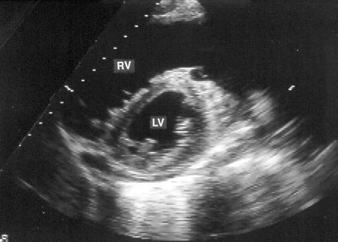
10 Pulmonary Hypertension Echo Findings 1. Right ventricular hypertrophy and/or dilatation 2. Abnl shape of LV in short axis ("D-shaped") 3. Right atrial dilatation 4. Dilated pulmonary artery 5. Abnormal systolic time intervals a. Prolonged RPEP/RVET b. Increased PV c - TV o interval 6. Abnormal pulmonic valve motion (M-mode)
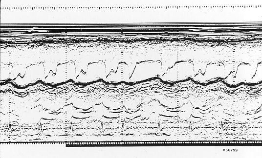
11 M-Mode
12 Pulmonary Hypertension M-Mode Echo Signs diminished "a" dip e f a b e rapid opening slope (b-c) c d flattened e-f slope Normal systolic notch
13

14 Flattened Ventricular Septum (D-Shaped )
15

16 Dilated coronary sinus Increased RA pressure
17 Determination of PA Pressure Echo-Doppler Methods 1. TR jet velocity method 2. Pulmonary acceleration time 3. Pulmonic regurgitant jet method 4. RV isovolumic relaxation time
18 TR Jet
19 Estimation of RV Systolic Pressure (RVSP) From Maximum Transtricuspid Gradient JVP P RVSP RVSP = P + JVP


20 Peak Velocity of Tricuspid Regurgitant Jet Determination of RV-RA pressure gradient RV-RA Gradient = 64 mmhg Est. of RA Pressure = 10 mmhg Pulm Art Pressure = 74 mmhg

21 Doppler Estimation of RV Systolic Pressure Simultaneous Doppler and Cath Tracings Currie JACC 6:750(1985)
 Max Gradient")
22 Max Gradient (Doppler), mmhg Doppler Estimation of RV Pressure Simultaneous Cath and Doppler Currie JACC 6:750(1981) Max Gradient (catheter), mmhg

23 Peak TR Jet Velocity

24 3 Methods of Estimating Right Atrial Pressure 1. Assume RA pressure of 5, 10, 15, or 20 mmhg 2. Clinical estimate of RA pressure (JVP) 3. IVC "collapsibility index"
 50% insp collapse < 50% insp collapse No collapse 6 10 mm Hg 10 15 mm Hg > 15 mm Hg Lang, et al Quantitation Guidelines J Am Soc Echocardiogr")
25 Estimation of Mean RA Pressure 2005 IVC size Collapsibility Index RA Pressure Normal IVC ( 1.7 cm) 50% decrease 0 5 mm Hg Dilated IVC (> 1.7 cm) 50% insp collapse < 50% insp collapse No collapse 6 10 mm Hg mm Hg > 15 mm Hg Lang, et al Quantitation Guidelines J Am Soc Echocardiogr 2005;17:
26 Estimation of Mean RA Pressure 2015 Normal RAP (0-5 [3] mm Hg) Intermediate (5-10 [8] mm Hg) High RAP (15 mm Hg) IVC diameter 2.1 cm 2.1 cm > 2.1 cm > 2.1 cm Collapse with sniff > 50% < 50% > 50% < 50% Lang, et al Quantitation Guidelines J Am Soc Echocardiogr 2015;28(1):1-39
27 Evaluation of RA Pressure IVC with insp RA pressure (cm) (%) (mm Hg) <2.1 >50% 0-5 (3) >2.1 <50% 5-10 (8) <2.1 >50% 5-10 (8) >2.1 <50% (15) Lang J Am Soc Echocardiogr 2015;28(1):1-39 Cardiac Chamber Quantification Guidelines (ASE/EAC)

28 IVC from Subcostal Long-AxisView IVC max IVC min IVC measured perpendicular to its long-axis Inner edge-to-inner edge Just proximal to junction of IVC with hepatic vein Or 1 2 cm proximal to IVC entrance into RA


29 IVC from Subcostal Long-Axis View End-expiration End-inspiration (with or without sniff)

30 Subcostal View M-mode Place cursor perpendicular to IVC Just proximal to junction b/w IVC and hepatic vein Use slow sweep speed to include 1 or 2 respirations

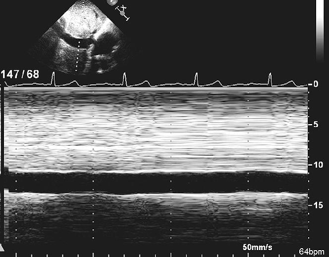
31 IVC Collapse

32 Normal size, normal collapse RA pressure 3 mm Hg
33 Mildly dilated, normal collapse RA pressure 8 mm Hg

34 Dilated, minimal or no collapse RA pressure 15 mm Hg
35 Limitations of "Sniff" Test Requires patient cooperation Tachypneic patients Mechanical ventilation Lack of standardization of insp effort Decrease sweep speed to include 1 or more resp cycles
36 Secondary Indices for RA Pressure Restrictive filling (TV inflow pattern) Tricuspid E/e > 6 Diastolic flow predominance in hepatic veins
 D VTI = Diastolic velocity time integral (or peak velocity)")
37 PW Doppler from Hepatic Vein SFF = systolic filling fraction (%) = S VTI S VTI + D VTI S VTI = Systolic velocity time integral (or peak velocity) D VTI = Diastolic velocity time integral (or peak velocity) x100

38 J Am Soc Echocardiogr 2015;28:1420-7
39 Impact of Body Size on IVC Parameters J Am Soc Echocardiogr 2015;28:1420-7

40 Limitations of "Sniff" Test Requires patient cooperation Tachypneic patients Mechanical ventilation Lack of standardization of insp effort Decrease sweep speed to include 1 or more resp cycles
 RA pressure estimate in ventilated patients Presence of pulmonic stenosis Inadequate signal in COPD patients Wide-open TR ( free TR")
41 Estimation of PA Pressure Limitations of TR Jet Method Absence of detectable TR (10%) Nonparallel intercept angle of TR jet Misidentification of jet signal (AS, MR) RA pressure estimate in ventilated patients Presence of pulmonic stenosis Inadequate signal in COPD patients Wide-open TR ( free TR )
42 Difference b/w Echo-Doppler and Right Heart Cath Measurement Pulmonary Artery Systolic Pressure Average of Echo-Doppler and RHC Measurement Excellent and good quality Doppler signal Fair and poor quality Doppler signal Mathai Advances in Pulm Hypertension 2008;7:1-7
43 Case
44 % Max Velocity Obtained Doppler Assessment of PA Systolic Pressure Importance of Using Multiple Views n = 614 pts some pts had >1 view some pts had >1 view with same max vel with same max vel 16% 15% 11% * 26% 21% * 11% 32% 0 V1 V2 V3 V4. V5 V6 V7 RV inflow mod'd RV inflow SAX Ap-4 Ap RV inflow Abramson J Am Soc Echocard 8:55(1995) subcost. Pedoff apical
45 Case
46 Adequate Spectral Doppler Signals Feasibility Without Contrast Feasibility With Contrast TR jet PR jet 94% 80% 98% 85% Tramarin Eur Heart J 1991;12:
47 PA Systolic Pressure TR Jet Method Feasibility high TR present in >85% normals Incidence higher in pulmonary hypertension If TR jet trivial or absent, can enhance TR velocity signal with agitated saline or echocontrast agents (eg Definity, Optison) Or use alternative methods...
48 PAcT
49 Pulmonary Systolic Flow Velocity Pattern PEP ET AT

50 Position of Sample Volume in Short-Axis View For Obtaining Pulmonary Flow-Velocity Profile
51 Sampling Sites in RVOT and PA RVOT PA
52 Pulmonary Flow-Velocity Patterns A B C

53 Pulmonary Flow Velocity Profiles PAcT =

54 Pulmonary Flow Velocity Profile

55 Pulmonary Flow Velocity Profile

56 Pulmonary Flow Velocity Profile
57 Pulmonary Acceleration Time Normal Borderline Usually PHTN Severe PHTN < 80 < 60 ms ms ms ms
58 PA Pressure Using PAcT Regression Equations PA mean PA mean PA mean PCW = 79 - (0.45)(PAcT) = 80-1/2(PAcT) = (PAcT) = (PAcT)
59 Mean Pulmonary Artery Pressure PA mean = PA s + 2 x PA d 3 PA mean = x PAcT Normal cutoff value for invasively measured mean PA pressure = 25 mm Hg
60 Estimation of PA Mean Pressure PA mean = 0.6 x PA SP + 2 mm Hg Chemla Chest 2004;126:
61 Pulmonary Acceleration Time Limitations/Pitfalls Waveform varies in different parts of PA Peak not always clearcut Poor RV function may decrease PAcT Inversely related to heart rate

62 Time to Peak Velocity (TPV) in 4 Sites in Pulmonary Artery Panidis Am J Cardiol 58:1145(1986)

63 Pulmonary Artery Flow Velocity Profile
64 Pulmonary Artery Acceleration Time Correction for Heart Rate PAcT c = PAcT x 75/HR
65 Simple visual assessment of the RV outflow tract Doppler pattern provides powerful insight into the hemodynamic basis of PHTN!

66 3 Patterns of Pulmonary Flow-Velocity Curves A B C A. Dome-like, max velocity in mid-systole, no notching B. Distinct notch in mid portion C. Triangular contour, sharp peak in early systole, late systolic notch
67 RV Outflow Tract Flow Velocity 3 Distinct Patterns No notch Mid-systolic notch Late-systolic notch Courtesy of Forfia - Hospital of the University of Pennsylvania

68 RV Outflow Tract Flow Velocity 3 Distinct Patterns No notch L-heart congestion PH largely 2º PCW Absence of significant pulm vasc disease Mid-systolic notch Markedly elevated PVR Low PA compliance RV dysfunction Late-systolic notch Intermediate PVR Mod pulm vasc disease Mod L-heart congestion Courtesy of Forfia - Hospital of the University of Pennsylvania

69 Pulm Vasc Resistance (Wood units) Pulmonary Hypertension Cohort Differences among 3 RVOT velocity patterns P=0.007 P< P< Arkles... Forfia Am J Resp Crit Care Med 2011;183:


70 TAPSE (cm) Pulmonary Hypertension Cohort Differences among 3 RVOT velocity patterns P=0.13 P< P<0.02 Arkles... Forfia Am J Resp Crit Care Med 2011;183:
71 Hemodynamic and Echo Data for Notch Groups (Mean ± SD) Parameter NN LSN MSN PAPs (mm Hg) PAPm (mm Hg) PVR (WU) E/A PAcT (ms) RA (cm) 53 ±16 33 ± ± ± ±29 4 ± ± ± ± ± ± ± ±17 50 ± ± ± ± ± 1.0 Arkles... Forfia Am J Resp Crit Care Med 2011;183:
72 Differentiate PAH from PVH Signs Favoring PVH LA enlargement (LA size > RA size) Atrial septum bows toward RA E/A ratio > 1.2 E/e (lateral) > 11; lateral e < 8 cm/s RVOT notching uncommon McLaughlin J Am Coll Cardiol 2015;68(18):
73 Differentiate PAH from PVH Signs Favoring PAH Marked RV enlargement LA size normal or small Atrial septum bows toward LA RVOT notching E/A ratio << 1 Lateral E/e < 8 McLaughlin J Am Coll Cardiol 2015;68(18):
74 PR Jet
75 RA RV PV PA LA

76 Pulmonic Regurgitant Jet
77 m/s Doppler Recording of Pulmonic Regurgitant Jet m/s 1.0 PR 0 PA EDP - RV EDP = 2 4V PI = 4(1.4) 2 PA EDP = 2 4V PI + RV EDP = PA EDP = 2 4V PI + RA EDP = 12mmHg
78 Adequate Spectral Doppler Signals Feasibility Without Contrast Feasibility With Contrast TR jet PR jet 94% 80% 98% 85% Tramarin Eur Heart J 1991;12:
79 Case
80 Pulmonary Artery Pressure PR PA diast = 4V 2 PR end + RA TR PA syst = 4V 2 TR + RA 4V 2


81
Right Heart Hemodynamics: Echo-Cath Discrepancies
 Department of cardiac, thoracic and vascular sciences University of Padua, School of Medicine Padua, Italy Right Heart Hemodynamics: Echo-Cath Discrepancies Luigi P. Badano, MD, PhD, FESC, FACC **Dr. Badano
2/4/2011. Nathan Kerner, M.D.
 Nathan Kerner, M.D. Definition Elevated pressures - cut off usually >40 mmhg pulmonary artery systolic pressure (PASP) Usually associated with elevated pulmonary vascular resistance (PVR) measured in dynessec/cm
Comprehensive Hemodynamics By Doppler Echocardiography. The Echocardiographic Swan-Ganz Catheter.
 Comprehensive Hemodynamics By Doppler Echocardiography. The Echocardiographic Swan-Ganz Catheter. Itzhak Kronzon, MD, FASE, FACC, FESC, FAHA, FACP, FCCP North Shore HS, LIJ/Lenox Hill Hospital, New York
Echo Doppler Assessment of Right and Left Ventricular Hemodynamics.
 Echo Doppler Assessment of Right and Left Ventricular Hemodynamics. Itzhak Kronzon, MD, FASE, FACC, FESC, FAHA, FACP, FCCP Northwell, Lenox Hill Hospital, New York Professor of Cardiology Hofstra University
Pulmonary Hypertension: Echocardiographic Evaluation of Pulmonary Hypertension and Right Ventricular Function. Irmina Gradus-Pizlo, MD
 Pulmonary Hypertension: Echocardiographic Evaluation of Pulmonary Hypertension and Right Ventricular Function Irmina Gradus-Pizlo, MD Disclosures: Nothing to disclose Overview Is pulmonary hypertension
Pulmonary Hypertension. Echocardiography: Pearls & Pitfalls
 Pulmonary Hypertension Echocardiography: Pearls & Pitfalls Αθανάσιος Γ. Κουτσάκης Ειδικευόμενος Καρδιολογίας Α Καρδιολογική Κλινική ΑΠΘ Σεμινάρια Ομάδων Εργασίας Ελληνικής Καρδιολογικής Εταιρείας Ιωάννινα,
Swan Song: Echocardiography as a Pulmonary Artery Catheter? Interdepartmental Division of Critical Care Medicine
 Swan Song: Echocardiography as a Pulmonary Artery Catheter? The swan is without spot, and it sings sweetly as it dies, that song ending its life Leonardo Da Vinci Curr Opin Anesthesiol 2016, 29:36 45 Circulation.
P = 4V 2. IVC Dimensions 10/20/2014. Comprehensive Hemodynamic Evaluation by Doppler Echocardiography. The Simplified Bernoulli Equation
 Comprehensive Hemodynamic Evaluation by Doppler Echocardiography Itzhak Kronzon, MD North Shore LIJ/ Lenox Hill Hospital New York, NY Disclosure: Philips Healthcare St. Jude Medical The Simplified Bernoulli
Adel Hasanin Ahmed 1
 Adel Hasanin Ahmed 1 PERICARDIAL DISEASE The pericardial effusion ends anteriorly to the descending aorta and is best visualised in the PLAX. PSAX is actually very useful sometimes for looking at posterior
Disclosure. RV is not the innocent bystander 10/1/16. Assessment and Management of Pulmonary Heart Disease in the Female Patient
 Assessment and Management of Pulmonary Heart Disease in the Female Patient Oct 1, 2016 Deborah Women s Heart Center Susan E Wiegers, MD, FASE, FACC Professor of Medicine Senior Associate Dean of Faculty
Assessing the Impact on the Right Ventricle
 Advances in Tricuspid Regurgitation Congress of the European Society of Cardiology (ESC) Munich, August 25-29, 2012 Assessing the Impact on the Right Ventricle Stephan Rosenkranz, MD Clinic III for Internal
DOPPLER HEMODYNAMICS (1) QUANTIFICATION OF PRESSURE GRADIENTS and INTRACARDIAC PRESSURES
 QUANTIFICATION OF PRESSURE GRADIENTS and INTRACARDIAC PRESSURES") THORAXCENTRE DOPPLER HEMODYNAMICS (1) QUANTIFICATION OF PRESSURE GRADIENTS and INTRACARDIAC PRESSURES J. Roelandt DOPPLER HEMODYNAMICS Intracardiac pressures and pressure gradients Volumetric measurement
THORAXCENTRE DOPPLER HEMODYNAMICS (1) QUANTIFICATION OF PRESSURE GRADIENTS and INTRACARDIAC PRESSURES J. Roelandt DOPPLER HEMODYNAMICS Intracardiac pressures and pressure gradients Volumetric measurement
Echocardiography. Guidelines for Valve and Chamber Quantification. In partnership with
 Echocardiography Guidelines for Valve and Chamber Quantification In partnership with Explanatory note & references These guidelines have been developed by the Education Committee of the British Society
Echocardiography: Guidelines for Valve Quantification
 Echocardiography: Guidelines for Echocardiography: Guidelines for Chamber Quantification British Society of Echocardiography Education Committee Richard Steeds (Chair), Gill Wharton (Lead Author), Jane
Hemodynamic Assessment. Assessment of Systolic Function Doppler Hemodynamics
 Hemodynamic Assessment Matt M. Umland, RDCS, FASE Aurora Medical Group Milwaukee, WI Assessment of Systolic Function Doppler Hemodynamics Stroke Volume Cardiac Output Cardiac Index Tei Index/Index of myocardial
HEMODYNAMIC ASSESSMENT
 HEMODYNAMIC ASSESSMENT INTRODUCTION Conventionally hemodynamics were obtained by cardiac catheterization. It is possible to determine the same by echocardiography. Methods M-mode & 2D echo alone can provide
Chamber Quantitation Guidelines - Update II
 Chamber Quantitation Guidelines - Update II Right Heart Measurements Steven A. Goldstein MD FACC FASE Professor of Medicine Georgetown University Medical Center MedStar Heart Institute Washington Hospital
Appendix II: ECHOCARDIOGRAPHY ANALYSIS
 Appendix II: ECHOCARDIOGRAPHY ANALYSIS Two-Dimensional (2D) imaging was performed using the Vivid 7 Advantage cardiovascular ultrasound system (GE Medical Systems, Milwaukee) with a frame rate of 400 frames
Evaluation of Left Ventricular Diastolic Dysfunction by Doppler and 2D Speckle-tracking Imaging in Patients with Primary Pulmonary Hypertension
 ESC Congress 2011.No 85975 Evaluation of Left Ventricular Diastolic Dysfunction by Doppler and 2D Speckle-tracking Imaging in Patients with Primary Pulmonary Hypertension Second Department of Internal
Pulmonary hypertension in clinical practice: are we focusing on the problem?
 Pulmonary hypertension in clinical practice: are we focusing on the problem? Odd Bech-Hanssen, MD, PhD Cardiology/Clinical Physiology Sahlgrenska University Hospital Gothenburg, Sweden Definition Mean
Chamber Quantitation Guidelines II Right Heart Measurements
 Chamber Quantitation Guidelines II Right Heart Measurements Steven A. Goldstein MD FACC FASE Director, Noninvasive Cardiology MedStar Heart Institute Washington Hospital Center Sunday, October 9, 2016
Right Heart Catheterization. Franz R. Eberli MD Chief of Cardiology Stadtspital Triemli, Zurich
 Right Heart Catheterization Franz R. Eberli MD Chief of Cardiology Stadtspital Triemli, Zurich Right Heart Catheterization Pressure measurements Oxygen saturation measurements Cardiac output, Vascular
Quantitation of right ventricular dimensions and function
 SCCS Basics of cardiac assessment Quantitation of right ventricular dimensions and function Tomasz Kukulski, MD PhD Dept of Cardiology, Congenital Heart Disease and Electrotherapy Silesian Medical University
Disclosures. Objectives 6/16/2016. A Look at the Other Side: Focus on the Right Ventricle and Pulmonary Hypertension
 A Look at the Other Side: Focus on the Right Ventricle and Pulmonary Hypertension Susan P. D Anna MSN, APN-BC, CHFN June 24, 2016 Disclosures Objectives Differentiate structure and function of RV and LV
How to Assess Diastolic Dysfunction?
 How to Assess Diastolic Dysfunction? Fausto J Pinto, MD, PhD, FESC, FACC, FASE Lisbon University Dyastolic Dysfunction Impaired relaxation Elevated filling pressures Ischemic heart disease Cardiomyopathies
The Doppler Examination. Katie Twomley, MD Wake Forest Baptist Health - Lexington
 The Doppler Examination Katie Twomley, MD Wake Forest Baptist Health - Lexington OUTLINE Principles/Physics Use in valvular assessment Aortic stenosis (continuity equation) Aortic regurgitation (pressure
Fig.1 Normal appearance of RV in SAX:
 Tutorial 7 - Assessment of the right heart Assessment of the Right heart The right heart assessment clinically and echocardiographically is not a very important part of mainstream cardiology. In the ICU,
Echocardiographic Cardiovascular Risk Stratification: Beyond Ejection Fraction
 Echocardiographic Cardiovascular Risk Stratification: Beyond Ejection Fraction October 4, 2014 James S. Lee, M.D., F.A.C.C. Associates in Cardiology, P.A. Silver Spring, M.D. Disclosures Financial none
Adult Echocardiography Examination Content Outline
 Adult Echocardiography Examination Content Outline (Outline Summary) # Domain Subdomain Percentage 1 2 3 4 5 Anatomy and Physiology Pathology Clinical Care and Safety Measurement Techniques, Maneuvers,
Tricuspid and Pulmonary Valve Disease
 Tricuspid and Pulmonary Valve Disease Lawrence Rudski MD FRCPC FACC FASE Professor of Medicine Director, Division of Cardiology Jewish General Hospital McGill University Question 1 All of the following
Right Ventricle Steven J. Lester MD, FACC, FRCP(C), FASE Mayo Clinic, Arizona
, FASE Mayo Clinic, Arizona") Right Ventricle Steven J. Lester MD, FACC, FRCP(C), FASE Mayo Clinic, Arizona 1. In which scenario will applying the simplified Bernoulli equation to the peak tricuspid regurgitation velocity and adding
Right Ventricle Steven J. Lester MD, FACC, FRCP(C), FASE Mayo Clinic, Arizona 1. In which scenario will applying the simplified Bernoulli equation to the peak tricuspid regurgitation velocity and adding
Tricuspid and Pulmonary Valve Disease
 Tricuspid and Pulmonary Valve Disease Lawrence Rudski MD FRCPC FACC FASE Professor of Medicine Director, Division of Cardiology Jewish General Hospital McGill University Right Sided Failure Edema Gut congestion
Bogdan A. Popescu. University of Medicine and Pharmacy Bucharest, Romania. EAE Course, Bucharest, April 2010
 Bogdan A. Popescu University of Medicine and Pharmacy Bucharest, Romania EAE Course, Bucharest, April 2010 This is how it started Mitral stenosis at a glance 2D echo narrow diastolic opening of MV leaflets
ECHO HAWAII. My home. Pulmonary Hypertension and Pulmonary Embolism: Role of Echo U.S.A. Japan. Hawaii Island 1/9/2018
 Pulmonary Hypertension and Pulmonary Embolism: Role of Echo ECHO HAWAII January 15 19, 2018 Kenya Kusunose, MD, PhD, FASE Tokushima University Hospital Japan My home Japan U.S.A Hawaii Island 1 Economy
Brief View of Calculation and Measurement of Cardiac Hemodynamics
 Cronicon OPEN ACCESS EC CARDIOLOGY Review Article Brief View of Calculation and Measurement of Cardiac Hemodynamics Samah Alasrawi* Pediatric Cardiologist, Al Jalila Children Heart Center, Dubai, UAE *
Mechanical Cardiac Support and Cardiac Transplant: The Role for Echocardiography
 Mechanical Cardiac Support and Cardiac Transplant: The Role for Echocardiography David Langholz, M.D., F.A.C.C. Co-Director Cardiovascular Imaging Fredrick Meijer Heart and Vascular Institute Spectrum
Doppler Basic & Hemodynamic Calculations
 Doppler Basic & Hemodynamic Calculations August 19, 2017 Smonporn Boonyaratavej MD Division of Cardiology, Department of Medicine Chulalongkorn University Cardiac Center, King Chulalongkorn Memorial Hospital
Ref 1. Ref 2. Ref 3. Ref 4. See graph
 Ref 1 Ref 2 Ref 3 1. Ages 6-23 y/o 2. Significant LVM differences by gender 3. For males 95 th percentiles: a. LVM/BSA = 103 b. LVM/height = 100 4. For females 95 th percentiles: a. LVM/BSA = 84 b. LVM/height
Left ventricular diastolic function and filling pressure in patients with dilated cardiomyopathy
 Left ventricular diastolic function and filling pressure in patients with dilated cardiomyopathy Bogdan A. Popescu University of Medicine and Pharmacy Bucharest, Romania My conflicts of interest: I have
Clinical implication of exercise pulmonary hypertension: when should we measure it?
 Clinical implication of exercise pulmonary hypertension: when should we measure it? Jang-Young, Kim Wonju College of Medicine, Yonsei Univ. Exercise pulmonary hypertension (EPH) Introduction of pulmonary
COMPLEX CONGENITAL HEART DISEASE: WHEN IS IT TOO LATE TO INTERVENE?
 COMPLEX CONGENITAL HEART DISEASE: WHEN IS IT TOO LATE TO INTERVENE? Aurora S. Gamponia, MD, FPPS, FPCC, FPSE OBJECTIVES Identify complex congenital heart disease at high risk or too late for intervention
Disclosures. Objectives. RV vs LV. Structure and Function 9/25/2016. A Look at the Other Side: Focus on the Right Ventricle and Pulmonary Hypertension
 Disclosures A Look at the Other Side: Focus on the Right Ventricle and Pulmonary Hypertension No financial relationships Susan P. D Anna MSN, APN BC, CHFN September 29, 2016 Objectives RV vs LV Differentiate
ASCeXAM / ReASCE. Practice Board Exam Questions Monday Morning
 ASCeXAM / ReASCE Practice Board Exam Questions Monday Morning Ultrasound Physics Artifacts Doppler Physics Imaging, Knobology, and Artifacts Echocardiographic Evaluation of the RV Tricuspid and Pulmonary
Diastology State of The Art Assessment
 Diastology State of The Art Assessment Dr. Mohammad AlGhamdi Assistant professor, KSAU-HS Consultant Cardiologist King AbdulAziz Cardiac Center Ministry of National Guard Health Affairs Diagnostic Clinical
MITRAL STENOSIS. Joanne Cusack
 MITRAL STENOSIS Joanne Cusack BSE Breakdown Recognition of rheumatic mitral stenosis Qualitative description of valve and sub-valve calcification and fibrosis Measurement of orifice area by planimetry
The Hemodynamics of PH Interpreting the numbers
 The Hemodynamics of PH Interpreting the numbers Todd M Bull MD Associate Professor of Medicine Division of Pulmonary Sciences and Critical Care Medicine Pulmonary Hypertension Center University of Colorado
Evalua&on)of)Le-)Ventricular)Diastolic) Dysfunc&on)by)Echocardiography:) Role)of)Ejec&on)Frac&on)
of)Le-)Ventricular)Diastolic) Dysfunc&on)by)Echocardiography:) Role)of)Ejec&on)Frac&on)") Evalua&on)of)Le-)Ventricular)Diastolic) Dysfunc&on)by)Echocardiography:) Role)of)Ejec&on)Frac&on) N.Koutsogiannis) Department)of)Cardiology) University)Hospital)of)Patras)! I have no conflicts of interest
Evalua&on)of)Le-)Ventricular)Diastolic) Dysfunc&on)by)Echocardiography:) Role)of)Ejec&on)Frac&on) N.Koutsogiannis) Department)of)Cardiology) University)Hospital)of)Patras)! I have no conflicts of interest
Evaluation of the Right Ventricle in Candidates for Right Ventricular Assist Device Implantation.
 Evaluation of the Right Ventricle in Candidates for Right Ventricular Assist Device Implantation. Evaluation of RVAD Function. Ioannis A Paraskevaidis Attikon University Hospital Historical Perspective
Right Heart Evaluation ASE Guidelines Review. Chris Mann RDCS, RCS, FASE Faculty, Echocardiography Pitt Community College Greenville, NC
 Right Heart Evaluation ASE Guidelines Review Chris Mann RDCS, RCS, FASE Faculty, Echocardiography Pitt Community College Greenville, NC Objectives Briefly review right atrial and right ventricular anatomy
25 different brand names >44 different models Sizes mm
 Types of Prosthetic Valves BIOLOGIC STENTED Porcine xenograft Pericardial xenograft STENTLESS Porcine xenograft Pericardial xenograft Homograft (allograft) Autograft PERCUTANEOUS MECHANICAL Bileaflet Single
Imaging Assessment of the Pulmonary Valve in Stenosis/Atresia and Regurgitation
 Imaging Assessment of the Pulmonary Valve in Stenosis/Atresia and Regurgitation Craig E Fleishman, MD FACC FASE The Heart Center at Arnold Palmer Hospital for Children SCAI Fall Fellows Course 2014 Las
DECLARATION OF CONFLICT OF INTEREST
 DECLARATION OF CONFLICT OF INTEREST ESC Congress 2011 Pathophysiology of HFPEF Vascular Remodeling & Pulmonary Hypertension Carolyn S.P. Lam MBBS, MRCP, MS Case Presentation 81 yo woman with dyspnoea &
Uncommon Doppler Echocardiographic Findings of Severe Pulmonic Insufficiency
 Uncommon Doppler Echocardiographic Findings of Severe Pulmonic Insufficiency Rahul R. Jhaveri, MD, Muhamed Saric, MD, PhD, FASE, and Itzhak Kronzon, MD, FASE, New York, New York Background: Two-dimensional
RVOTO adult and post-op
 Right ventricular outflow tract obstruction in the adult: native and post-op Helmut Baumgartner Westfälische Wilhelms-Universität Münster Adult Congenital and Valvular Heart Disease Center University of
HISTORY. Question: What category of heart disease is suggested by the fact that a murmur was heard at birth?
 HISTORY 23-year-old man. CHIEF COMPLAINT: Decreasing exercise tolerance of several years duration. PRESENT ILLNESS: The patient is the product of an uncomplicated term pregnancy. A heart murmur was discovered
Choose the grading of diastolic function in 82 yo woman
 Question #1 Choose the grading of diastolic function in 82 yo woman E= 80 cm/s A= 70 cm/s LAVI < 34 ml/m 2 1= Grade 1 2= Grade 2 3= Grade 3 4= Normal 5= Indeterminate 2018 MFMER 3712003-1 Choose the grading
Transthoracic echocardiography in the evaluation of pediatric pulmonary hypertension and ventricular dysfunction
 REVIEW ARTICLE Transthoracic echocardiography in the evaluation of pediatric pulmonary hypertension and ventricular dysfunction Martin Koestenberger, 1 Mark K. Friedberg, 2 Eirik Nestaas, 3 Ina Michel-Behnke,
Atrial Septal Defects
 Supplementary ACHD Echo Acquisition Protocol for Atrial Septal Defects The following protocol for echo in adult patients with atrial septal defects (ASDs) is a guide for performing a comprehensive assessment
Back to Basics: Common Errors In Quantitation In Everyday Practice
 Back to Basics: Common Errors In Quantitation In Everyday Practice Deborah Agler, ACS, RDCS, FASE October 9, 2017 ASE: Echo Florida Rebecca T. Hahn, MD Director of Interventional Echocardiography Professor
Value of echocardiography in chronic dyspnea
 Value of echocardiography in chronic dyspnea Jahrestagung Schweizerische Gesellschaft für /Schweizerische Gesellschaft für Pneumologie B. Kaufmann 16.06.2016 Chronic dyspnea Shortness of breath lasting
Right Ventricular Failure and Pulmonary Hypertension 2011
 Right Ventricular Failure and Pulmonary Hypertension 2011 George G. Sokos, DO FACC Assistant Professor of Medicine, Temple University Director, Advanced Heart Failure and Cardiac Transplant Fellowship
RIGHT VENTRICULAR SIZE AND FUNCTION
 RIGHT VENTRICULAR SIZE AND FUNCTION Edwin S. Tucay, MD, FPCC, FPCC, FPSE Philippine Society of Echocardiography Quezon City, Philippines Echo Mission, BRTTH, Legaspi City, July 1-2, 2016 NO DISCLOSURE
Echocardiographic Evaluation of the Cardiomyopathies. Stephanie Coulter, MD, FACC, FASE April, 2016
 Echocardiographic Evaluation of the Cardiomyopathies Stephanie Coulter, MD, FACC, FASE April, 2016 Cardiomyopathies (CMP) primary disease intrinsic to cardiac muscle Dilated CMP Hypertrophic CMP Infiltrative
Pericardial Diseases. Smonporn Boonyaratavej, MD. Division of Cardiology, Department of Medicine Chulalongkorn University
 Pericardial Diseases Smonporn Boonyaratavej, MD Division of Cardiology, Department of Medicine Chulalongkorn University Cardiac Center, King Chulalongkorn Memorial Hospital 21 AUGUST 2016 Pericardial
Cath Lab Essentials: Basic Hemodynamics for the Cath Lab and ICU
 Cath Lab Essentials: Basic Hemodynamics for the Cath Lab and ICU Ailin Barseghian El-Farra, MD, FACC Assistant Professor, Interventional Cardiology University of California, Irvine Department of Cardiology
British Society of Echocardiography
 British Society of Echocardiography Affiliated to the British Cardiac Society A Minimum Dataset for a Standard Adult Transthoracic Echocardiogram From the British Society of Echocardiography Education
Pulmonary Hypertension: Another Use for Viagra
 Pulmonary Hypertension: Another Use for Viagra Kathleen Tong, MD Director, Heart Failure Program Assistant Clinical Professor University of California, Davis Disclosures I have no financial conflicts A
Valvular Regurgitation: Can We Do Better Than Colour Doppler?
 Valvular Regurgitation: Can We Do Better Than Colour Doppler? A/Prof David Prior St Vincent s Hospital Melbourne Sports Cardiology Valvular Regurgitation Valve regurgitation volume loads the ventricles
Outline. Echocardiographic Assessment of Pericardial Effusion/Tamponade: The Essentials
 Echocardiographic Assessment of Pericardial Effusion/Tamponade: The Essentials John R Schairer DO FACC Henry Ford Heart and Vascular Institute No Disclosures Outline Normal Anatomy and Physiology Pathophysiology
Prof. JL Zamorano Hospital Universitario Ramón y Cajal
 Prof. JL Zamorano Hospital Universitario Ramón y Cajal Should we forget TR? Nath J et al. Impact of tricuspid regurgitation on long-term survival. J Am Coll Cardiol. 2004; 43:405-409 Why is it difficult
Normal TTE Examination, Doppler Echocardiography and Normal Antegrade Flow Patterns
 Normal TTE Examination, Doppler Echocardiography and Normal Antegrade Flow Patterns Pravin Patil, MD FACC FASE Associate Professor of Medicine Director, Cardiovascular Disease Training Program Lewis Katz
Prognostic value of echocardiographic parameters in patients with pulmonary arterial hypertension (PAH) treated with targeted therapies
 treated with targeted therapies") Prognostic value of echocardiographic parameters in patients with pulmonary arterial hypertension (PAH) treated with targeted therapies E. Beciani, M. Palazzini, C. Bachetti, F. Sgro, E. Conficoni, E.
Prognostic value of echocardiographic parameters in patients with pulmonary arterial hypertension (PAH) treated with targeted therapies E. Beciani, M. Palazzini, C. Bachetti, F. Sgro, E. Conficoni, E.
PERICARDIAL DIAESE. Kaijun Cui Associated professor Sichuan University
 PERICARDIAL DIAESE Kaijun Cui Associated professor Sichuan University CLASSIFICATION acute pericarditis pericardial effusion cardiac tamponade constrictive pericarditis congenitally absent pericardium
Giovanni Di Salvo MD, PhD, FESC Second University of Naples Monaldi Hospital
 Giovanni Di Salvo MD, PhD, FESC Second University of Naples Monaldi Hospital VSD is one of the most common congenital cardiac abnormalities in the newborn. It can occur as an isolated finding or in combination
An Inconvenient Choice Pulmonary Artery Systolic Pressure of 43 mmhg: Is a Work Up for Pulmonary Hypertension Warranted?
 An Inconvenient Choice Pulmonary Artery Systolic Pressure of 43 mmhg: Is a Work Up for Pulmonary Warranted? Michael D. McGoon, MD Professor of Medicine Consultant in Cardiovascular Diseases Mayo Clinic
Diastolic Function Overview
 Diastolic Function Overview Richard Palma BS, RDCS, RCS, APS, FASE Director and Clinical Coordinator The Hoffman Heart and Vascular Institute School of Cardiac Ultrasound None Disclosures Learning Objectives
Evaluation of the Right Ventricle and Risk Stratification for Sudden Cardiac Death
 Evaluation of the Right Ventricle and Risk Stratification for Sudden Cardiac Death Presenters: Sabrina Phillips, MD FACC FASE Director, Adult Congenital Heart Disease Services The University of Oklahoma
THE RIGHT VENTRICLE IN PULMONARY HYPERTENSION R. DRAGU
 THE RIGHT VENTRICLE IN PULMONARY HYPERTENSION R. DRAGU Cardiology Dept. Rambam Health Care Campus Rappaport Faculty of Medicine Technion, Israel Why the Right Ventricle? Pulmonary hypertension (PH) Right
The Patient with Atrial Fibrilation
 Assessment of Diastolic Function The Patient with Atrial Fibrilation Assoc. Prof. Adriana Ilieşiu, FESC University of Medicine Carol Davila Bucharest, Romania Associated Conditions with Atrial Fibrillation
The right ventricle in chronic heart failure
 The right ventricle in chronic heart failure ESC 2012 Christian Opitz, Berlin There are no conflicts of interest relevant to this presentation Percent of Population Prevalence of Heart Failure by Age and
Advanced Applica,on of Point- of- Care Echocardiography in Cri,cal Care. Dr. Mark Tutschka Dr. Rob ArnAield
 Advanced Applica,on of Point- of- Care Echocardiography in Cri,cal Care Dr. Mark Tutschka Dr. Rob ArnAield OBJECTIVES Provide an overview of common advanced echocardiographic techniques suitable for use
Basic Approach to the Echocardiographic Evaluation of Ventricular Diastolic Function
 Basic Approach to the Echocardiographic Evaluation of Ventricular Diastolic Function J A F E R A L I, M D U N I V E R S I T Y H O S P I T A L S C A S E M E D I C A L C E N T E R S T A F F C A R D I O T
Echocardiography Conference
 Echocardiography Conference David Stultz, MD Cardiology Fellow, PGY-6 September 20, 2005 Atrial Septal Aneurysm Bulging of Fossa Ovalis Associated commonly with Atrial septal defect or small perforations
Constrictive Pericarditis Pitfalls in MR Diagnosis Cylen Javidan-Nejad Associate Professor Mallinckrodt Institute of Radiology Washington University
 Constrictive Pericarditis Pitfalls in MR Diagnosis Cylen Javidan-Nejad Associate Professor Mallinckrodt Institute of Radiology Washington University in St. Louis Goal o To review the imaging criteria of
Aortic Stenosis: Spectrum of Disease, Low Flow/Low Gradient and Variants
 Aortic Stenosis: Spectrum of Disease, Low Flow/Low Gradient and Variants Martin G. Keane, MD, FASE Professor of Medicine Lewis Katz School of Medicine at Temple University Basic root structure Parasternal
Normal TTE/TEE Examinations
 Normal TTE/TEE Examinations Geoffrey A. Rose, MD FACC FASE Sanger Heart & Vascular Institute Before you begin imaging... Obtain the patient s Height Weight BP PLAX View PLAX View Is apex @ 9-10 o clock?
Diastolic Heart Function: Applying the New Guidelines Case Studies
 Diastolic Heart Function: Applying the New Guidelines Case Studies Mitral Regurgitation The New ASE William Guidelines: A. Zoghbi Role MD, of FASE, 2D/3D MACCand CMR Professor and Chairman, Department
What is controversial in diagnostic imaging?
 Controversies in the management of pulmonary hypertension What is controversial in diagnostic imaging? G. Derumeaux Lyon University Hospices Civils de Lyon France Déclaration de Relations Professionnelles
Pericardial Disease: Case Examples. Echo Fiesta 2017
 Pericardial Disease: Case Examples Echo Fiesta 2017 2014 2014 MFMER MFMER 3346252-1 slide-1 Objectives Have a systematic approach to evaluation of constriction 2014 MFMER 3346252-2 CASE 1 2013 MFMER 3248567-3
The Fontan circulation. Folkert Meijboom
 The Fontan circulation Folkert Meijboom What to expect? Why a Fontan-circulation Indications How does it work Types of Fontan circulation Historical overview Role of echocardiography What to expect? Why
가천의대길병원소아심장과최덕영 PA C IVS THE EVALUATION AND PRINCIPLES OF TREATMENT STRATEGY
 가천의대길병원소아심장과최덕영 PA C IVS THE EVALUATION AND PRINCIPLES OF TREATMENT STRATEGY PA c IVS (not only pulmonary valve disease) Edwards JE. Pathologic Alteration of the right heart. In: Konstam MA, Isner M, eds.
TAVR: Echo Measurements Pre, Post And Intra Procedure
 2017 ASE Florida, Orlando, FL October 10, 2017 8:00 8:25 AM 25 min TAVR: Echo Measurements Pre, Post And Intra Procedure Muhamed Sarić MD, PhD, MPA Director of Noninvasive Cardiology Echo Lab Associate
The background of the Cardiac Sonographer Network News masthead is a diagnostic image:
 Number 5 Welcome Number 5 Welcome to the newsletter created just for you: sonographers who perform pediatric echocardiograms in primarily adult echo labs. Each issue features tips on echocardiography of
ECHOCARDIOGRAPHY DATA REPORT FORM
 Patient ID Patient Study ID AVM - - Date of form completion / / 20 Initials of person completing the form mm dd yyyy Study period Preoperative Postoperative Operative 6-month f/u 1-year f/u 2-year f/u
How to Assess and Treat Obstructive Lesions
 How to Assess and Treat Obstructive Lesions Erwin Oechslin, MD, FESC, FRCPC, Director, Congenital Cardiac Centre for Adults Peter Munk Cardiac Centre University Health Network/Toronto General Hospital
Dr. Md. Rajibul Alam Prof. of Medicine Dinajpur Medical college
 Dr. Md. Rajibul Alam Prof. of Medicine Dinajpur Medical college PULMONARY HYPERTENSION Difficult to diagnose early Because Not detected during routine physical examination and Even in advanced cases symptoms
PART II ECHOCARDIOGRAPHY LABORATORY OPERATIONS ADULT TRANSTHORACIC ECHOCARDIOGRAPHY TESTING
 PART II ECHOCARDIOGRAPHY LABORATORY OPERATIONS ADULT TRANSTHORACIC ECHOCARDIOGRAPHY TESTING STANDARD - Primary Instrumentation 1.1 Cardiac Ultrasound Systems SECTION 1 Instrumentation Ultrasound instruments
THE PERICARDIUM: LOOKING OUTSIDE THE HEART
 THE PERICARDIUM: LOOKING OUTSIDE THE HEART DISCLOSURE Relevant relationships with commercial entities none Potential for conflicts of interest within this presentation none Steps taken to review and mitigate
Diagnosis and assessment of pulmonary vascular disease by Doppler echocardiography
 Review Article Diagnosis and assessment of pulmonary vascular disease by Doppler echocardiography Justin D. Roberts and Paul R. Forfia Department of Medicine, Division of Cardiovascular Medicine, University
GENERAL PRINCIPLES FOR ECHO ASSESSMENT OF DIASTOLIC FUNCTION (For full recommendation refer to the Left Ventricular Diastolic Function Guideline)
") 1 THE AMERICAN SOCIETY OF ECHOCARDIOGRAPHY RECOMMENDATIONS FOR THE EVALUATION OF LEFT VENTRICULAR DIASTOLIC FUNCTION BY ECHOCARDIOGRAPHY: A QUICK REFERENCE GUIDE FROM THE ASE WORKFLOW AND LAB MANAGEMENT
1 THE AMERICAN SOCIETY OF ECHOCARDIOGRAPHY RECOMMENDATIONS FOR THE EVALUATION OF LEFT VENTRICULAR DIASTOLIC FUNCTION BY ECHOCARDIOGRAPHY: A QUICK REFERENCE GUIDE FROM THE ASE WORKFLOW AND LAB MANAGEMENT