Calcification Of Bovine Pericardial Aortic Heart Valves
|
|
|
- Marion Cummings
- 6 years ago
- Views:
Transcription

1 Western University Electronic Thesis and Dissertation Repository September 2015 Calcification Of Bovine Pericardial Aortic Heart Valves Asha Parekh The University of Western Ontario Supervisor Prof. Wankei Wan The University of Western Ontario Graduate Program in Biomedical Engineering A thesis submitted in partial fulfillment of the requirements for the degree in Doctor of Philosophy Asha Parekh 2015 Follow this and additional works at: Part of the Biomedical Engineering and Bioengineering Commons Recommended Citation Parekh, Asha, "Calcification Of Bovine Pericardial Aortic Heart Valves" (2015). Electronic Thesis and Dissertation Repository This Dissertation/Thesis is brought to you for free and open access by Scholarship@Western. It has been accepted for inclusion in Electronic Thesis and Dissertation Repository by an authorized administrator of Scholarship@Western. For more information, please contact tadam@uwo.ca.
2 CALCIFICATION OF BOVINE PERICARDIAL AORTIC HEART VALVES (Thesis format: Integrated Article) by Asha Parekh Graduate Program in Biomedical Engineering A thesis submitted in partial fulfillment of the requirements for the degree of Doctor of Philosophy The School of Graduate and Postdoctoral Studies The University of Western Ontario London, Ontario, Canada Asha Parekh 2015
3 Abstract Heart valve disease is prevalent among Canadian population and worldwide; and for failing valves, the ultimate solution is valve replacement surgery. Bovine pericardial tissue is commonly used as a biomaterial to fabricate bioprosthetic heart valves (BHVs), however calcification of the soft tissue is an ongoing concern for its long-term performance. Calcification ultimately results in device failure due to regurgitation, stenosis, or both, which is caused by stiffening, tearing and rupturing of the tissue valve leaflets. This project investigates parameters related to bovine pericardial heart valve calcification. Three in vitro methods of calcium quantification in soft tissue were assessed using bovine pericardium (BP) all three methods proved to be interchangeable with reliable results. We investigated the use of dimethyl sulfoxide (DMSO) and sodium dodecyl sulfate (SDS) as mediums to effectively remove cell membrane phospholipid debris in efforts to inhibit or decrease calcification - calcium reduction of approximately 50% was achieved with the use of DMSO. Lastly, we microscopically examined fresh and glutaraldehyde (GA) treated BP to examine the inherent forms of calcium present calcium sites associated with sulfur were discovered, which have not been reported in literature. These insights could lead to significant advances in BHVs. Keywords Heart valves, bioprosthetic heart valves, bovine pericardium, calcium, calcification, atomic absorption spectroscopy, inductively coupled plasma mass spectrometry, micro-computed tomography, dimethyl sulfoxide, sodium dodecyl sulfate, calcium sulfate, hydroxyapatite, ectopic calcification, soft tissue mineralization ii
4 Acknowledgments I d like to start by sincerely thanking my supervisor, Dr. Wankei Wan, for all of his support throughout my academic career at Western. He has gone above and beyond my expectations as a supervisor, continuously providing both academic and non-academic guidance, support, and encouragement. I have learnt a tremendous amount from him during my graduate study years and it is impossible for me to put a value on those life experiences and lessons learned. I am also very indebted to my industry sponsor and advisor, Dr. Eric Talman, whom I thank for his invaluable contributions to this work and for his continuous advice and support. This project was achievable due to his continuous support, including the constant supply of pericardium, telephone meetings and in-person meetings. I would like to thank my advisory committee, Dr. Derek Boughner, Dr. Ray Guo, and Dr. David Holdsworth for their time and guidance. I thank Dr. Holdsworth for his contributions to my µct work, but arguably more importantly, for his invaluable advice and his constant motivation and inspiration to always put my best foot forward. The following people have contributed a significant amount in helping me complete this work and I would like to thank them: Clayton Cook and Dan Sweiger for help with the design and the construction of my calcification testing apparatus. Their continuous support throughout the years has been tremendous and is very much appreciated. Joseph Umoh for his time spent helping me and training me to do µct and bone mineral analysis on my samples. Hristo Nikolov for his assistance in designing my µct sample holder and for his continuous support. Dr. Charles Wu for use of the cryogenic mill and the Biotron for ICP-MS analysis. Helium Mak for his assistance in many things around the lab and for training me to do tensile mechanical testing; Dr. Jian Liu for doing SEM and EDX on my samples; Betty Li for SEM and XRD; and the whole lab group for their support throughout the years. iii
5 I would also like to thank Mount Brydges Abattoir for the supply of bovine hearts and pericardium. I m grateful for the many friendships that I ve formed at Western over the years. I d like to thank all of my friends here at Western and also my friends and family outside of my Western world, for their love, support, motivation, patience, and understanding throughout these years. Thank-you for being with me on this journey. To my family: I d like to thank my sister Seema who is always there for me, as a sibling and also a close friend. She is constantly showing me support and giving me positive motivation to do well in all of my life endeavours. I am thankful for everything she does and continues to do for me. And undoubtedly the most important thank-you I would like to give is to my parents. I thank them for all of the opportunities they ve given me in life, which have led me to where I am today. They have always supported me in every way possible, but their unwavering support throughout my PhD years has been especially plentiful, and for that I will be forever grateful. I dedicate this thesis to them. This work was supported by the Natural Sciences and Engineering Research Council of Canada (NSERC), the Canadian Institutes of Health Research (CIHR), the Western Graduate Research Scholarship and Sorin Group Canada Inc. iv
6 Table of Contents Abstract... ii Acknowledgments... iii Table of Contents... v List of Tables... ix List of Figures... x List of Appendices... xii List of Abbreviations... xiii Chapter Introduction Background Objectives References... 5 Chapter Literature Review Anatomy of the Heart and Blood Flow Heart Valve Structure and Functions Heart Valve Disease Aortic Stenosis Aortic Insufficiency Prosthetic Heart Valves Mechanical Valves Bioprosthetic Valves Pericardium as a Heart Valve Replacement Material Structure and Composition v
7 2.5 Tissue Mechanical Properties Loading Stress - Strain Pericardium Mechanical Testing and Properties Chemical Crosslinking Glutaraldehyde Ectopic and Dystrophic Calcification Calcification of Heart Valve Leaflets Anti-Calcification Strategies Motivation for Thesis References Chapter In vitro Quantification Methods for Calcium in Soft Tissue Introduction Materials and Methods Preparation of Pericardial Tissue Sample Preparation for Calcium Analysis Calcium Determination by AAS Calcium Determination by ICP-MS Calcium Visualization Using Micro-computed Tomography (µct) Statistical Data Analysis Results and Discussion Conclusions References Chapter The Effects of Dimethyl Sulfoxide (DMSO) on Calcification of Bovine Pericardium 68 vi
8 4.1 Introduction Materials and Methods Pericardium Processing Treatment Protocols Design of Pressurized System µct Imaging and Calcium Quantification Mechanical Testing Statistical Data Analysis Results and Discussion Comparison of Calcification Rates Calcium Distribution in BP Tensile Property Testing Conclusions References Chapter Forms of Calcium Present in Fresh and GA-Fixed Bovine Pericardium Introduction Materials and Methods Preparation of Pericardial Tissue Sample Dehydration Scanning Electron Microscopy (SEM) Energy Dispersive X-ray (EDX) Spectroscopy X-Ray Diffraction (XRD) Results and Discussion Conclusions References vii
9 Chapter Discussion, Conclusions and Future Work Discussion Limitations Future Work References Appendices viii
10 List of Tables Table 3.1 Calcium Uptake by Individual BP Samples (n=6) as Determined by AAS, ICP-MS, and µct at t=21 days Table 3.2 Comparison of the Relative Advantages and Disadvantages of AAS, ICP-MS, and µct Methods for Calcium Determination in Soft Tissues Table 5.1 Comparison of Ca/S, Ca/O, S/O Ratios Between BP Sample in Figure 5.2 and Forms of CS Table 5.2 Comparison of Ca/P, Ca/O, P/O Ratios Between BP Sample in Figure 5.4 and HA, CDHA Table 5.3 Elemental Ratios of BP Sample in Figure 5.6 with Mixed Composition ix
11 List of Figures Figure 2.1 Normal Human Heart Illustrating Position of Valves and Direction of Blood Flow... 8 Figure 2.2 Aortic Valve Leaflet Structure Figure 2.3 Different Types of Prosthetic Valves: Figure 2.4 Pericardium Location and Structure Figure 2.5 Schematic of Tensile, Compressive and Shear Forces Figure 2.6 Schematic of Mechanical Forces on Aortic Valve during Peak Systole and Peak Diastole Figure 2.7 Possible Forms of GA in Aqueous Solution Figure 2.8 Possible Reactions of GA with Proteins Figure 3.1 Scanning Electron Microscopy (SEM) Image of Cryo-milled BP Sample Figure 3.2 Calcium Distribution in BP Using High-resolution µct. White Spots Depict Calcium (arrows for examples) Figure 3.3 Calcium Uptake by BP Samples Measured by AAS, ICP-MS, and µct over a 28- day period, n=6 for each time point, p> Figure 4.1 BP Sample Holder for µct Imaging Figure 4.2 Calcium Uptake in GA and DMSO treated BP, Zero Pressure, p< Figure 4.3 Calcium Uptake in BP for Groups A, B, 40 mmhg pressure, p< Figure 4.4 Calcium Uptake in Groups A, B, C, 40 mmhg pressure, p< Figure 4.5 Calcium Uptake in BP for Groups A, B, C, D, 40 mmhg, p< x
12 Figure 4.6 µct Image of Sample from Group A Treatment Protocol, 40 mmhg, White Spots Indicate Sites of Calcium Deposition Figure 4.7 µct Image of Sample from Group B Treatment Protocol, 40 mmhg, White Spots Indicate Sites of Calcium Deposition Figure 4.8 µct Image of Sample from Group D Treatment Protocol, 40 mmhg, White Spots Indicate Sites of Calcium Deposition Figure 4.9 Fitted Stress-Strain Curves for Groups A, B, C, D, t=28 days, p> Figure 5.1 Collagen in Fresh BP Sample Figure 5.2 a) High Resolution SEM Image of Hexagonal Calcium Crystals in BP b) Image Indicating Region Selected for EDX Analysis Figure 5.3 EDX Analysis of Hexagonal Crystals in 5.2 Indicating the Presence of Calcium and Sulfur Figure 5.4 a) SEM Image of Phosphorus-Based Calcium Mineralization in GA-fixed BP b) Image Indicating Region Selected for EDX Analysis Figure 5.5 EDX Analysis of Site in 5.4 Indicating Presence of Calcium and Phosphorus Figure 5.6 a) SEM Image of Mineralized Site of Mixed Composition in GA-fixed BP b) Image Indicating Region Selected for EDX Analysis Figure 5.7 EDX Analysis of Site in 5.6 Indicating Presence of Calcium, Sulfur, and Phosphorus xi
13 List of Appendices Appendix A: Preparation of Solutions Appendix B: Details of the Design and Construction of Apparatus Used for Pressurized Testing Appendix C: Calcium Reduction Testing Data Appendix D: Additional SEM Images of Calcium Sites on BP Appendix E: Copyright Permissions xii
14 List of Abbreviations Atomic Absorption Spectroscopy Bioprosthetic Heart Valves Bone Mineral Content Bovine Pericardium Calcium-Deficient Hydroxyapatite Calcium Sulfate Calcium Sulfate Anhydrous Calcium Sulfate Dihydrate Calcium Sulfate Hemihydrate Carbonic Anyhdrase II Dimethyl Sulfoxide Energy Dispersive X-Ray Extra Cellular Matrix Glycosaminoglycan Glutaraldehyde Hydroxyapatite Inductively Coupled Plasma Mass Spectroscopy Inferior Vena Cava Microcomputed Tomography Osteopontin Phosphate Buffered Solution Saline Solution Scanning Electron Microscopy Simulated Blood Plasma Sodium Dodecyl Sulfate Superior Vena Cava Synthetic Bone Valvular Interstitial Cells X-Ray Diffraction AAS BHVs BMC BP CDHA CS CSA CSD CSH CAII DMSO EDX ECM GAG GA HA ICP-MS IVC µct OPN PBS SS SEM SBP SDS SVC SB3 VICs XRD xiii
15 1 Chapter 1 1 Introduction This chapter will describe why heart valve research is important and necessary, followed by the objectives of this project. 1.1 Background A significant population is affected by heart valve disease, ranging from children in more infrequent cases to the predominantly affected aging population (Schoen & Levy 2005; Simionescu 2004). Approximately 300, ,000 heart valve replacement surgeries are performed worldwide annually (Chambers 2014). There are two broad categories used to describe prosthetic heart valves: mechanical and bioprosthetic. Mechanical valves have superior durability, whereas bioprosthetic heart valves (BHVs) suffer from deterioration for a multitude of reasons. There is an increasing use of BHVs (Kulik et al. 2006; Hoerstrup & Weber 2015), however over 50% of BHVs fail within years (Siddiqui et al. 2009), demonstrating a need for substantial improvements. The function of heart valves is to allow unimpeded unidirectional blood flow throughout the four chambers of the heart during the cardiac cycle, and to prevent backflow. On average a human heart valve will experience over approximately 1 billion cardiac cycles over its lifetime (Balachandran et al. 2011), which emphasizes the importance of heart valve durability. The aortic valve specifically prevents retrograde flow into the left ventricle during diastole and allows blood to flow from the heart to the entire body.
16 2 (Misfeld & Sievers 2007) The implications are serious if the valve fails to open and close properly, as it can limit the ability of the heart to pump blood to the body s organs sufficiently. Major heart valve diseases include: stenosis, which is the stiffening, thickening or narrowing of a valve or when the flaps of a valve fuse together; insufficiency, referring to regurgitation or back flow of blood through the valve; and less commonly, atresia, which is a condition where the valve opening does not develop at all, therefore lacking an opening for blood to pass through. (Rozeik et al. 2014) Heart valve diseases can be congenital or acquired later in life and can be comprised of one or more problem. The causes behind congenital heart diseases are challenging to identify, as they occur during the development of the heart before birth. Acquired heart valve disease can be caused by a number of factors including: age-related changes, rheumatic fever or infection (John & Liao 2006). Mineral deposition, most commonly calcium, is a process that normally presents itself on the aortic valve with age-related changes (Schoen & Levy 2005). Calcification of the aortic valve is one of the most common reasons that necessitates heart valve replacement; and although calcification is a well-known pathology, the mechanisms behind it are still largely unknown (Hopkins 2005; Giachelli 1999). There are two main types of biomaterials that are currently used for the manufacturing of BHVs: porcine valves and bovine pericardium (Singhal et al. 2013). Since porcine valves are most similar in size to human valves, they can be explanted from pigs, chemically treated, and subsequently implanted into the patient in need. Bovine valves are too large
17 3 to exercise this procedure; therefore the pericardium (the sac that surrounds the heart) is alternatively used to fabricate valves of desired sizes. It is difficult to draw generalized direct comparison conclusions on the performance of porcine versus bovine valves due to the variation in valve manufacturers, patient populations and other limitations. However, bovine valves have exhibited superiority compared to porcine valves in incidents of complications and hemodynamic profile and are reported to be of better quality (Yap et al. 2013; Azarnoush et al. 2013). In order to sustain long-term durability and reduce antigenicity in the human body, bioprosthetic heart valves must undergo treatment(s) prior to implantation. The most common form of fixation is by chemically crosslinking the tissue with glutaraldehyde (GA) (Rémi et al. 2011; Chandran et al. 2011). GA has been the standard treatment method for heart valve tissue for over 50 years; it acts to suppress any immune host response and to stabilize the collagen in the tissue (Schoen & Levy 2005; Simionescu 2004). Despite its extensive use, GA has also been shown to be a cause of calcification on the heart valve leaflets (Lee 2009; Weska et al. 2010), which ultimately results in device failure. This demonstrates a need for the modification of GA treatment of the tissue. There is a necessity for heart valve researchers to find novel treatment methods and advance with potential new heart valve models to improve the current situation surrounding BHVs; there must be progression in this direction in order to increase durability, reduce the rates of BHV failure and to expand the patient population that can be considered for BHV replacement surgery.
18 4 Overall this work focuses on bovine pericardial aortic valve replacements, investigating methods of calcium quantification in the soft tissue, potential anti-calcification treatments for BHVs, and the inherent properties of bovine pericardium. 1.2 Objectives The overall objective of this thesis was to study calcification of bovine pericardium in relation to aortic heart valve replacements, ultimately working towards creating a valve with improved durability. The specific objectives of this research were: 1. To develop a definitive method to assay calcium in soft tissue 2. To assess the incorporation of a polar aprotic solvent (DMSO) treatment step into pericardial tissue processing for BHVs 3. To assess the incorporation of an anionic surfactant (SDS) treatment step into pericardial tissue processing for BHVs 4. To study the different forms of calcium present in fresh pericardial tissue and GA treated pericardial tissue
19 5 1.3 References Azarnoush, K. et al., Comparison between three types of stented pericardial aortic valves (Trivalve trial): study protocol for a randomized controlled trial. Trials, 14(July 2011), p.413. Available at: Balachandran, K., Sucosky, P. & Yoganathan, A.P., Hemodynamics and mechanobiology of aortic valve inflammation and calcification. International Journal of Inflammation, 2011, p Chambers, J., Prosthetic heart valves. International Journal of Clinical Practice, 68(10), pp Chandran, K.B., Udaykumar, H.S. & Reinhardt, J.M., Image-based computational modeling of the human circulatory and pulmonary systems: Methods and applications. Image-Based Computational Modeling of the Human Circulatory and Pulmonary Systems: Methods and Applications, pp Kulik, A. et al Mechanical versus bioprosthetic valve replacement in middle-aged patients. European Journal of Cardio-thoracic Surgery, 30, pp Giachelli, C.M., Ectopic calcification. The American Journal of Pathology, 154(3), pp Hoerstrup, S.P., Weber, B Biological heart valves. European Heart Journal, 36, pp doi: /eurheartj/ehu483 Hopkins, R.A., Cardiac Reconstructions with Allograft Tissues, Springer-Verlag, New York John, R. & Liao, K., Heart Valves, Springer Science+Business Media, New York Lee, C.H., Physiological variables involved in heart valve substitute calcification. Expert Opinion on Biological Therapy, 9(8), pp
20 6 Misfeld, M. & Sievers, H.-H., Heart valve macro- and microstructure. Philosophical Transactions of the Royal Society of London. Series B, Biological Sciences, 362(1484), pp Rémi, E. et al., Pericardial Processing: Challenges, Outcomes and Future Prospects, Biomaterials Science and Engineering., pp Available at: Rozeik, M., Wheatley, D. & Gourlay, T., The aortic valve: structure, complications and implications for transcatheter aortic valve replacement. Perfusion, 29(4), pp Available at: Schoen, F.J. & Levy, R.J., Calcification of tissue heart valve substitutes: Progress toward understanding and prevention. Annals of Thoracic Surgery, 79(3), pp Siddiqui, R.F., Abraham, J.R. & Butany, J., Bioprosthetic heart valves: Modes of failure. Histopathology, 55(2), pp Simionescu, D.T., Prevention of calcification in bioprosthetic heart valves: challenges and perspectives. Expert Opinion on Biological Therapy, 4(12), pp Singhal, P., Luk, A. & Butany, J., Bioprosthetic Heart Valves: Impact of Implantation on Biomaterials. ISRN Biomaterials, 2013, pp Available at: Weska, R.F. et al., Natural and prosthetic heart valve calcification: Morphology and chemical composition characterization. Artificial Organs, 34(4), pp Yap, K.H. et al., Aortic valve replacement: Is porcine or bovine valve better? Interactive Cardiovascular and Thoracic Surgery, 16(3), pp
21 7 Chapter 2 2 Literature Review This chapter will review and summarize literature that is relevant to calcification of bioprosthetic heart valves. The importance of native heart valve functions will be examined by reviewing the anatomy of the heart, followed by the diseases that heart valves may possess or acquire. In order to understand the tissue that is being replaced and the biomaterials that are used for their replacement, properties of native heart valve tissue and properties of the replacement tissue will be reviewed. Current replacement valves, their treatment procedures, and possible modes of failure will be examined in order to evaluate needs for improved durability of future bioprosthetic heart valves. 2.1 Anatomy of the Heart and Blood Flow The heart is a vital organ that delivers oxygen-enriched blood throughout the entire body, allowing all other organs to work efficiently. The heart consists of two sides with four chambers; the left side is comprised of the left atria and the left ventricle, encompassing the aortic valve and mitral valve, while the right side consists of the right atria and right ventricle, consisting of the tricuspid valve and pulmonary valve (Figure 2.1). Deoxygenated blood that has circulated throughout the body enters the heart through the right atrium, passes through the tricuspid valve into the right ventricle and then is pumped through the pulmonary valve to the lungs. Once the blood is re-oxygenated in the lungs, it is directed to the left atrium, pumped through the mitral valve into the left
 Figure 2.")
22 8 ventricle. The left ventricle then contracts, forcing the oxygenated blood through the aortic valve, initiating the cycle of systemic circulation to repeat. In a proper functioning heart, the four encompassed valves ensure unidirectional blood flow throughout circulation. (Whitaker 2014; Iaizzo 2009) Figure 2.1 Normal Human Heart Illustrating Position of Valves and Direction of Blood Flow (Stanford Children's Health 2015) Heart Valve Structure and Functions There are two types of heart valves present in the heart, atrioventricular and semi-lunar. The atrioventricular valves are situated between the right atrium and right ventricle
23 9 (tricuspid valve) and between the left atrium and left ventricle (mitral valve), whereas the semi-lunar valves lie between the right ventricle and pulmonary artery (pulmonary valve) and between the left ventricle and the aorta (aortic valve). (Iaizzo 2013) Atrioventricular Valves The two atrioventricular valves are the tricuspid valve, located on the right side of the heart between the right atrium and the right ventricle, and the mitral valve on the left side between the left atrium and the left ventricle. The tricuspid valve has three irregularly shaped flaps, while the mitral valve, also known as the bicuspid valve, has two irregularly shaped flaps. Both atrioventricular valves have the prominent support structure comprised of strong chordae tendineae and fibrous support cords. The chordae tendineae extend from the papillary muscles and myocardium on the ventricular wall to the middle layer of each flap. The function of the atrioventricular valves is to control blood flow from the atria into the ventricles. The chordae tendineae pull the valve leaflets together in order to prevent retrograde flow into the atria as the ventricle contracts. (Iaizzo 2009; Iaizzo 2013) Semi-lunar Valves The pulmonary and aortic valves possess three crescent shaped cusps, which respectively attach to the points where the pulmonary artery and the aorta leave the ventricles. They are less complex than the atrioventricular valves; they are also thinner and do not have chordae tendineae. The pulmonary valve regulates blood flow between the right ventricle and the pulmonary artery, and the aortic valve is the control between the left ventricle and
24 10 the aorta. Pressure increases as the ventricles contract, which passively pushes the valve leaflets upward, enabling blood to leave the ventricle. The leaflets are comprised of three distinctive layers: the ventricularis, spongiosa, and fibrosa. Details of the aortic valve composition are described below. (Iaizzo 2013; Rozeik et al. 2014) Composition of Aortic Valve Cusps The layer of the valve that faces the ventricle is called the ventricularis; it is composed of mainly elastin, aligned radially throughout the leaflet and some collagen. The elastin component allows for the endurance of high strain rates between cycles as the leaflets extend and also provides the necessary tension required to close the valve. The middle layer is known as the spongiosa, which is comprised mainly of glycosaminoglycans (GAGs) and loosely arranged collagen fibers that connect to the outer layers. The spongiosa functions to reduce tensile and compressive stress during the cardiac cycle due to its jelly-like nature, which permits internal shearing within the leaflet. The fibrosa is the layer that faces the aorta and it is made up of mostly collagen with a moderate amount of elastin. The collagen fibers in the fibrosa are circumferentially aligned, providing stiffness and strength to each leaflet. This arrangement maintains the arcs of the leaflets and also prevents back flow. The intricate network of these fibrous layers interacts to allow for proper valve functionality, and valvular interstitial cells (VICs) maintain this extracellular matrix (ECM). (Misfeld & Sievers 2007; Iaizzo 2013) Figure 2.2 illustrates the layers described above. The structure and composition of the native heart valve are important to study and take into account while considering potential materials for heart valve replacements.
 Any of the four heart valves can acquire disease from a number of physiological conditions; however the most commonly disease affected valve")
25 11 Figure 2.2 Aortic Valve Leaflet Structure 2.2 Heart Valve Disease Adapted from (Wirrig & Yutzey 2014) Any of the four heart valves can acquire disease from a number of physiological conditions; however the most commonly disease affected valve is the aortic valve and then the mitral valve (John & Liao 2006). Both valves are located on the left side of the heart where higher pressure loads are experienced, making them more prone to disease (Balachandran et al. 2011). The two most common forms of disease are stenosis and insufficiency. They will be discussed in further detail below Aortic Stenosis Aortic stenosis is a common and serious form of heart valve disease. It occurs when the aortic valve opening is restricted, preventing proper blood flow from the left ventricle to the aorta and subsequently to the rest of the body (Dweck et al. 2012). Aortic stenosis is
26 12 initiated when there is an imbalanced distribution of mechanical stress on the valve; this triggers damage to the endothelium and allows for lipid infiltration, which further instigates inflammatory response signalling. With the contribution of signalling molecules and cells, this pathway eventually advances to fibrosis and calcification, restricting the valve (Rayner et al. 2014). This results in increased pressure to the left ventricle during the cardiac cycle. In order to compensate for the restricted opening, the left ventricle enlarges (hypertrophy), but this temporary solution only exacerbates the malfunctioning of the whole heart due to the continued increasing demand on the heart s components (Martin 2013) Aortic Insufficiency Aortic insufficiency, also known as regurgitation, occurs when the aortic valve allows back flow from the aorta into the left ventricle. An abnormal structure of the valve or weakening of the valve leaflets can cause this, and in the case of acute aortic regurgitation it can also be caused by infection, injury or high blood pressure. Any of these occurrences will prevent a full seal of the valve. Similar to the process that occurs in aortic stenosis, the heart tries to compensate for the lack of blood flow via an increase in ventricle size to retain the required output, which initiates the same detrimental pathway that eventually leads to failure if left untreated. (Rozeik et al. 2014; John & Liao 2006)
27 Prosthetic Heart Valves Prosthetic heart valves provide an alternative for diseased valves, offering the patients a chance at survival. Although this is not an indefinite cure, valve replacement is often the patient s last treatment option and at least improves their life expectancy and symptoms. Ideally, a valve replacement would mimic the features of our native valve as closely as possible. If the perfect heart valve replacement existed, it would have a high resistance to thrombosis, superior hemodynamics, it would be easy to implant and would have an indefinite lifetime. Regrettably, to date no such valve exists (Pibarot & Dumesnil 2009). Surrounding the time that prosthetic heart valves emerged there were progressions made that had a significant effect on their clinical outcome. Unfortunately the efforts that have been made in the last few decades have been producing minimal improvements (Zilla et al. 2008). There are two broad categories for current heart valve replacements: mechanical and bioprosthetic. Mechanical valves are manufactured from man-made materials such as metal (titanium, stainless steel) or ceramics. These valves are very durable and have a long lifetime, however they also require a lifelong administration of anticoagulants since thrombogenecity is of high risk (Schoen & Levy 2005; Pibarot & Dumesnil 2009). Bioprosthetic valves are constructed from biological tissue, namely human tissue (if available), porcine valves, or bovine pericardium; and though they are more similar in nature to and can resemble more closely our native heart valve than mechanical valves, their largest disadvantage is poor durability (Simionescu 2004; Vesely 2003). Both types of valves have their benefits and drawbacks, however every case is different and must be assessed individually to determine what type of valve best fits the criteria for the specific patient. There is also ongoing research surrounding the

28 14 development of tissue engineered heart valves and though there may seem to be promising results, there is still a long way to go before tissue-engineered valves become common practice. Figure 2.3 depicts different types of available prosthetic valves, ranging from mechanical to bioprosthetic. Figure 2.3 Different Types of Prosthetic Valves: A, Bileaflet mechanical valve (St Jude); B, Monoleaflet mechanical valve (Medtronic Hall); C, Caged ball valve (Starr-Edwards); D, Stented porcine bioprosthesis (Medtronic Mosaic); E, Stented pericardial bioprosthesis (Carpentier-Edwards Magna); F, Stentless porcine bioprosthesis (Medtronic Freestyle); G, Percutaneous bioprosthesis expanded over a balloon (Edwards Sapien); H, Self-expandable percutaneous bioprosthesis (CoreValve) (Pibarot & Dumesnil 2009) Mechanical Valves The three main types of mechanical valves are shown in Figure 2.3 (A, B, C). The first edition of mechanical valves was the ball valve, which was first successfully implanted by Charles Hufnagel in 1952 by placing the valve heterotopically in the descending aorta
29 15 (Kwasny et al. 2013). Albert Starr was first to place the ball valve in the mitral position in The ball valve that was designed by Starr and M. Lowell Edwards underwent several modifications in an effort to reduce its thrombogenecity, however anticoagulation therapy was a need that continued for patients (Chaikof 2007). Disc valves emerged, which have monoleaflet (single disk, Figure 2.3, B) and bileaflet (2 semilunar disks, Figure 2.3, A) designs. When the tilting disk was first introduced in the late 1960 s, it was a step forward since the resistance to forward flow was minimized in comparison, there was a decrease in turbulence, it limited regions of stagnation and reduced shear stress. This design reduced anticoagulation therapy requirements, however it did not completely eliminate the need. The bileaflet valve emerged in 1977, with the idea of having unimpeded central flow. The use of thromboresistant alloys and advanced ceramics was beneficial and the valve possessed superior hemodynamics in comparison to its predecessors, however the need for anticoagulation therapy still remained (Chaikof 2007). In general, mechanical valves are known for their superior structural integrity and high durability, however the obstruction of blood flow by the leaflets poses a significant problem. Firstly, the physical presence of an occlusion presents blood flow abnormalities and secondly, the blood contacting material being non-physiologic poses a compatibility issue. The obstruction causes inconsistency of stress through the valve, which results in cell rupture and thrombosis. In order to counteract the risk of thrombosis, the use of anticoagulants is necessary. The side effects of this therapy come with their own risks, including fatal hemorrhagic incidents. As with any manufactured device there also lies a risk of faulty design and/or construction, which can lead to the failure of the device. (Simionescu 2004; Siddiqui et al. 2009)
30 Bioprosthetic Valves Bioprosthetic valves are either fully or at least partially composed of biological tissue. There are three main sources of bioprosthetic valve tissue: human, porcine (pig origin), and bovine (cow origin). Human tissue valves would undoubtedly be most suitable and desirable, however the availability of human valves is low. When obtainable, human tissue can either be harvested from the same patient (autograft), or it can be from another human (homograft). Donald Ross implanted the first allograft aortic valve in the subcoronary position in 1962 (Hoerstrup & Weber 2015). It was at this time that the biological and hemodynamic advantages of using cadaveric heart valves became apparent (Chaikof 2007). As previously mentioned, these valves are limited in supply, therefore it was necessary to look at other tissue sources as a substitute material. Another approach that is used with human tissue valves is known as the Ross procedure, which entails moving the patient s pulmonary valve to replace the aortic valve and replacing the pulmonary valve with another homograft. The rationale behind this procedure stems from the pressure differential between the right and left side of the heart. The right side, where the pulmonary valve lies, withstands approximately 1/5 of the pressure that the left side experiences. Therefore by placing the patient s own living valve in the high-pressure region, it holds better promise for long-term durability. (John & Liao 2006; Chambers 2014) The first experience with heterografts/xenografts was in 1965 by Jean-Paul Binet and Alain Carpentier. They had 5 patients that had undergone heart valve replacements with heterografts and all survived (Ratner et al. 2013). Although promising, these valves
31 17 suffered from deterioration. Carpentier continued to work on advancing the technology and eventually found that GA was superiorly effective in increasing the tissue stability and decreasing immunoreactivity in comparison to other tested compounds (Acton 2013). In 1968 Carpentier implanted the first successful GA treated xenografts and the first commercialized valves were introduced in 1970 (Carpentier Edwards porcine aortic valve) and in 1972 (Hancock). The modifications made since that time include improving design flaws, such as misplaced suspension stitches, efforts to reduce the valve profile, and the addition of anti-calcification treatments. (Zilla et al. 2008) Due to farming of animals, xenograft material is abundant in comparison to human tissue. The two main types of xenografts that are currently used, porcine valves and bovine pericardial valves, are different in nature. Porcine replacement valves are made from the porcine heart valve tissue itself, whereas the bovine tissue that is used to fabricate valves comes from the pericardium (sac that surrounds the heart). Both porcine valves and pericardial valves can be stented (mounted on a metallic/polymeric support stent) or stentless (no external support stent). Stentless valves were developed in efforts to improve biocompatibility and the long-term durability of tissue valves by reducing nonphysiological materials. Unfortunately they have not been as successful as hoped or predicted (Chambers 2014; Zilla et al. 2008). The use of these soft tissues allows for the valve design to be substantially more similar to our native heart valve, alleviating some of the problems that arise from mechanical valves. Thrombogenecity becomes less of a concern due to superior biocompatibility and the semi-lunar design allows for the conventional stream of blood flow (Chandran et al.
32 ). These improvements also render anticoagulation therapy unnecessary, which is a great benefit to a significant portion of the patient population. Although bioprosthetic valves may seem desirable for a number of cases as opposed to mechanical valves, they do pose some challenges of their own - their most tragic shortcoming being their limited lifespan, ranging only from approximately years (Iaizzo 2013). The principal cause for bioprosthetic heart valve failure is structural deterioration, either from calcification or non-calcific incidences such as cusp tearing as a result of collagen degradation and lack of repair mechanisms (Schoen & Levy 2005; Simionescu 2004). In order to suppress the body s immune response to foreign tissue, bioprosthetic valves must be treated prior to implantation. The standard treatment uses GA crosslinking as a fixative, blocking immune responsive sites and stabilizing the tissue. Although this treatment is necessary, it also has been shown to contribute to calcification of the valve, leading to failure (Lee 2009; Manji et al. 2014). Valve design is also an important factor to consider in non-calcific deterioration occurrences. Since most tissue valves are stented, there are regions that are subjected to increased stress, for example where the sutures lie. Repetitive abrasion between the fabric and pericardium could result in tissue damage (Siddiqui et al. 2009; Butany et al. 2011). Other complications can also arise with bioprosthetic heart valves such as infection, namely infective endocarditis, causing inflammation and subsequent degeneration (Singhal et al. 2013; Butany et al. 2011). Since calcification is a large focus of this thesis, it will be discussed in further detail later in this chapter (sections 2.6, 2.7). This work also focuses on bovine pericardium as a biomaterial for BHV fabrication and will be emphasized accordingly from here on in.
33 Pericardium as a Heart Valve Replacement Material Pericardium is the tissue sac that surrounds the heart. It is a widely used biomaterial for a number of bioprostheses, including and most commonly for heart valves (Iaizzo 2009; Rémi et al. 2011). The pericardium serves multiple purposes, including but not limited to: prevention of adhesion to surrounding tissues, providing a natural barrier to the heart, maintaining the heart s anatomical position, and preventing over dilation of the heart (Whitaker 2014). As a heart valve substitute material, pericardium offers the benefit of providing sheets of material to fabricate valves of multiple sizes with, which may make it easier to handle for manufacturing compared to porcine valve tissue. The mechanical behaviour of pericardial tissue under stress is determined by its structure and composition Structure and Composition The pericardium consists of two layers: the fibrous pericardium, which is the outermost layer and the serous pericardium, the inner layer, which is in fact a sac itself. The layer of the serous pericardium that surfaces the heart is known as the visceral layer and is also the heart s epicardium; the side that neighbours (and is attached to) the fibrous pericardium is called the parietal layer. Between the fibrous and serous pericardium is a region known as the pericardial cavity, which contains a lubricating fluid. The layers of the pericardium are illustrated in Figure 2.4. The pericardium is essentially connective tissue lined with mesothelial cells and is composed primarily of collagen (approximately 90%, predominantly type I), some elastin (<5%), glycoproteins, and GAGs. The portion of the pericardium that is removable is the parietal layer with the fibrous pericardium -

34 20 this provides a two-layered structure for biomaterials applications. The dynamic nature of pericardium allows it to alter itself according to its environment and mechanical loading under physiological conditions. This contributes to the variation of tissue properties within the sac, including the thickness of the material, fiber alignment, and thus mechanical properties. (Rémi et al. 2011; Iaizzo 2009; Whitaker 2014) Figure 2.4 Pericardium Location and Structure (Vegas 2012) 2012 Springer Science+Business Media, LLC. Used by Permission. 2.5 Tissue Mechanical Properties An important aspect of using biological tissues as biomaterials in the human body is having an understanding how they respond to forces that they will endure under physiological conditions. These biomechanics characterize the performance and durability of a material. Studying mechanical properties of tissues is normally done in a
35 21 well-controlled environment using a geometrically simple material sample; a defined load is applied and the tissue response is measured. In order to understand the application of biomechanical property testing to heart valve tissue, this section describes some important terms and concepts relevant to the aortic valve Loading Loading is referred to the application of a force to an object. There are several different types of loading, such as: tension, compression, shear, and bending. In tension loading, both ends of a material are pulled in opposite directions (elongated). Compression testing involves the exact opposite of this, basically squeezing a material. Bending essentially consists of both tension and compression, as it applies a load that arcs a material, causing one side to be stretched and the other side to be compressed. Shear loading occurs when the force applied is parallel to the plane of a material. This force causes layers or parts within a material to slide in opposite directions. How these four loading conditions apply to the aortic valve will be explained in further detail below. (Jia 2014; Ratner et al. 2013) Stress - Strain The term stress refers to the loading of an applied force to a specific cross-sectional area of an object, while strain refers to the response of a system to an applied stress (Javidinejad 2015).
36 Tensile and Compressive Stress Strain During diastole the aortic valve experiences tensile stretching, and the pressure felt by the aortic valve also puts a compressive stress on the valve leaflets. The valves are simultaneously being stretched as the valve closes, and the aortic surface (fibrosa) of the valve is subjected to pressure, causing internal compression. The abundance of collagen fibers in the fibrosa provides the strength required to withstand the stretching. (Balachandran et al. 2011). The modulus of elasticity (E), also known as they Young s modulus, is a measure of the stiffness of a material. This can be measured using the stress and strain values using the following equation: E= stress/strain (2.1) Shear Stress Strain An example of shear stress and resulting deformation is shown in Figure 2.5. Shear stress is also the force divided by the area over which it acts. Shear strain is the result of the relative displacement of the surfaces divided by the thickness of the material. An important parameter related to shear stress-strain is the shear modulus or modulus of rigidity, denoted by G; G is the ratio of change in shear stress over the change in shear strain. Shear stress strain is pertinent to the aortic heart valve, as during systole the ventricular surface of the leaflets is subjected to shear stress as the blood flows past the leaflets.
37 23 There is also shear stress on the aortic surface of the valve during diastole as blood accumulates in the sinuses (Balachandran et al. 2011). Figure 2.5 Schematic of Tensile, Compressive and Shear Forces Bending Bending is a reoccurring motion for heart valves as they open and close. As the curvature of the leaflets change, the valve is subjected to bending stress. If the stiffness of the leaflet increases, as is the case with some diseased valves (both native and replacement valves), the bending stress also increases, which can contribute to early failure of the valve. Sharp bending produces large amounts of stress on the valve leaflets that can lead to mechanical fatigue and local structural collapse, a phenomenon known as buckling. In comparison to other mechanical testing methods, bending is difficult to measure uniaxially in heart valve leaflets. This is due to the material properties, as the tissue is not a rigid material and instead is quite pliable. Biaxial testing and three point bending techniques have been used alternatively. (Shah & Vyavahare 2008) An illustration of the mechanical forces that the aortic valve experiences is shown below in Figure 2.6.

38 24 Figure 2.6 Schematic of Mechanical Forces on Aortic Valve during Peak Systole and Peak Diastole (Balachandran et al. 2011) Anisotropy If a material exhibits properties of varying mechanical properties when measured in different directions, it is known as an anisotropic material. Since the properties of these materials are determined and are dependent on the direction in which they are measured, anisotropic materials are more difficult to describe than isotropic (not directionally dependent) materials (Ratner et al. 2013). Both native heart valve tissue and bovine pericardial tissue are anisotropic materials (Zioupos & Barbenel 1994; Chandran et al. 2011; Zioupos et al. 1994) Viscoelasticity Viscoelasticity refers to the property of a material that demonstrates conjoint characteristics of a viscous (fluid) material and an elastic (solid) material. The rate of stress or strain that is inflicted on the material dictates the relationship of stress and strain,
39 25 indicating a time dependency. This can also be seen in the occurrence of creep, when strain continues to change in the direction of deformation while being held at a given stress. A common method used to characterize viscoelastic behaviour is by stressing the material cyclically (sinusoidally), exposing a phase lag between the stress applied and resultant strain. The unloading curve is also lower than that of the loading curve in viscoelastic materials, demonstrating an energy loss. Most biological tissues, including heart valve tissue, exhibit viscoelastic behaviour. (Findley et al. 1976) Pericardium Mechanical Testing and Properties Several studies have been conducted to evaluate the mechanical properties of heart valve tissue, including human tissue, porcine tissue, and bovine pericardial tissue. These types of experiments are necessary in order to determine the native tissue characteristics as well as to assess the suitability and safety of these biomaterials. A review of some of these tests and their findings will be summarized below, focusing on pericardium. Uniaxial tensile testing is a relatively easy method used to characterize tissue. Basically, a strip of material is clamped at both ends and stretched to a defined load. The load and the tissue extension response is recorded, which can then be converted into stress and strain. With known values of tensile stresses under physiological conditions, these tests are useful in determining whether a material will be able to maintain their stability in a given environment. Stress strain curves obtained from tensile testing has shown nonlinear behaviour of pericardium (Balachandran et al. 2011; Mavrilas et al. 2005; Thubrikar et al. 1983).
40 26 Unidirectional strain is not an accurate depiction of what is experienced by a heart valve, thus there is some criticism of uniaxial tensile testing. Biaxial testing employs the use of a square piece of tissue that is clamped at the edges and can be loaded either on one side or both. By analyzing the deformation of markers on the surface of the sample, the change in deformation can be assessed (Billiar & Sacks 2000; Zhu & Barthelat 2011). Pericardium is a good candidate for biaxial testing, since larger sample areas are more easily acquired as compared to (human or porcine) valve leaflets. Biaxial testing has been shown to simulate physiological conditions more accurately than uniaxial (Paez & Jorge- Herrero 1999). Furthermore, multiaxial testing is another option that can subject a multitude of forces to a material simultaneously (Sacks & Sun 2003; Arcidiacono et al. 2005). Additionally, the conditions and parameters used in tensile testing have been shown to be of importance. For example, Lee et al. (1994) demonstrated using both small and large deformations that higher strain rates for testing offered a better evaluation of viscoelastic properties (Lee, J.M 1994). Cyclical loading also provides insight to tissue fatigue properties (Wells et al. 2005). Shear testing has also provided insight into the material properties of bovine pericardium. Boughner et al. (2000) studied the change in shear properties of pericardium after GA treatment. They found that at low shear stresses, GA fixed pericardium had a high resistance to shear that fresh pericardium did not exhibit. This could be attributed to the disruption of collagen fibers that occurs during GA fixation. The shear properties of pericardium are important to consider for bioprosthetic valve use, since in order for the valve leaflet to bend smoothly internal shearing must take place.
41 27 Alternatively, Mirnajafi et al. (2005) have studied flexural properties of pericardium by means of a three-point bending technique and compared results between fresh and fixed tissue. The findings suggest that the flexural properties of bovine pericardium are largely attributed to the inter-fiber crosslinks, rather than the actual collagen fiber stiffness (Mirnajafi et al. 2005). Overall, a range of mechanical testing studies have given insight into the material properties of pericardium, revealing it s non-linear, anisotropic, and viscoelastic nature. 2.6 Chemical Crosslinking As stated previously, it is essential to crosslink bovine pericardium before implantation into patients in order to stabilize the tissue. Although GA is the most frequently used and standard agent for BHVs, there are additional methods that have been researched and used, such as carbodiimides, genipin, epoxides, acyl azides and photo-oxidative crosslinking (Vasudev, S.C. et al. 1997; Jorge-Herrero, E. et al. 1999; Bernacca et al. 1992; Vyavahare, N. et al. 1997). Since GA is the gold standard for use in BHVs, it will be discussed in further detail below Glutaraldehyde GA (pentane-1,5-dial, C 5 H 8 O 2 ) is an organic compound that is commonly used as a crosslinking agent for a variety of applications, including the fixation of BHVs. GA can form several possible structures in aqueous solution (Figure 2.7), making its crosslinking mechanisms difficult to explain. Thus, over the years, several hypotheses have been put forward in attempt to explain GA crosslinking in aqueous solution. For proteins
42 28 specifically, there have also been several postulations as to how they crosslink with GA, and the mechanism is yet still poorly understood (Migneault et al. 2004). In general, the amino groups of proteins are implicated in the reaction, as their nucleophilic nature renders them highly reactive (Jayakrishnan & Jameela 1996; Hopkins 2005; Cheung et al. 1990). Figure 2.8 illustrates the possible reactions of proteins with GA in aqueous solution. Within the structure of pericardium, collagen offers the amine functionality required for crosslinking and involves the amino acid residues lysine and/or hydroxylysine. Seemingly, a mechanism that is commonly noted for the crosslinking of collagencontaining tissue is the formation of a Schiff base by the aldehydes upon the exposure to the amino groups. However, considering that GA can exist in various aqueous forms, it is reasonably plausible that more than one reaction mechanism could be simultaneously contributing to the crosslinking (Migneault et al. 2004). Figure 2.7 Possible Forms of GA in Aqueous Solution (Migneault et al. 2004) 2009 BioTechniques. Used by Permission.
43 29 Figure 2.8 Possible Reactions of GA with Proteins (Migneault et al. 2004) 2009 BioTechniques. Used by Permission. GA is used as a crosslinking agent in biological tissues to stabilize the material by the crosslinking of collagen, a reduction in antigenicity, and prevention of enzymatic tissue digestion. It is has also been shown to aid in sterilization (Golomb et al. 1987; Schoen & Levy 2005). The effectiveness of GA crosslinking in tissue is dependent on certain parameters, such as: the concentration of GA, the purity of GA, the time of tissue exposure to GA, and the temperature and ph of reaction (Vyavahare, PhD et al. 1997; Cheung et al. 1990). Low concentrations of GA are effective in keeping the stiffness of the tissue at an appropriate level, however the extent of sterilization and stabilization is reduced. Higher concentrations produce the opposite results. High-temperature fixation has been shown to allow for lower concentrations of GA, due to increased crosslinking and higher diffusion rates. More uniformity of crosslinking was also demonstrated with higher temperatures (Jayakrishnan & Jameela 1996). The effect of pressure during
44 30 fixation has also shown to be of consideration. Collagen organization within the tissue appeared to be compressed when high-pressure fixation was employed, while lowpressure fixed samples resembled more similar to native valve morphology (Hopkins 2005). The significance of pressurized fixation is not definite, however the supposition is that retaining native aortic morphology (as much as possible) assists in the ability of the heterologous material to endure the mechanical forces that it is subjected to in the heart. All of the abovementioned fixation parameters could contribute to the long-term durability of heart valve bioprostheses. Although tissue crosslinking with GA is necessary and has its benefits, it does not come without consequences. GA fixation has been shown to cause inflammatory response and is known for its cytotoxicity, exposing the tissue to infection and inhibiting tissue remodeling (Giachelli 1999; Kim et al. 1999). Beyond the point of stability, the increase in tissue stiffness is also undesirable and in some cases induces tissue buckling. Conventional GA fixation induces calcification, which ultimately leads to failure of the valve (Manji et al. 2014; Cunanan et al. 2001). This has been attributed to residual cytotoxic GA and cell debris remaining in the tissue post-fixation, which can lead to toxic effects and biomineralization. Since calcification is a major effect and challenge of GA crosslinking and is also a large focus of this thesis, it will be discussed further in the next section. 2.7 Ectopic and Dystrophic Calcification Inappropriate biomineralization occurring in soft tissues is known as ectopic calcification (Giachelli 1999), whereas calcification that occurs in degenerated or necrotic tissue is
45 31 referred to as dystrophic calcification (Bonucci 2007; Li & Uitto 2013). Native heart valve calcification is always ectopic and is sometimes dystrophic; it does occur that valvular calcification develops in inflamed or damaged tissue, implying dystrophic calcification, but it is not always the case. Conversely, bioprosthetic heart valve calcification is almost always termed dystrophic, as deposition of the valve itself is inflicting damage to the local area. The following section will discuss ectopic and dystrophic calcification of soft tissues, with an emphasis on heart valve tissue Calcification of Heart Valve Leaflets When ectopic calcification occurs on vital organs such as heart valves, the consequences are severe and can be fatal. There are a number of possible mechanisms that attempt to explain heart valve calcification, since any one precise mechanism is unknown; and there is also evidence that multiple mechanisms may simultaneously contribute to the occurrence of calcification. Factors influencing calcification of natural and bioprosthetic heart valves are explored below Regulatory Mechanisms of Calcification Previously, calcification of heart valves was believed to be a passive, degenerative process; however more recently, studies have shown evidence that it is an active, cellmediated process that involves a vast range of molecules (Stones 2007; Ronchetti et al. 2013). Several regulatory mechanisms have been proposed in literature, most of which implicate osteopontin (OPN). OPN is an acidic phosphoprotein that is found in mineralized tissues such as teeth and bone; and in such hard tissues, OPN regulates the rate of bone formation and bone resorption via osteoblasts and osteoclasts respectively
46 32 (Sodek et al. 1994). Although OPN is not as extensively expressed in soft tissues, studies have reported OPN richness in ectopically calcified sites (Rajachar et al. 2009). A number of research groups have now implied OPN as an ectopic calcification inhibitor (Kazama et al. 2006; Lee 2009). In 2002, Steitz et al. suggested that in addition to inhibiting mineral deposition, OPN promotes regression of calcification via carbonic anhydrase II (CAII), which induces acidification of the local environment and dissolution of residual bioapatite. Reverting mechanisms were enhanced due to OPN s ability to recruit and migrate additional macrophages, and it was also speculated that bone resorption was facilitated by CAII regulation (Steitz et al. 2002). Ohri et al. (2005) also investigated OPN as an inhibitor of ectopic calcification and drew similar conclusions, indicating CAII expression was important in the mechanism of action (Ohri et al. 2005). In 2009, Rajachar et al. also tested this hypothesis using a CAII knockout method. The conclusions were consistent in both manners and demonstrated mitigation of calcification with OPN, and also showed that the mitigation was dependent on CAII, since inhibition was not achieved by OPN expression alone. (Rajachar et al. 2009) Angiogenesis has also been associated with ectopic calcification. The theory behind the link is that the new blood vessels can act as a conduit for osteoprogenitor cells. Endothelial cells releasing cytokines can induce the differentiation of osteoprogenitor cells, and with the provision of an encouraging environment, it is rather plausible that an osteogenic pathway could ensue. This association attempts to provide a cellular link between angiogenesis and ectopic calcification, as there is evidence that the
47 33 osteoprogenitor cells produced by the cytokines are the pericytes that exist in the new blood vessels. (Collett & Canfield 2005) Other studies have proposed mechanisms involving collagen remodeling of the fibrosa layer of heart valve leaflets. Aortic disease has been shown to increase the collagen remodeling, however this fibrosis and calcification correlation is still poorly understood (Wirrig & Yutzey 2014). Although there are similarities between ectopic calcification of various soft tissues, there seem to be underlying differences on a molecular level, for example even between valvular calcification and vascular calcification, as the cell types involved are expected to be different. This shows that while efforts are being made to inhibit ectopic calcification of soft tissues in general, successful therapies will need to account for unique regulatory mechanisms and cellular influences that are tissue specific. (Wirrig & Yutzey 2011) Overall, mouse knockout models and evidence of gene expression from bone cells have lent to a new definition and understanding of ectopic calcification on a molecular level, indicating that it is an active and regulated process; and although there has been significant progress in identification of potential regulatory pathways and mechanisms, much remains to be revealed about specific functionality and utility for disease comprehension and treatment Effects of Glutaraldehyde Crosslinking on Calcification As mentioned earlier, the benefits of GA crosslinking come with consequence - a major drawback being the initiation of calcium deposition. The calcium influx theory has been a frequently proposed mechanism of action by GA to induce calcification, where an
48 34 increase in calcium concentration being exposed to high phosphate levels causes precipitation of calcium phosphate minerals (Giachelli 1999). GA toxicity is also problematic and may lead to dystrophic calcification by leaching out of BHVs, causing damage to surrounding tissue (Stones 2007). It has been demonstrated that there is indeed a quantitative relationship between the amount of GA and calcium deposits (Lee 2009). Crosslinking renders the tissue cells nonviable, imparting a loss of regulatory action from cells; and as mentioned above, regulatory mechanisms could be substantially involved in calcification Effects of Mechanical Stress on Calcification Heart valves are subjected to repetitive mechanical stress as they open and close. This has been suggested to intensify calcification in BHVs, as high degrees of mineralization have been shown to correlate to areas of high mechanical stress (Thubrikar 1983). Tensile stresses during diastole were initially hypothesized to inflict cuspal tearing, however further studies revealed that flexural fatigue is associated with tissue rupture (Vesely 2003). Stress has also been implicated in BHV calcification and subsequent failure since studies have found higher rates of failure in the left side of the heart, where there are higher pressures and stresses inflicted on the valves versus the less stressed right side (Simionescu 2004). The cyclic compressive stress on the valve leaflets has also been linked to the disruption of collagen architecture, inducing calcification by exposing calcium-binding sites (Vesely 2003).
49 Host Factors that Affect Calcification It is known that there are inherent host factors that affect the success or failure of heart valve replacements. Age is of high concern in that regard, as children and adolescents have the highest rate of early primary tissue failure. The risk is substantially increased for patients <35 years old, as failure occurs within 5 years in almost every case of a patient younger than 35, whereas approximately only 10% fail in patients >65 years of age. Although the reasoning behind this factor is not completely understood, it can be attributed to superior immune system proficiency of younger individuals (Siddiqui et al. 2009) and/or higher rates of metabolism (Simionescu 2004). Other host factors that can affect valve replacements are congenital anomalies or if the patient has another condition that can cause inflammatory responses, such as renal disease and rheumatic fever. Genetic factors can also play a role in soft tissue calcification (Goldbarg et al. 2007) The Role of the Organic Matrix of Heart Valve Tissue on Calcification As can be seen from the abovementioned points, different constituents of heart valve tissue (collagen, elastin, VICs) have all been implicated in the origin of calcification. The breakdown of collagen has been associated with the initiation of calcification in a number of studies, ranging from the effect of mechanical stress to an effect of macrophage deposition on the valve (Siddiqui et al. 2009). Elastin has also been implicated in earlier years as an origin for calcification and has been shown to calcify (Stones 2007). The role of elastin in calcification is still a point of interest (Perrotta et al. 2011). Conversely, cells control the molecular regulatory mechanisms in the valve tissue and there is an increasing
50 36 emphasis being placed on these pathways; as they are implicated in differentiating into osteoblast-like cells, contributing to calcification (Sage et al. 2010; Duer et al. 2008); furthermore, GA treatment renders the tissue cells unable to regulate and function normally, which has also been shown to lead to calcification (Schoen & Levy 2005). Overall, although many studies have been performed in efforts to understand the source of calcification of native and bioprosthetic heart valves, much remains undetermined and no single mechanism can explain the occurrence. Considering all of the potential mechanisms, it is indeed conceivable that multiple mechanisms work concurrently in the event of calcification. Further studies must be conducted to assist in understanding these mechanisms, enabling the use of knowledge to prevent and/or treat disease. 2.8 Anti-Calcification Strategies Despite the poor understanding behind the mechanisms of valve calcification, several efforts have been made to inhibit or at least decrease the extent of calcification, attempting to increase the durability of BHVs. Systemic therapy has been explored, however anti-calcification measures that are administered systemically have demonstrated to have a negative effect on physiological bone formation (Simionescu 2004; Schoen & Levy 2005). Drug delivery has also been moderately explored, however the most attempted method to alleviate heart valve calcification is surface modification of biomaterials. For example, Ohri et al. (2004) used a subcutaneous mouse knockout model to demonstrate the effectiveness of grafting functionalized hyaluronic acid (HA) to the free
51 37 aldehyde groups in glutaraldehyde treated bovine pericardium in attempt to reduce calcification. They reported an 84.5% reduction in calcium in comparison to tissue that had not had HA modification (Ohri et al. 2004). A number of groups have endeavored the incorporation of ethanol in their pretreatment protocol. Pathak et al. (2004) used a short and long chain alcohol combination in ethanol buffered solution, hypothesizing this to reduce phospholipid content, thus inhibiting calcification potential. This was tested in rat subcutaneous model and had positive results (Pathak et al. 2004). Ethanol-aluminum chloride treatment was also shown to significantly decrease calcification in porcine valve cusps and the aortic wall when studied in juvenile sheep for 150 days (Clark et al. 2005). When combined with octanediol, ethanol pretreatment was shown to remove lipids, reduce calcification and uphold collagen stability when implanted in rats for days (Pettenazzo et al. 2008). In 2011, Connolly et al. used triglycidal amine (TGA) in conjunction with ethanol to test for anti-calcification potential in a subdermal rat model and subsequently in a sheep mitral valve replacement study. Calcification resistance was achieved, however structural instability was also observed after 150 days of circulatory exposure (Connolly et al. 2011). Ethanol pretreatment has been known to permanently modify the collagen structure in tissue, however as can be seen from select studies, combining ethanol with supplementary solutions can potentially allow for stability to be achieved. Kasimir et al. (2003) studied multiple detergents (trypsin, SDS, Triton-X 100 and sodium-deoxycholate) for decellularization to compare their effectiveness of cell removal and maintaining structural integrity. Trypsin vastly modified the structural matrix with incomplete decellularization; SDS was efficient in cell removal but also imposed
52 38 structural changes; Triton-X 100 and sodium-deoxycholate both appeared superior in cell removal and did not change the structure of the matrix (Kasimir et al. 2003). Some research groups have explored the importance of GAG stability in structural deterioration. A recent endeavour of such involved comparing GA treated pericardium with pericardium treated with higher concentrations of GAGs and with a commercial pericardial patch (Glycar). Using a subcutaneous rat model for 8 weeks, GA treated pericardium exhibited the highest levels of calcium deposition and had lower tensile strength. There were no significant differences between the tissues in regards to enzymatic degradation and immune response. Although the GAG treatment seemed to hold promise, unfortunately this study also revealed that the stabilization of GAGs was not completely effective and penetration into the pericardium was limited, therefore GAG leaching occurred and surrounding tissue was damaged. They concluded that this treatment would not be safe for clinical use (Van Den Heever et al. 2013). Alternative crosslinking treatments have also been employed in attempt to eliminate the effects of GA treatment. Some of these have included epoxy compounds, dye-mediated photo-oxidative reactions and carbodiimide compounds (Moore & Adams 2001; Moore et al. 1998). Calcification has still posed concern in these treatments, underscoring the multiple factors that contribute to the manifestation of calcium on heart valves. Research is also being conducted in search of new/alternate biomaterials. Ghanbari et al. (2010) suggested incorporation of a nanocomposite polymeric biomaterial. They studied the synthetic material polyhedral oligomeric silsesquioxane poly(carbonate-urea) urethane (POSS-PCU) in comparison to bovine pericardium and also polyurethane. They
53 39 achieved a reduction in calcification and also maintained mechanical properties, suggesting that this synthetic material is superior for bioprosthetic heart valve use (Ghanbari et al. 2010). In 2014, Bracaglia et al. developed a different novel synthetic material that incorporates the use of bovine pericardium and poly(propylene fumarate) (PPF). Essentially, the pericardium is coated with the polymer, proposing physical protection from enzyme degradation while maintaining the structural matrix in its original form for long-term durability. This method also avoids the use of chemical crosslinking, evading the undesirable side effects that are known to follow. This model was tested in vivo using a subdermal rat model and results showed less calcification in the PPF reinforced pericardium. (Bracaglia et al. 2014). Using a support material could prove to be an effective strategy for increasing the durability of BHVs, however further research needs to be completed in order to evaluate the long-term efficacy and safety of these materials. As can be seen from all of the abovementioned studies, despite the abundance of different approaches that have been attempted, to date there is no single procedure that has made a significant clinical impact. It may be difficult to compare and contrast these methodologies, as some are targeted towards different mechanisms and although others may seem similar, the results are highly process dependent. As such, these successes are prosperous only in their focus, and combination therapies are expectedly needed to simultaneously address calcification of heart valves.
54 Motivation for Thesis Although BHVs have been in use since the 1960 s, there have been minimal substantial improvements in their long-term durability (Zilla et al. 2008) this statement encapsulates the incentive for this research project, as it demonstrates the strong need for substantial developments in the field. The main focus of this thesis is on calcification, since it is a major cause of failure in BHVs. If we can extend the lifetime of a BHV, it would make a significant impact on a large patient population, which is the ultimate goal.
55 References Acton, Q. A Cyclic Ethers Advances in Research and Application. Scholarly Editions, Atlanta Arcidiacono, G., Corvi, A. & Severi, T., Functional analysis of bioprosthetic heart valves. Journal of Biomechanics, 38(7), pp Azimi, G., Papangelakis, V.G. & Dutrizac, J.E., Modelling of calcium sulphate solubility in concentrated multi-component sulphate solutions. Fluid Phase Equilibria, 260(2), pp Balachandran, K., Sucosky, P. & Yoganathan, A.P., Hemodynamics and mechanobiology of aortic valve inflammation and calcification. International Journal of Inflammation, 2011, p Bernacca, G.M., et al., Chemical modification of bovine pericardium and its effect on calcification in the rat subdermal model. Biomaterials,13(6), pp Bertazzo, S. et al., Nano-analytical electron microscopy reveals fundamental insights into human cardiovascular tissue calcification. Nature Materials, 12(6), pp Available at: Billiar, K.L. & Sacks, M.S., Biaxial mechanical properties of the natural and glutaraldehyde treated aortic valve cusp--part I: Experimental results. Journal of Biomechanical Engineering, 122(1), pp Bohner, M., New hydraulic cements based on??-tricalcium phosphate-calcium sulfate dihydrate mixtures. Biomaterials, 25(4), pp Bonucci, E., Biological Calcification, Available at: Boughner, D.R., et al., The pericardial bioprosthesis: altered tissue shear properties following glutaraldehyde fixation. Journal of Heart Valve Disease, 9(6), pp
56 42 Bracaglia, L.G. et al., Reinforced Pericardium as a Hybrid Material for Cardiovascular Applications. Tissue Engineering Part A, 20(21-22), pp Available at: Butany, J. et al., Modes of failure in explanted Mitroflow pericardial valves. Annals of Thoracic Surgery, 92(5), pp Available at: Chaikof, E.L., The development of prosthetic heart valves. New England Journal of Medicine, 357(14), pp Chambers, J., Prosthetic heart valves. International Journal of Clinical Practice, 68(10), pp Chandran, K.B., Udaykumar, H.S. & Reinhardt, J.M., Image-based computational modeling of the human circulatory and pulmonary systems: Methods and applications. Image-Based Computational Modeling of the Human Circulatory and Pulmonary Systems: Methods and Applications, pp Cheung, D.T. et al., Mechanism of crosslinking of proteins by glutaraldehyde. IV: In vitro and in vivo stability of a crosslinked collagen matrix. Connective Tissue Research, 25(1), pp Clark, J.N. et al., Prevention of calcification of bioprosthetic heart valve cusp and aortic wall with ethanol and aluminum chloride. Annals of Thoracic Surgery, 79(3), pp Collett, G.D.M. & Canfield, A. E., Angiogenesis and pericytes in the initiation of ectopic calcification. Circulation Research, 96(9), pp Connolly, J.M. et al., Triglycidyl amine crosslinking combined with ethanol inhibits bioprosthetic heart valve calcification. Annals of Thoracic Surgery, 92(3), pp Available at:
57 43 Crawford, G.U. of M., Managing Sulfur Concentrations in Feed and Water. Minnesota Nutrition Conference. Cunanan, C.M. et al., Tissue Characterization and Calcification Potential of Commercial Bioprosthetic Heart Valves. Control, 4975(01). Delogne, C. et al., Characterization of the calcification of cardiac valve bioprostheses by environmental scanning electron microscopy and vibrational spectroscopy. Journal of Microscopy, 228(1), pp Drewnoski, M.E. et al., Assessment of ruminal hydrogen sulfide or urine thiosulfate as diagnostic tools for sulfur induced polioencephalomalacia in cattle. Journal of Veterinary Diagnostic Investigation, 24(4), pp Drewnoski, M.E., Pogge, D.J. & Hansen, S.L., High-sulfur in beef cattle diets : A review. Journal of Animal Science, 92, pp Dweck, M.R., Boon, N. a. & Newby, D.E., Calcific aortic stenosis: A disease of the valve and the myocardium. Journal of the American College of Cardiology, 60(19), pp Available at: Findley, W.N. et al Creep and Relaxation of Nonlinear Viscoelastic Materials: With an Introduction to Linear Viscoelasticity, Dover Publications, New York Ghanbari, H. et al., The anti-calcification potential of a silsesquioxane nanocomposite polymer under in vitro conditions: Potential material for synthetic leaflet heart valve. Acta Biomaterialia, 6(11), pp Available at: Giachelli, C.M., Ectopic Calcification. The American Journal of Pathology, 154(3), pp Gilinskaya, L.G. et al., Investigation of Pathogenic Mineralization on Human Heart Valves. Materials. Methods of Investigation., 44(5), pp
58 44 Goldbarg, S.H. et al., Insights Into Degenerative Aortic Valve Disease. Journal of the American College of Cardiology, 50(13), pp Golomb, G. et al., The role of glutaraldehyde-induced cross-links in calcification of bovine pericardium used in cardiac valve bioprostheses. The American journal of pathology, 127(1), pp Gross, J.M., Calcification of bioprosthetic heart valves and its assessment. Journal of Thoracic and Cardiovascular Surgery, 121(3), pp Gürbüz, S. et al., A Systematic Study to Understand the Effects of Particle Size Distribution of Magnetic Fingerprint Powders on Surfaces with Various Porosities. Journal of Forensic Sciences, 60(3), pp Available at: Hassoulas, J., Rose, A.G., Experimental Evaluation of the Mitroflow Pericardial Heart Valve Prosthesis. Part II. Pathologic Examination., pp Hoerstrup, S.P., Weber, B Biological heart valves. European Heart Journal, 36, pp doi: /eurheartj/ehu483 Van Den Heever, J.J. et al., The effect of different treatment modalities on the calcification potential and cross-linking stability of bovine pericardium. Cell and Tissue Banking, 14(1), pp Hopkins, R.A., Cardiac Reconstructions with Allograft Tissues, Springer-Verlag, New York Iaizzo, P.A., Handbook of Cardiac Anatomy, Physiology, and Devices. 2nd Edition, Springer Science+Business Media, New York Iaizzo, P.A., Heart valves From Design to Clinical Implantation. 1 st ed, Springer Science+Business Media New York
59 45 Javidinejad, A Essentials of Mechanical Stress Analysis. ISBN: 13: Jayakrishnan, a. & Jameela, S.R., Glutaraldehyde as a fixative in bioprostheses and drug delivery matrices. Biomaterials, 17(5), pp Jia, J., Essentials of Applied Dynamic Analysis., pp Available at: John, R. & Liao, K., Heart Valves, Springer Science+Business Media, New York Jorge-Herrero, E., et al., Influence of different chemical cross-linking treatments on the properties of bovine pericardium and collagen. Biomaterials, 20(6), pp Kasimir, M.T. et al., Comparison of different decellularization procedures of porcine heart valves. International Journal of Artificial Organs, 26(5), pp Kazama, J.J., Amizuka, N. & Fukagawa, M., Ectopic calcification as abnormal biomineralization. Therapeutic Apheresis and Dialysis, 10(SUPPL. 1), pp Kim, K.M., Herrera, G. a & Battarbee, H.D., Role of glutaraldehyde in calcification of porcine aortic valve fibroblasts. The American journal of pathology, 154(3), pp Kwasny, L.B., Bianco, R.W. & Toledo-Pereyra, L.H., History of Heart Valve Repair, Springer Science+Business Media, New York Lee, C.H., Physiological variables involved in heart valve substitute calcification. Expert Opinion on Biological Therapy, 9(8), pp Lee, J.M., High Strain Rate Testing and Structural Analysis of Pericardial Bioprosthetic Materials. Biomaterials Mechanical Properties ASTM STP 1173, ed. Philadelphia: American Society for Testing and Materials
60 46 Li, Q. & Uitto, J., Mineralization/anti-mineralization networks in the skin and vascular connective tissues. American Journal of Pathology, 183(1), pp Available at: Manji, R. A. et al., Bioprosthetic heart valves of the future. Xenotransplantation, 21(1), pp Martin, C.M., Heart Valves. The American Biology Teacher, 8(7), pp Mavrilas, D. et al., Dynamic mechanical characteristics of intact and structurally modified bovine pericardial tissues. Journal of Biomechanics, 38(4), pp Mavrilas, D., Screening biomaterials with a new in vitro method for potential calci cation : Porcine aortic valves and bovine pericardium., 5, pp Migneault, I. et al., Glutaraldehyde: Behavior in aqueous solution, reaction with proteins, and application to enzyme crosslinking. BioTechniques, 37(5), pp Mirnajafi, A. et al., The effects of collagen fiber orientation on the flexural properties of pericardial heterograft biomaterials. Biomaterials, 26(7), pp Misfeld, M. & Sievers, H.-H., Heart valve macro- and microstructure. Philosophical Transactions of the Royal Society of London. Series B, Biological Sciences, 362(1484), pp Moore, M. a. et al., Evaluation of porcine valves prepared by dye-mediated photooxidation. Annals of Thoracic Surgery, 66(6 SUPPL.). Moore, M. a. & Adams, a. K., Calcification resistance, biostability, and low immunogenic potential of porcine heart valves modified by dye-mediated photooxidation. Journal of Biomedical Materials Research, 56(1), pp Munnelly, A.E. et al., Porcine vena cava as an alternative to bovine pericardium in bioprosthetic percutaneous heart valves. Biomaterials, 33(1), pp.1 8. Available at:
61 47 Ohri, R. et al., Hyaluronic acid grafting mitigates calcification of glutaraldehydefixed bovine pericardium. Journal of biomedical materials research. Part A, 70(2), pp Ohri, R. et al., Mitigation of ectopic calcification in osteopontin-deficient mice by exogenous osteopontin. Calcified Tissue International, 76(4), pp Paez, J.M.G. & Jorge-Herrero, E., Assessment of Pericardium in Cardiac Bioprostheses. Journal of biomaterial applications, 13. Pathak, C.P. et al., Treatment of bioprosthetic heart valve tissue with long chain alcohol solution to lower calcification potential. Journal of biomedical materials research. Part A, 69(1), pp Perrotta, I. et al., New evidence for a critical role of elastin in calcification of native heart valves: Immunohistochemical and ultrastructural study with literature review. Histopathology, 59(3), pp Pettenazzo, E., Valente, M. & Thiene, G., Octanediol treatment of glutaraldehyde fixed bovine pericardium: evidence of anticalcification efficacy in the subcutaneous rat model. European Journal of Cardio-thoracic Surgery, 34(2), pp Pibarot, P. & Dumesnil, J.G., Prosthetic heart valves: Selection of the optimal prosthesis and long-term management. Circulation, 119(7), pp Rajachar, R.M. et al., Role of carbonic anhydrase II in ectopic calcification. Cardiovascular Pathology, 18(2), pp Available at: Ratner et al Biomaterials Science: An Introduction to Materials in Medicine. 3 rd edition, Elsevier, Waltham, MA Rayner, J. et al., Aortic valve disease. International Journal of Clinical Practice, (October), pp
62 48 Rémi, E. et al., Pericardial Processing: Challenges, Outcomes and Future Prospects, Biomaterials Science and Engineering., pp Available at: Ronchetti, I. et al., Fibroblast involvement in soft connective tissue calcification. Frontiers in Genetics, 4(MAR), pp Rozeik, M., Wheatley, D. & Gourlay, T., The aortic valve: structure, complications and implications for transcatheter aortic valve replacement. Perfusion, 29(4), pp Available at: Sacks, M.S. & Sun, W., Multiaxial mechanical behavior of biological materials. Annual review of biomedical engineering, 5, pp Schoen, F.J. & Levy, R.J., Calcification of tissue heart valve substitutes: Progress toward understanding and prevention. Annals of Thoracic Surgery, 79(3), pp Shah, S.R. & Vyavahare, N.R., The effect of glycosaminoglycan stabilization on tissue buckling in bioprosthetic heart valves. Biomaterials, 29(11), pp Siddiqui, R.F., Abraham, J.R. & Butany, J., Bioprosthetic heart valves: Modes of failure. Histopathology, 55(2), pp Simionescu, D.T., Prevention of calcification in bioprosthetic heart valves: challenges and perspectives. Expert opinion on biological therapy, 4(12), pp Singhal, P., Luk, A. & Butany, J., Bioprosthetic Heart Valves: Impact of Implantation on Biomaterials. ISRN Biomaterials, 2013, pp Available at: Sodek, J. et al., Osteopontin., 150(i), pp
63 49 Stanford Children's Health 2015, Transposition of the Great Arteries. Available from: < [31 August 2015]. Steitz, S. a et al., Osteopontin inhibits mineral deposition and promotes regression of ectopic calcification. The American journal of pathology, 161(6), pp Available at: Stones, U.T., CalcifyingMatrices: Pathological Calcifications Thai, V.V. & Lee, B.T., Fabrication of calcium phosphate-calcium sulfate injectable bone substitute using hydroxy-propyl-methyl-cellulose and citric acid. Journal of Materials Science: Materials in Medicine, 21(6), pp Tiemann, H. et al., Calcium sulfate hemihydrate (bassanite) statoliths in the cubozoan Carybdea sp. Zoologischer Anzeiger, 245(1), pp Thubrikar, M.J. et al., Role of mechanical stress in calcification of aortic bioprosthetic valves. Journal of Thoracic and Cardiovascular Surgery, 86(1), pp Vallet-Regí, M. & González-Calbet, J.M., Calcium phosphates as substitution of bone tissues. Progress in Solid State Chemistry, 32(1-2), pp Vasudev, S.C., Moses, L.R. & Sharma, C.P., Covalently bonded heparin to alter the pericardial calcification. Artificial Cells, Blood Substitutes, and Immobilization Biotechnology, 28(3), pp Vasudev, S.C. and T. Chandy, Effect of alternative crosslinking techniques on the enzymatic degradation of bovine pericardia and their calcification. J Biomed Mater Res, 35(3), pp Vegas, A., Perioperative Two-Dimensional Transesophageal Echocardiography. Springer Science+Business Media, LLC. Available at:
64 50 Vesely, I., The evolution of bioprosthetic heart valve design and its impact on durability. Cardiovascular Pathology, 12(5), pp Vyavahare, PhD, N.R. et al., Current Progress in Anticalcif ication for Bioprosthetic and Polymeric Heart Valves. Cardiovascular Pathology, 6(4), pp Vyavahare, N., et al., Prevention of bioprosthetic heart valve calcification by ethanol preincubation. Efficacy and mechanisms. Circulation, 95(2), pp Wang, G. et al., A facile method to in situ formation of hydroxyapatite single crystal architecture for enhanced osteoblast adhesion. Journal of Materials Chemistry, 22(36), p Wells, S.M., Sellaro, T. & Sacks, M.S., Cyclic loading response of bioprosthetic heart valves: Effects of fixation stress state on the collagen fiber architecture. Biomaterials, 26(15), pp Whitaker, R.H., Anatomy of the heart. Medicine (United Kingdom), 42(8), pp Available at: Wirrig, E.E. & Yutzey, K.E., Conserved transcriptional regulatory mechanisms in aortic valve development and disease. Arteriosclerosis, Thrombosis, and Vascular Biology, 34(4), pp Wirrig, E.E. & Yutzey, K.E., Transcriptional regulation of heart valve development and disease. Cardiovascular Pathology, 20(3), pp Available at: Zhu, D. & Barthelat, F., Mechanics of Biological Systems and Materials, Volume 2. Experimental Mechanics, 2(c), pp Available at: Zilla, P. et al., Prosthetic heart valves: Catering for the few. Biomaterials, 29(4), pp
65 51 Zioupos, P. & Barbenel, J.C., Mechanics of native bovine pericardium. Biomaterials, 15(5), pp Zioupos, P., Barbenel, J.C. & Fisher, J., Anisotropic elasticity and strength of glutaraldehyde fixed bovine pericardium for use in pericardial bioprosthetic valves. Journal of Biomedical Materials Research, 28(1), pp
66 52 Chapter 3 3 In vitro Quantification Methods for Calcium in Soft Tissue Introduction Ectopic mineralization, generally referred to as the aberrant biomineralization of soft tissues via deposition of calcium compounds, has been linked to several clinical conditions, such as injury, aging, cancer, diabetes, and autoimmune diseases. Of particular interest in cardiovascular device development, bioprosthetic heart valves (BHVs) have also been shown to be prone to ectopic calcification, which has been attributed to be an important factor leading to their failure. (Giachelli 1999; Li & Uitto 2013) Since their development, bioprosthetic heart valves (BHVs) have undergone many modifications in efforts to improve their reliability and durability, however none have yet demonstrated absolute long-term clinical success for all patient groups (Simionescu 2004; Schoen & Levy 2005). Patients with valvular heart disease certainly have a longer and enhanced quality of life due to the advances in prosthetic heart valves (when valve repair is not an option), however there is still substantial room and a significant need for optimizing BHVs. 1 This chapter has been submitted as a manuscript as titled to the Journal of Physiological Measurements - Article reference: PMEA Authors: Asha Parekh, David W. Holdsworth, Eric Talman, Wankei Wan
67 53 One of the most frequent reasons necessitating heart valve replacement surgery is calcification of the heart valve leaflets (Gross 2001). The growth of calcium deposits on heart valves interferes with their motion leading to incomplete leaflet opening and/or leaflet tearing, which are conditions that may result in valvular stenosis and/or valvular insufficiency (Vesely 2003). Many factors contribute to calcification; and although calcification is a well-known phenomenon, the mechanisms behind it are not completely understood (Gross 2001). Quantifying the amount of calcium in heart valve tissue is an important aspect in this area of research, as the extent of calcification plays a significant role in the ultimate functioning of the soft tissue based devices. Imaging techniques such as Scanning Electron Microscopy (SEM) and Transmission Electron Microscopy (TEM) are commonly used procedures to detect and confirm the presence of calcium in explanted tissues, medical devices and biomaterials (Vasudev et al. 2000; Bertazzo et al. 2013). The presence of calcium in soft tissues has also been characterized by spectroscopic methods such as Fourier Transform Infrared Spectroscopy (FTIR) (Delogne et al. 2007). Some further quantitative methods have involved measuring the calcium in solution upon sample hydrolysis using colorimetric methods and atomic spectroscopy (Ohri et al. 2004; Munnelly et al. 2012). Among these techniques, the most commonly used approaches to quantify calcium in soft tissues are atomic absorption spectroscopy (AAS) and inductively coupled plasma spectroscopy with a mass spectrometer as a detector (ICP- MS). However, due to the difficulties encountered in sample preparation, results reported show large variations (Pettenazzo et al. 2008; Steitz et al. 2002). In order to prepare soft tissue samples into a form that could yield useful reproducible results from AAS, ICP-
68 54 MS or similar modes of analysis, processing that allows complete sample dissolution is needed. One approach that can enhance tissue sample dissolution is by increasing the surface area per unit sample mass. For calcium quantification within heart valve tissue, past efforts used in sample preparation do not define the specific procedure for mincing the tissue samples. Gilinskaya et al. (2003), Delogne et al. (2007) and presumably others have employed a mortar and pestle to grind lyophilized samples. This study aims at developing a reliable and reproducible method to quantify calcium in soft tissues. Bovine pericardium that is commonly used in bioprosthetic heart valve manufacturing will be the tissue being investigated. Tissue samples will be processed using a cryo-milling procedure for quantification using AAS and ICP-MS. A novel methodology using µct will be explored. This approach has the potential of simultaneous determination of the spatial distribution of calcium within soft tissue samples. 3.2 Materials and Methods Preparation of Pericardial Tissue Bovine hearts with pericardium attached were obtained from a local slaughterhouse (Mount Brydges Abattoir, Ontario, Canada). Upon transportation to our laboratory, the pericardium layer was carefully removed, rinsed with cold saline solution and excess fat from the tissue was carefully removed. A pre-treatment step of immersion in 0.2% PBS buffered glutaraldehyde (GA) (t = 3-4 hours) followed by treatment with 0.5% PBS buffered GA (t = 2-3 days) was employed for fixation. The tissue was then cut into 3x3 (cm) sections and placed in simulated blood plasma (SBP) solution in a temperature-
69 55 regulated shaker bath at 37.5 C. To prepare SBP with calcium and phosphate levels at the same total concentration of electrolytes as present in human blood plasma, CaCl 2, MgCl 2, NaCl, K 2 HPO 4 and NaHCO 3 were dissolved in distilled water, with concentrations of electrolytes as follows: Ca , Mg , Na , Cl , K + 2.0, HPO , HCO [mmol L -1 ]. Any unused GA treated tissue was stored in saline solution at 4 C. Samples were extracted from the SBP at 7 days, 14 days, 21 days. Samples were first analyzed under µct and subsequently via AAS and ICP-MS Sample Preparation for Calcium Analysis Bovine pericardium patches were pat dried to remove excess solution, and were then placed in a cryogenic mill (SPEX SamplePrep LLP., Model 6770). The patches were precooled with liquid nitrogen for five minutes and subsequently milled for one minute in the temperature-controlled vessel. The resulting powdered tissue was recovered from the sample holder and stored in vials for AAS and ICP-MS analysis Calcium Determination by AAS Approximately 0.2 g of the cryo-milled samples were placed in digestion vials and 10 ml of aqua regia and 15 ml deionized water was added to each vial. The samples were digested at 120 C for approximately 2 hours upon which the milled samples completely dissolved. These solutions were used for calcium analysis using atomic absorption spectrometry (Varian Spectra AA 55). A calibration curve of calcium standard solution (Fluka, Sigma Aldrich) was used to convert the absorbance into concentration. Results are reported as grams of calcium per milligram of dry sample weight.
70 Calcium Determination by ICP-MS Approximately 0.2g of each cryo-milled sample was digested using aqua regia for 2 hours at 95 C. The resulting solution was analyzed using an inductively coupled plasma mass spectrometer (Agilent 7700 Series ICP-MS) Calcium Visualization Using Micro-computed Tomography (µct) For visualization of calcium using micro-computed tomography (µct), whole tissue samples were used. Each tissue sample was mounted in a custom designed sample holder to hold and keep the tissue samples in place for imaging. Each sample was sandwiched between two rectangular foam frames and in the assembly was placed in a conical tube. Hot water was injected at the bottom of the tube in order to prevent tissue dehydration for the duration of the scan. This design allowed for the tissue and water to be separated (i.e. not submerged), allowing for differentiation between the two, since their x-ray attenuation coefficients are very similar. This design also provided more ease and accuracy in computing the bone mineral content (BMC) Imaging Parameters Samples were imaged using a micro-computed tomography (µct) scanner (GE Healthcare, Explore Locus) with an x-ray voltage of 80kV and a current of 450 µa for 2.75 hours. Data were acquired at a voxel size of mm and reconstructed into a 3D volume at a voxel size of mm. For clearer visualization of the mineral deposition, maximum intensity projection views were acquired and will be shown in the results section.
71 Bone Mineral Content Analysis With a known volume of the sample and calibration using water and synthetic bone material (SB3), the bone mineral content in the tissue was calculated in mg of hydroxyapatite (mg HA), from which the amount of calcium was computed (mg Ca) Statistical Data Analysis A comparison analysis of the experimental values was conducted using a one-way ANOVA test (Prism 6). The differences between the three methods of analysis were considered statistically significant for values of p< Results and Discussion For the reliable determination of calcium contents in soft tissue using AAS or ICP-MS, it is critical that the tissue samples are completely digested in the digesting reagent. Typically, the solution is usually prepared by dissolving a known mass of dried tissue, such as bovine pericardium, in a known volume of a strong oxidizing acidic medium such as aqua regia. Although soft tissues consist mainly of proteins, their composition and structure vary and consequently their solubility characteristics may vary in the solvent used. Any component of the tissue that is not digested would lead to erroneous results in calcium determination. One approach that can be used to maximize tissue sample solubility and enhance the rate of dissolution for complete calcium release is to increase the surface area of a given sample size. The traditional mortar and pestle technique had been tried with some success (Gilinskaya 2003). A ball mill grinder is a better alternative as it can grind a sample into particles with sizes in the micrometer range (Gürbüz et al. 2015). However, associated with size reduction in the ball mill grinding process, a large

72 58 amount of heat is generated. For soft tissue, this could lead to denaturization and decomposition, which can alter its digestion characteristics. In the present work, we chose to use a cryo-mill grinder that operates at liquid nitrogen temperature. This would ensure that the sample does not overheat; the tissue also becomes brittle at such a low temperature, which facilitates the grinding process for increased uniformity of particle size in sample particle production. A representative view of the tissue sample after cryomilling is shown in Figure 3.1. The particle size is ~1-3µm. This greatly increases its surface area and ensures rapid and complete dissolution for calcium determination using AAS and ICP-MS. Figure 3.1 Scanning Electron Microscopy (SEM) Image of Cryo-milled BP Sample µct is an established method used in the determination and mapping of calcium/phosphorus ratio at bone sites (Speller et al. 2005; Dong et al. 2014) and imaging of soft tissue (Wathen et al. 2013). Recently, this method has been used to assess aortic calcification (Huesa et al. 2013). Another study detailed its application in pericardial tissue, where enhanced µct was used to visualize the progression of calcification using a
73 59 subcutaneous rat model over a period of 60 days (Liu et al. 2014). It was therefore worthwhile to assess the possibility of using this technique to quantify calcium in soft tissues. The advantages of the use of µct are that bulk tissue can be used directly without a multi-step sample preparation process as in the cases of AAS and ICP-MS. It would also allow for visualization of the spatial distribution of calcium in the tissue. Although quantification of calcium is fairly straightforward with µct, depending on their sizes, mapping of the calcification sites may require resolutions in the range of tens of micrometers. For the purpose of calcium quantification only, a relatively low resolution and shorter scan time can be used. If visualization of the spatial distribution of the calcium sites is desired, a higher resolution with a longer scan time is required. Figure 3.2 is an example of a high-resolution µct scan, used to show the maximum intensity projection of each plane of a pericardium sample. Figure 3.2 Calcium Distribution in BP Using High-resolution µct. White Spots Depict Calcium (arrows for examples) Calcium in tissue data were collected on glutaraldehyde fixed pericardium samples after immersion in SBP using the AAS, ICP-MS and µct methods on a weekly basis over a
74 60 period of four weeks. As immersion time increases, we expect an increase in calcium uptake in the pericardium samples. This increase in calcium uptake over time had been reported extensively and was one of the procedures used to assess the relative calcification tendency of treated soft tissues (Mavrilas 2004). To demonstrate the precision and reproducibility of the analysis techniques, we chose to focus on data collected on day 21 of the experiment. Table 3.1 summarizes the data for all samples (n=6) determined with AAS, ICP-MS and µct together with their means and standard deviations. As can be seen in the Table 3.1, a one-way ANOVA analysis indicates that there is no statistical difference (p>0.05) of the mean calcium content as determined using all three techniques used. Table 3.1 Calcium Uptake by Individual BP Samples (n=6) as Determined by AAS, ICP-MS, and µct at t=21 days Method of Analysis AAS ICP-MS µct Ca (ppm) Mean ± St Dev ± ± ± The calcium uptake results of the bovine pericardium samples over the course of 28 days as determined by AAS, ICP-MS and µct are shown in Figure 3.3. As expected, calcium
75 61 content of the tissue increased with increasing exposure time in the SBP. More importantly, one-way ANOVA analysis indicated that there is no statistical difference (p>0.05) of the mean calcium determined using all three techniques at all time points. Figure 3.3 Calcium Uptake by BP Samples Measured by AAS, ICP-MS, and µct over a 28-day period, n=6 for each time point, p>0.05 Many of the early reports on calcium in soft tissues determination were performed using AAS (Hassoulas, J., Rose 1988). In fact, with the ability to detect calcium in the ppm range, AAS was one of the first methods used for this purpose. However, due to the difficulty to completely digest the tissue samples, analysis results were often unreliable with large sample-to-sample variations. With the advent of ICP-MS, which can detect metals in the ppb and even ppt levels, it has also been used. The ICP-MS technique was often preferred as atomic mass of the isotopes of calcium, principally 20 Ca, can be accurately detected and quantified. However, with ICP-MS, the tissue samples still have
76 62 to be prepared the same way as when AAS is used and consequently would suffer the same uncertainty due to sample digestion issues. Hence, unless the tissue samples have very low levels of calcium, in the ppb and lower ranges, there is no clear advantage of ICP-MS over AAS. As seen in Figure 3.3, the use of µct for calcium determination gave results that are statistically equivalent to that of AAS and ICP-MS. Since no sample preparation was required, the procedure is highly simplified and time saving. In addition, it also provided information on the spatial distribution of calcium within the tissue sample (see Figure 3.2), which is not possible using AAS or ICP-MS. This information could be invaluable in some instances. In bioprosthetic aortic heart valve manufacturing, calcification reduction treatments are often applied to the tissue used. Since one of the causes of failure of these valves is calcification (Lee 2009), it may be desirable to determine if their failure is related to the spatial distribution of the residual calcium in the tissue. It is interesting to note that even though statistically, the calcium quantification results are not different for all three methods, a closer look at Figure 3.3 revealed that the mean values for the ICP-MS data are systematically higher than that determined by the other two techniques. This is most likely due to the natural abundance of the isotope of 18 Ar in the plasma generated, which has an identical atomic mass as 20 Ca (Tan and Horlick 1986; Beauchemin et al. 1987). As a result, this would lead to a positive bias of calcium content determined in the sample and more importantly, depending on the amount of the atomic mass Ar isotopic is present in the plasma, a larger variation of the results.
77 63 Based on the results presented, all three methods we studied for calcium determination in soft tissues gave equally good results and can be used interchangeably. The choice of one method over another may depend on a number of factors such as the amount of calcium in the sample, requirement for determining the spatial distribution of calcium in tissue and other factors. The relative advantages and disadvantages of these methods are summarized in Table 3.2. Depending on the type of information desired, one or more of these three methods can be chosen. Table 3.2 Comparison of the Relative Advantages and Disadvantages of AAS, ICP- MS, and µct Methods for Calcium Determination in Soft Tissues Method Advantages Disadvantages AAS ICP-MS µct - Simple - Fast - Economical - Concentration as low as ppb and ppt level - No sample preparation required - Non-destructive - Spatial distribution can also be determined - Concentrations at ppm and higher only - Tedious sample preparation - Interference by atomic mass 40 18Ar - Tedious sample preparation - Expensive - Concentration at ppm level only - Expensive 3.4 Conclusions In this chapter, we compared three different methods for the quantification of calcium in bovine pericardium. Methods evaluated were atomic absorption spectroscopy (AAS), inductively coupled plasma mass spectrometry (ICP-MS) and micro-ct (µct). By
78 64 using a cryo-miller to achieve reduction of tissue samples to micron size particles, consistent calcium determination using AAS and ICP-MS was achieved. Use of µct not only gave consistent and reproducible results, but 3D spatial distribution in the tissue sample was also visualized and the sample was preserved since no cryo-milling was necessary. All three techniques gave results that were statistically equivalent. The choice of a specific method therefore is a function of calcium concentration, time required, cost and if calcium mapping is desired. The methods reported are useful in the assessment of calcification of soft tissues and in the development of improved prosthetic devices such as porcine and pericardial heart valves.
79 References Beauchemin, D. et al Study of the effects of concomitant elements in inductively coupled plasma mass spectrometry. Spectrochimica Acta, 42B, pp Bertazzo, S. et al., Nano-analytical electron microscopy reveals fundamental insights into human cardiovascular tissue calcification. Nature materials, 12(6), pp Available at: Delogne, C. et al., Characterization of the calcification of cardiac valve bioprostheses by environmental scanning electron microscopy and vibrational spectroscopy. Journal of Microscopy, 228(1), pp Dong et al High-resolution micro-ct scanning as an innovative tool for evaluating dental hard tissue development. Journal of Applied Clinical Medical Physics, 15, pp Giachelli, C.M., Ectopic Calcification. The American Journal of Pathology, 154(3), pp Gilinskaya, L.G. et al., Investigation of Pathogenic Mineralization on Human Heart Valves. Materials. Methods of Investigation., 44(5), pp Gross, J.M., Calcification of bioprosthetic heart valves and its assessment. Journal of Thoracic and Cardiovascular Surgery, 121(3), pp Gürbüz, S. et al., A Systematic Study to Understand the Effects of Particle Size Distribution of Magnetic Fingerprint Powders on Surfaces with Various Porosities. Journal of Forensic Sciences, 60(3), pp Available at: Hassoulas, J. and Rose, A.G., Experimental Evaluation of the Mitroflow Pericardial Heart Valve Prosthesis. Part II. Pathologic Examination., pp
80 66 Huesa et al A new method for the quantification of aortic calcification by threedimensional micro-computed tomography. International Journal of Molecular Medicine, 32, pp Lee, C.H., Physiological variables involved in heart valve substitute calcification. Expert opinion on biological therapy, 9(8), pp Li, Q. & Uitto, J., Mineralization/anti-mineralization networks in the skin and vascular connective tissues. American Journal of Pathology, 183(1), pp Available at: Liu, J. et al Mapping the calcification of bovine pericardium in rat model by enhanced micro-computed tomography. Biomaterials, 35(29), pp Available at: Mavrilas, D., Screening biomaterials with a new in vitro method for potential calcification : Porcine aortic valves and bovine pericardium. Journal of Materials Science: Materials in Medicine.15(6), pp Munnelly, A.E. et al., Porcine vena cava as an alternative to bovine pericardium in bioprosthetic percutaneous heart valves. Biomaterials, 33(1), pp.1 8. Available at: Ohri, R. et al., Hyaluronic acid grafting mitigates calcification of glutaraldehydefixed bovine pericardium. Journal of Biomedical Materials Research. Part A, 70(2), pp Pettenazzo, E., Valente, M. & Thiene, G., Octanediol treatment of glutaraldehyde fixed bovine pericardium: evidence of anticalcification efficacy in the subcutaneous rat model. European Journal of Cardio-thoracic Surgery, 34(2), pp Schoen, F.J. & Levy, R.J., Calcification of tissue heart valve substitutes: Progress toward understanding and prevention. Annals of Thoracic Surgery, 79(3), pp
81 67 Simionescu, D.T., Prevention of calcification in bioprosthetic heart valves: challenges and perspectives. Expert opinion on biological therapy, 4(12), pp Speller, R. et al MicroCT analysis of calcium/phosphorus ratio maps at different bone sites. Nuclear Instruments and Methods in Physics Research A: Accelerators, Spectrometers, Detectors and Associated Equipment 548, pp Steitz, S. a et al., Osteopontin inhibits mineral deposition and promotes regression of ectopic calcification. The American journal of pathology, 161(6), pp Available at: Tan, S.H. and Horlick, G Background spectral features in inductively coupled plasma/mass spectrometry. Applied Spectroscopy, 40, pp Vasudev, S.C., Moses, L.R. & Sharma, C.P., Covalently bonded heparin to alter the pericardial calcification. Artificial cells, blood substitutes, and immobilization biotechnology, 28(3), pp Vesely, I., The evolution of bioprosthetic heart valve design and its impact on durability. Cardiovascular Pathology, 12(5), pp Wathen, C.A. et al. (2013) In vivo X-Ray Computed Tomographic Imaging of Soft Tissue with Native, Intravenous, or Oral Contrast. Sensors, 13, pp
82 68 Chapter 4 4 The Effects of Dimethyl Sulfoxide (DMSO) on Calcification of Bovine Pericardium 4.1 Introduction Over time bioprosthetic heart valves (BHVs) inevitably suffer from structural deterioration, which primarily stems from calcification (John & Liao 2006); and although calcification is a well-known topic, the exact mechanisms of ectopic calcification remain largely unknown. Extensive research has been conducted in an effort to develop a treatment process that could at best eliminate, or at least significantly reduce calcification of soft tissue valves. Although none have fully succeeded, there have been improvements that have increased the long-term durability to a lifetime of approximately years (Iaizzo 2013). Treating BHVs before implantation is indisputably necessary for the long-term success of the valve. The use of glutaraldehyde (GA), a highly reactive water-soluble dialdehyde, has been standard practice for cross-linking since its emergence in the 1960 s (Zilla et al. 2008). GA stabilizes the collagen structure, prevents tissue digestion by enzymes or bacteria, and reduces the antigenicity of the material (Schoen & Levy 2005). The suppression of host immunological reactivity and collagen stabilization are essential components to the GA fixation process, however GA has also been shown to promote dystrophic calcification (Stones 2007).
83 69 Several postulations exist in attempt to explain the mechanisms of valvular calcification. One hypothesis is that the mineralization process in the cusps of BHVs is initiated within tissue cells that have been devitalized but not removed by glutaraldehyde pretreatment (Chandy et al. 1998; Vyavahare, et al. 1997). The physiological mechanisms that exist for normal extrusion of calcium ions would be disrupted in cells that have been rendered nonviable by glutaraldehyde fixation, leading to a much higher calcium concentration surrounding the valve (Simionescu 2004). Cell membranes are high in phosphorus; they can bind calcium and also serve as nucleation sites. Initial calcification deposits eventually enlarge and coalesce; the proliferation resulting in grossly mineralized nodules that stiffen and weaken the tissue, therefore causing malfunction in the prosthesis by means of stenosis, regurgitation, or both (Chambers 2014; Dweck et al. 2012). Furthermore, mineralization can be enhanced at the sites of intense mechanical deformation generated by motion, such as the points of flexion in BHVs (Vesely 2003; Thubrikar 1983). Various treatments have been tested in efforts to reduce and ultimately eliminate valvular calcification. Some studies have modified the standard GA chemical crosslinking treatment in efforts to neutralize the toxicity of aldehyde residues and/or extract lipids, by using GA acetals (GAA) (Jorge-Herrero et al. 2010), or by the addition of ethanol (Connolly et al. 2011), or by use of diphosphonates and amino oleic acid (Simionescu 2004; Weska et al. 2010) for example. Several decellularization methods such as detergent and enzyme extraction (DEE), trypsin (TS), and Triton X-100 and sodiumdeoxycholate (TSD), have also been investigated to assess their cell removal efficiency, and their effect on mechanical properties and structure of the resulting tissue (Yang et al.
84 ). Potentially, the extraction of cell membrane phospholipid debris and modulation of collagen fiber mechanical properties could result in calcification reduction and a device with increased durability. Dimethyl sulfoxide (DMSO) is a dipolar aprotic solvent that has been shown to be effective in improving the internal shear properties of porcine heart valve tissue (Wan & Boughner 1999). It has also led to a reduction in lipid content in tissue (Wan & Boughner 1999). DMSO has been approved by the FDA for treatment of interstitial cystitis and is also used for cryopreservation of mammalian cells and tissue. Although pericardial tissue does not have a high lipid content, the processing of pericardial tissue for BHVs results in phospholipids containing cell debris left in the tissue. With the high phosphorous contents, this cell debris can act as sites for calcification (Golomb et al. 1987). DMSO could be an effective medium to remove the phospholipids to reduce the rate of calcification. A commonly researched method to control calcification in BHVs is the use of a surfactant. Various surfactants (cationic, anionic, non-ionic) and methods have been proposed, however their mechanism of action remains unclear (Schoen et al. 1986; Siddiqui et al. 2009). Some BHV manufacturers employ the use of surfactants as part of their BHV manufacturing process; sodium dodecyl sulfate (SDS) is an anionic surfactant, widely used for various purposes including emulsifying fat, as a wetting agent, and as a research tool in protein biochemistry. It has also been studied for use in BHVs in an effort to reduce calcification (Collatusso et al. 2012; Mendoza-Novelo et al. 2011).
85 71 The goal of this study was to evaluate and compare the efficacy of DMSO and SDS as anti-calcification treatments for GA fixed bovine pericardium (BP) using an in vitro model. The effects of the treatment protocols were characterized in terms of calcium uptake over a 28-day period using µct imaging, and mechanical testing was performed to evaluate the tissue tensile properties. 4.2 Materials and Methods GA was purchased from Electron Microscopy Sciences, DMSO was obtained from Caledon Laboratory Chemicals and SDS was purchased from Sigma Aldrich (St. Louis, MO, USA). All chemicals purchased were of reagent grade. Saline solution (SS), phosphate buffer solution (PBS) and simulated blood plasma (SBP) were made in our laboratory as per Appendix A. Distilled water was used when required for all experiments Pericardium Processing Bovine pericardium (fresh and 0.2% GA fixed) was obtained from Sorin Group Inc. and was processed as detailed in Chapter 3 (3.2.1) Treatment Protocols Four treatment protocols were investigated and samples were categorized into groups as follows:
86 72 Group A: 0.2% GA (t = 3-4 hours) + 0.5% GA (t = 2-3 days); Group B: 40% DMSO (t = 24 hours) + 0.2% GA (t = 3-4 hours) + 0.5% GA (t = 2-3 days); Group C: 0.2% GA (t = 3-4 hours) + 40% DMSO (t = 24 hours) + 0.5% GA (t = 2-3 days); Group D: 0.1% SDS (t = 24 hours) + 0.2% GA (t = 3-4 hours) + 0.5% GA (t = 2-3 days) All treatments were done by zero pressure immersion. Post-fixation, the tissue was cut into 3x3 (cm) sections and placed in SBP solution (Appendix A). Calcification testing was first conducted under zero pressure using a shaker bath and subsequently in a custom designed and constructed pressurized apparatus (Appendix B), both temperature regulated in a bath at 37.5 C. Excess treated tissue was stored in SS (short-term) or GA (long-term) at 4 C. Samples were extracted from the SBP at 7 days, 14 days, 21 days and 28 days for analysis. Six samples were collected at each time point for determination of the statistics of the treatment data Design of Pressurized System In order to better simulate the heart environment, we designed and built a pulsatile system. This system generates a pressure difference of 40 mmhg at a frequency of 60Hz and subjects the tissue to the calcium containing SBP. Details of this system can be found in Appendix B.
 4.2.4.2 Calcium Quantification The amount of calcium deposited on the tissue samples was calculated as described in Chapter 3 (3.")
87 µct Imaging and Calcium Quantification A sample holder was assembled to conduct µct imaging as described in Chapter 3 (3.2.5). The setup of the sample holder is shown below in Figure 4.1 below. Figure 4.1 BP Sample Holder for µct Imaging Data Acquisition Procedure µct images were acquired as described in Chapter 3 ( ) Calcium Quantification The amount of calcium deposited on the tissue samples was calculated as described in Chapter 3 ( ).
88 Mechanical Testing Sample Preparation Tissue samples were cut into 5x15 mm strips for tensile testing. A previously built custom tissue grip using sand paper was used to keep the tissue in place without slipping during testing. Thickness of the tissue samples was measured using an in-house built Mitutoyo gauge (Gordon, MJ. 1999) Uniaxial Tensile Testing The tensile properties of the tissue were measured using a servo-hydraulic uniaxial material testing system (Instron Model 8872). This system is equipped with a 1 kg load cell and has an interface to a computer system for control and data acquisition. The tensile testing procedure was adapted from previous work done in our lab (Millon, L.E. 2006). Samples were placed with a 10 mm distance between the grips in order to keep the distance between the two grips constant. Cyclic testing at a crosshead speed of 40 mm/s under pre-tension conditions was applied, corresponding to systolic and diastolic pressures. A sine excitation wave with a sampling rate of 1Hz was used, with amplitude of 1.5mm. 10 cycles of pre-conditioning were carried out in order to remove any residual stress in the tissue.
89 Statistical Data Analysis A two-way ANOVA test (Prism 6) was performed on the calcification uptake data and a one-way ANOVA test (Prism 6) was used to compare the tensile testing results. The results for both tests were considered statistically significant for values of p< Results and Discussion Comparison of Calcification Rates The composition of calcium in all samples was analyzed using atomic absorption spectroscopy (AAS), inductively coupled plasma spectroscopy with a mass spectrometer as a detector (ICP-MS), and µct. Based on our findings in Chapter 3, only µct results are reported in this chapter. The corresponding AAS and ICPS results can be found in Appendix C. Upon subjecting the pericardial samples to SBP, the amount of calcium in the tissue continually increased over the 28-day period, as expected. In both zero pressure and pressurized experiments, Group B accumulated approximately 50% less calcium compared to Group A (Figures 4.2 and 4.3) by the end of the 28-day period. Beyond the 7-day time point, the results between the two groups are statistically significant (p<0.05). The reduction in calcium in the DMSO treated samples is hypothesized to be attributed to the removal of phospholipid content and cell debris from the tissue. While the relative difference in calcium uptake between Group A and Group B samples are similar after 28 days, the difference between the zero pressure and pressurized experiments can be seen in the amount of calcium in the treated tissues, as the values acquired in the pressurized
90 76 samples are approximately four times those obtained using zero pressure. This is indicative of the role of mechanical stresses on calcification as has been reported in the literature (Rayner et al. 2014). Figure 4.2 Calcium Uptake in GA and DMSO treated BP, Zero Pressure, p<0.05 Figure 4.3 Calcium Uptake in BP for Groups A, B, 40 mmhg pressure, p<0.05
91 77 Upon completion of pressurized tests for Groups A and B, we decided to see if there was an observable effect on the order of steps in the treatment protocols. We therefore added Group C, a third treatment protocol for which the first two steps in Group B were reversed, giving a treatment of: 0.2% GA + 40% DMSO + 0.5% GA. The results are shown in Figure 4.4 and indicate that implementing the DMSO treatment after GA (Group C) yields a lower effectiveness in calcification reduction. Again, beyond the 7- day time point, the results between the three groups are statistically significant (p<0.05). We believe that the initial 0.2% GA crosslinking could inhibit the DMSO from effectively removing the cell debris and phospholipid content, yielding a higher number of potential calcification sites and thus a higher extent of calcification at each time point. Ca (ppm) Group A Group B Group C t (days) Figure 4.4 Calcium Uptake in Groups A, B, C, 40 mmhg pressure, p<0.05
92 78 Based on the results obtained from Group C as compared to Group B, we decided to subsequently investigate the use of surfactant (SDS) as a first step in the treatment protocol for Group D. The results are presented in Figure 4.5 and as can be seen, the use of SDS did suppress calcification better than Groups A and C, however it was not as efficient as Group B. We would expect an improved result compared to Group C, since the tissue had not undergone initial GA crosslinking, allowing for improved effectiveness of the surfactant. The proposed mechanism of action for SDS is similar to that of DMSO, namely the removal of phospholipids (Vyavahare, PhD et al. 1997; Hirsch et al. 1993). This implies that either DMSO is simply more effective than SDS, or that there is another mechanism in effect that is enhancing the efficiency of DMSO. Investigating a treatment protocol that employs both DMSO and SDS to observe whether there is an additive effect could lend further insight into the mechanisms. Ca (ppm) Group A Group B Group C Group D t (days) Figure 4.5 Calcium Uptake in BP for Groups A, B, C, D, 40 mmhg, p<0.05
93 79 Figures demonstrate that standard GA crosslinking (Group A) induced the highest extent of calcification, followed by GA-DMSO-GA and SDS-GA-GA treatments (Group C and Group D) and subsequently DMSO-GA-GA treatment (Group B). As with the previous studies, statistically significant differences between the four treatment groups were observed beyond the 7-day time point (p<0.05). From these experiments it can be seen that although GA crosslinking may be necessary, it is not an effective means to keep calcification at a minimum. This is consistent with reports in literature (Butany et al. 2011). There are pre-treatment steps that can be employed to assist in calcification reduction, such as the use of DMSO and/or SDS. Of note is that even with these pretreatment steps there is still an uptake of calcium that occurs. This could be for a number of reasons. The residual effects of GA toxicity could play a role in calcification (Bonucci 2007; Iaizzo 2013). Another detail is that these treatments are specifically targeting the cell debris in the tissue and there are plausibly additional calcification mechanisms in effect, since there are many potential contributing factors to soft tissue calcification (Schoen & Levy 2005; Simionescu 2004). The fact that there is also an initial amount of calcium present on the tissue reasonably also has an influence on the rate of calcification, since calcium deposits continue to grow and coalesce with time. Although both zero pressure and pressurized samples show similar trends in calcification, the pressurized experiments that simulate one aspect of physiological conditions indicate a significant increase in calcium uptake, about four folds, after 28 days. This illustrates the importance of further experiments under realistic physiological conditions.
94 Calcium Distribution in BP As shown in Chapter 3, µct imaging can be used not only to quantify calcium in soft tissues, but also to visualize its distribution in the samples. To show the extent of calcification in the tissue samples can be visually realized, examples of µct images of samples from Groups A, B and D are shown in Figures It can be seen that in addition to the difference in calcium concentration, there is also a difference in the distribution of calcium in the tissue between Group B and Group D. In Group B, the calcium is quite uniformly distributed throughout the cross-section of the tissue sample. In Group D, more calcium appears to stay closer to the surface of the sample (Figure 4.8). A point of interest was that by visual inspection it was evident that Group D (SDS-GA) treated tissue was thicker than the other groups. In this regard, we hypothesize that the observed difference could at least be partially attributed to the tissue swelling effect of SDS treatment. Swelling may allow more effective penetration of the surfactant into the tissue and consequently be able to more effectively remove the calcium in the center of the tissue. Going forward, this could be tested by exploiting a mild shaking mechanism during treatment since the pre-treatment steps in these experiments were all carried out by simple immersion. This has been employed in previous studies, however, for comparison purposes, we sought to carry out all of our experiments using the same conditions and therefore chose not to elicit any agitation mechanism.


95 81 Figure 4.6 µct Image of Sample from Group A Treatment Protocol, 40 mmhg, White Spots Indicate Sites of Calcium Deposition Figure 4.7 µct Image of Sample from Group B Treatment Protocol, 40 mmhg, White Spots Indicate Sites of Calcium Deposition Figure 4.8 µct Image of Sample from Group D Treatment Protocol, 40 mmhg, White Spots Indicate Sites of Calcium Deposition
96 Tensile Property Testing Uniaxial tensile testing was performed in order to assess the tensile strength of the treated tissues. An exponential model with two fitting parameters was used to fit the data (equation 4.1). σ = Aexp(Bε) (4.1) Figure 4.9 Fitted Stress-Strain Curves for Groups A, B, C, D, t=28 days, p>0.05
97 83 Figure 4.9 indicates that there are no significant differences regarding the tensile strength among the treatment groups. This is demonstrated statistically for all data points, across the full strain range (p>0.05). This can be due to a number of reasons. It is possible that the calcium deposits do not drastically affect the tensile properties. It is also possible that the time frame employed limits the extent of calcification and therefore no differences are seen. Longer testing times could be evaluated in the future to further explore these assumptions. Another important mechanical stress that is endured by the heart valve leaflets is shear stress (Talman & Boughner 1996); as the tissue leaflets undergo repetitive bending, there is cyclic internal shearing that occurs (Balachandran et al. 2011). Although these mechanical properties have not been evaluated in this study, this is a consideration for future mechanical testing that can be performed. 4.4 Conclusions In this study, we have examined the anti-calcification potentials of DMSO and SDS on GA treated bovine pericardium. Pericardial patches were pre-treated with GA, DMSO and SDS (Groups A, B, C, D) and successively analyzed visually and quantitatively using µct. The results showed that DMSO pre-treatment as the first step (Group B) is most effective in decreasing calcium accumulation compared to DMSO as a second step (Group C) and also in comparison to the use of a surfactant, SDS (Group D). Groups B, C, and D were all superior in anti-calcification efficacy compared to tissue that had only had GA treatment (Group A). We have also revealed the distribution of calcium deposition in the pericardial tissue samples, demonstrating the variation in dispersion of
98 84 calcium residuals, which indicates that the distribution of calcium within the tissue could be treatment dependent. Further testing of these protocols, namely Group B and Group D and their combination, could potentially increase the durability of BHVs currently in use. As previously mentioned and demonstrated in this study, GA treatment alone does not mitigate calcification. Since this study demonstrated that DMSO treatment is the most effective in reducing the rate of calcification; further validation of its effect should be conducted. Incorporation of this treatment procedure could be implemented in the pericardial heart valve manufacturing procedure to increase its lifespan.
99 References Balachandran, K., Sucosky, P. & Yoganathan, A.P., Hemodynamics and mechanobiology of aortic valve inflammation and calcification. International Journal of Inflammation, 2011, pp Bonucci, E., Biological Calcification. Springer-Verlag Berlin Heidelberg Available at: Butany, J. et al., Modes of failure in explanted Mitroflow pericardial valves. Annals of Thoracic Surgery, 92(5), pp Available at: Chambers, J., Prosthetic heart valves. International Journal of Clinical Practice, 68(10), pp Chandy, T. et al., Pericardial Calcification: Changes due to Antiplatelet Agents. Cardiovascular Engineering, 3(8), pp Collatusso, C. et al., Efeito da descelularização com SDS na prevenção da calcificação em pericárdio bovino fixado em glutaraldeído : estudo em ratos. Revista Brasileira de Cirurgia Cardiovascular, 27(1), pp Connolly, J.M. et al., Triglycidyl amine crosslinking combined with ethanol inhibits bioprosthetic heart valve calcification. Annals of Thoracic Surgery, 92(3), pp Available at: Dweck, M.R., Boon, N. a. & Newby, D.E., Calcific aortic stenosis: A disease of the valve and the myocardium. Journal of the American College of Cardiology, 60(19), pp Available at: Golomb, G. et al., The role of glutaraldehyde-induced cross-links in calcification of bovine pericardium used in cardiac valve bioprostheses. The American Journal of Pathology, 127(1), pp
100 86 Gordon MJ., Controlling the mechanical properties of PVA hydrogels for biomedical applications. MESc Thesis, University of Western Ontario. Hirsch, D. et al., Inhibition of calcification of glutaraldehyde pretreated porcine aortic valve cusps with sodium dodecyl sulfate: Preincubation and controlled release studies. Journal of Biomedical Materials Research, 27(12), pp Iaizzo, P.A., Heart valves From Design to Clinical Implantation J. D. Iaizzo, P.A., Bianco, R. W., Hill, A. J., St.Louis, ed., John, R. & Liao, K., Heart Valves, Springer Science+Business Media, New York Jorge-Herrero, E. et al., Biocompatibility and calcification of bovine pericardium employed for the construction of cardiac bioprostheses treated with different chemical crosslink methods. Artificial Organs, 34(5), pp Mendoza-Novelo, B. et al., Decellularization of pericardial tissue and its impact on tensile viscoelasticity and glycosaminoglycan content. Acta Biomaterialia, 7(3), pp Millon, L.E., Isotropic and Anisotropic Polyvinyl Alcohol Based Hydrogels for Biomedical Applications. PhD Thesis, University of Western Ontario. Rayner, J. et al., Aortic valve disease. International Journal of Clinical Practice, (October), pp Schoen, F.J. & Levy, R.J., Calcification of tissue heart valve substitutes: Progress toward understanding and prevention. Annals of Thoracic Surgery, 79(3), pp Schoen, F.J., Tsao, J.W. & Levy, R.J., Calcification of bovine pericardium used in cardiac valve bioprostheses. Implications for the mechanisms of bioprosthetic tissue mineralization. The American journal of pathology, 123(1), pp
101 87 Siddiqui, R.F., Abraham, J.R. & Butany, J., Bioprosthetic heart valves: Modes of failure. Histopathology, 55(2), pp Simionescu, D.T., Prevention of calcification in bioprosthetic heart valves: challenges and perspectives. Expert opinion on biological therapy, 4(12), pp Stones, U.T., CalcifyingMatrices: Pathological Calcifications Talman, E.A and Boughner, D.R Internal shear properties of fresh porcine aortic valve cusps: implications for normal valve function. J Heart Valve Disease, 5(2), pp Thubrikar, M.J. et al., Role of mechanical stress in calcification of aortic bioprosthetic valves. J Thorac Cardiovasc Surg, 86(1), pp Vesely, I., The evolution of bioprosthetic heart valve design and its impact on durability. Cardiovascular Pathology, 12(5), pp Vyavahare, PhD, N.R. et al., Current Progress in Anticalcif ication for Bioprosthetic and Polymeric Heart Valves. Cardiovascular Pathology, 6(4), pp Wan, W.K. and Boughner, D.R Improved Bioprosthetic Heart Valve. Patent WO 99/58166 Weska, R.F. et al., Natural and prosthetic heart valve calcification: Morphology and chemical composition characterization. Artificial Organs, 34(4), pp Yang, M. et al., Favorable effects of the detergent and enzyme extraction method for preparing decellularized bovine pericardium scaffold for tissue engineered heart valves. Journal of Biomedical Materials Research - Part B Applied Biomaterials, 91(1), pp
102 88 Zilla, P. et al., Prosthetic heart valves: Catering for the few. Biomaterials, 29(4), pp
103 89 Chapter 5 5 Forms of Calcium Present in Fresh and GA-Fixed Bovine Pericardium 5.1 Introduction Most soft tissues, including cardiovascular tissue, can undergo calcification. In the heart, the valves are particularly prone to calcification. The growth of calcium deposits on the valve cusps limits their motion, which causes tearing that results in valvular stenosis and/or valvular deficiency (Martin 2013; Singhal et al. 2013) Aortic valve stenosis is the most common among all valve diseases and the ultimate definitive treatment is valve replacement (Dweck et al. 2012). Among the two common types of replacement valves (mechanical and bioprosthetic), the mechanical valve has good durability but is procoagulant and prothrombotic; thus necessitating long term anticoagulation therapy, which limits its use (Pibarot & Dumesnil 2009). The majority of bioprosthetic heart valves (BHVs) are fabricated from chemically crosslinked bovine pericardium (BP) and porcine heart valve cusps; and even though they are thromboresistant and possess better hemodynamics they have higher failure rates and a shorter lifespan than mechanical valves (Schoen & Levy 2005; Simionescu 2004). Failure is most often attributed to calcification of the tissue in BHVs (Giachelli 1999; Li & Uitto 2013). This further highlights the need to have a better understanding of the nature of the tissue, which could allow for the development of optimal methods to control and reduce calcification in soft
104 90 tissues of both human and animal origin. For BHVs, such as the pericardial BHV, this could lead to enhanced durability and benefits to patients. Since calcification is the most common reason for BHV deterioration and eventual failure (Gross 2001), many efforts have been concentrated in attempting to diminish mineral deposition. Various treatment procedures have been explored and employed in commercial BHV production. These include the use of surfactants, such as sodium dodecyl sulfate (SDS) (Den et al. 2006); solvents, such supercritical CO 2 (Kafesjian & Howanec 2001); and the use of a combination of surfactants and solvents, such as Tween 80 and ethanol (Nashef & Ahmed 1993). There are also numerous research studies that have investigated anti-calcification treatments in efforts to increase BHV durability (Ohri et al. 2005; Zilla et al. 2008; Siddiqui et al. 2009). Despite all these efforts, there is no conclusive evidence of increased durability. Ectopic calcification in BHVs entails the possibility of several mechanisms in action. Previous studies have revealed that heart valve calcification is not likely to be a passive process as previously thought, but more of an active process that is regulated by cellular mechanisms (Stones 2007; Ronchetti et al. 2013). In the case of BHVs, post-implantation calcification could be due to several different mechanisms. In addition to the inevitable immune response by the body at the site of implantation (Manji et al. 2014), glutaraldehyde (GA), the most commonly used crosslinking agent for BHV tissues, has also been implicated in inducing calcification. This relates to the normal physiological mechanisms to regulate calcium being interrupted, and the increase in calcium concentration being exposed to high phosphate levels in blood serum causes precipitation of calcium phosphate minerals (Giachelli 1999; Stones 2007). The toxicity of GA has
105 91 also demonstrated to potentially cause calcification by damaging the tissue, allowing for sites of dystrophic calcification (Weska et al. 2010). Studies have shown that there is a quantitative relationship between the amount of GA and calcium deposits (Lee 2009). In addition, various components and locations of functioning BHVs are continuously under different forms of stress, such as tension, compression and flexure (Balachandran et al. 2011). Areas that are under higher mechanical stresses have been shown to also have a higher degree of mineralization (Vesely 2003). Despite the fact that BHVs have been in use for almost 50 years, the durability of these valves has essentially remained unchanged (Zilla et al. 2008). This points to an inadequate understanding of the critical mechanisms of BHV failure and in particular, calcification of tissues such as bovine pericardium and porcine heart valves. Interestingly, since it is well established that calcium is present in various forms of phosphates in human soft tissues (most prominently calcium hydroxyapatite) (Bertazzo et al. 2013; Ronchetti et al. 2013), there is an implicit assumption in the literature that one can safely accept that calcium also exists in similar chemical forms in animal tissue such as bovine pericardium. This would imply that the mechanism of continuous calcification in BHVs post-implantation is closely parallel to that of calcium hydroxyapatite (HA) formation. However, if the chemical forms of calcium present in these tissues are not the same or similar to those present in human soft tissues, the eventual chemical forms and their rate of deposition could be different. More importantly, the approaches currently used to control calcium content and its deposition in BHV may not be optimal. In this study, we investigated the morphology, the chemical composition, and the possible forms of calcium that are present in fresh and GA treated BP. Implications of
106 92 these results in terms of mechanisms and how they potentially contribute to BHV calcification under physiological conditions will be discussed. 5.2 Materials and Methods Preparation of Pericardial Tissue Bovine hearts were obtained as described in Chapter 3 (3.2.1) and several tissue patches were excised for analysis. Analysis was conducted on fresh pericardium and GA treated pericardium (as described in Chapter 4, Section 4.2.2, Group A). The cows (n=6) were all aged between months Sample Dehydration Each BP sample was dried by successive solvent exchange in water and ethanol (50%, 70%, 80%, 90%, 95%, 100%, 100%). Samples were subsequently placed in a closed environment for 24 hours to allow for ethanol evaporation and complete drying prior to analysis Scanning Electron Microscopy (SEM) To assess the morphology and microstructure of calcium deposits on the tissue, images were obtained using a Leo 1530 scanning electron microscope at an accelerating voltage ranging between 2~10 kv.
107 Energy Dispersive X-ray (EDX) Spectroscopy In order to determine the chemical composition of the calcium deposits on the tissue samples, elemental analysis was conducted using an energy-dispersive x-ray microanalysis system that is connected to the SEM machine. Using the backscatter mode on the SEM, elemental analysis was performed on chosen regions of interest X-Ray Diffraction (XRD) In an effort to identify the crystalline structures found in the tissue, samples were dehydrated as described in Section and x-ray diffraction (XRD) was performed. XRD analysis was conducted using a Rigaku-Rotaflex Diffractometer (RU- 200BH) with Co-kα radiation (λ =1.79 Å) at 30kV and 44mA. Spectra with a 2θ diffraction angle were acquired using a 0.02 step size. Spectra were plotted for 2θ values of 5 to Results and Discussion Bovine pericardium that is excised for BHV fabrication is a two-layered structure comprised of approximately 90% type I collagen, with some elastin, glycosaminoglycans (GAGs) and glycoproteins. The inner surface is known as the (parietal) serous pericardium, whereas the outer surface is referred to as the fibrous pericardium (Figure 2.4) (Iaizzo 2009; Rémi et al. 2011). Figure 5.1 illustrates the characteristic banding structure of collagen found in bovine pericardium. Despite any structural differences between bovine pericardium and native human heart valve tissue, several studies that have investigated ectopic calcification have proposed that the mineralization in both tissues is similar in nature to bone formation (Bonucci 2007), implicating calcium

108 94 phosphate crystals forming apatite (namely HA) of poor crystallinity along collagen fibers (Bertazzo et al. 2013). These studies have shown that the elemental composition of calcium deposits is similar to that of bone mineral. Figure 5.1 Collagen in Fresh BP Sample Our studies using high resolution SEM of fresh and GA fixed pericardium showed multiple sites of calcific deposits on the tissue, with a broad range of morphology. Figures 5.2a, 5.4a and 5.6a are typical of the morphologies observed. There were sites of mineralization that resembled typical HA (Figure 5.4a), but of the most interest were the apparent clusters of well-defined crystals with a hexagonal cross-section of ~200 nm and length of ~100 to 500 nm (Figure 5.2a, additional images are in Appendix D). Previous studies of soft tissues, including BHV materials, have invariably reported calcium morphologies similar to that shown in Figure 5.4a with ill-defined crystal morphology (Cottignoli et al. 2015). The discovery of deposits shown in Figure 5.2a with apparent
, sulfur-based calcium (including calcium sulfate-like (CS)) and compositions that were consistent with a mixture")

109 95 highly crystalline morphology is therefore rather unexpected. EDX analysis on our samples indicated the presence of calcium in various forms, shown in Figures 5.2b, 5.4b and 5.6b. A range of compositions was found, such as phosphate-based calcium (including HA-like), sulfur-based calcium (including calcium sulfate-like (CS)) and compositions that were consistent with a mixture of calcium (Ca), sulfur (S), and phosphorus (P). The hexagonal crystal clusters invariably had a composition very close to that of CS. The most interesting results, as shown in Figure 5.3, is that the EDX spectrum showed the presence of calcium and sulfur, but no phosphorus. This can be contrasted with the EDX results shown in Figure 5.5, where calcium is present together with phosphorus as expected of HA type of calcification. EDX shown in Figure 5.7 is a calcium-containing site where calcium is present together with both sulfur and phosphorus. Details of the EDX analysis are collected in Tables 5.1, 5.2 and 5.3 below. Figure 5.2 a) High Resolution SEM Image of Hexagonal Calcium Crystals in BP b) Image Indicating Region Selected for EDX Analysis


110 96 Figure 5.3 EDX Analysis of Hexagonal Crystals in Figure 5.2 Indicating the Presence of Calcium and Sulfur Figure 5.4 a) SEM Image of Phosphorus-Based Calcium Mineralization in GA-fixed BP b) Image Indicating Region Selected for EDX Analysis Figure 5.5 EDX Analysis of Mineralization Site in Figure 5.4 Indicating Presence of Calcium and Phosphorus
 SEM Image of Mineralized Site of Mixed Composition in GA-fixed BP b) Image Indicating Region Selected for EDX Analysis Figure 5.")

.")
111 97 Figure 5.6 a) SEM Image of Mineralized Site of Mixed Composition in GA-fixed BP b) Image Indicating Region Selected for EDX Analysis Figure 5.7 EDX Analysis of Mineralization Site in Figure 5.6 Indicating Presence of Calcium, Sulfur, and Phosphorus Similar to many types of soft tissue in humans and animals that have been studied, we found the presence of calcium with compositions and morphologies similar to HA (Figure 5.4a/b). In addition to these calcification sites, we have also identified sites containing calcium in association with sulfate (Figure 5.2a/b). This is rather unexpected, as we are not able to find any report of its existence in soft tissue in the literature. The presence of this form of calcium in bovine pericardium suggests that there may be an entirely different mechanism in effect during the initiation of ectopic calcification in
112 98 BHVs than that which occurs in human heart valves (Bertazzo et al. 2013). Moreover, this could also be a reason why the current methods used in calcification reduction for BHV manufacturing may not be the most effective. One common form of a sulfur-containing calcium compound is calcium sulfate. Calcium sulfate exists in three different crystallized forms: anhydrous, dihydrate, and hemihydrate. Phosphorus-containing calcium compounds that are commonly associated with tissues are calcium hydroxyapatite and calcium-deficient hydroxyapatite (Bertazzo et al. 2013; Cottignoli et al. 2015). Chemical formulas and abbreviations of these calcium compounds are shown below. Calcium sulfate anyhydrous (CSA) - CaSO 4 Calcium sulfate dihydrate (CSD) - CaSO 4.2H 2 O Calcium sulfate hemihydrate (CSH) - CaSO 4.0.5H 2 O, Hydroxyapatite (HA) - Ca 10 (PO 4 ) 6 (OH) 2 Calcium-deficient hydroxyapatite (CDHA) - Ca 9 (PO 4 ) 5 (HPO 4 )OH A comparison of the Ca/S, Ca/O and S/O ratios determined from the EDX results in Figure 5.3 and those calculated for the three different calcium sulfates indicate that the results are consistent with a calcium compound that could be either CSA or CSH. On the other hand, results shown in Table 5.2 is consistent with that of some form of calcium HA, possibly a mixture of HA and CDHA. Results in Table 5.3 show calcification sites that have Ca/S and Ca/P ratios that are neither CS nor HA, but rather possibly mixtures of sulfur and phosphorus containing calcium compounds.
113 99 Table 5.1 Comparison of Ca/S, Ca/O, S/O Ratios Between BP Sample in Figure 5.2 and Forms of CS Fresh BP Sample CSA CSD CSH Ca/S Ca/O S/O Table 5.2 Comparison of Ca/P, Ca/O, P/O Ratios Between BP Sample in Figure 5.4 and HA, CDHA GA- Fixed BP Sample HA CDHA Ca/P Ca/O P/O Table 5.3 Elemental Ratios of BP Sample in Figure 5.6 with Mixed Composition GA- Fixed CSA CSD CSH BP Sample Ca/S Ca/O S/O HA CDHA Ca/P P/O
114 100 The large variation of the Ca/P/S ratio as found in many sites of mineralization (such as Figure 5.6, Table 5.3) is an indication that calcium phosphates and CS can possibly cocrystallize. Recent studies in bone regeneration indicated that in the presence of phosphate ions, CS can react with phosphate by gradual dissociation to form various forms of phosphate based calcium structures, including HA structures (Bohner 2004; Thai & Lee 2010). There are also various studies that demonstrate several forms of phosphate-based calcium can precipitate into HA and CDHA, and that forms of CS are inter-convertible (CSA - CSD - CSH) depending on the environmental conditions (Azimi et al. 2007; Bohner 2004; Thai & Lee 2010; Nilsson et al. 2002). These studies demonstrate that the CS found in fresh and GA-fixed bovine pericardium could undergo conversions to form various calcium compounds, including HA, which could eventually grow and plausibly contribute to failure of BHVs. XRD analysis was performed in efforts to identify the forms of calcium present in the fresh and GA-fixed tissue. However, due to the high organic component of the samples, a diffuse background was obtained. This is due to the large ratio of collagen to calcium in fresh tissue, making it difficult to discern intensity peaks. Some possible peaks may correlate to calcium sulfate and further testing will be conducted. The presence of these discovered calcium sulfate crystals, shown in Figure 5.2a, could be related to the fairly high level of sulfur in beef cattle feed and water in various forms, such as elemental sulfur, sulfates, and sulfuric acid (Drewnoski et al. 2012; Drewnoski et al. 2014; Crawford 2007). Also the digestion, absorption and eventual metabolism of sulfur in cattle may be very different from that in humans. The presence of CS in bovine pericardium could also possibly be a way for the cow to create a reservoir of calcium in
115 101 its body whenever it is needed for repair or regeneration in the form of HA. It is interesting to note that the presence of sulfate in humans is often associated with renal disease and malfunctioning of the kidney, but the pericardium samples we studied were from healthy cows of months old; this indicates that the presence of some level of sulfate in cows, and in particular soft tissue such as pericardium, is not detrimental to their health. Evidently, cows are able to tolerate the presence of calcium in the form of sulfate in their tissue. In the pericardial heart valve industry, fixation with GA of BP is an invariable processing step. Our results indicated that in addition to the phosphate containing calcium salts, the CS identified also survived the fixation process. If the tissue we investigated is typical, and there is no reason to believe it is not, the CS will survive the GA treatment step and be left in the pericardial heart valve. It has been shown that phosphate ions can interact with CS and results in its gradual substitution, leading to the formation of calcium compounds with varying ratios of S and P and eventually phosphates, including HAs. This implies that CS can act as sites for calcification in pericardial valves. This would be in addition to the conventional calcium HA-containing calcification sites. The impact on pericardial heart valve durability and therefore patient benefits are unknown, but quite possibly not positive. Current techniques in calcification control of pericardial BHVs use either surfactant or solvent treatments or a combination of both. These approaches are based on the assumption that calcium is present in the form of phosphates, mainly calcium HA, which the current study demonstrates may not be the case. In order to make meaningful advances in improving bovine pericardial BHV durability, we need to take into account
116 102 the presence of calcium in the form of sulfate in the pericardial tissue when evaluating potential anti-calcification strategies. 5.4 Conclusions In this chapter, we have microscopically examined fresh and GA treated BP for the presence of calcium and examined the various forms of calcium mineralization. Samples were examined under high-resolution SEM to study the morphology of the calcium structures, and backscattered electron mode was used in conjunction with EDX to determine their elemental composition. This study revealed the presence of sulfur-based calcium compounds, most likely calcium sulfates, in addition to the conventional HA type calcium phosphates. An important aspect to consider is that GA treatment did not remove these sulfates that could act as sites for calcification in pericardial BHVs. Any future strategy in calcification reduction and durability increase in pericardial heart valve would have to take this into account.
117 References Azimi, G., Papangelakis, V.G. & Dutrizac, J.E., Modelling of calcium sulphate solubility in concentrated multi-component sulphate solutions. Fluid Phase Equilibria, 260(2), pp Balachandran, K., Sucosky, P. & Yoganathan, A.P., Hemodynamics and mechanobiology of aortic valve inflammation and calcification. International journal of inflammation, 2011, p Bertazzo, S. et al., Nano-analytical electron microscopy reveals fundamental insights into human cardiovascular tissue calcification. Nature materials, 12(6), pp Available at: Bohner, M., New hydraulic cements based on??-tricalcium phosphate-calcium sulfate dihydrate mixtures. Biomaterials, 25(4), pp Bonucci, E., Biological Calcification, Available at: Crawford, G.U. of M., Managing Sulfur Concentrations in Feed and Water. Minnesota Nutrition Conference. Cottignoli et al Morphological and Chemical Study of Pathological Deposits in Human Aortic and Mitral Valve Stenosis: A Biomineralogical Contribution. Pathology Research International, Article ID Available at: Den, H.F. et al Decellurisation processes for making bioprostheses. Patent WO A3 Drewnoski, M.E. et al., Assessment of ruminal hydrogen sulfide or urine thiosulfate as diagnostic tools for sulfur induced polioencephalomalacia in cattle. Journal of Veterinary Diagnostic Investigation, 24(4), pp
118 104 Drewnoski, M.E., Pogge, D.J. & Hansen, S.L., High-sulfur in beef cattle diets : A review., pp Dweck, M.R., Boon, N. a. & Newby, D.E., Calcific aortic stenosis: A disease of the valve and the myocardium. Journal of the American College of Cardiology, 60(19), pp Available at: Giachelli, C.M., Ectopic Calcification. The American journal of pathology, 154(3), pp Gross, J.M., Calcification of bioprosthetic heart valves and its assessment. Journal of Thoracic and Cardiovascular Surgery, 121(3), pp Iaizzo, P. a., Handbook of cardiac anatomy, physiology, and devices: Second edition. Handbook of Cardiac Anatomy, Physiology, and Devices: Second Edition, pp Kafesjian, R. and Howanec, M. Jr., Supercritical fluid extraction process for tissue preparation. Patent US B2 Lee, C.H., Physiological variables involved in heart valve substitute calcification. Expert opinion on biological therapy, 9(8), pp Li, Q. & Uitto, J., Mineralization/anti-mineralization networks in the skin and vascular connective tissues. American Journal of Pathology, 183(1), pp Available at: Manji, R. a. et al., Bioprosthetic heart valves of the future. Xenotransplantation, 21(1), pp Martin, C.M., Heart Valves. The American Biology Teacher, 8(7), pp Nashef, A. and Ahmed, A.I., Surfactant treatment of implantable biological tissue to inhibit calcification. Patent US A
119 105 Ohri, R. et al., Mitigation of ectopic calcification in osteopontin-deficient mice by exogenous osteopontin. Calcified Tissue International, 76(4), pp Pibarot, P. & Dumesnil, J.G., Prosthetic heart valves: Selection of the optimal prosthesis and long-term management. Circulation, 119(7), pp Rémi, E. et al., Pericardial Processing: Challenges, Outcomes and Future Prospects, Biomaterials Science and Engineering., pp Available at: Ronchetti, I. et al., Fibroblast involvement in soft connective tissue calcification. Frontiers in Genetics, 4(MAR), pp Schoen, F.J. & Levy, R.J., Calcification of tissue heart valve substitutes: Progress toward understanding and prevention. Annals of Thoracic Surgery, 79(3), pp Siddiqui, R.F., Abraham, J.R. & Butany, J., Bioprosthetic heart valves: Modes of failure. Histopathology, 55(2), pp Simionescu, D.T., Prevention of calcification in bioprosthetic heart valves: challenges and perspectives. Expert opinion on biological therapy, 4(12), pp Singhal, P., Luk, A. & Butany, J., Bioprosthetic Heart Valves: Impact of Implantation on Biomaterials. ISRN Biomaterials, 2013, pp Available at: Stones, U.T., CalcifyingMatrices: Pathological Calcifications Thai, V.V. & Lee, B.T., Fabrication of calcium phosphate-calcium sulfate injectable bone substitute using hydroxy-propyl-methyl-cellulose and citric acid. Journal of Materials Science: Materials in Medicine, 21(6), pp
120 106 Thubrikar, M.J. et al., Role of mechanical stress in calcification of aortic bioprosthetic valves. J Thorac Cardiovasc Surg, 86(1), pp Tiemann, H. et al., Calcium sulfate hemihydrate (bassanite) statoliths in the cubozoan Carybdea sp. Zoologischer Anzeiger, 245(1), pp Vallet-Regí, M. & González-Calbet, J.M., Calcium phosphates as substitution of bone tissues. Progress in Solid State Chemistry, 32(1-2), pp Vesely, I., The evolution of bioprosthetic heart valve design and its impact on durability. Cardiovascular Pathology, 12(5), pp Wang, G. et al., A facile method to in situ formation of hydroxyapatite single crystal architecture for enhanced osteoblast adhesion. Journal of Materials Chemistry, 22(36), p Weska, R.F. et al., Natural and prosthetic heart valve calcification: Morphology and chemical composition characterization. Artificial Organs, 34(4), pp Zilla, P. et al., Prosthetic heart valves: Catering for the few. Biomaterials, 29(4), pp
121 107 Chapter 6 6 Discussion, Conclusions and Future Work 6.1 Discussion With the lifetime of BHVs currently at approximately years (Siddiqui et al. 2009), there is a need to improve their durability. Since calcification is a major cause of BHV failure, anti-calcification treatments seem promising to increase their durability and hence have been the focus of many BHV manufacturers and research studies (Schoen & Levy 2005; Simionescu 2004). If the rate of calcification in BHVs can be reduced, the impact would be significant as this could increase the patient population considered for BHV replacement. In this study, we focused on the calcification of bovine pericardial bioprosthetic heart valves by: examining methods of calcium analysis in soft tissue, exploring potential anti-calcification treatment protocols, and investigating the different forms of calcium present in fresh bovine pericardium. We have successfully demonstrated that the quantity of calcium in soft tissue can be measured using AA, ICPS, or µct interchangeably yielding reliable results, with µct allowing for visualizing the spatial distribution of calcium within the tissue. This allows for substitutable use of these methods, depending on the desired result and available options to a researcher. We have also determined that the order of chemical treatments applied to the tissue has an effect on the extent of calcification, and that the use of DMSO and SDS can
122 108 potentially reduce calcification appreciably. The use of DMSO prior to GA treatment yielded the best results (approximately 50% reduction in calcium uptake), indicating that this could be a suitable treatment protocol to investigate further for use in BHV fabrication. While examining bovine pericardium under high magnification, we discovered a wide range of calcific deposits on the fresh tissue and GA treated tissue. These consisted of a large number of sulfur-based calcium precipitates, including hexagonal calcium crystals that are not reported in literature, some phosphorus-based calcium precipitates, and some calcium precipitates with the presence of both sulfur and phosphorus. This revelation could lead to a significant decrease in calcification in BHVs, if the sulfur-based calcium can be targeted and removed or reduced. In conclusion, we have studied a few aspects related to the calcification of bovine pericardial BHVs, hoping that these revelations assist, influence, and expedite the muchneeded improvements to BHV durability. 6.2 Limitations Limitations of this study include that we were not particularly selective in the pericardium used for testing. This is important since location on the pericardium selected influences the tissue properties due to its anisotropic nature. It also has a clinical significance since in reality, valve manufacturers are extremely selective of the portions of pericardium used and thickness is a key criteria used for selection. The use of an in vitro model also imposes a limit on this study, as it does not accurately depict what
123 109 transpires under physiological conditions. Additionally, the 28-day time consideration of this model does not necessarily mean that the results would be equivalent for a longer time period. 6.3 Future Work In terms of future work, there are several aspects from this study that could be investigated further. With regards to the calcification reduction study carried out in Chapter 4, we could investigate the use of a treatment protocol that employs both DMSO and SDS to determine whether we could further enhance the anti-calcification effect. We could also consider additional mechanical testing methods beyond tensile testing. Furthermore, based on the best results, we could build prototype BHVs and test their durability in a left ventricle simulator. Subsequently, the next logical step would be the use of an animal model to determine their durability in vivo. In terms of the study performed in Chapter 5, we would like to continue to explore the different forms of calcium present in fresh bovine pericardium by: further endeavouring to characterize the calcific deposits, increasing the number of samples, and expanding the sources of pericardium.
124 References Schoen, F.J. & Levy, R.J., Calcification of tissue heart valve substitutes: Progress toward understanding and prevention. Annals of Thoracic Surgery, 79(3), pp Siddiqui, R.F., Abraham, J.R. & Butany, J., Bioprosthetic heart valves: Modes of failure. Histopathology, 55(2), pp Simionescu, D.T., Prevention of calcification in bioprosthetic heart valves: challenges and perspectives. Expert opinion on biological therapy, 4(12), pp
125 111 Appendices Appendix A: Preparation of Solutions Saline Solution (SS) Chemical Name Mass (g) Sodium chloride (NaCl) 9 The chemical was added to distilled water to make a final volume of 1L of SS. Phosphate Buffer Solution (PBS) Chemical Name Mass (g) Potassium phosphate (KH 2 PO 4 ) 2.4 Potassium chloride (KCl) 2 Sodium chloride (NaCl) 80 Sodium phosphate dibasic heptahydrate (Na 2 HPO 4 7H 2 O) 26.8 The listed chemicals were added to distilled water to make a final volume of 1L stock 10x PBS solution. The solution was diluted as necessary before use. Simulated Blood Plasma Solution (SBP) Chemical Name Mass (g) Potassium hydrogen phosphate (KH2PO4) Potassium chloride (KCl) Sodium chloride (NaCl) Sodium bicarbonate (NaHCO 3 ) 26.8 Calcium Chloride (CaCl 2 ) Magnesium Chloride (MgCl 2 ) The listed chemicals were added to distilled water to make a final volume of 1L SBP solution.
126 112 Appendix B: Details of the Design and Construction of Apparatus Used for Pressurized Testing In order to mimic 40mmHg of pressure, a motor was used in conjunction with a stainless steel shaft key to align 6 separate cams, with spacers separating them, each exerting 40 mmhg on each tissue sample. The cams were offset radially to ensure a balance of pressure on the motor. The circular profile of the cams was offset by (measured distance to exert 40 mmhg by manometer). To ensure the rotation speed of 60rpm would be consistent, each piston stroke of the individual cams was timed separately. We also wanted to be able to retrieve individual samples at different time points (i.e. 1 week, 2 weeks, etc.). For this reason we decided to use six separate pistons and we implemented valves to allow for individual retraction of sample holders. Through the PVC tubing, the samples were exposed to calcium in the SBP allowing for the uptake of calcium to occur. We also wanted the tissue samples to be at physiological temperature, which we controlled using a temperature-regulated shaker bath set to 37.5 C. Materials used for construction: All manufactured plastic components are made of Delrin (Acetal). All valves and fittings are made of Polyvinyl Choride (PVC). The clear tubing used is PVC. All metal components are 6061 aluminum. Springs, bearings and shaft connected to the motor are stainless steel. Emery paper is 100 grit. O-rings and seals are Buna Rubber. Reservoir is made of Acrylic plastic. Figures shown below:


127 113 Pressurized Flow System for Calcification Studies Individual Sample Holders used in Pressurized System
128 114 Appendix C: Calcium Reduction Testing Data Zero Pressure Calcification Reduction Test Data using AAS, ICP-MS, µct(groups A,B) Pressurized Calcification Reduction Test Data using AAS, ICP-MS, µct (Groups A,B)
129 115 Calcium uptake in BP samples in Group A Zero Pressure Time (days) AA (ppm) ICPS (ppm) µct (ppm) ± ± ± ± ± ± ± ± ± ± ± ± Calcium uptake in BP samples in Group B Zero Pressure Time (days) AA (ppm) ICPS (ppm) µct (ppm) ± ± ± ± ± ± ± ± ± ± ± ± 66.5 Calcium uptake in BP samples in Group A Pressurized Time (days) AA (ppm) ICPS (ppm) µct (ppm) ± ± ± ± ± ± ± ± ± ± ± ± Calcium uptake in BP samples in Group B Pressurized Time (days) AA (ppm) ICPS (ppm) µct (ppm) ± ± ± ± ± ± ± ± ± ± ± ± 193.9
130 116 Calcium uptake in BP samples in Group C - Pressurized Time (days) AA (ppm) ICPS (ppm) µct (ppm) ± ± ± ± ± ± ± ± ± ± ± ± Calcium uptake in BP samples in Group D - Pressurized Time (days) µct (ppm) ± ± ± ±

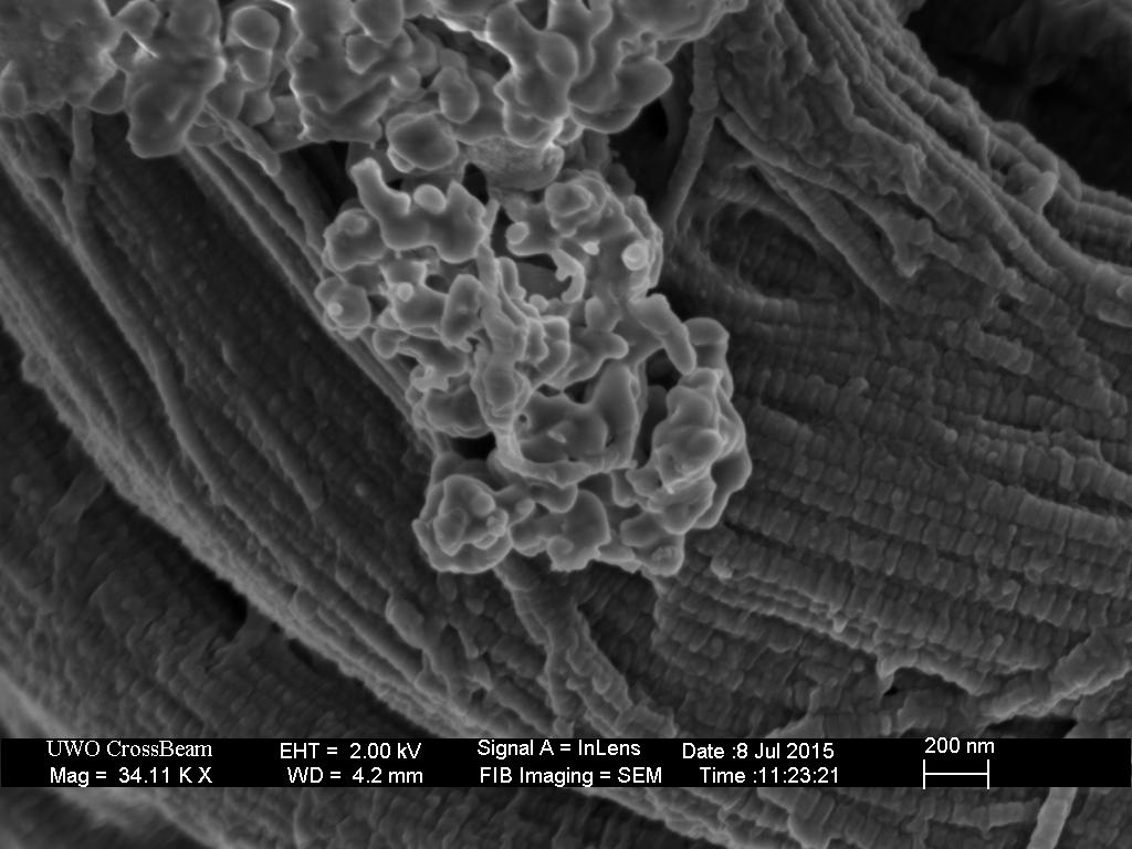
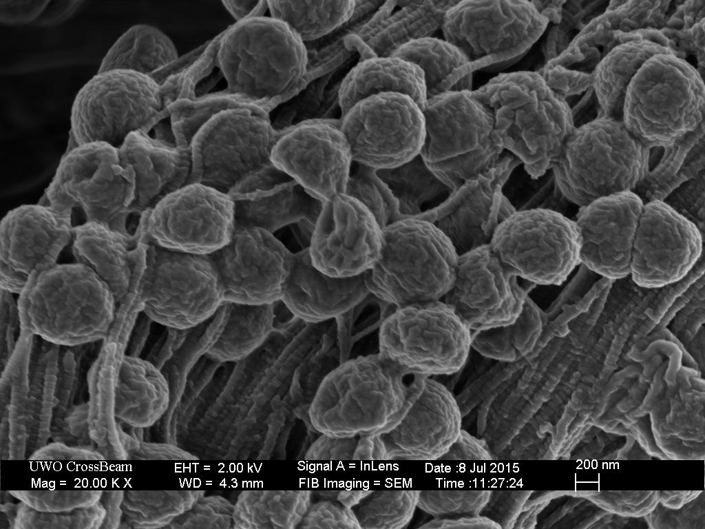
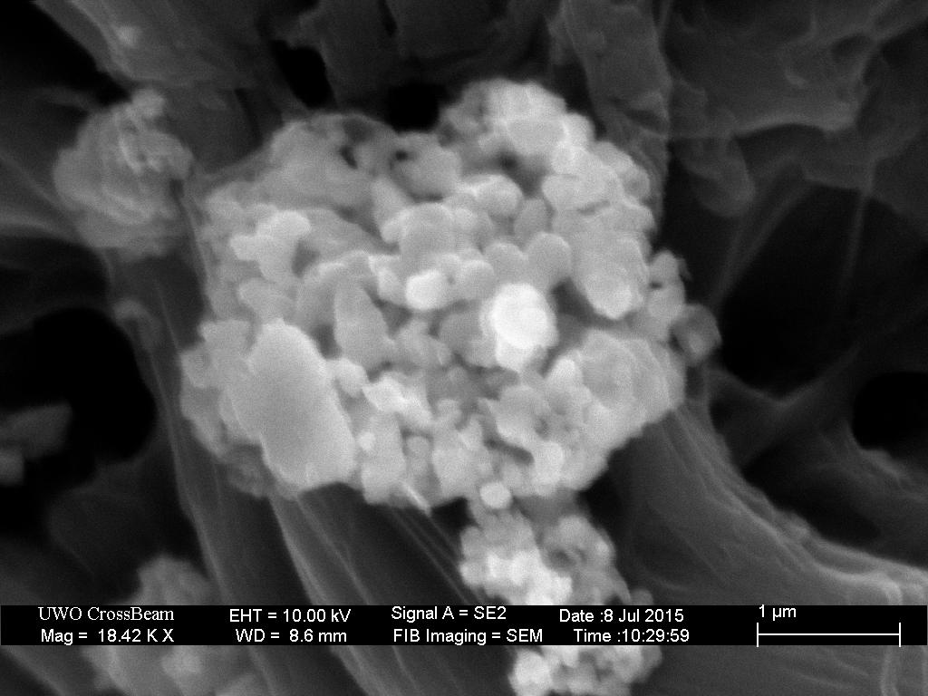
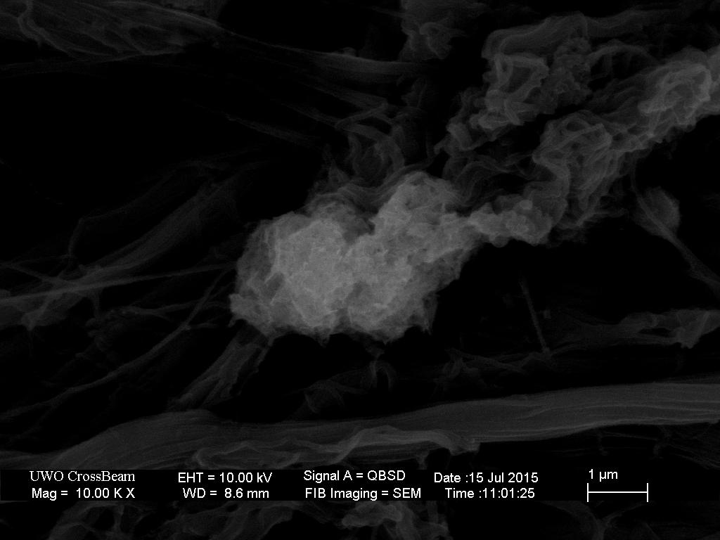
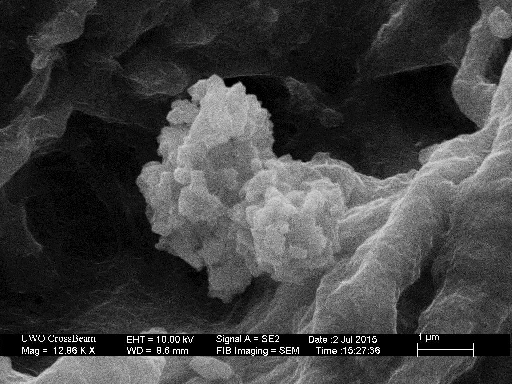
131 117 Appendix D: Additional SEM Images of Calcium Sites on BP
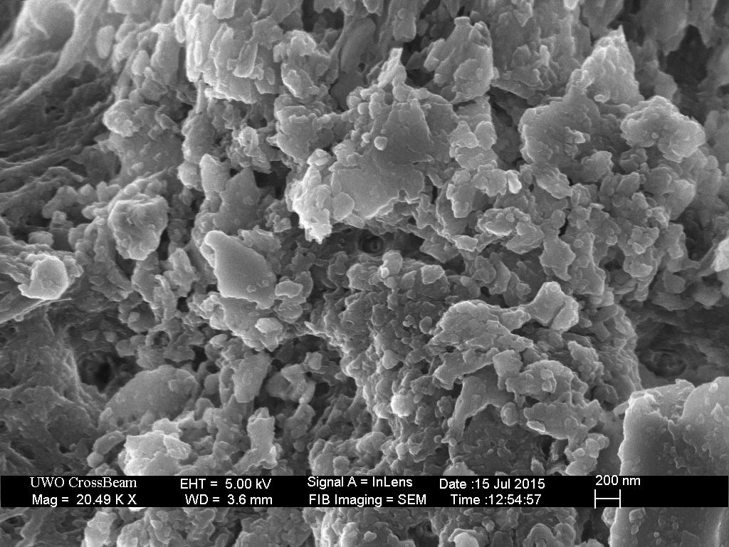
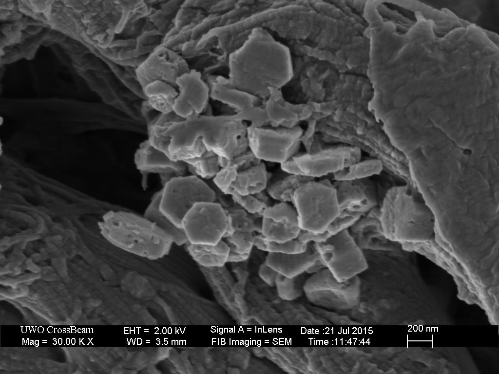
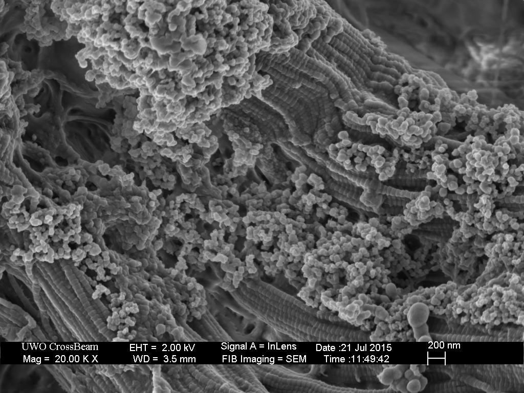
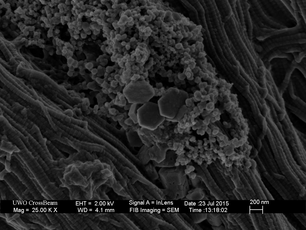
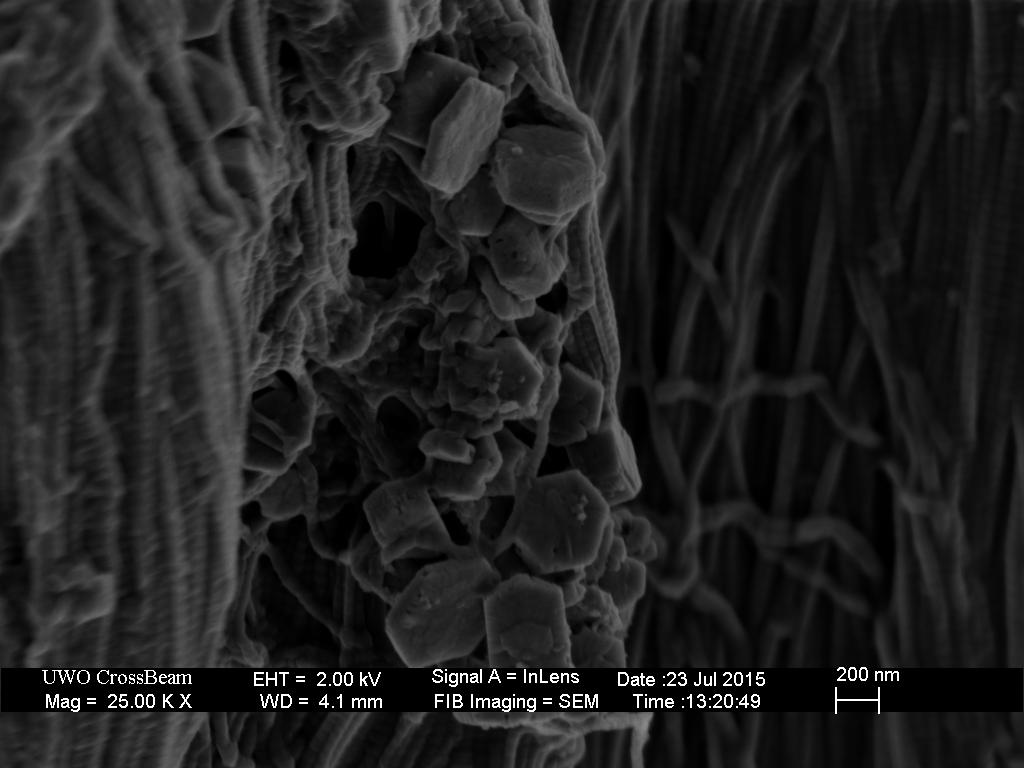
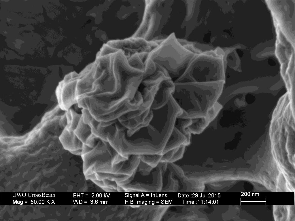
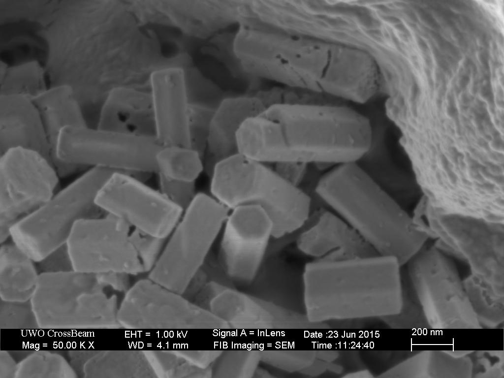
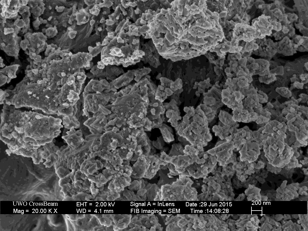
132 118
133 119 Appendix E: Copyright Permissions Figure 2.3
134 120
135 121
136 Figure
137 123
138 124
Image Analysis and Cytometry in Three-Dimensional Digital Reconstruction of Porcine Native Aortic Valve Leaflets
 Image Analysis and Cytometry in Three-Dimensional Digital Reconstruction of Porcine Native Aortic Valve Leaflets Introduction Chi Zheng, M1 - University of Pittsburgh; BSE - University of Michigan In association
THE HEART OBJECTIVES: LOCATION OF THE HEART IN THE THORACIC CAVITY CARDIOVASCULAR SYSTEM
 BIOLOGY II CARDIOVASCULAR SYSTEM ACTIVITY #3 NAME DATE HOUR THE HEART OBJECTIVES: Describe the anatomy of the heart and identify and give the functions of all parts. (pp. 356 363) Trace the flow of blood
Heart Dissection. 5. Locate the tip of the heart or the apex. Only the left ventricle extends all the way to the apex.
 Heart Dissection Page 1 of 6 Background: The heart is a four-chambered, hollow organ composed primarily of cardiac muscle tissue. It is located in the center of the chest in between the lungs. It is the
CIRCULATORY SYSTEM BLOOD VESSELS
 Name: Block: CIRCULATORY SYSTEM Multicellular organisms (above the level of roundworms) rely on a circulatory system to bring nutrients to, and take wastes away from, cells. In higher organisms such as
Chapter 20 (1) The Heart
 The Heart") Chapter 20 (1) The Heart Learning Objectives Describe the location and structure of the heart Describe the path of a drop of blood from the superior vena cava or inferior vena cava through the heart out
Chapter 20 (1) The Heart Learning Objectives Describe the location and structure of the heart Describe the path of a drop of blood from the superior vena cava or inferior vena cava through the heart out
The Cardiovascular System Part I: Heart Outline of class lecture After studying part I of this chapter you should be able to:
 The Cardiovascular System Part I: Heart Outline of class lecture After studying part I of this chapter you should be able to: 1. Describe the functions of the heart 2. Describe the location of the heart,
Chapter 14. The Cardiovascular System
 Chapter 14 The Cardiovascular System Introduction Cardiovascular system - heart, blood and blood vessels Cardiac muscle makes up bulk of heart provides force to pump blood Function - transports blood 2
the Cardiovascular System I
 the Cardiovascular System I By: Dr. Nabil A Khouri MD, MsC, Ph.D MEDIASTINUM 1. Superior Mediastinum 2. inferior Mediastinum Anterior mediastinum. Middle mediastinum. Posterior mediastinum Anatomy of
TSDA Boot Camp September 13-16, Introduction to Aortic Valve Surgery. George L. Hicks, Jr., MD
 TSDA Boot Camp September 13-16, 2018 Introduction to Aortic Valve Surgery George L. Hicks, Jr., MD Aortic Valve Pathology and Treatment Valvular Aortic Stenosis in Adults Average Course (Post mortem data)
The Cardiovascular System
 11 PART A The Cardiovascular System PowerPoint Lecture Slide Presentation by Jerry L. Cook, Sam Houston University ESSENTIALS OF HUMAN ANATOMY & PHYSIOLOGY EIGHTH EDITION ELAINE N. MARIEB The Cardiovascular
Cardiovascular System. Heart Anatomy
 Cardiovascular System Heart Anatomy 1 The Heart Location & general description: Atria vs. ventricles Pulmonary vs. systemic circulation Coverings Walls The heart is found in the mediastinum, the medial
Read Me. covering the Heart Anatomy. Labs. textbook. use. car: you
 Heart Anatomy Lab Pre-Lab Exercises Read Me These exercises should be done before coming to lab, after watching the videos covering the Heart Anatomy Labs. Answer the questions in this guide using the
The Cardiovascular System (Heart)
") The Cardiovascular System The Cardiovascular System (Heart) A closed system of the heart and blood vessels The heart pumps blood Blood vessels allow blood to circulate to all parts of the body The function
The Cardiovascular System The Cardiovascular System (Heart) A closed system of the heart and blood vessels The heart pumps blood Blood vessels allow blood to circulate to all parts of the body The function
Heart Anatomy. 7/5/02 Stephen G Davenport 1
 Heart Anatomy Copyright 1999, Stephen G. Davenport, No part of this publication may be reproduced, stored in a retrieval system, or transmitted, in any form without prior written permission. 7/5/02 Stephen
Anatomy of the Heart. Figure 20 2c
 Anatomy of the Heart Figure 20 2c Pericardium & Myocardium Remember, the heart sits in it s own cavity, known as the mediastinum. The heart is surrounded by the Pericardium, a double lining of the pericardial
The Heart. Size, Form, and Location of the Heart. 1. Blunt, rounded point; most inferior part of the heart.
 12 The Heart FOCUS: The heart is composed of cardiac muscle cells, which are elongated, branching cells that appear striated. Cardiac muscle cells behave as a single electrical unit, and the highly coordinated
The Heart and Cardiovascular System
 The Heart and Cardiovascular System What you will learn The location of the heart 3 layers and covering of the heart Explain the function of the heart as 2 separate pumps Identify the 4 chambers of the
This lab activity is aligned with Visible Body s A&P app. Learn more at visiblebody.com/professors
 1 This lab activity is aligned with Visible Body s A&P app. Learn more at visiblebody.com/professors 2 PRE-LAB EXERCISES: A. Watch the video 29.1 Heart Overview and make the following observations: 1.
The Cardiovascular System
 Essentials of Human Anatomy & Physiology Elaine N. Marieb Seventh Edition Chapter 11 The Cardiovascular System Slides 11.1 11.19 Lecture Slides in PowerPoint by Jerry L. Cook The Cardiovascular System
THE HEART. A. The Pericardium - a double sac of serous membrane surrounding the heart
 THE HEART I. Size and Location: A. Fist-size weighing less than a pound (250 to 350 grams). B. Located in the mediastinum between the 2 nd rib and the 5 th intercostal space. 1. Tipped to the left, resting
Iatrogenic pathology of the heart:
 Iatrogenic pathology of the heart: Complications of mitral valve plasty and replacement Patrick Bruneval D pt of Pathology Hôpital Européen Georges Pompidou Enterprise Interest None Mitral valve surgery
Two semilunar valves. Two atrioventricular valves. Valves of the heart. Left atrioventricular or bicuspid valve Mitral valve
 The Heart 3 Valves of the heart Two atrioventricular valves Two semilunar valves Right atrioventricular or tricuspid valve Left atrioventricular or bicuspid valve Mitral valve Aortic valve Pulmonary valve
Ch 19: Cardiovascular System - The Heart -
 Ch 19: Cardiovascular System - The Heart - Give a detailed description of the superficial and internal anatomy of the heart, including the pericardium, the myocardium, and the cardiac muscle. Trace the
CorMatrix ECM Bioscaffold
 CorMatrix ECM Bioscaffold REMODEL. REGROW. RESTORE. CorMatrix ECM Bioscaffold provides a natural bioscaffold matrix that enables the body s own cells to repair and remodel damaged cardio-vascular tissue.
STRUCTURES OF THE CARDIOVASCULAR SYSTEM
 STRUCTURES OF THE CARDIOVASCULAR SYSTEM CARDIOVASCULAR SYSTEM Also called the circulatory system Consists of the heart, arteries, veins, and capillaries Main function is to pump/circulate oxygenated blood
SCPA602 Cardiovascular System
 SCPA602 Cardiovascular System Associate Professor Dr. Wannee Jiraungkoorskul Department of Pathobiology, Faculty of Science, Mahidol University Tel: 02-201-5563, E-mail: wannee.jir@mahidol.ac.th 1 Objectives
Late failure of transcatheter heart valves: An open question
 Late failure of transcatheter heart valves: An open question A comparison with surgically implanted bioprosthetic heart valves. A. Rashid The Cardiothoracic Centre Liverpool, UK. Conflict of Interest Statement
Cardiovascular Anatomy Dr. Gary Mumaugh
 Cardiovascular Anatomy Dr. Gary Mumaugh Location of Heart Approximately the size of your fist Location o Superior surface of diaphragm o Left of the midline in mediastinum o Anterior to the vertebral column,
Anatomy of the Heart
 Biology 212: Anatomy and Physiology II Anatomy of the Heart References: Saladin, KS: Anatomy and Physiology, The Unity of Form and Function 8 th (2018). Required reading before beginning this lab: Chapter
PATIENT BOOKLET MEDTRONIC SURGICAL VALVE REPLACEMENT. Tissue Valve for Aortic and Mitral Valve Replacement
 PATIENT BOOKLET MEDTRONIC SURGICAL VALVE REPLACEMENT Tissue Valve for Aortic and Mitral Valve Replacement ARE MEDTRONIC SURGICAL TISSUE HEART VALVES RIGHT FOR YOU? Medtronic surgical heart valves are for
The Heart. The Heart A muscular double pump. The Pulmonary and Systemic Circuits
 C H A P T E R 19 The Heart The Heart A muscular double pump circuit takes blood to and from the lungs Systemic circuit vessels transport blood to and from body tissues Atria receive blood from the pulmonary
37 1 The Circulatory System
 H T H E E A R T 37 1 The Circulatory System The circulatory system and respiratory system work together to supply cells with the nutrients and oxygen they need to stay alive. a) The respiratory system:
Human Anatomy, First Edition
 Human Anatomy, First Edition McKinley & O'Loughlin Chapter 22 : Heart 1 Functions of the Heart Center of the cardiovascular system, the heart. Connects to blood vessels that transport blood between the
Unit 10 Cardiovascular System
 Unit 10 Cardiovascular System I. Functions Deliver nutrients to cells > O 2, sugars, amino acids, lipids, ions, H 2 O... Remove waste from cells > CO 2, pathogens, toxins, lactic acid... Fight off infection
Read Chapters 21 & 22, McKinley et al
 ACTIVITY 9: BLOOD AND HEART OBJECTIVES: 1) How to get ready: Read Chapters 21 & 22, McKinley et al., Human Anatomy, 5e. All text references are for this textbook. Read dissection instructions BEFORE YOU
Chapter 18 - Heart. I. Heart Anatomy: size of your fist; located in mediastinum (medial cavity)
") Chapter 18 - Heart I. Heart Anatomy: size of your fist; located in mediastinum (medial cavity) A. Coverings: heart enclosed in double walled sac called the pericardium 1. Fibrous pericardium: dense connective
Chapter 18 - Heart I. Heart Anatomy: size of your fist; located in mediastinum (medial cavity) A. Coverings: heart enclosed in double walled sac called the pericardium 1. Fibrous pericardium: dense connective
LAB 12-1 HEART DISSECTION GROSS ANATOMY OF THE HEART
 LAB 12-1 HEART DISSECTION GROSS ANATOMY OF THE HEART Because mammals are warm-blooded and generally very active animals, they require high metabolic rates. One major requirement of a high metabolism is
THE CIRCULATORY SYSTEM
 Biology 30S THE CIRCULATORY SYSTEM Name: This module adapted from bblearn.merlin.mb.ca 1 Introduction to Circulation The first organ to form, and the last organ to die. The heart is the pump of life. The
Cardiovascular System. I. Structures of the heart A. : Pericardium sack that surrounds the heart
 Cardiovascular System I. Structures of the heart A. : Pericardium sack that surrounds the heart 1. : Pericardial Cavity serous fluid filled space between the heart and the pericardium B. Heart Wall 1.
Section 5.1 The heart and heart disease
 Section 5.1 The heart and heart disease Mammals are too large to rely on diffusion. They need a circulatory system to move substances around the body. Blood moves down pressure gradients, from high to
Approximately the size of your fist Location Superior surface of diaphragm Left of the midline in mediastinum Anterior to the vertebral column,
 Dr. Gary Mumaugh Approximately the size of your fist Location Superior surface of diaphragm Left of the midline in mediastinum Anterior to the vertebral column, posterior to the sternum Posteriorly the
THE CARDIOVASCULAR SYSTEM. Part 1
 THE CARDIOVASCULAR SYSTEM Part 1 CARDIOVASCULAR SYSTEM Blood Heart Blood vessels What is the function of this system? What other systems does it affect? CARDIOVASCULAR SYSTEM Functions Transport gases,
LECTURE 5. Anatomy of the heart
 LECTURE 5. Anatomy of the heart Main components of the CVS: Heart Blood circulatory system arterial compartment haemomicrocirculatory (=microvascular) compartment venous compartment Lymphatic circulatory
The HEART. What is it???? Pericardium. Heart Facts. This muscle never stops working It works when you are asleep
 This muscle never stops working It works when you are asleep The HEART It works when you eat It really works when you exercise. What is it???? Located between the lungs in the mid thoracic region Apex
By the end of this session, the student should be able to:
 Valvular Heart disease HVD By Dr. Ashraf Abdelfatah Deyab VHD- Objectives By the end of this session, the student should be able to: Define and classify valvular heart disease. Enlist the causes of acquired
Human Anatomy and Physiology Chapter 19 Worksheet 1- The Heart
 Human Anatomy and Physiology Chapter 19 Worksheet 1- The Heart Name Date Period 1. The "double pump" function of the heart includes the right side, which serves as the circuit pump, while the left side
The Heart and Heart Disease
 The Heart and Heart Disease Illustration of the heart by Leonardo DaVinci heart-surgeon.com/ history.html 2/14/2010 1 I. Location, Size and Position of the Heart A. Triangular organ located 1. of mass
2. right heart = pulmonary pump takes blood to lungs to pick up oxygen and get rid of carbon dioxide
 A. location in thorax, in inferior mediastinum posterior to sternum medial to lungs superior to diaphragm anterior to vertebrae orientation - oblique apex points down and to the left 2/3 of mass on left
Test Review Circulatory System Chapters
 Test Review Circulatory System Chapters 13-2010 1. The tissue that forms the tight fitting sac around the heart is the a. parietal pericardium c. myocardium b. visceral pericardium d. endocardium 2. Which
Cardiac Surgery A Resource of Experimental Design
 Cardiac Surgery A Resource of Experimental Design Complete Transposition: a. Atrial switch the chronically systemic right ventricle b. Arterial switch the suddenly systemic left ventricle Fontan operation
Echocardiographic Evaluation of Mitral Valve Prostheses
 Echocardiographic Evaluation of Mitral Valve Prostheses Dennis A. Tighe, M.D., FACC, FACP, FASE Cardiovascular Medicine University of Massachusetts Medical School Worcester, MA www.asecho.org 1 Nishimura
The Cardiovascular System
 Essentials of Human Anatomy & Physiology Elaine N. Marieb Slides 11.1 11.19 Seventh Edition Chapter 11 The Cardiovascular System Functions of the Cardiovascular system Function of the heart: to pump blood
Introduction to soft tissues
 Modelli Costitutivi dei Materiali Corso di Laurea in Ingegneria Biomedica Pavia, 2013 Introduction to soft tissues Ferdinando Auricchio 1 2 3 4 1 Dipartimento di Ingegneria Civile e Architettura, Università
The cardiovascular system is composed of the heart and blood vessels that carry blood to and from the body s organs. There are 2 major circuits:
 1 The cardiovascular system is composed of the heart and blood vessels that carry blood to and from the body s organs. There are 2 major circuits: pulmonary and systemic. The pulmonary goes out to the
By: Stephanie Bendtsen, Joseph Calderan, and Celeste Dupont Team 17 Client: Dr. Sun
 By: Stephanie Bendtsen, Joseph Calderan, and Celeste Dupont Team 17 Client: Dr. Sun Flow Loop Design Introduction Purpose: to build a pulsatile flow loop that allows for implementation and testing of mechanical
Circulatory Systems. All cells need to take in nutrients and expel metabolic wastes.
 Circulatory Systems All cells need to take in nutrients and expel metabolic wastes. Single celled organisms: nutrients from the environment can diffuse (or be actively transported) directly in to the cell
Circulation. Circulation = is a process used for the transport of oxygen, carbon! dioxide, nutrients and wastes through-out the body
 Circulation Circulation = is a process used for the transport of oxygen, carbon! dioxide, nutrients and wastes through-out the body Heart = muscular organ about the size of your fist which pumps blood.
ACTIVITY 9: BLOOD AND HEART BLOOD
 ACTIVITY 9: BLOOD AND HEART OBJECTIVES: 1) How to get ready: Read Chapters 21 & 22, McKinley et al., Human Anatomy, 4e. All text references are for this textbook. Read dissection instructions BEFORE YOU
Blood and Heart. Student Learning Objectives:
 Blood and Heart Student Learning Objectives: Identify the major components of the blood. Identify the primary structures associated with the heart Follow the blood through the path of the circulation.
Chapter 20: Cardiovascular System: The Heart
 Chapter 20: Cardiovascular System: The Heart I. Functions of the Heart A. List and describe the four functions of the heart: 1. 2. 3. 4. II. Size, Shape, and Location of the Heart A. Size and Shape 1.
THE HEART. Unit 3: Transportation and Respiration
 THE HEART Unit 3: Transportation and Respiration The Circulatory System Also called the Cardiovascular System Circulates blood in the body Transports nutrients, oxygen, carbon dioxide, hormones, and blood
Cardiovascular System
 Cardiovascular System Purpose Transport oxygen and nutrients Take waste products away from tissues & organs Things we learned Blood pressure: the force of blood pushing against the walls of blood vessels
Unit 6: Circulatory System. 6.2 Heart
 Unit 6: Circulatory System 6.2 Heart Functions of Circulatory System 1. The heart is the pump necessary to circulate blood to all parts of the body 2. Arteries, veins and capillaries are the structures
The Heart. Happy Friday! #takeoutyournotes #testnotgradedyet
 The Heart Happy Friday! #takeoutyournotes #testnotgradedyet Introduction Cardiovascular system distributes blood Pump (heart) Distribution areas (capillaries) Heart has 4 compartments 2 receive blood (atria)
The Circulatory System (p )
") The Circulatory System (p. 268-281) How Does Gravity Affect Blood Circulation? As with all land animals, the giraffe and the corn snake are constantly subject to the force of gravity The circulatory system
The Circulatory System (p. 268-281) How Does Gravity Affect Blood Circulation? As with all land animals, the giraffe and the corn snake are constantly subject to the force of gravity The circulatory system
The Cardiovascular System. Chapter 15. Cardiovascular System FYI. Cardiology Closed systemof the heart & blood vessels. Functions
 Chapter 15 Cardiovascular System FYI The heart pumps 7,000 liters (4000 gallons) of blood through the body each day The heart contracts 2.5 billion times in an avg. lifetime The heart & all blood vessels
Cardiovascular Structure & Function
 Cardiovascular Structure & Function Cardiovascular system: The heart Arteries Veins Capillaries Lymphatic vessels Weighting of the heart ceremony: Ancient Egyptians William Harvey and Blood Flow April
25 different brand names >44 different models Sizes mm
 Types of Prosthetic Valves BIOLOGIC STENTED Porcine xenograft Pericardial xenograft STENTLESS Porcine xenograft Pericardial xenograft Homograft (allograft) Autograft PERCUTANEOUS MECHANICAL Bileaflet Single
The Cardiovascular System
 PowerPoint Lecture Slide Presentation by Patty Bostwick-Taylor, Florence-Darlington Technical College The Cardiovascular System 11 PART A The Cardiovascular System A closed system of the heart and blood
Hemodynamics Benefit of Supra-Annular Design in Failed Bio-Prosthetic Valves
 Hemodynamics Benefit of Supra-Annular Design in Failed Bio-Prosthetic Valves Speaker's name: I have the following potential conflicts of interest to report: Proctorship for Medtronic Agenda Failure modes
CARDIOVASCULAR SYSTEM
 CARDIOVASCULAR SYSTEM Overview Heart and Vessels 2 Major Divisions Pulmonary Circuit Systemic Circuit Closed and Continuous Loop Location Aorta Superior vena cava Right lung Pulmonary trunk Base of heart
Anatomy: Cardiovascular System
 In the name of God Anatomy: Cardiovascular System Moradian MD, MPH, PhD candidate Tehran University of Medical Sciences drmoradian@sums.ac.ir 2015 The Cardiovascular System A closed system of the heart
Heart. Structure Physiology of blood pressure and heartbeat
 Heart Structure Physiology of blood pressure and heartbeat Location and Anatomy Location and Anatomy Pericardial cavity: surrounds, isolates, and anchors heart Parietal pericardium lined with serous membrane
11/10/2014. Muscular pump Two atria Two ventricles. In mediastinum of thoracic cavity 2/3 of heart's mass lies left of midline of sternum
 It beats over 100,000 times a day to pump over 1,800 gallons of blood per day through over 60,000 miles of blood vessels. During the average lifetime, the heart pumps nearly 3 billion times, delivering
UNDERSTANDING YOUR HEART VALVE. Mosaic Tissue Valve
 UNDERSTANDING YOUR HEART VALVE Mosaic Tissue Valve A Message to You from the Employees at Medtronic, Inc. We understand that having heart valve replacement surgery is an important change in your life.
The Cardiovascular and Lymphatic Systems Cardiovascular System Blood Vessels Blood Vessels Arteries Arteries Arteries
 CH 12 The Cardiovascular and s The Cardiovascular and s OUTLINE: Cardiovascular System Blood Vessels Blood Pressure Cardiovascular System The cardiovascular system is composed of Blood vessels This system
Practical Histology. Cardiovascular System. Dr Narmeen S. Ahmad
 Practical Histology Cardiovascular System Dr Narmeen S. Ahmad The Cardiovascular System A closed system of the heart and blood vessels Functions of cardiovascular system: Transport nutrients, hormones
CJ Shuster A&P2 Lab Addenum Beef Heart Dissection 1. Heart Dissection. (taken from Johnson, Weipz and Savage Lab Book)
") CJ Shuster A&P2 Lab Addenum Beef Heart Dissection 1 Heart Dissection. (taken from Johnson, Weipz and Savage Lab Book) Introduction When you have finished examining the model, you are ready to begin your
CJ Shuster A&P2 Lab Addenum Beef Heart Dissection 1 Heart Dissection. (taken from Johnson, Weipz and Savage Lab Book) Introduction When you have finished examining the model, you are ready to begin your
Chapter 27 -The Heart & Blood Vessels
 Chapter 27 -The Heart & Blood Vessels 3.2 Learning Objectives 3.2.2 Organisational Complexity of the human 1. Describe the structures and organisation of tissues in the closed circulatory system. 2. Discuss
RESEARCH AND REVIEWS: JOURNAL OF PHARMACOLOGY AND TOXICOLOGICAL STUDIES
 e-issn:2322-0139 RESEARCH AND REVIEWS: JOURNAL OF PHARMACOLOGY AND TOXICOLOGICAL STUDIES Comparative Evaluation of Safety Outcomes of Different Prosthetic Valves in Indian Subjects. Kama Raval 1 *, Reena
Circulatory System 10.1
 1 Circulatory System 10.1 2 ARTERIES Arteries-blood vessels that carry blood away from the heart Thick walls Inner & Outer layers: connective tissue Middle layers are muscle and elastic connective tissue
Danil Hammoudi.MD 1/12/2009
 Danil Hammoudi.MD Aorta the biggest and longest artery (a blood vessel carrying blood away from the heart) in the body. It carries oxygen rich blood from the left ventricle of the heart to the body.inferior
AP2 Lab 1 - Blood & Heart
 AP2 Lab 1 - Blood & Heart Project 1 - Formed Elements Identification & Recognition See fig. 17.10 and Table 17.2. Instructor may also provide other images. Note: See Fig. 17.11 All formed elements are
CV Anatomy Quiz. Dr Ella Kim Dr Pip Green
 CV Anatomy Quiz Dr Ella Kim Dr Pip Green Q1 The location of the heart is correctly described as A) lateral to the lungs. B) medial to the sternum. C) superior to the diaphragm. D) posterior to the spinal
The Cardiovascular System
 The Cardiovascular System The Cardiovascular System A closed system of the heart and blood vessels The heart pumps blood Blood vessels allow blood to circulate to all parts of the body The function of
Heart Valves: Before and after surgery
 Heart Valves: Before and after surgery Tim Sutton, Consultant Cardiologist Middlemore Hospital, Auckland Auckland Heart Group Indications for intervention in Valvular disease To prevent sudden death and
Principles of Anatomy and Physiology
 Principles of Anatomy and Physiology 14 th Edition CHAPTER 20 The Cardiovascular System: The Heart Introduction The purpose of the chapter is to: 1. Learn about the components of the cardiovascular system
Echo Evaluation of a Mitral Valve Prostheses Sunday, February 14, :50 2:10 PM 20 min
 2016 ASE State of the Art Echocardiography Course Tucson, AZ Echo Evaluation of a Mitral Valve Prostheses Sunday, February 14, 2016 1:50 2:10 PM 20 min 1 M U H A M E D S A R I Ć, M D, P H D D i r e c t
3D Printing & Echocardiography
 ASE SOTA Feb 19, 2018 3D Printing & Echocardiography Stephen H. Little, MD John S. Dunn Chair in Cardiovascular Research and Education, Associate professor, Weill Cornell Medicine Disclosures Personal
The Circulatory System. The Heart, Blood Vessels, Blood Types
 The Circulatory System The Heart, Blood Vessels, Blood Types The Closed Circulatory System Humans have a closed circulatory system, typical of all vertebrates, in which blood is confined to vessels and
Cardiovascular System Note-Taking Guide
 FUNctions: Name: 3-27-14 Cardiovascular System Note-Taking Guide Heart: Pumps and delivers through the body Blood: Vessels: Delivers and to the body Carries waste and Maintains homeostasis - Carries blood
Do Now. Get out work from last class to be checked
 Do Now Get out work from last class to be checked Heart Actions Cardiac Cycle: One complete heartbeat. The contraction of a heart chamber is called systole and the relaxation of a chamber is called diastole.
The Cardiovascular System
 The Cardiovascular System The Manila Times College of Subic Prepared by: Stevens B. Badar, RN, MANc THE HEART Anatomy of the Heart Location and Size approx. the size of a person s fist, hollow and cone-shaped,
1. Which of the following blood vessels has a thin elastic layer? A. Aorta. B. Pulmonary artery. C. Posterior vena cava. D. Mesenteric capillary.
 CIRCULATORY SYSTEM 1. Which of the following blood vessels has a thin elastic layer? A. Aorta. B. Pulmonary artery. C. Posterior vena cava. D. Mesenteric capillary. 2. Capillary beds are equipped with
Introduction to Biomedical Engineering
 Introduction to Biomedical Engineering FW 16/17, AUT Biomechanics of tendons and ligaments G. Rouhi Biomechanics of tendons and ligaments Biomechanics of soft tissues The major soft tissues in musculoskeletal
An Illustrated 1. Dissection Guide. To The... Mammalian. rr= Heart. Right ventricle+---, by David H. Hall
 An Illustrated 1. Dissection Guide. To The... Mammalian rr= Heart ventricle+---, by David H. Hall The Mam.malian Heart Because mammals are warm blooded (endothermic) and generally very active animals,
Outcomes of Mitral Valve Repair for Mitral Regurgitation Due to Degenerative Disease
 Outcomes of Mitral Valve Repair for Mitral Regurgitation Due to Degenerative Disease TIRONE E. DAVID, MD ; SEMIN THORAC CARDIOVASC SURG 19:116-120c 2007 ELSEVIER INC. PRESENTED BY INTERN 許士盟 Mitral valve
CIRCULATORY SYSTEM TASK CARDS Worksheet
 CIRCULATORY SYSTEM TASK CARDS Worksheet Name: Date: Instructions: Put the answers to each task card in the numbered boxes on the chart. 1 a) left semilunar valve / aortic valve b) blood would backflow
I will not discuss off label use or investigational use in my presentation.
 I will not discuss off label use or investigational use in my presentation. Surgical valves Design and Durability Testing Potential Concerns Real Practice 1952-1962 1963-1966 1967-1969 1969-1977 1977-1984