Martin G. Keane, MD, FASE Temple University School of Medicine
|
|
|
- Agatha Potter
- 6 years ago
- Views:
Transcription

1 Martin G. Keane, MD, FASE Temple University School of Medicine
2 Measurement of end-diastolic LV internal diameter (LVIDd) made by properly-oriented M-Mode techniques in the Parasternal Long Axis View (PLAX): A.Are identical to those made from 2D images B. Are larger than those made from 2D images C. Are less discrepant from 2D measures with advancing age D.Are identical if trailing edge to leading edge convention is used E. Are completely unreliable compared to 2D measurements
3 In males, the geometric pattern of left ventricular concentric remodeling is present when: A. LVMI 115 g/m 2 and RWT 0.42 B. LVMI >115 g/m 2 and RWT >0.42 C. LVMI 115 g/m 2 and RWT >0.42 D. LVMI >115 g/m 2 and RWT 0.42 E. LVMI 115 g/m 2 and RWT <0.34
4 Volumetric measurements of LV cavity size (Simpson s Method) are considered superior to strictly Linear techniques (Rotational Ellipse) because: A. Small errors in linear measurements are greatly magnified by squaring terms in linear techniques. B. Complex mathematical modeling of volumetric techniques insures precision C. Linear measurement techniques were developed for M-mode echocardiography and have decreased accuracy when applied to 2D echocardiography. D. Volumetric techniques directly measure volumes, whereas linear techniques measure only length and width E. Volumetric techniques correct for shape distortions better than linear techniques.
5 Ventricular Chamber Size Chamber Dimensions Chamber Volume Ventricular Muscle Mass Ventricular Wall Thickness Myocardial Hypertrophy Ventricular Geometry Ventricular Function Systolic Diastolic


6 M-Mode 2D guided M-mode in PLAX view Leading edge to leading edge convention 2-Dimensional Useful in cases of off-axis M-mode Requires good endocardial definition

7 Use leading edge to leading edge convention 2D guidance to orient M-mode perpendicular to LV
8 IVS d = <1.1 cm LVID d = <5.6 cm PWT d = <1.1 cm LVID s = variable
9 Measured in freeze-frame End-diastole First frame after mitral valve closure or Frame in which LV diameter is the largest End-systole First frame after aortic valve closure or Frame in which LV dimension is smallest Ideally in PLAX view PSAX only if positioned perpendicular


10 IVS d What Criterion did I use? LVID d PWT d End Dia stole LVID s End Systole

11 Lang R, et al. J Am Soc Ec ho (2015)
12 Measurement of systolic function Calculated from M-mode dimensions LVID = d - LVID s LVID d Normal 25% X 100 Inherently limited Assessing 3D function using 1dimensional measurement Inaccurate in presence of regional wall motion abnormalities especially at the apex
13 More accurate assessment of LV size LV Ejection Fraction (%) can be calculated 2D Techniques based on geometric assumptions Simple assumptions - easier to use but less accurate Complex assumptions - more accurate but less easy to use 3D Techniques very accurate As yet, infrequently utilized in clinical practice
14 All are based on assumption of symmetry Neglects focal abnormalities More complex geometric models are the most accurate Rotational Ellipse Prolate Ellipse - Bullet shape

15 A B D c a n b e simp lified to Volume = 4/ 3π * A * B * D Volume D 3
")
16 Volume D 3 Simplest geometric approximation Can be calculated using M-mode only Not accurate with abnormal LV shape Large-scale / epidemiologic studies Framingham and other large-scale population studies (M-mode!!)
 * Length (apical)")
17 A endocardial L endocardial Volume 5/6 * Area (SAX) * Length (apical)
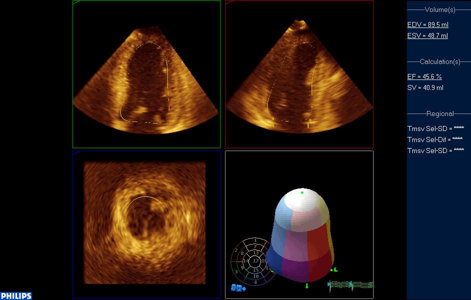
18 Eliminates need for (most) geometric assumptions Volume of asymmetric ventricle can be calculated Ejection fraction can be easily calculated Old Days off-line computer analysis Now? On-line on digital systems
19 Subdivide LV: series of discs finite thickness measurable area Disc volume = * r 2 * h Sum of disc volumes = LV volume

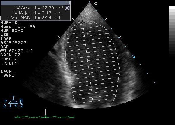
20
21 Most accurate LV volume Particularly with abnormal LV shape Apical 4 Chamber + Apical 2 Chamber Biplane approximation is best
22 LV diastolic LV diastolic Lang R, et al. J Am Soc Ec ho (2015)

23 Use LV Volumes LVEDV = End Diastolic Volume LVESV = End Systolic Volume EF (%) = LVEDV - LVESV LVEDV X 100 Can use any LV volume technique Simpson s Method of Discs is preferred
24 Lang R, et al. J Am Soc Ec ho (2015)
25

26 You will hea r a bout this in deta il during Stress Echo Lectures
27 Left Ventricular Hypertrophy Abnormal increase in LV mass Important prognostic indicator Basic concept for measurement: LV Mass = Mass of Cardiac Muscle Cardiac Muscle Mass = Volume of Muscle * Specific Gravity of Muscle Cardiac Muscle Mass = (LV Vol epi - LV Vol endo ) * 1.05 g/ cm 3
28 Using Rotational Ellipse: [(IVS d +LVID d +PWT d ) 3 - (LVID d ) 3 ] * 1.05 Using Area-Length: [(5/6*Area epi *L epi ) - (5/6*Area end *L end )] * 1.05 Simpson s Method of Discs NOT USED!!! Cannot define all epicardial surfaces
29 Adaptive response Volume and/or Pressure overload Wall thickening normalizes LV wall stress Optimizes myocardial oxygen consumption Increase in Myocyte mass No change in myocyte number

30 Abnormal myocardium Fetal / alternate protein isomers Abnormal subcellular organelles Decreased capillary density Abnormal systolic / diastolic function Subclinical initially Ultimately leads to CHF

31 Gender M-Mode Derived 2D Derived Male >115 g/m 2 >102 g/ m 2 Female >95 g/m 2 >88 g/ m 2

32 Based on 2D wall thickness only: Normal = < 1.1 cm Mild LVH = cm Moderate LVH = cm Severe LVH = > 1.4 cm
33 Wall stress = pressure x radius wall thickness Index of LV function Approximates afterload


34 LV Geometry in LVH Beyond Simple Mass Hypertrophy minimizes wall stress: T w P * R h P = Intracavitary Pressure R = Chamber Radius h = Ventricular Wall Thickness Wall thickness (h) in BOTH pressure or volume load Cavity radius (R) in volume load Different loads create different geometry Reflected by Relative Wall Thickness
35 RWT = 2 * PWT LVID LVID Nl 0.34 WT >


36 Increase in systolic pressure w/o major change in cavity radius By LaPlace: T w P * R h End Result: LV Wall thickness LV Mass Relative Wall Thickness


37 LVIDd = 4.7 PWTd = 1.5 RWT = 0.62


38 Increase in diastolic chamber size By LaPlace: T w P * R h End Result: LV wall thickness LV mass NO CHANGE in RWT


39 LVIDd = 7.0 PWTd = 1.2 RWT = 0.34


40 Combinations of pressure and volume overload result in a spectrum of LV geometry in the general population.
41 Short axis/long axis Spherical ventricle is at mechanical disadvantage Aortic regurgitation, dilated CM most common causes of increased sphericity index
42 Severe AR
43 Hemodynamic (Doppler) Assessment World Renowned talk by Itzhak Kronzon Global Longitudinal Strain (and other strain) Fabulously explained by Steve Lester Diastolic Function and Dysfunction Exquisitely delineated by Gerry Aurigemma, Miguel Quiñonez, Natesa Pandian Three-Dimensional (3D) Evaluation Brought to you by Dr. 3D Sunil Mankad
44 Measurement of end-diastolic LV internal diameter (LVIDd) made by properly-oriented M-Mode techniques in the Parasternal Long Axis View (PLAX): A. Are identical to those made from 2D images B. Are larger than those made from 2D images C. Are less discrepant from 2D measures with advancing age D. Are identical if trailing edge to leading edge convention is used E. Are completely unreliable compared to 2D measurements
45 A. Incorrect - M-mode imaging and 2D imaging represent different modalities, and measurements derived will not be identical B. Correct - Due to angulation of the ventricle in the PLAX, subtle degrees of obliquity results in LVIDd measurements that are between 6 and 12 mm larger than measured directly on 2D images. C. Incorrect - The heart typically angulates to a more apex-upward orientation with age in the parasternal long axis view, M-Mode derived measurements become MORE discrepant over time. D. Incorrect LEADING edge to leading edge measurements are conventional on M-mode. Even if trailing edge to leading edge measurement is made on M-mode, inherent differences in edge detection and technique result in non-identical measurements E. Incorrect M-mode imaging affords extremely accurate spacial resolution. Performed properly in correct orientation, M-mode measurements are extremely accurate and reliable.
46 In males, the geometric pattern of left ventricular concentric remodeling is present when: A. LVMI 115 g/m 2 and RWT 0.42 B. LVMI >115 g/m 2 and RWT >0.42 C. LVMI 115 g/m 2 and RWT >0.42 D. LVMI >115 g/m 2 and RWT 0.42 E. LVMI 115 g/m 2 and RWT <0.34
47 A. Incorrect This is normal LV mass index and normal relative wall thickness. This would be classified as Normal LV Anatomy. B. Incorrect LV mass index is increased above threshold norms for males, indicating LV hypertrophy. Relative wall thickness is greater than threshold norm. This would be classified as Concentric Hypertrophy a finding most common in cases of predominant pressure overload. C. Correct In the situation of normal LV mass index for males, but when relative wall thickness exceeds accepted norm values, is classified as Concentric Remodeling. This is considered by some as a pre-hypertrophic state, and is common in hypertensive populations. D. Incorrect LV mass index is greater than established population norms for males, indicating left ventricular hypertrophy. Relative wall thickness is in normal range. This is classified as Eccentric Hypertrophy a finding most common in cases of predominant volume overload. E. Incorrect This situation represents normal LV mass index and a relative wall thickness below the mean normal value of RWT. This would be classifies at Normal LV Anatomy.
48 Volumetric measurements of LV cavity size (Simpson s Method) are considered superior to strictly Linear techniques (Rotational Ellipse) because: A. Small errors in linear measurements are greatly magnified by squaring terms in linear techniques. B. Complex mathematical modeling of volumetric techniques insures precision C. Linear measurement techniques were developed for M-mode echocardiography and have decreased accuracy when applied to 2D echocardiography. D. Two-Dimensional volumetric techniques directly measure volumes, whereas linear techniques measure only length and width E. Volumetric techniques correct for shape distortions better than linear techniques.
49 A. Incorrect Linear techniques and volumetric techniques utilize measurements raised to second or third power, resulting in magnification of measurement errors in both. B. Incorrect - Complex models of ventricular volume are still subject to significant lack of precision, particularly with poor endocardial definition and off-axis imaging. C. Incorrect Although developed for M-mode echo, linear techniques for LV volume can be accurately applied to 2-dimensional echo imaging. Frequently, measurement of LV lengths/diameters are MORE accurately performed on 2D imaging. D. Incorrect 2D volumetric techniques calculate overall LV volume using a compilation of smaller, measurable volumes. Linear measurements are still frequently a component in volumetric techniques. Thus volume is not directly measured E. Correct Volumetric techniques, particularly when applied in a biplane fashion, can incorporate significant cavity shape abnormalities and focal wall motion abnormalities into estimation of diastolic and systolic ventricular volume. Linear techniques rely on broad assumptions of symmetry of cavity size and function. Depending on where abnormalities are located, linear technique assumptions of


50
Evaluation of Left Ventricular Function and Hypertrophy Gerard P. Aurigemma MD
 Evaluation of Left Ventricular Function and Hypertrophy Gerard P. Aurigemma MD Board Review Course 2017 43 year old health assistant Severe resistant HTN LT BSA 2 Height 64 1 Here is the M mode echocardiogram
MAYON VOLCANO: FAST FACTS
 MAYON VOLCANO: FAST FACTS Type of Volcano: Stratovolcano Elevation: 2.46 km Base Diameter: 20 km Base Circumference: 62.8 km Area: 314.1 km 2 Reference: http://www.phivolcs.dost.gov.ph/html/update_vmepd/volcano/volcanolist/mayon.htm
Echocardiographic Assessment of the Left Ventricle
 Echocardiographic Assessment of the Left Ventricle Theodora Zaglavara, MD, PhD, BSCI/BSCCT Department of Cardiovascular Imaging INTERBALKAN EUROPEAN MEDICAL CENTER 2015 The quantification of cardiac chamber
Assessment of LV systolic function
 Tutorial 5 - Assessment of LV systolic function Assessment of LV systolic function A knowledge of the LV systolic function is crucial in the undertanding of and management of unstable hemodynamics or a
Quantification of Cardiac Chamber Size
 2017 KSE 2017-11-25 Quantification of Cardiac Chamber Size Division of Cardiology Keimyung University Dongsan Medical Center In-Cheol Kim M.D., Ph.D. LV size and function Internal linear dimensions PLX
좌심실수축기능평가 Cardiac Function
 Basic Echo Review Course 좌심실수축기능평가 Cardiac Function Seonghoon Choi Cardiology Hallym university LV systolic function Systolic function 좌심실수축기능 - 심근의수축으로심실에서혈액을대동맥으로박출하는기능 실제임상에서 LV function 의의미 1Diagnosis
LV FUNCTION ASSESSMENT: WHAT IS BEYOND EJECTION FRACTION
 LV FUNCTION ASSESSMENT: WHAT IS BEYOND EJECTION FRACTION Jamilah S AlRahimi Assistant Professor, KSU-HS Consultant Noninvasive Cardiology KFCC, MNGHA-WR Introduction LV function assessment in Heart Failure:
Aortic Stenosis: Spectrum of Disease, Low Flow/Low Gradient and Variants
 Aortic Stenosis: Spectrum of Disease, Low Flow/Low Gradient and Variants Martin G. Keane, MD, FASE Professor of Medicine Lewis Katz School of Medicine at Temple University Basic root structure Parasternal
Adel Hasanin Ahmed 1 LV MORPHOLOGY
 Adel Hasanin Ahmed 1 LV MORPHOLOGY The left ventricular wall comprises three layers- middle circumferential layer and superficial and deep longitudinal layers: 1. Subepicardial longitudinal layer (25%
RIGHT VENTRICULAR SIZE AND FUNCTION
 RIGHT VENTRICULAR SIZE AND FUNCTION Edwin S. Tucay, MD, FPCC, FPCC, FPSE Philippine Society of Echocardiography Quezon City, Philippines Echo Mission, BRTTH, Legaspi City, July 1-2, 2016 NO DISCLOSURE
Chamber Quantitation Guidelines: What is New?
 Chamber Quantitation Guidelines: What is New? Roberto M Lang, MD J AM Soc Echocardiogr 2005; 18:1440-1463 1 Approximately 10,000 citations iase in itune Cardiac Chamber Quantification: What is New? Database
LV geometric and functional changes in VHD: How to assess? Mi-Seung Shin M.D., Ph.D. Gachon University Gil Hospital
 LV geometric and functional changes in VHD: How to assess? Mi-Seung Shin M.D., Ph.D. Gachon University Gil Hospital LV inflow across MV LV LV outflow across AV LV LV geometric changes Pressure overload
Cardiac Chamber Quantification by Echocardiography
 Cardiac Chamber Quantification by Echocardiography Maryam Bokhamseen, RCS, RCDS, EACVI Echotechnologist ǁ, Non invasive Cardiac Laboratory King Abdulaziz Cardiac Center. Outline: Introduction. Background
Prospect Cardiac Packages. S-Sharp
 Prospect Cardiac Packages S-Sharp B mode: Teichholz: Teichholz formula LV Volume 2D: modified Simpson's rule method ALM: area length method LV Volume (Intg.): integral method M mode: Long axis: Teichholz
Value of echocardiography in chronic dyspnea
 Value of echocardiography in chronic dyspnea Jahrestagung Schweizerische Gesellschaft für /Schweizerische Gesellschaft für Pneumologie B. Kaufmann 16.06.2016 Chronic dyspnea Shortness of breath lasting
10/7/2013. Systolic Function How to Measure, How Accurate is Echo, Role of Contrast. Thanks to our Course Director: Neil J.
 Systolic Function How to Measure, How Accurate is Echo, Role of Contrast Neil J. Weissman, MD MedStar Health Research Institute & Professor of Medicine Georgetown University Washington, D.C. No Disclosures
Basic Assessment of Left Ventricular Systolic Function
 WINFOCUS BASIC ECHO (WBE) Basic Assessment of Left Ventricular Systolic Function Ritesh Dhar, MD Director, Echocardiography Lab and Staff Cardiologist Intermountain Medical Center Murray, Utah Outline
Recommendations for chamber quantification *
 Eur J Echocardiography (2006) 7, 79e108 GUIDELINES Recommendations for chamber quantification * Roberto M. Lang, Michelle Bierig, Richard B. Devereux, Frank A. Flachskampf *, Elyse Foster, Patricia A.
B-Mode measurements protocols:
 Application Note How to Perform the Most Commonly Used Measurements from the Cardiac Measurements Package associated with Calculations of Cardiac Function using the Vevo Lab Objective The Vevo LAB offline
THE LEFT ATRIUM HOW CAN ECHO HELP US?
 THE LEFT ATRIUM HOW CAN ECHO HELP US? Dr. Dragos COZMA BACKGROUND Left atrium (LA) dilation can occur in a broad spectrum of cardiovascular diseases including hypertension, left ventricular dysfunction,
HYPERTROPHY: Behind the curtain. V. Yotova St. Radboud Medical University Center, Nijmegen
 HYPERTROPHY: Behind the curtain V. Yotova St. Radboud Medical University Center, Nijmegen Disclosure of interest: none Relative wall thickness (cm) M 0.22 0.42 0.43 0.47 0.48 0.52 0.53 F 0.24 0.42 0.43
Conflict of Interests
 The Left Ventricle: How Should We Quantify Its Size and Function; Is It Time for 3D in Everyone? Roberto M Lang, MD Conflict of Interests Philips Medical Imaging Research Grants Speakers bureau Advisory
Heart Failure in Women: Dr Goh Ping Ping Cardiologist Asian Heart & Vascular Centre
 Heart Failure in Women: More than EF? Dr Goh Ping Ping Cardiologist Asian Heart & Vascular Centre Overview Review pathophysiology as it relates to diagnosis and management Rational approach to workup:
Quantitation of right ventricular dimensions and function
 SCCS Basics of cardiac assessment Quantitation of right ventricular dimensions and function Tomasz Kukulski, MD PhD Dept of Cardiology, Congenital Heart Disease and Electrotherapy Silesian Medical University
Table of Contents RECOMMENDATION PAPER
 European Heart Journal Cardiovascular Imaging (2015) 16, 577 605 doi:10.1093/ehjci/jev076 RECOMMENDATION PAPER Recommendations on the use of echocardiography in adult hypertension: a report from the European
Adult Echocardiography Examination Content Outline
 Adult Echocardiography Examination Content Outline (Outline Summary) # Domain Subdomain Percentage 1 2 3 4 5 Anatomy and Physiology Pathology Clinical Care and Safety Measurement Techniques, Maneuvers,
Left Ventricle Remodeling for Patients with Heart Failure and its Influence on Cardiac Performance
 Original Article Left Ventricle Remodeling for Patients with Heart Failure and its Influence on Cardiac * Mutaz F. Hussain** Anmar Z. Saleh* BSc FICMS BSc,PhD J Fac Med Baghdad 2013; Vol.55, No. 2 Received:
PRELIMINARY STUDIES OF LEFT VENTRICULAR WALL THICKNESS AND MASS OF NORMOTENSIVE AND HYPERTENSIVE SUBJECTS USING M-MODE ECHOCARDIOGRAPHY
 Malaysian Journal of Medical Sciences, Vol. 9, No. 1, January 22 (28-33) ORIGINAL ARTICLE PRELIMINARY STUDIES OF LEFT VENTRICULAR WALL THICKNESS AND MASS OF NORMOTENSIVE AND HYPERTENSIVE SUBJECTS USING
Little is known about the degree and time course of
 Differential Changes in Regional Right Ventricular Function Before and After a Bilateral Lung Transplantation: An Ultrasonic Strain and Strain Rate Study Virginija Dambrauskaite, MD, Lieven Herbots, MD,
Review of Cardiac Imaging Modalities in the Renal Patient. George Youssef
 Review of Cardiac Imaging Modalities in the Renal Patient George Youssef ECHO Left ventricular hypertrophy (LVH) assessment Diastolic dysfunction Stress ECHO Cardiac CT angiography Echocardiography - positives
Questions on Chamber Quantitation
 Questions on Chamber Quantitation @RobertoMLang Which of the following statements is true? 1. The aortic annulus should be measured in midsystole. 2. The aortic annulus should be measured in enddiastole.
Left atrial function. Aliakbar Arvandi MD
 In the clinic Left atrial function Abstract The left atrium (LA) is a left posterior cardiac chamber which is located adjacent to the esophagus. It is separated from the right atrium by the inter-atrial
Evaluation of Systolic Function of the Left Ventricle
 Evaluation of Systolic Function of the Left Ventricle Roxy Senior MD DM FRCP FESC FACC and Vinay Kumar Bhatia PhD MRCP Department of Cardiovascular Medicine, Northwick Park Hospital and Institute for Medical
Imaging Guide Echocardiography
 Imaging Guide Guide to Small Animal Echocardiography using the Vevo Imaging Systems System Compatibility: This guide contains instructions and suggestions for work on the Vevo2100, VevoLAZR, Vevo 3100
Hemodynamic Assessment. Assessment of Systolic Function Doppler Hemodynamics
 Hemodynamic Assessment Matt M. Umland, RDCS, FASE Aurora Medical Group Milwaukee, WI Assessment of Systolic Function Doppler Hemodynamics Stroke Volume Cardiac Output Cardiac Index Tei Index/Index of myocardial
Η ηχωκαρδιολογία στην διάγνωση κα πρόγνωση της καρδιακής ανεπάρκειας µε µειωµένο και φυσιολογικό κλάσµα εξώθησης
 Η ηχωκαρδιολογία στην διάγνωση κα πρόγνωση της καρδιακής ανεπάρκειας µε µειωµένο και φυσιολογικό κλάσµα εξώθησης Βασίλειος Σαχπεκίδης Επιµελητής Β Καρδιολογίας Γ.Ν. Παπαγεωργίου Θεσσαλονίκη ESC Guidelines
Echo assessment of the failing heart
 Echo assessment of the failing heart Mark K. Friedberg, MD The Labatt Family Heart Center The Hospital for Sick Children Toronto, Ontario, Canada Cardiac function- definitions Cardiovascular function:
Coronary artery disease (CAD) risk factors
 risk factors") Background Coronary artery disease (CAD) risk factors CAD Risk factors Hypertension Insulin resistance /diabetes Dyslipidemia Smoking /Obesity Male gender/ Old age Atherosclerosis Arterial stiffness precedes
Background Coronary artery disease (CAD) risk factors CAD Risk factors Hypertension Insulin resistance /diabetes Dyslipidemia Smoking /Obesity Male gender/ Old age Atherosclerosis Arterial stiffness precedes
Echocardiographic Evaluation of the Cardiomyopathies. Stephanie Coulter, MD, FACC, FASE April, 2016
 Echocardiographic Evaluation of the Cardiomyopathies Stephanie Coulter, MD, FACC, FASE April, 2016 Cardiomyopathies (CMP) primary disease intrinsic to cardiac muscle Dilated CMP Hypertrophic CMP Infiltrative
Patterns of Left Ventricular Remodeling in Chronic Heart Failure: The Role of Inadequate Ventricular Hypertrophy
 Abstract ESC 82445 Patterns of Left Ventricular Remodeling in Chronic Heart Failure: The Role of Inadequate Ventricular Hypertrophy FL. Dini 1, P. Capozza 1, P. Fontanive 2, MG. Delle Donne 1, V. Santonato
Cardiac Magnetic Resonance in pregnant women
 Cardiac Magnetic Resonance in pregnant women Chen SSM, Leeton L, Dennis AT Royal Women s Hospital and The University of Melbourne, Parkville, Australia alicia.dennis@thewomens.org.au Quantification of
ECHOCARDIOGRAPHY DATA REPORT FORM
 Patient ID Patient Study ID AVM - - Date of form completion / / 20 Initials of person completing the form mm dd yyyy Study period Preoperative Postoperative Operative 6-month f/u 1-year f/u 2-year f/u
Strain and Strain Rate Imaging How, Why and When?
 Strain and Strain Rate Imaging How, Why and When? João L. Cavalcante, MD Advanced Cardiac Imaging Fellow Cleveland Clinic Foundation Disclosures: No conflicts of interest Movement vs Deformation Movement
2/2/2011. Strain and Strain Rate Imaging How, Why and When? Movement vs Deformation. Doppler Myocardial Velocities. Movement. Deformation.
 Strain and Strain Rate Imaging How, Why and When? João L. Cavalcante, MD Advanced Cardiac Imaging Fellow Cleveland Clinic Foundation Disclosures: No conflicts of interest Movement vs Deformation Movement
How NOT to miss Hypertrophic Cardiomyopathy? Adaya Weissler-Snir, MD University Health Network, University of Toronto
 How NOT to miss Hypertrophic Cardiomyopathy? Adaya Weissler-Snir, MD University Health Network, University of Toronto Introduction Hypertrophic cardiomyopathy is the most common genetic cardiomyopathy,
Imaging Assessment of Aortic Stenosis/Aortic Regurgitation
 Imaging Assessment of Aortic Stenosis/Aortic Regurgitation Craig E Fleishman, MD FACC FASE The Heart Center at Arnold Palmer Hospital for Children, Orlando SCAI Fall Fellows Course 2014 Las Vegas Disclosure
Echocardiography: Guidelines for Valve Quantification
 Echocardiography: Guidelines for Echocardiography: Guidelines for Chamber Quantification British Society of Echocardiography Education Committee Richard Steeds (Chair), Gill Wharton (Lead Author), Jane
Tissue Doppler and Strain Imaging
 Tissue Doppler and Strain Imaging Steven J. Lester MD, FRCP(C), FACC, FASE Relevant Financial Relationship(s) None Off Label Usage None 1 Objective way with which to quantify the minor amplitude and temporal
Reproducibility and Accuracy of Echocardiographic Measurements of Left Ventricular Parameters Using Real-Time Three-Dimensional Echocardiography
 Journal of the American College of Cardiology Vol. 44, No. 4, 2004 2004 by the American College of Cardiology Foundation ISSN 0735-1097/04/$30.00 Published by Elsevier Inc. doi:10.1016/j.jacc.2004.05.050
Global left ventricular circumferential strain is a marker for both systolic and diastolic myocardial function
 Global left ventricular circumferential strain is a marker for both systolic and diastolic myocardial function Toshinari Onishi 1, Samir K. Saha 2, Daniel Ludwig 1, Erik B. Schelbert 1, David Schwartzman
Alicia Armour, MA, BS, RDCS
 Alicia Armour, MA, BS, RDCS No disclosures Review 2D Speckle Strain (briefly) Discuss some various patient populations & disease pathways where Strain can be helpful Discuss how to acquire images for Strain
Tissue Doppler and Strain Imaging
 Tissue Doppler and Strain Imaging Steven J. Lester MD, FRCP(C), FACC, FASE Relevant Financial Relationship(s) None Off Label Usage None 1 Objective way with which to quantify the minor amplitude and temporal
Pulmonary Hypertension. Echocardiography: Pearls & Pitfalls
 Pulmonary Hypertension Echocardiography: Pearls & Pitfalls Αθανάσιος Γ. Κουτσάκης Ειδικευόμενος Καρδιολογίας Α Καρδιολογική Κλινική ΑΠΘ Σεμινάρια Ομάδων Εργασίας Ελληνικής Καρδιολογικής Εταιρείας Ιωάννινα,
The athlete s heart: Different training responses in African and Caucasian male elite football players
 The athlete s heart: Different training responses in African and Caucasian male elite football players Gard Filip Gjerdalen Oslo University Hospital, Aker. Bjørknes College Co-writers: Hisdal J, Solberg
Vevo 2100 System Cardio Measurements. Dieter Fuchs, PhD FUJIFILM VisualSonics, Inc.
 Vevo 2100 System Cardio Measurements Dieter Fuchs, PhD FUJIFILM VisualSonics, Inc. dfuchs@visualsonics.com Instructions This document is a guideline on how to assess cardiac function in rodents imaged
Nancy Goldman Cutler, MD Beaumont Children s Hospital Royal Oak, Mi
 Nancy Goldman Cutler, MD Beaumont Children s Hospital Royal Oak, Mi Identify increased LV wall thickness (WT) Understand increased WT in athletes Understand hypertrophic cardiomyopathy (HCM) Enhance understanding
Index of subjects. effect on ventricular tachycardia 30 treatment with 101, 116 boosterpump 80 Brockenbrough phenomenon 55, 125
 145 Index of subjects A accessory pathways 3 amiodarone 4, 5, 6, 23, 30, 97, 102 angina pectoris 4, 24, 1l0, 137, 139, 140 angulation, of cavity 73, 74 aorta aortic flow velocity 2 aortic insufficiency
Evaluation of Left Ventricular Diastolic Dysfunction by Doppler and 2D Speckle-tracking Imaging in Patients with Primary Pulmonary Hypertension
 ESC Congress 2011.No 85975 Evaluation of Left Ventricular Diastolic Dysfunction by Doppler and 2D Speckle-tracking Imaging in Patients with Primary Pulmonary Hypertension Second Department of Internal
Tissue Doppler and Strain Imaging. Steven J. Lester MD, FRCP(C), FACC, FASE
, FACC, FASE") Tissue Doppler and Strain Imaging Steven J. Lester MD, FRCP(C), FACC, FASE Relevant Financial Relationship(s) None Off Label Usage None a. Turn the wall filters on and turn down the receiver gain. b. Turn
Tissue Doppler and Strain Imaging Steven J. Lester MD, FRCP(C), FACC, FASE Relevant Financial Relationship(s) None Off Label Usage None a. Turn the wall filters on and turn down the receiver gain. b. Turn
British Society of Echocardiography
 British Society of Echocardiography Affiliated to the British Cardiac Society A Minimum Dataset for a Standard Adult Transthoracic Echocardiogram From the British Society of Echocardiography Education
Right Heart Evaluation ASE Guidelines Review. Chris Mann RDCS, RCS, FASE Faculty, Echocardiography Pitt Community College Greenville, NC
 Right Heart Evaluation ASE Guidelines Review Chris Mann RDCS, RCS, FASE Faculty, Echocardiography Pitt Community College Greenville, NC Objectives Briefly review right atrial and right ventricular anatomy
Diagnostic approach to heart disease
 Diagnostic approach to heart disease Initial work up History Physical exam Chest radiographs ECG Special studies Echocardiography Cardiac catheterization Echocardiography principles Technique of producing
Altered left ventricular geometry and torsional mechanics in high altitude-induced pulmonary hypertension:
 Altered left ventricular geometry and torsional mechanics in high altitude-induced pulmonary hypertension: a 3-D echocardiographic study B.W. De Boeck,* S. Kiencke, C. Dehnert, K. Auinger, # M. Maggiorini,
Cardiac ultrasound protocols
 Cardiac ultrasound protocols IDEXX Telemedicine Consultants Two-dimensional and M-mode imaging planes Right parasternal long axis four chamber Obtained from the right side Displays the relative proportions
5 Working With Measurements
 5 Working With Measurements Measurement Overview Measurements accompanying ultrasound images supplement other clinical procedures available to the attending physician. Accuracy of the measurements is determined
Echocardiographic Cardiovascular Risk Stratification: Beyond Ejection Fraction
 Echocardiographic Cardiovascular Risk Stratification: Beyond Ejection Fraction October 4, 2014 James S. Lee, M.D., F.A.C.C. Associates in Cardiology, P.A. Silver Spring, M.D. Disclosures Financial none
Athlete s Heart vs. Cardiomyopathy
 Athlete s Heart vs. Cardiomyopathy Linda D. Gillam, MD, MPH, FASE Chair, Department of Cardiovascular Medicine Medical Director, Cardiovascular Service Line Former Team Cardiologist to the New York Jets
Rotation: Echocardiography: Transthoracic Echocardiography (TTE)
") Rotation: Echocardiography: Transthoracic Echocardiography (TTE) Rotation Format and Responsibilities: Fellows rotate in the echocardiography laboratory in each clinical year. Rotations during the first
Rotation: Echocardiography: Transthoracic Echocardiography (TTE) Rotation Format and Responsibilities: Fellows rotate in the echocardiography laboratory in each clinical year. Rotations during the first
Three-dimensional echocardiography in the clinical world
 Three-dimensional echocardiography in the clinical world Dr. JL Zamorano Director CV Institute University Clinic SC, Madrid Advantages of 3D. Spatial manipulation. Optimal alineation of structures. Views
Effect of loading and geometry on functional parameters
 Effect of loading and geometry on functional parameters Piet Claus Cardiovascular Imaging and Dynamics Department of Cardiovascular Diseases Leuven University, Leuven, Belgium 5 th European Echocardiography
Echocardiography. Guidelines for Valve and Chamber Quantification. In partnership with
 Echocardiography Guidelines for Valve and Chamber Quantification In partnership with Explanatory note & references These guidelines have been developed by the Education Committee of the British Society
Right Ventricular Strain in Normal Healthy Adult Filipinos: A Retrospective, Cross- Sectional Pilot Study
 Right Ventricular Strain in Normal Healthy Adult Filipinos: A Retrospective, Cross- Sectional Pilot Study By Julius Caesar D. de Vera, MD Jonnah Fatima B. Pelat, MD Introduction Right ventricle contributes
The Athlete s Heart. Role of Echo. Neil J. Weissman, MD MedStar Health Research Institute & Professor of Medicine Georgetown University
 The Athlete s Heart Role of Echo Neil J. Weissman, MD MedStar Health Research Institute & Professor of Medicine Georgetown University Washington, D.C. Disclosures Grant support (to institution) for Core
THE HEART AND HYPERTENSION. Philippe Gosse Hypertension Unit University Hospital Bordeaux
 THE HEART AND HYPERTENSION Philippe Gosse Hypertension Unit University Hospital Bordeaux INCREASED LVM Cardiomyocytes hypertrophy is a response to pressure overload This response is influenced by many
International Journal of Scientific & Engineering Research, Volume 6, Issue 4, April ISSN Left ventricle function in systemic
 International Journal of Scientific & Engineering Research, Volume 6, Issue 4, April-2015 1536 Left ventricle function in systemic hypertension from M-Mode to speckle tracking echocardiography Noha Hassanin
Aortic valve Stenosis: Insights in the evaluation of LV function. Erwan DONAL Cardiologie CHU Rennes
 Aortic valve Stenosis: Insights in the evaluation of LV function Erwan DONAL Cardiologie CHU Rennes erwan.donal@chu-rennes.fr Preload Afterload Myocardial Fiber Shortening Circumferential Longitudinal
Appendix II: ECHOCARDIOGRAPHY ANALYSIS
 Appendix II: ECHOCARDIOGRAPHY ANALYSIS Two-Dimensional (2D) imaging was performed using the Vivid 7 Advantage cardiovascular ultrasound system (GE Medical Systems, Milwaukee) with a frame rate of 400 frames
Evaluation of Ejection Fraction in Patients with Cardiac Resynchronization Therapy by Two and Three Dimensional Echocardiography
 58 Original article Evaluation of Ejection Fraction in Patients with Cardiac Resynchronization Therapy by Two and Three Dimensional Echocardiography Anil OM Department of cardiology, Manmohan Cardiothoracic
Cardiac Risk Assessment Using 2D and 3D Transthoracic Echocardiography in Patients Undergoing Haemodialysis
 Cardiac Risk Assessment Using 2D and 3D Transthoracic Echocardiography in Patients Undergoing Haemodialysis A thesis submitted to The University of Manchester for the degree of Doctor of Philosophy (phd)
Practical Echocardiography: ECHOES in the REAL WORLD Know When to Hold Em and When to Fold Em
 Practical Echocardiography: ECHOES in the REAL WORLD Know When to Hold Em and When to Fold Em Introduction The use of ultrasound in private veterinary practice is continuing to grow. The popularity of
Adel Hasanin Ahmed 1
 Adel Hasanin Ahmed 1 PERICARDIAL DISEASE The pericardial effusion ends anteriorly to the descending aorta and is best visualised in the PLAX. PSAX is actually very useful sometimes for looking at posterior
What are the best diagnostic tools to quantify aortic regurgitation?
 What are the best diagnostic tools to quantify aortic regurgitation? Agnès Pasquet, MD, PhD Pôle de Recherche Cardiovasculaire Institut de Recherche Expérimentale et Clinique Université catholique de Louvain
Chamber Quantitation Guidelines - Update II
 Chamber Quantitation Guidelines - Update II Right Heart Measurements Steven A. Goldstein MD FACC FASE Professor of Medicine Georgetown University Medical Center MedStar Heart Institute Washington Hospital
Physical Stress Echocardiography Evaluation in Athletes with Bicuspid Aortic Valves
 International Journal of Clinical Medicine, 2010, 1, 24-30 doi:10.4236/ijcm.2010.11005 Published Online August 2010 (http://www.scirp.org/journal/ijcm) Physical Stress Echocardiography Evaluation in Athletes
LA Function analysis Marcia Barbosa Vice Presidente - Brazilian Soc of Cardiology President-elect - Interamerican Soc of Cardiology
 LA Function analysis Marcia Barbosa Vice Presidente - Brazilian Soc of Cardiology President-elect - Interamerican Soc of Cardiology Belo Horizonte Brazil DECLARATION OF CONFLICT OF INTEREST Nothing to
Managing Hypertrophic Cardiomyopathy with Imaging. Gisela C. Mueller University of Michigan Department of Radiology
 Managing Hypertrophic Cardiomyopathy with Imaging Gisela C. Mueller University of Michigan Department of Radiology Disclosures Gadolinium contrast material for cardiac MRI Acronyms Afib CAD Atrial fibrillation
The Athlete s Heart. Critical Role of Echo. Neil J. Weissman, MD MedStar Health Research Institute & Professor of Medicine Georgetown University
 The Athlete s Heart Critical Role of Echo Neil J. Weissman, MD MedStar Health Research Institute & Professor of Medicine Georgetown University Washington, D.C. Disclosures Grant support (to institution)
Top 10 Facts in Contrast Echocardiography. Pamela R. Burgess, BS, RDCS, RDMS, RVT, FASE
 Top 10 Facts in Contrast Echocardiography Pamela R. Burgess, BS, RDCS, RDMS, RVT, FASE Presenter Disclosure The following relationship exist related to this presentation: Pamela R. Burgess, BS, RDCS, RDMS,
Effect of the Geometry of the Left Ventricle
 EF AD LV GEOMETRY/Dumesnil and Shoucri 91 Rearranging equation 2 and solving for the activity yields =C u * T S e-ud (I - eut) (3) If the activity within the left ventricle is treated as a point source
Cardiac hypertrophy : differentiating disease from athlete
 Cardiac hypertrophy : differentiating disease from athlete Ario Soeryo Kuncoro, MD, Cardiologist Echocardiography Division, National Cardiovascular Centre Harapan Kita-Jakarta Departement of Cardiology
Advanced imaging of the left atrium - strain, CT, 3D, MRI -
 Advanced imaging of the left atrium - strain, CT, 3D, MRI - Monica Rosca, MD Carol Davila University of Medicine and Pharmacy, Bucharest, Romania Declaration of interest: I have nothing to declare Case
Atrial Septal Defects
 Supplementary ACHD Echo Acquisition Protocol for Atrial Septal Defects The following protocol for echo in adult patients with atrial septal defects (ASDs) is a guide for performing a comprehensive assessment
The importance of left atrium in LV diastolic function
 II Baltic Heart Failure Meeting and Congress of Latvian Society of Cardiology The importance of left atrium in LV diastolic function Dr. Artem Kalinin Eastern Clinical University Hospital Riga 30.09.2010.
How does the heart pump? From sarcomere to ejection volume
 How does the heart pump? From sarcomere to ejection volume Piet Claus Cardiovascular Imaging and Dynamics Department of Cardiovascular Diseases University Leuven, Leuven, Belgium Course on deformation
Introduction. In Jeong Cho, MD, Wook Bum Pyun, MD and Gil Ja Shin, MD ABSTRACT
 ORIGINAL ARTICLE DOI 10.4070 / kcj.2009.39.4.145 Print ISSN 1738-5520 / On-line ISSN 1738-5555 Copyright c 2009 The Korean Society of Cardiology The Influence of the Left Ventricular Geometry on the Left
Københavns Universitet
 university of copenhagen Københavns Universitet Total average diastolic longitudinal displacement by colour tissue doppler imaging as an assessment of diastolic function de Knegt, Martina Chantal; Biering-Sørensen,
Echocardiography for the Electrophysiologist: Day-to-day practice. Emmanuel Fares, MD
 Echocardiography for the Electrophysiologist: Day-to-day practice Emmanuel Fares, MD EP and pacing service, Department of Cardiovascular Medicine, Cairo University Agenda Role of echo in arrhythmia management:
Certificate in Clinician Performed Ultrasound (CCPU) Syllabus. Rapid Cardiac Echo (RCE)
 Syllabus. Rapid Cardiac Echo (RCE)") Certificate in Clinician Performed Ultrasound (CCPU) Syllabus Rapid Cardiac Echo (RCE) Purpose: Rapid Cardiac Echocardiography (RCE) This unit is designed to cover the theoretical and practical curriculum
Certificate in Clinician Performed Ultrasound (CCPU) Syllabus Rapid Cardiac Echo (RCE) Purpose: Rapid Cardiac Echocardiography (RCE) This unit is designed to cover the theoretical and practical curriculum
A Study of Regional and Global Myocardial Morphology and Function in Various Substrates of Cardiac Remodelling
 UNIVERSITY OF ZAGREB SCHOOL OF MEDICINE Maja Čikeš A Study of Regional and Global Myocardial Morphology and Function in Various Substrates of Cardiac Remodelling PhD THESIS Zagreb, 2009. The scientific
Evalua&on)of)Le-)Ventricular)Diastolic) Dysfunc&on)by)Echocardiography:) Role)of)Ejec&on)Frac&on)
of)Le-)Ventricular)Diastolic) Dysfunc&on)by)Echocardiography:) Role)of)Ejec&on)Frac&on)") Evalua&on)of)Le-)Ventricular)Diastolic) Dysfunc&on)by)Echocardiography:) Role)of)Ejec&on)Frac&on) N.Koutsogiannis) Department)of)Cardiology) University)Hospital)of)Patras)! I have no conflicts of interest
Evalua&on)of)Le-)Ventricular)Diastolic) Dysfunc&on)by)Echocardiography:) Role)of)Ejec&on)Frac&on) N.Koutsogiannis) Department)of)Cardiology) University)Hospital)of)Patras)! I have no conflicts of interest