MAYON VOLCANO: FAST FACTS
|
|
|
- Phebe Park
- 6 years ago
- Views:
Transcription
1 MAYON VOLCANO: FAST FACTS Type of Volcano: Stratovolcano Elevation: 2.46 km Base Diameter: 20 km Base Circumference: 62.8 km Area: km 2 Reference: jdam2016


2 jdam2016


3


4 Sizing up the situation: Jose Donato A. Magno, MD Philippine Society of Echocardiography
5 Session Outline Understanding the importance of correct chamber quantification techniques Knowing which aspects to quantify: chamber dimensions, parameters of function* Mastering the technique of quantification:** image optimization, basic strategies, tips and pitfalls *Will not include RV assessment (separate session) **2015 ASE/EACI Recommendations

6 Session Roadmap Technical Tips

7 Main Reference

8 Main Reference: why update? Rapid flux of technological and methodological advances Clamor for normative values Existence of discrepancies between previous guidelines Need for general guidance across centers in the world
9 Importance of Chamber Quantification
10 Why measure? Measurements reduce doubt about size conclusions. Measurements can be compared objectively. Measurements transcend language barriers.
11 Why measure properly? Incorrect measurements lead to incorrect assumptions and conclusions Echo reports can greatly affect the end-users (physician, sonographer, laboratory, patient, community) decisions and behavior Harmonized and accurate techniques minimize variability and allow for comparison
12 Why measure chambers properly? Cardiac size is important to the lay Heart function is crucial to the physician Cardiomegaly may be the first clue to pathology Chamber size and function reflect hemodynamics and overall cardiac physiology Chamber dimensions on echo usually serve as reference standard for basic non-invasive imaging tests (e.g. electrocardiogram, chest radiograph)
13 Clinical implications of abnormal dimensions Left ventricular mass (LVM) can independently predict adverse cardiovascular events and premature death. 1-3 LA size is a powerful prognosticator independent of left ventricular systolic dysfunction during stress echo 4 and in patients with preserved ejection fraction 5 LV ejection fraction is a powerful predictor of mortality among patients with LV systolic dysfunction. 1. Koren MJ et al. Ann Intern Med 1991;114: Drazner MHet al. J Am Coll Cardiol 2004;43: Verma A et al. JACC Cardiovasc Imaging 2008;1: Bangalore S et al. J Am Coll Cardiol Sep 25;50(13): Patel DA, et al. Mayo Clinic Proceedings. 2011;86(8):
14 Key Insight The quantification of chamber size and function is the cornerstone of cardiac imaging. -- ASE/EACI 2015

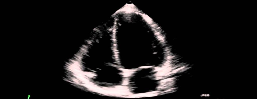
15 Where did my patient go???
16 Items to measure on echocardiography
17 What to quantify Chamber dimensions Global systolic function Regional function Left ventricle Right ventricle Atria Aortic territory Vena cava
18 What to quantify LEFT VENTRICLE Cavity size: LVEDD, LVEDD indexed to BSA Wall thickness: IVSD, PWD, RWT Mass: LVM, LVMI Volumes: LVEDV, LVESV Systolic function: LVEF (biplane Simpson s)
19 What to quantify LEFT ATRIUM Length: LA length 4C, LA length 2C Area: LA area 4C, LA area 2C Volume: LAV, LAV indexed to BSA
20 What to quantify AORTIC ROOT (diamters) LV outflow tract (LVOT) AV annulus Sinus of Valsalva (SOV) Sinotubular junction (STJ) Prox. Ascending aorta
21 The debate: thresholds for severity Partition values for severity of abnormality Normative data on classification of severity Standard cut-offs The resolution Experience-based partition values only for: LVEF and LA volume

22

23 Correct quantification techniques
24 General guidelines All measurements should be performed on more than 1 cardiac cycle (to account for interbeat variability) e.g. average of 3 beats for sinus, and at least 5 beats for AF However, the use of representative bets is acceptable in the clinical setting The same range of normal values generally apply for TTE and TEE, but certain views are more optimal in selected scenarios Range of normal values are provided for LV size, function and mass as well as LA volume, For other parameters, measurements exceeding SDs should be classified as abnormal.
25 General guidelines Get basic clinical profile to come up with a pre-test analysis. Accurately measure the patient s height and weight, and compute the body surface area. Check the patient s rhythm and ensure a good ECG tracing.
26 Left ventricle: Size Internal linear dimensions Linear dimensions are used to provide geometric conclusions about the left ventricle

27 Left ventricle: Size Internal linear dimensions Two methods: M-mode approach and direct 2D measurement M-mode approach has high temporal resolution but is prone to tangentiality issues

28 Left ventricle: Size Internal linear dimensions M-mode measurements guided by targeted SAX or PLAX view Simple for screening large populations Beam orientation may be off-axis Inaccurate in abnormal LV geometry

29 Left ventricle Size Internal linear dimensions get good 2D PLAX view measure LVEDD at end-diastole, when MV leaflets are fully open draw caliper perpendicular to long axis of LV, at level of MV leaflet tips locate landmarks properly: endocardiumcavity interface LV internal dimensions: directly measure on 2D PLAX
30 Left ventricle Size LV mass Perform all measurements at end-diastole 3 methods Linear or Cube method 2D truncated ellipsoid 3D based formula


31 Left ventricle Size LV mass Perform all measurements at end-diastole 3 methods Linear or Cube method 2D truncated ellipsoid 3D based formula

32 Left ventricle Size LV mass Perform all measurements at end-diastole 3 methods Linear or Cube method 2D truncated ellipsoid 3D based formula LV mass: 2D-guided, or 3D ideally

33 Left ventricle Size LV mass index Reference values
34 Left ventricle Size LV geometrical conclusions Based on: LVMI RWT sex LV geometry conclusion is important
35 Key Insight LV linear dimensions provide information regarding geometrical conclusions.
36 Sample Computations Clinical scenario 45 male Hypertensive Non-adherent to meds Height: 1.5 m Weight: 60 kg BSA: 1.58 m 2
37 Sample Computations LV dimensions LVEDD: 4.0 cm IVSD : 1.3 cm PWD : 1.6 cm
 / 4.0 = 0.")
38 Sample Computations LV dimensions LVEDD indexed to BSA = 4.0 cm / 1.58 m2 = 2.5 cm/m 2 RWT = (1.6 x 2) / 4.0 = 0.80
39 Sample Computations LV dimensions LV mass = 0.8 x 1.04 [ (IVSD + LVEDD + PWD) 3 LVEDD 3 ] g = 0.8 x 1.04 [ ( ) ] g = 221 g LV mass index = LV mass / BSA = 221 g / 1.58 m 2 = 140 g/m 2 Patient data LVEDD: 4.0 cm IVSD : 1.3 cm PWD : 1.6 cm
 LVEDD BSA : 2.5 cm/m2 (N) LVMI : 140 g/m 2 RWT : 0.8 LVMI: 140 g/m 2 RWT: 0.")
40 Sample Computations LV geometrical conclusions Patient data: 45 male LVEDD : 4.0 cm (N) LVEDD BSA : 2.5 cm/m2 (N) LVMI : 140 g/m 2 RWT : 0.8 LVMI: 140 g/m 2 RWT: 0.8 Concentric LV hypertrophy
41 Left ventricle Size Volumes Volumes are used for EF calculation
42 Left ventricle Size Volumes Two methods: using 2DE or 3DE Linear derivations based on geometric assumptions may be inaccurate Teichholz and Quinones methods are no longer recommended for clinical use

43 Left ventricle Size Volumes: Biplane disk summation Corrects for shape distortions Apex frequently foreshortened

44 Left ventricle Size Volumes: Area-length Partial correction for shape distortion Heavily based on geometrical assumptions

45 Left ventricle Size Volumes: Endocardial enhancement Useful for suboptimal windows Acoustic shadowing in LV basal segments with excess contrast

46 Left ventricle Size Volumes: 3D data sets No geometrical assumption Unaffected by foreshortening More accurate and reproducible Lower temporal resolution Image quality dependent
 and enddiastole (MLd).")
47 Left ventricle: Global systolic function Global longitudinal strain Angle and vendor independent with established prognostic value where ML is myocardial length at end-systole (MLs) and enddiastole (MLd).

 or")
48 Left ventricle: Global systolic function 2DE-derived LVEF Severity classification Adjusted for sex LV systolic function: 2DE-derived LVEF (biplane Simpson s formula) or 3Dderived, if applicable
49 Key Insight LV volumes are used to indicate systolic function via the ejection fraction.

50 Left ventricle: Regional function LV segmentation


51 Left ventricle: Regional function Correlation Keep perfusion territories in mind when regional wall motion
52 Key Insight When numbers do not fit, re-assess and correlate with visual estimation and clinical data.
53 Left atrial dimensions LA functions Contractile pump Reservoir Conduit Echo uses of LA size Diastolic function LV function Intracardiac pressure Valve lesions TTE is preferred approach

54 Left atrial dimensions Correlation s


55 Left atrial dimensions LA volumetric measurement Area-length Disc summation 3D volumes
56 Left atrial dimensions Biplane area length method Enables accurate assessment of the asymmetric remodeling of the left atrium More robust predictor of cardiovascular events than linear or area measurements


57 Left atrial dimensions Biplane area length method LA volume index: biplane area-length is preferred

58 Left atrial dimensions LA volume index 2006 cut-off: 28 ml/m cut-off: 34 ml/m 2 much more studies to back up new cutoff Volume index is the preferred measure of LA size
59 Key Insight Take equally great care in measuring the LA size, since this will have several echo and clinical implications.

60 Right atrial dimensions RA size measurement Less research and fewer clinical outcomes Apical 4 chamber view is preferred Diameters and volumes

61 Right atrial dimensions RA size measurement Less research and fewer clinical outcomes Apical 4 chamber view is preferred Diameters and volumes

62 Right atrial dimensions RA size measurement RA volume Calculated using singleplane area-length or disk summation techniques normal ranges 25+7 ml/m 2 in men 21+6 ml/m 2 in women RA size: recommended parameter is RA volume
63 Aortic territory General rules Get good long-axis view with a central AV coaptation point calcium protuberances should be considered as part of the lumen, not of the aortic wall, and therefore excluded from the diameter measurement AV annulus: Inner edge to inner edge technique Other parts: leading edge to leading edge
64 Aortic root dimensions Correlation Annulus root


65 Aortic root dimensions Proper measurement AV annulus SOV STJ Asc AV annulus: Inner edge technique; the rest: leading edge technique
66 Aortic root dimensions Feigenbaum H, Armstrong WF, Ryan T, eds. Feigenbaum s Echocardiography. 6th ed. Philadelphia, PA: Lippincott Williams & Wilkins; p673; Ascending aorta values from Erbel et al


67 Key Insight Optimize the view before taking aortic measurements.
68 Recap of Session Outline Understanding the importance of correct chamber quantification techniques Knowing which aspects to quantify: chamber dimensions, parameters of function Mastering the technique of quantification: image optimization, basic strategies, tips and pitfalls

69
70 Questions Sigmoid septum: how to address? HCM: how to report? When EF values differ: how to decide? When numbers and visual estimate contradict LV mass: when LVMI is increased, LVEDD and RWT are normal: how to conclude? RWT: double PWD or average IVSD and PWD?
Quantification of Cardiac Chamber Size
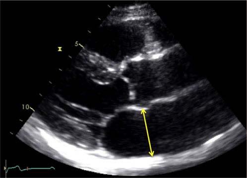
 2017 KSE 2017-11-25 Quantification of Cardiac Chamber Size Division of Cardiology Keimyung University Dongsan Medical Center In-Cheol Kim M.D., Ph.D. LV size and function Internal linear dimensions PLX
Cardiac Chamber Quantification by Echocardiography
 Cardiac Chamber Quantification by Echocardiography Maryam Bokhamseen, RCS, RCDS, EACVI Echotechnologist ǁ, Non invasive Cardiac Laboratory King Abdulaziz Cardiac Center. Outline: Introduction. Background
Chamber Quantitation Guidelines: What is New?
 Chamber Quantitation Guidelines: What is New? Roberto M Lang, MD J AM Soc Echocardiogr 2005; 18:1440-1463 1 Approximately 10,000 citations iase in itune Cardiac Chamber Quantification: What is New? Database
Martin G. Keane, MD, FASE Temple University School of Medicine
 Martin G. Keane, MD, FASE Temple University School of Medicine Measurement of end-diastolic LV internal diameter (LVIDd) made by properly-oriented M-Mode techniques in the Parasternal Long Axis View (PLAX):
Echocardiographic Assessment of the Left Ventricle
 Echocardiographic Assessment of the Left Ventricle Theodora Zaglavara, MD, PhD, BSCI/BSCCT Department of Cardiovascular Imaging INTERBALKAN EUROPEAN MEDICAL CENTER 2015 The quantification of cardiac chamber
좌심실수축기능평가 Cardiac Function
 Basic Echo Review Course 좌심실수축기능평가 Cardiac Function Seonghoon Choi Cardiology Hallym university LV systolic function Systolic function 좌심실수축기능 - 심근의수축으로심실에서혈액을대동맥으로박출하는기능 실제임상에서 LV function 의의미 1Diagnosis
LV FUNCTION ASSESSMENT: WHAT IS BEYOND EJECTION FRACTION
 LV FUNCTION ASSESSMENT: WHAT IS BEYOND EJECTION FRACTION Jamilah S AlRahimi Assistant Professor, KSU-HS Consultant Noninvasive Cardiology KFCC, MNGHA-WR Introduction LV function assessment in Heart Failure:
Adel Hasanin Ahmed 1 LV MORPHOLOGY
 Adel Hasanin Ahmed 1 LV MORPHOLOGY The left ventricular wall comprises three layers- middle circumferential layer and superficial and deep longitudinal layers: 1. Subepicardial longitudinal layer (25%
Assessment of LV systolic function
 Tutorial 5 - Assessment of LV systolic function Assessment of LV systolic function A knowledge of the LV systolic function is crucial in the undertanding of and management of unstable hemodynamics or a
RIGHT VENTRICULAR SIZE AND FUNCTION
 RIGHT VENTRICULAR SIZE AND FUNCTION Edwin S. Tucay, MD, FPCC, FPCC, FPSE Philippine Society of Echocardiography Quezon City, Philippines Echo Mission, BRTTH, Legaspi City, July 1-2, 2016 NO DISCLOSURE
ECHOCARDIOGRAPHY DATA REPORT FORM
 Patient ID Patient Study ID AVM - - Date of form completion / / 20 Initials of person completing the form mm dd yyyy Study period Preoperative Postoperative Operative 6-month f/u 1-year f/u 2-year f/u
Value of echocardiography in chronic dyspnea
 Value of echocardiography in chronic dyspnea Jahrestagung Schweizerische Gesellschaft für /Schweizerische Gesellschaft für Pneumologie B. Kaufmann 16.06.2016 Chronic dyspnea Shortness of breath lasting
Imaging Guide Echocardiography
 Imaging Guide Guide to Small Animal Echocardiography using the Vevo Imaging Systems System Compatibility: This guide contains instructions and suggestions for work on the Vevo2100, VevoLAZR, Vevo 3100
Evaluation of Left Ventricular Function and Hypertrophy Gerard P. Aurigemma MD
 Evaluation of Left Ventricular Function and Hypertrophy Gerard P. Aurigemma MD Board Review Course 2017 43 year old health assistant Severe resistant HTN LT BSA 2 Height 64 1 Here is the M mode echocardiogram
Right Heart Evaluation ASE Guidelines Review. Chris Mann RDCS, RCS, FASE Faculty, Echocardiography Pitt Community College Greenville, NC
 Right Heart Evaluation ASE Guidelines Review Chris Mann RDCS, RCS, FASE Faculty, Echocardiography Pitt Community College Greenville, NC Objectives Briefly review right atrial and right ventricular anatomy
Advanced imaging of the left atrium - strain, CT, 3D, MRI -
 Advanced imaging of the left atrium - strain, CT, 3D, MRI - Monica Rosca, MD Carol Davila University of Medicine and Pharmacy, Bucharest, Romania Declaration of interest: I have nothing to declare Case
LV geometric and functional changes in VHD: How to assess? Mi-Seung Shin M.D., Ph.D. Gachon University Gil Hospital
 LV geometric and functional changes in VHD: How to assess? Mi-Seung Shin M.D., Ph.D. Gachon University Gil Hospital LV inflow across MV LV LV outflow across AV LV LV geometric changes Pressure overload
THE LEFT ATRIUM HOW CAN ECHO HELP US?
 THE LEFT ATRIUM HOW CAN ECHO HELP US? Dr. Dragos COZMA BACKGROUND Left atrium (LA) dilation can occur in a broad spectrum of cardiovascular diseases including hypertension, left ventricular dysfunction,
Left atrial function. Aliakbar Arvandi MD
 In the clinic Left atrial function Abstract The left atrium (LA) is a left posterior cardiac chamber which is located adjacent to the esophagus. It is separated from the right atrium by the inter-atrial
Hemodynamic Assessment. Assessment of Systolic Function Doppler Hemodynamics
 Hemodynamic Assessment Matt M. Umland, RDCS, FASE Aurora Medical Group Milwaukee, WI Assessment of Systolic Function Doppler Hemodynamics Stroke Volume Cardiac Output Cardiac Index Tei Index/Index of myocardial
Conflict of Interests
 The Left Ventricle: How Should We Quantify Its Size and Function; Is It Time for 3D in Everyone? Roberto M Lang, MD Conflict of Interests Philips Medical Imaging Research Grants Speakers bureau Advisory
Basic Assessment of Left Ventricular Systolic Function
 WINFOCUS BASIC ECHO (WBE) Basic Assessment of Left Ventricular Systolic Function Ritesh Dhar, MD Director, Echocardiography Lab and Staff Cardiologist Intermountain Medical Center Murray, Utah Outline
Adult Echocardiography Examination Content Outline
 Adult Echocardiography Examination Content Outline (Outline Summary) # Domain Subdomain Percentage 1 2 3 4 5 Anatomy and Physiology Pathology Clinical Care and Safety Measurement Techniques, Maneuvers,
What is the Definition of Small Systemic Ventricle. Hong Ryang Kil, MD Department of Pediatrics, College of Medicine, Chungnam National University
 What is the Definition of Small Systemic Ventricle Hong Ryang Kil, MD Department of Pediatrics, College of Medicine, Chungnam National University Contents Introduction Aortic valve stenosis Aortic coarctation
NEW GUIDELINES. A Guideline Protocol for the Assessment of Aortic Regurgitation From the British Society of Echocardiography Education Committee
 NEW GUIDELINES A Guideline Protocol for the Assessment of Aortic Regurgitation From the British Society of Echocardiography Education Committee Gill Wharton, Prathap Kanagala (Lead Authors) Richard Steeds
Part II: Fundamentals of 3D Echocardiography: Acquisition and Application
 Part II: Fundamentals of 3D Echocardiography: Acquisition and Application Dr. Bruce Bollen 3D matrix array TEE probes provide options for both 2D and 3D imaging. Indeed, their utility in obtaining multiple
Vevo 2100 System Cardio Measurements. Dieter Fuchs, PhD FUJIFILM VisualSonics, Inc.
 Vevo 2100 System Cardio Measurements Dieter Fuchs, PhD FUJIFILM VisualSonics, Inc. dfuchs@visualsonics.com Instructions This document is a guideline on how to assess cardiac function in rodents imaged
cardiac imaging planes planning basic cardiac & aortic views for MR
 cardiac imaging planes planning basic cardiac & aortic views for MR Dianna M. E. Bardo, M. D. Assistant Professor of Radiology & Cardiovascular Medicine Director of Cardiac Imaging cardiac imaging planes
Basic Approach to the Echocardiographic Evaluation of Ventricular Diastolic Function
 Basic Approach to the Echocardiographic Evaluation of Ventricular Diastolic Function J A F E R A L I, M D U N I V E R S I T Y H O S P I T A L S C A S E M E D I C A L C E N T E R S T A F F C A R D I O T
Comprehensive Echo Assessment of Aortic Stenosis
 Comprehensive Echo Assessment of Aortic Stenosis Smonporn Boonyaratavej, MD, MSc King Chulalongkorn Memorial Hospital Bangkok, Thailand Management of Valvular AS Medical and interventional approaches to
New Cardiovascular Devices and Interventions: Non-Contrast MRI for TAVR Abhishek Chaturvedi Assistant Professor. Cardiothoracic Radiology
 New Cardiovascular Devices and Interventions: Non-Contrast MRI for TAVR Abhishek Chaturvedi Assistant Professor Cardiothoracic Radiology Disclosure I have no disclosure pertinent to this presentation.
British Society of Echocardiography
 British Society of Echocardiography Affiliated to the British Cardiac Society A Minimum Dataset for a Standard Adult Transthoracic Echocardiogram From the British Society of Echocardiography Education
Echo Assessment Pre-TAVI
 Disclosure Statement of Financial Interest Within the past 12 months, I or my spouse/partner have had a financial Interest /arrangement or affiliation with the organization(s) listed below Echocardiographic
Echocardiography for the Electrophysiologist: Day-to-day practice. Emmanuel Fares, MD
 Echocardiography for the Electrophysiologist: Day-to-day practice Emmanuel Fares, MD EP and pacing service, Department of Cardiovascular Medicine, Cairo University Agenda Role of echo in arrhythmia management:
How NOT to miss Hypertrophic Cardiomyopathy? Adaya Weissler-Snir, MD University Health Network, University of Toronto
 How NOT to miss Hypertrophic Cardiomyopathy? Adaya Weissler-Snir, MD University Health Network, University of Toronto Introduction Hypertrophic cardiomyopathy is the most common genetic cardiomyopathy,
Certificate in Clinician Performed Ultrasound (CCPU) Syllabus. Rapid Cardiac Echo (RCE)
 Syllabus. Rapid Cardiac Echo (RCE)") Certificate in Clinician Performed Ultrasound (CCPU) Syllabus Rapid Cardiac Echo (RCE) Purpose: Rapid Cardiac Echocardiography (RCE) This unit is designed to cover the theoretical and practical curriculum
Certificate in Clinician Performed Ultrasound (CCPU) Syllabus Rapid Cardiac Echo (RCE) Purpose: Rapid Cardiac Echocardiography (RCE) This unit is designed to cover the theoretical and practical curriculum
Echocardiographic Cardiovascular Risk Stratification: Beyond Ejection Fraction
 Echocardiographic Cardiovascular Risk Stratification: Beyond Ejection Fraction October 4, 2014 James S. Lee, M.D., F.A.C.C. Associates in Cardiology, P.A. Silver Spring, M.D. Disclosures Financial none
Tissue Doppler and Strain Imaging
 Tissue Doppler and Strain Imaging Steven J. Lester MD, FRCP(C), FACC, FASE Relevant Financial Relationship(s) None Off Label Usage None 1 Objective way with which to quantify the minor amplitude and temporal
B-Mode measurements protocols:
 Application Note How to Perform the Most Commonly Used Measurements from the Cardiac Measurements Package associated with Calculations of Cardiac Function using the Vevo Lab Objective The Vevo LAB offline
Prospect Cardiac Packages. S-Sharp
 Prospect Cardiac Packages S-Sharp B mode: Teichholz: Teichholz formula LV Volume 2D: modified Simpson's rule method ALM: area length method LV Volume (Intg.): integral method M mode: Long axis: Teichholz
ECHOCARDIOGRAPHY CHAPTER INTRODUCTION
 CHAPTER 2 ECHOCARDIOGRAPHY 2.1 INTRODUCTION The use of ultrasound in the diagnosis of cardiac disease has been available for more than four decades with the diagnostic potential of this modality first
Heart Failure in Women: Dr Goh Ping Ping Cardiologist Asian Heart & Vascular Centre
 Heart Failure in Women: More than EF? Dr Goh Ping Ping Cardiologist Asian Heart & Vascular Centre Overview Review pathophysiology as it relates to diagnosis and management Rational approach to workup:
When Does 3D Echo Make A Difference?
 When Does 3D Echo Make A Difference? Wendy Tsang, MD, SM Assistant Professor, University of Toronto Toronto General Hospital, University Health Network 1 Practical Applications of 3D Echocardiography Recommended
Evaluation of Left Ventricular Diastolic Dysfunction by Doppler and 2D Speckle-tracking Imaging in Patients with Primary Pulmonary Hypertension
 ESC Congress 2011.No 85975 Evaluation of Left Ventricular Diastolic Dysfunction by Doppler and 2D Speckle-tracking Imaging in Patients with Primary Pulmonary Hypertension Second Department of Internal
Echocardiography: Guidelines for Valve Quantification
 Echocardiography: Guidelines for Echocardiography: Guidelines for Chamber Quantification British Society of Echocardiography Education Committee Richard Steeds (Chair), Gill Wharton (Lead Author), Jane
10/7/2013. Systolic Function How to Measure, How Accurate is Echo, Role of Contrast. Thanks to our Course Director: Neil J.
 Systolic Function How to Measure, How Accurate is Echo, Role of Contrast Neil J. Weissman, MD MedStar Health Research Institute & Professor of Medicine Georgetown University Washington, D.C. No Disclosures
Top 10 Facts in Contrast Echocardiography. Pamela R. Burgess, BS, RDCS, RDMS, RVT, FASE
 Top 10 Facts in Contrast Echocardiography Pamela R. Burgess, BS, RDCS, RDMS, RVT, FASE Presenter Disclosure The following relationship exist related to this presentation: Pamela R. Burgess, BS, RDCS, RDMS,
Prof. JL Zamorano Hospital Universitario Ramón y Cajal
 Prof. JL Zamorano Hospital Universitario Ramón y Cajal Fully Automated Quantification Software Adaptive analytical algorithm consists in knowledge-based identification of global shape and specific adaptation
Revealing new insights. irotate electronic rotation and xplane adjustable biplane imaging. Ultrasound cardiology. irotate and xplane
 Ultrasound cardiology irotate and xplane Revealing new insights irotate electronic rotation and xplane adjustable biplane imaging Annemien van den Bosch and Jackie McGhie Department of Cardiology, Erasmus
5 Working With Measurements
 5 Working With Measurements Measurement Overview Measurements accompanying ultrasound images supplement other clinical procedures available to the attending physician. Accuracy of the measurements is determined
PROSTHETIC VALVE BOARD REVIEW
 PROSTHETIC VALVE BOARD REVIEW The correct answer D This two chamber view shows a porcine mitral prosthesis with the typical appearance of the struts although the leaflets are not well seen. The valve
Recommendations for chamber quantification *
 Eur J Echocardiography (2006) 7, 79e108 GUIDELINES Recommendations for chamber quantification * Roberto M. Lang, Michelle Bierig, Richard B. Devereux, Frank A. Flachskampf *, Elyse Foster, Patricia A.
Tissue Doppler and Strain Imaging
 Tissue Doppler and Strain Imaging Steven J. Lester MD, FRCP(C), FACC, FASE Relevant Financial Relationship(s) None Off Label Usage None 1 Objective way with which to quantify the minor amplitude and temporal
Quantifying LV function how good are we?
 Quantifying LV function how good are we? Professor Alan G Fraser Wales Heart Research Institute Cardiff University, U.K. Support for research from Hitachi Aloka, & GE Ultrasound Visual assessment of synchronicity
4D Auto LAQ (Left Atrial Quantification)
") 4D Auto LAQ (Left Atrial Quantification) Introduction There has been an increased interest in quantification of the left atrium (LA) for various types of diseases; e.g. heart failure, heart valve diseases,
4D Auto LAQ (Left Atrial Quantification) Introduction There has been an increased interest in quantification of the left atrium (LA) for various types of diseases; e.g. heart failure, heart valve diseases,
Ιπποκράτειες μέρες καρδιολογίας Θεσσαλονίκη, 9-10 Μαρτίου Φωτεινή Α. Λαζαρίδου Επιμελήτρια Α Γενικό Νοσοκομείο Αγιος Παύλος, Θεσσαλονίκη
 Ιπποκράτειες μέρες καρδιολογίας Θεσσαλονίκη, 9-10 Μαρτίου 2018 Φωτεινή Α. Λαζαρίδου Επιμελήτρια Α Γενικό Νοσοκομείο Αγιος Παύλος, Θεσσαλονίκη RV shape Triangular shape in frontal plane crescent shape in
Review of Cardiac Imaging Modalities in the Renal Patient. George Youssef
 Review of Cardiac Imaging Modalities in the Renal Patient George Youssef ECHO Left ventricular hypertrophy (LVH) assessment Diastolic dysfunction Stress ECHO Cardiac CT angiography Echocardiography - positives
POSITION PAPER. Keywords Adult echocardiography Transthoracic echocardiography Ventricular function Normal values
 European Heart Journal Cardiovascular Imaging (2015) 16, 233 271 doi:10.1093/ehjci/jev014 POSITION PAPER Recommendations for Cardiac Chamber Quantification by Echocardiography in Adults: An Update from
Assessment of cardiac function with 3D echocardiography. Đánh giá chức năng tim bằng siêu âm tim 3D
 Assessment of cardiac function with 3D echocardiography Đánh giá chức năng tim bằng siêu âm tim 3D TS. BS. Nguyễn Thị Thu Hoài Viện Tim Mạch Quốc Gia Việt Nam TỪ SIÊU ÂM M-mode ĐẾN SIÊU ÂM 3D TỪ SIÊU ÂM
Appendix II: ECHOCARDIOGRAPHY ANALYSIS
 Appendix II: ECHOCARDIOGRAPHY ANALYSIS Two-Dimensional (2D) imaging was performed using the Vivid 7 Advantage cardiovascular ultrasound system (GE Medical Systems, Milwaukee) with a frame rate of 400 frames
Imaging Assessment of Aortic Stenosis/Aortic Regurgitation
 Imaging Assessment of Aortic Stenosis/Aortic Regurgitation Craig E Fleishman, MD FACC FASE The Heart Center at Arnold Palmer Hospital for Children, Orlando SCAI Fall Fellows Course 2014 Las Vegas Disclosure
The importance of left atrium in LV diastolic function
 II Baltic Heart Failure Meeting and Congress of Latvian Society of Cardiology The importance of left atrium in LV diastolic function Dr. Artem Kalinin Eastern Clinical University Hospital Riga 30.09.2010.
Nancy Goldman Cutler, MD Beaumont Children s Hospital Royal Oak, Mi
 Nancy Goldman Cutler, MD Beaumont Children s Hospital Royal Oak, Mi Identify increased LV wall thickness (WT) Understand increased WT in athletes Understand hypertrophic cardiomyopathy (HCM) Enhance understanding
Cardiac Magnetic Resonance in pregnant women
 Cardiac Magnetic Resonance in pregnant women Chen SSM, Leeton L, Dennis AT Royal Women s Hospital and The University of Melbourne, Parkville, Australia alicia.dennis@thewomens.org.au Quantification of
Questions on Chamber Quantitation
 Questions on Chamber Quantitation @RobertoMLang Which of the following statements is true? 1. The aortic annulus should be measured in midsystole. 2. The aortic annulus should be measured in enddiastole.
Evaluation of Ejection Fraction in Patients with Cardiac Resynchronization Therapy by Two and Three Dimensional Echocardiography
 58 Original article Evaluation of Ejection Fraction in Patients with Cardiac Resynchronization Therapy by Two and Three Dimensional Echocardiography Anil OM Department of cardiology, Manmohan Cardiothoracic
HYPERTROPHY: Behind the curtain. V. Yotova St. Radboud Medical University Center, Nijmegen
 HYPERTROPHY: Behind the curtain V. Yotova St. Radboud Medical University Center, Nijmegen Disclosure of interest: none Relative wall thickness (cm) M 0.22 0.42 0.43 0.47 0.48 0.52 0.53 F 0.24 0.42 0.43
Functional anatomy of the aortic root. ΔΡΟΣΟΣ ΓΕΩΡΓΙΟΣ Διεσθσνηής Καρδιοθωρακοτειροσργικής Κλινικής Γ.Ν. «Γ. Παπανικολάοσ» Θεζζαλονίκη
 Functional anatomy of the aortic root ΔΡΟΣΟΣ ΓΕΩΡΓΙΟΣ Διεσθσνηής Καρδιοθωρακοτειροσργικής Κλινικής Γ.Ν. «Γ. Παπανικολάοσ» Θεζζαλονίκη What is the aortic root? represents the outflow tract from the LV provides
Myocardial Strain Imaging in Cardiac Diseases and Cardiomyopathies.
 Myocardial Strain Imaging in Cardiac Diseases and Cardiomyopathies. Session: Cardiomyopathy Tarun Pandey MD, FRCR. Associate Professor University of Arkansas for Medical Sciences Disclosures No relevant
Echocardiography as a diagnostic and management tool in medical emergencies
 Echocardiography as a diagnostic and management tool in medical emergencies Frank van der Heusen MD Department of Anesthesia and perioperative Care UCSF Medical Center Objective of this presentation Indications
Index of subjects. effect on ventricular tachycardia 30 treatment with 101, 116 boosterpump 80 Brockenbrough phenomenon 55, 125
 145 Index of subjects A accessory pathways 3 amiodarone 4, 5, 6, 23, 30, 97, 102 angina pectoris 4, 24, 1l0, 137, 139, 140 angulation, of cavity 73, 74 aorta aortic flow velocity 2 aortic insufficiency
Pregnancy, Heart Disease and Imaging. Hemodynamics. Decreased systemic vascular resistance. Physiology anemia
 Pregnancy, Heart Disease and Imaging Sangeeta Shah, MD, FASE, FACC Associate Professor, Ochsner Clinical School of Medicine Advanced CV Imaging and Adult Congenital Heart Disease New Orleans, LA Hemodynamics
Tissue Doppler and Strain Imaging. Steven J. Lester MD, FRCP(C), FACC, FASE
, FACC, FASE") Tissue Doppler and Strain Imaging Steven J. Lester MD, FRCP(C), FACC, FASE Relevant Financial Relationship(s) None Off Label Usage None a. Turn the wall filters on and turn down the receiver gain. b. Turn
Tissue Doppler and Strain Imaging Steven J. Lester MD, FRCP(C), FACC, FASE Relevant Financial Relationship(s) None Off Label Usage None a. Turn the wall filters on and turn down the receiver gain. b. Turn
Η ηχωκαρδιολογία στην διάγνωση κα πρόγνωση της καρδιακής ανεπάρκειας µε µειωµένο και φυσιολογικό κλάσµα εξώθησης
 Η ηχωκαρδιολογία στην διάγνωση κα πρόγνωση της καρδιακής ανεπάρκειας µε µειωµένο και φυσιολογικό κλάσµα εξώθησης Βασίλειος Σαχπεκίδης Επιµελητής Β Καρδιολογίας Γ.Ν. Παπαγεωργίου Θεσσαλονίκη ESC Guidelines
The Athletic Heart Takes Shape: Overview of Cardiac Remodeling Rory B. Weiner, MD
 The Athletic Heart Takes Shape: Overview of Cardiac Remodeling Rory B. Weiner, MD Cardiovascular Performance Program, MGH Assistant Professor of Medicine, Harvard Medical School Outline Athlete s Heart
Highlights from EuroEcho 2009 Echo in cardiomyopathies
 Highlights from EuroEcho 2009 Echo in cardiomyopathies Bogdan A. Popescu University of Medicine and Pharmacy, Bucharest, Romania ESC Congress 2010 Hypertrophic cardiomyopathy To determine the differences
Normal TTE/TEE Examinations
 Normal TTE/TEE Examinations Geoffrey A. Rose, MD FACC FASE Sanger Heart & Vascular Institute Before you begin imaging... Obtain the patient s Height Weight BP PLAX View PLAX View Is apex @ 9-10 o clock?
PRELIMINARY STUDIES OF LEFT VENTRICULAR WALL THICKNESS AND MASS OF NORMOTENSIVE AND HYPERTENSIVE SUBJECTS USING M-MODE ECHOCARDIOGRAPHY
 Malaysian Journal of Medical Sciences, Vol. 9, No. 1, January 22 (28-33) ORIGINAL ARTICLE PRELIMINARY STUDIES OF LEFT VENTRICULAR WALL THICKNESS AND MASS OF NORMOTENSIVE AND HYPERTENSIVE SUBJECTS USING
Outline. EuroScore II. Society of Thoracic Surgeons Score. EuroScore II
 SURGICAL RISK IN VALVULAR HEART DISEASE: WHAT 2D AND 3D ECHO CAN TELL YOU AND WHAT THEY CAN'T Ernesto E Salcedo, MD Professor of Medicine University of Colorado School of Medicine Director of Echocardiography
Echocardiography. Guidelines for Valve and Chamber Quantification. In partnership with
 Echocardiography Guidelines for Valve and Chamber Quantification In partnership with Explanatory note & references These guidelines have been developed by the Education Committee of the British Society
Normal values for cardiovascular magnetic resonance in adults and children
 Kawel-Boehm et al. Journal of Cardiovascular Magnetic Resonance (2015) 17:29 DOI 10.1186/s12968-015-0111-7 REVIEW Normal values for cardiovascular magnetic resonance in adults and children Nadine Kawel-Boehm
Aortic Stenosis: Spectrum of Disease, Low Flow/Low Gradient and Variants
 Aortic Stenosis: Spectrum of Disease, Low Flow/Low Gradient and Variants Martin G. Keane, MD, FASE Professor of Medicine Lewis Katz School of Medicine at Temple University Basic root structure Parasternal
PART II ECHOCARDIOGRAPHY LABORATORY OPERATIONS ADULT TRANSTHORACIC ECHOCARDIOGRAPHY TESTING
 PART II ECHOCARDIOGRAPHY LABORATORY OPERATIONS ADULT TRANSTHORACIC ECHOCARDIOGRAPHY TESTING STANDARD - Primary Instrumentation 1.1 Cardiac Ultrasound Systems SECTION 1 Instrumentation Ultrasound instruments
Regurgitant Lesions. Bicol Hospital, Legazpi City, Philippines July Gregg S. Pressman MD, FACC, FASE Einstein Medical Center Philadelphia, USA
 Regurgitant Lesions Bicol Hospital, Legazpi City, Philippines July 2016 Gregg S. Pressman MD, FACC, FASE Einstein Medical Center Philadelphia, USA Aortic Insufficiency Valve anatomy and function LVOT and
Pulmonary Hypertension. Echocardiography: Pearls & Pitfalls
 Pulmonary Hypertension Echocardiography: Pearls & Pitfalls Αθανάσιος Γ. Κουτσάκης Ειδικευόμενος Καρδιολογίας Α Καρδιολογική Κλινική ΑΠΘ Σεμινάρια Ομάδων Εργασίας Ελληνικής Καρδιολογικής Εταιρείας Ιωάννινα,
A New Method to Rapidly Evaluate LVEF from a Contractility Polar Map. Lebeau et al.
 A New Method to Rapidly Evaluate LVEF from a Contractility Polar Map Lebeau et al. Good afternoon It is my pleasure to present to you a new method to rapidly evaluate LVEF from a contractility polar map
Managing Hypertrophic Cardiomyopathy with Imaging. Gisela C. Mueller University of Michigan Department of Radiology
 Managing Hypertrophic Cardiomyopathy with Imaging Gisela C. Mueller University of Michigan Department of Radiology Disclosures Gadolinium contrast material for cardiac MRI Acronyms Afib CAD Atrial fibrillation
Dr Winnie Sze-Wun Chan. Cardiac Team Deputy Team Head Department of Radiology and Imaging Queen Elizabeth Hospital Hong Kong
 Dr Winnie Sze-Wun Chan Cardiac Team Deputy Team Head Department of Radiology and Imaging Queen Elizabeth Hospital Hong Kong Why? Is CT reliable? How to perform the CT study? How to interpret the CT study?
Aortic Valve Practice Guidelines: What Has Changed and What You Need to Know
 Aortic Valve Practice Guidelines: What Has Changed and What You Need to Know James F. Burke, MD Program Director Cardiovascular Disease Fellowship Lankenau Medical Center Disclosure Dr. Burke has no conflicts
LA Function analysis Marcia Barbosa Vice Presidente - Brazilian Soc of Cardiology President-elect - Interamerican Soc of Cardiology
 LA Function analysis Marcia Barbosa Vice Presidente - Brazilian Soc of Cardiology President-elect - Interamerican Soc of Cardiology Belo Horizonte Brazil DECLARATION OF CONFLICT OF INTEREST Nothing to
VMS Quick Reference Guide
 VMS Quick Reference Guide Connecting to the VMS Workstation Ensure Ultrasound system is up and running prior to starting VMS. Connect the video cable from the ultrasound machine to the VMS Integrated Station.
Conflict of Interests
 New Approaches to Systolic Function: 4D Roberto M Lang, MD Conflict of Interests Philips Medical Imaging Research Grants Speakers bureau Advisory bureau Tomtec Research Grants Epsilon Research Grants 1
Mixed aortic valve disease
 Mixed aortic valve disease IOANNIS NTALAS MD, PhD Cardiologist, Clinical Fellow in Cardiovascular Imaging & Non-Invasive Cardiology, St Thomas Hospital School of Biomedical Engineering & Imaging Sciences
Aortic stenosis aetiology: morphology of calcific AS,
 How to improve patient selection in aortic stenosis? Fausto J. Pinto, FESC Aortic stenosis aetiology: morphology of calcific AS, bicuspid valve, and rheumatic AS (Adapted from C. Otto, Principles of
ARTIFACTS: THEORY AND ILLUSTRATIVE EXAMPLES
 ARTIFACTS: THEORY AND ILLUSTRATIVE EXAMPLES Robert A. Levine, M.D. Marielle Scherrer-Crosbie, M.D. Eric M. Isselbacher, M.D. No conflicts of interest Philippe Bertrand, Pieter Vendervoort, Hasselt and
Back to Basics: Common Errors In Quantitation In Everyday Practice
 Back to Basics: Common Errors In Quantitation In Everyday Practice Deborah Agler, ACS, RDCS, FASE October 9, 2017 ASE: Echo Florida Rebecca T. Hahn, MD Director of Interventional Echocardiography Professor
Evaluation of Systolic Function of the Left Ventricle
 Evaluation of Systolic Function of the Left Ventricle Roxy Senior MD DM FRCP FESC FACC and Vinay Kumar Bhatia PhD MRCP Department of Cardiovascular Medicine, Northwick Park Hospital and Institute for Medical
Stephen Glen ISCHAEMIC HEART DISEASE AND LEFT VENTRICULAR FUNCTION
 Stephen Glen ISCHAEMIC HEART DISEASE AND LEFT VENTRICULAR FUNCTION Overview Coronary arteries Terminology to describe contractility Measuring ventricular function Systolic dysfunction Practice cases- LV
Global left ventricular circumferential strain is a marker for both systolic and diastolic myocardial function
 Global left ventricular circumferential strain is a marker for both systolic and diastolic myocardial function Toshinari Onishi 1, Samir K. Saha 2, Daniel Ludwig 1, Erik B. Schelbert 1, David Schwartzman
Degenerative Mitral Regurgitation: Etiology and Natural History of Disease and Triggers for Intervention
 Degenerative Mitral Regurgitation: Etiology and Natural History of Disease and Triggers for Intervention John N. Hamaty D.O. FACC, FACOI November 17 th 2017 I have no financial disclosures Primary Mitral