PISA Evaluation of Mitral Regurgitation. Raymond Graber, MD Cardiac Anesthesia Group University Hospitals Case Medical Center 4/07/2011
|
|
|
- Liliana Hunt
- 6 years ago
- Views:
Transcription

1 PISA Evaluation of Mitral Regurgitation Raymond Graber, MD Cardiac Anesthesia Group University Hospitals Case Medical Center 4/07/2011
2 Introduction Evaluation of MR. What is PISA? Physiologic basis Issues How to do it with GE Vivid 7.
3 Zoghbi WA, et al. Recommendations for Evaluation of the Severity of Native Valvular Regurgitation with Two-dimensional and Doppler Echocardiography. J Am Soc Echocardiogr 2003;16: In the OR, typical methods for MR evaluation include qualitative measures such as color jet area, chamber size, and pulmonary vein flow. Quantitative measures include vena contracta and measures of EROA and regurgitant volume.

4 Regurgitant Color Doppler Flow Pattern: Distal jet. Vena contracta Proximal flow convergence
5 Jet Area Issues Evaluating color jet area would seem to be an easy thing to do, but there are multiple caveats: Assumes that regurgitant velocities correlates with regurgitant volumes. Is one point in time, whereas volume includes duration of flow. Behavior of jet depends on receiving chamber can be constrained by side of atrium Jets can course outside of ultrasound plane. Depend on machine settings. Depends on driving pressure across the mitral valve.
6 Phenylephrine used to elevate BPsys by mm Hg in group A. Gain settings changed.
7 Fehske W et al: Am J Cardiol 73: , 1994 Note the overlap between groups!
8 What is PISA? PISA = Proximal Isovelocity Surface Area A concept that can be used to help determine size of a regurgitant or stenotic orifice. Based on flow dynamics, use of aliasing and continuity principle.
9 Flow Dynamics 1 When fluid is forced from a chamber thru an orifice, the fluid accelerates towards the orifice, and velocity is greatest at the narrowest point of the orifice.

10 Flow Dynamics 2 Conceptually, this results in a series of concentric hemispheres of increasing velocity as the orifice is approached. These are the proximal isovelocity hemispheric shells that we will calculate the surface area of.


11 With color Doppler, as flow accelerates towards orifice at some point, velocity may exceed the aliasing velocity, and color will reverse from red to blue. At this hemisphere, the velocity is thus known (it equals the aliasing velocity). Use of Aliasing
 A 1 x V 1 = A 2")
12 Continuity Principle Because of the conservation of mass principle, flow rate must remain constant along the length of a conduit (assuming the absence of any leaks or additional input) A 1 x V 1 = A 2 x V 2
 A hemisphere = 2πr 2 V hemisphere = V Aliasing EROA x V max = 2 πr 2 x V")
13 Applying this to MR: Regurgitant Orifice Flow = Flow at hemisphere of color change EROA x V orifice = A hemisphere V hemisphere V orifice =V max (by CWD) A hemisphere = 2πr 2 V hemisphere = V Aliasing EROA x V max = 2 πr 2 x V Aliasing
14 The end result: EROA = 2 πr 2 (V Aliasing / V max ) RV mr = EROA x TVI mr
15 Assumptions: Assumes accurate Doppler measurement of regurgitant velocity. Assumes regurgitant orifice is circular. Assumes that the orifice is on a planar surface, and that the incoming flow forms a complete hemisphere. Assumes single orifice. Depends on accurate measurement of radius. Assumes that regurgitant orifice is constant in size.


16 Can we measure regurgitant velocity accurately with Doppler?

17 Central jet: good CWD curve.

18 Eccentric jet: suboptimal CWD curve.

19 Are Regurgitant Orifices Circular? This 3D TEE shows a regurgitant orifice that is elongated.

20 Rifkin and Sharma. Alternative Isovelocity Surface Model. J A C C : Cardiovascular Imaging 2010 Other mathematical models being developed for PISA in non-hemispherical orifices.
 can be used. (Multiply this times the calculated EROA and RV)")
21 Is the orifice on a planar surface? Eccentric jets frequently don t have a planar surface. A correction factor (a/180) can be used. (Multiply this times the calculated EROA and RV)

22 Is There A Single Orifice? Frequently not!

23 Getting an Accurate Measurement of Radius: Adjusting the aliasing velocity by shifting the color Doppler baseline towards the direction of flow increases the measured radius and improves accuracy.

24 Is the Regurgitant Orifice Constant In Size? Orifices can change in size over time, especially with prolapse. It is recommended to use the PISA radius that corresponds to the time of peak regurgitant velocity.

25 (J Am Coll Cardiol Img 2010; 3:235 43) How good are these measurements? Can we agree upon them?

26 In the ideal situation clinicians would look at a measure, and all would agree that the MR was severe, or agree that it was not. Yet in this study, was not the case. For example, looking at Jet size in patient 1, 39% rated it as severe, and 61% rated it as not severe!

27 Cardiologist Agreement The authors defined substantial agreement as >80% of cardiologists were in agreement with a finding for a specific patient. In what % of images was there substantial agreement? : Jet Area: 44% Vena Contracta: 44% EROA: 38%
28 Reasons for Variability of Assessments > 30% variation of PISA radius during the course of the MR jet: 44% > 30% variation of VC width during the course of the MR jet : 44% Effective MR orifice identifiable: 44% Eccentric were much harder to evaluate quantitativly then central jets
29 Their Conclusions: The VC and PISA measurements for distinction of severe versus non-severe MR are only modestly reliable and associated with suboptimal interobserver agreement. The presence of an identifiable effective regurgitant orifice improves reproducibility of VC and a central regurgitant jet predicts substantial agreement among multiple observers of PISA assessment.
30 Example: PISA Step By STEP

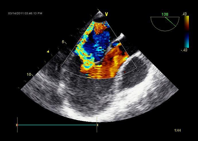
31 Use MELAX view, obtain image of MR jet that includes PISA shell, flow convergence and vena contracta. Also obtain image of CWD thru mitral valve, lining up with the regurgitant jet. Note the timing of the peak velocity of MR jet.

32 Bring up the MELAX view, and scroll to image that shows PISA shell. Ideally, this should correspond to the time of peak MR velocity.
33 Zoom to magnify the image, adjust the color Doppler baseline towards jet direction to achieve an aliasing velocity of m/sec.

34 In the Measurement menu find PISA MR under the PISA folder.
35 Turn color off to visualize ventricular side of orifice center.
36 Place cursor at this location to start radius measurement.
37 Turn color back on, and draw radius to PISA shell.

38 Bring up CWD of mitral regurg jet. Find PISA MR under the PISA folder.

39 Trace MR curve. ERO and RV are calculated by the machine.
40 EROA = 2 πr 2 (V Aliasing / V max ) EROA = 2 (3.14)(.8 cm) 2 (.29 m/sec)/(4.35 m/sec) EROA =.269 cm 2 RV = EROA x TVI mr =.269 cm 2 x cm RV = cm 3 Note rounding by the machine!


41 Putting it All Together: Jet Area: 7 cm 2 Vena Contracta:.3 cm EROA:.269 cm 2 RV: 37 ml
42 Jet Area: 7 cm 2 Vena Contracta:.3 cm EROA:.269 cm 2 RV: 37 ml Moderate MR (low end)

43 Notes: If you are doing calculations by hand, make sure you convert units as needed. Some machines use cm/sec, others use m/sec. GE Vivid 7: m/sec
44 Notes: Use angle correction factor as needed: Generally don t need to correct central jets, but becomes an issue with eccentric jets and also when used in mitral stenosis. Multiply machine calculated EROA and RV by (a/180) to get corrected numbers. If doing your own calculations, use this factor only in the EROA calculation. Then this corrected EROA x TVI mr = corrected RV.
45 Modifications: Derivation of Angle Correction: If hemisphere is not complete because of impingement by wall or leaflet (leading to a funnel constraining flow), area of hemisphere is modified: A hemisphere = 2πr 2 (a/180) Where a is the proximal flow convergence angle Thus: EROA = 2 πr 2 (V Aliasing / V max ) (a/180)

 EROA x V max = 2 πr 2 x V Aliasing EROA = 2(3.")
46 Modifications: No CWD Measures If you can t get a good CWD waveform, here is a method to estimate EROA. Set aliasing velocity to 40 cm/sec. Assume Vmax = 500 cm/sec (This works when LV systolic pressure is greater than left atrial pressure by about 100 mm Hg) EROA x V max = 2 πr 2 x V Aliasing EROA = 2(3.14)r 2 (40)/(500) EROA = r 2 /2
47 Conclusions: We discussed basis of PISA calculations. Discussed pitfalls of PISA. Showed an example how to do PISA measurements with the GE Vivid 7.
48 In the end, one must integrate all the qualitative and quantitative measures to come up with a good MR severity assessment. It seems that we have a lot of room for improvement, and that current echocardiographic grading of MR severity is more art than science. Paul A. Grayburn, MD, Paul Bhella, MD 2010
49 References: Zoghbi WA, et al. Recommendations for Evaluation of the Severity of Native Valvular Regurgitation with Two-dimensional and Doppler Echocardiography. J Am Soc Echocardiogr 2003;16: Lambert S. Proximal Isovelocity Surface Area Should Be Routinely Measured in Evaluating Mitral Regurgitation: A Core Review. Anesth Analg 2007;105:940 3 Shanewise JS. PRO: Proximal Isovelocity Surface Area Should Be Routinely Measured in Evaluating Mitral Regurgitation. Anesth Analg 2007;105:947-8 Savage RM, Konstadt S. CON: Proximal Isovelocity Surface Area Should Not Be Measured Routinely in All Patients with Mitral Regurgitation Anesth Analg 2007;105:944-6 Paul A. Grayburn, MD, Paul Bhella, MD Grading Severity of Mitral Regurgitation byechocardiography: Science or Art? JACC: Cardiovascular Imaging : Biner S, Rafique A, Rafii F, et al. Reproducibility of proximal isovelocity surface area, vena contracta, and regurgitant jet area for assessment of mitral regurgitation severity. J Am Coll Cardiol Img 2010;3:
New 3D Quantification of Mitral Regurgitation Severity. Judy Hung, MD Cardiac Ultrasound Laboratory Massachusetts General Hospital Boston, MA
 New 3D Quantification of Mitral Regurgitation Severity Judy Hung, MD Cardiac Ultrasound Laboratory Massachusetts General Hospital Boston, MA No Financial Disclosures No off label discussion of devices
Valvular Regurgitation: Can We Do Better Than Colour Doppler?
 Valvular Regurgitation: Can We Do Better Than Colour Doppler? A/Prof David Prior St Vincent s Hospital Melbourne Sports Cardiology Valvular Regurgitation Valve regurgitation volume loads the ventricles
Marti McCulloch, BS, MBA, RDCS, FASE Houston, Texas
 Marti McCulloch, BS, MBA, RDCS, FASE Houston, Texas Mitral Regurgitation What to Expect Review Specific Signs of Severity Supportive Signs of Severity Qualitative Parameters Structural Doppler Quantitative
Professors Carpentier and McGoon Mechanism, resulting from the disease Severity of regurgitation, resulting from the mechanism Echo
 Professors Carpentier and McGoon Mechanism, resulting from the disease Severity of regurgitation, resulting from the mechanism Echo define the mechanism, quantify the regurgitation severity CP1293058-3
Prof. JL Zamorano Hospital Universitario Ramón y Cajal
 Prof. JL Zamorano Hospital Universitario Ramón y Cajal Should we forget TR? Nath J et al. Impact of tricuspid regurgitation on long-term survival. J Am Coll Cardiol. 2004; 43:405-409 Why is it difficult
Quantifying Aortic Regurgitation
 Quantifying Aortic Regurgitation Linda D. Gillam, MD, MPH Morristown Medical Center Dorothy and Lloyd Huck Chair Cardiovascular Medicine Atlantic Health System No Disclosures 1 Valve Dysfunction Functional
How to assess ischaemic MR?
 ESC 2012 How to assess ischaemic MR? Luc A. Pierard, MD, PhD, FESC, FACC Professor of Medicine Head, Department of Cardiology University Hospital Sart Tilman, Liège ESC 2012 No conflict of interest Luc
Hemodynamic Assessment. Assessment of Systolic Function Doppler Hemodynamics
 Hemodynamic Assessment Matt M. Umland, RDCS, FASE Aurora Medical Group Milwaukee, WI Assessment of Systolic Function Doppler Hemodynamics Stroke Volume Cardiac Output Cardiac Index Tei Index/Index of myocardial
The Doppler Examination. Katie Twomley, MD Wake Forest Baptist Health - Lexington
 The Doppler Examination Katie Twomley, MD Wake Forest Baptist Health - Lexington OUTLINE Principles/Physics Use in valvular assessment Aortic stenosis (continuity equation) Aortic regurgitation (pressure
Disclosures Rebecca T. Hahn, MD, FASE
 The New ASE Guidelines for Native Valvular Regurgitation Mitral Regurgitation The New ASE Guidelines: Role of 2D/3D and CMR (With caveats and comments from R. Hahn) William A. Zoghbi MD, FASE, MACC Professor
Reproducibility of Proximal Isovelocity Surface Area, Vena Contracta, and Regurgitant Jet Area for Assessment of Mitral Regurgitation Severity
 JACC: CARDIOVASCULAR IMAGING VOL. 3, NO. 3, 2010 2010 BY THE AMERICAN COLLEGE OF CARDIOLOGY FOUNDATION ISSN 1936-878X/10/$36.00 PUBLISHED BY ELSEVIER INC. DOI:10.1016/j.jcmg.2009.09.029 Reproducibility
Direct Planimetry of Mitral Valve Regurgitation Orifice Area by Real-time 3D Transesophageal Echocardiography
 Direct Planimetry of Mitral Valve Regurgitation Orifice Area by Real-time 3D Transesophageal Echocardiography Ertunc Altiok, Sandra Hamada, Silke van Hall, Mehtap Hanenberg, Eva Grabskaya, Michael Becker,
Guidelines in perspective?
 EuroValve 2016 Challenges in the management Secondary MR Guidelines in perspective? Luc A. Pierard, MD, PhD Professor of Medicine Head of the Department of Cardiology, Liège, Belgium Heart Valve Clinic,
Quantification of MR
 Valvular Regurgitation: Putting the New Guidelines into Practice James D. Thomas, MD, FACC, FASE, FESC Director, Center for Heart Valve Disease Bluhm Cardiovascular Institute Professor of Medicine, Feinberg
CHAPTER 4 AN EFFICACIOUS APPROACH FOR THE QUANTIFICATION OF MITRAL REGURGITATION USING IMAGE PROCESSING AND PROXIMAL FLOW CONVERGENCE METHOD
 CHAPTER 4 AN EFFICACIOUS APPROACH FOR THE QUANTIFICATION OF MITRAL REGURGITATION USING IMAGE PROCESSING AND PROXIMAL FLOW CONVERGENCE METHOD 4.1. Introduction Mitral Regurgitation also called Mitral Insufficiency
What are the best diagnostic tools to quantify aortic regurgitation?
 What are the best diagnostic tools to quantify aortic regurgitation? Agnès Pasquet, MD, PhD Pôle de Recherche Cardiovasculaire Institut de Recherche Expérimentale et Clinique Université catholique de Louvain
LUST trial. Echocardiography USER S MANUAL
 LUST trial Echocardiography USER S MANUAL Rosa Sicari, Luna Gargani Ins1tute of Clinical Physiology Na1onal Council of Research, Pisa, Italy Parameters required (1) Aortic root Measurement of aortic root
True morphology of mitral regurgitant flow assessed by three- dimensional transesophageal echocardiography
 DOI: 10.1111/echo.13395 ORIGINAL INVESTIGATION True morphology of mitral regurgitant flow assessed by three- dimensional transesophageal echocardiography Martin Lombardero M.D. Ruth Henquin D.L.S.H.T.M.,
Echocardiographic evaluation of mitral stenosis
 Echocardiographic evaluation of mitral stenosis Euroecho 2011 Philippe Unger, MD, FESC Erasme Hospital, ULB, Brussels, Belgium I have nothing to declare EuroHeart Survey Etiology of single native left-sided
Case Reviews: Hemodynamic Calculations in Valvular Regurgitation
 Case Reviews: Hemodynamic Calculations in Valvular Regurgitation Case 5 History: 69-year-old man with orthotopic heart transplant 15 years ago. Inferior MI several years ago. Recurrent CHF. Currently dyspneic
Doppler Basic & Hemodynamic Calculations
 Doppler Basic & Hemodynamic Calculations August 19, 2017 Smonporn Boonyaratavej MD Division of Cardiology, Department of Medicine Chulalongkorn University Cardiac Center, King Chulalongkorn Memorial Hospital
Functional Mitral Regurgitation
 Club 35 - The best in heart valve disease - Functional Mitral Regurgitation Steven Droogmans, MD, PhD UZ Brussel, Jette, Belgium 08-12-2011 Euroecho & other Imaging Modalities 2011 No conflicts of interest
Improvements in outcomes for heart valve surgery, in particular mitral valve repair, have dictated
 Imaging techniques DOPPLER ECHOCARDIOGRAPHIC ASSESSMENT OF VALVAR REGURGITATION c COLOUR Correspondence to: James D Thomas MD, Department of Cardiology, Desk F15, 9500 Euclid Avenue, The Cleveland Clinic
Mitral regurgitation (MR) is a
 is a") Review Article Hellenic J Cardiol 2013; 54: 448-454 3D Vena Contracta Area to Quantify Severity of Mitral Regurgitation: A Practical New Tool? Dimitrios Maragiannis, Stephen H. Little The Methodist DeBakey
Review Article Hellenic J Cardiol 2013; 54: 448-454 3D Vena Contracta Area to Quantify Severity of Mitral Regurgitation: A Practical New Tool? Dimitrios Maragiannis, Stephen H. Little The Methodist DeBakey
AATS Annual Meeting Seattle, WA Irving Kron, M.D. Professor and Chairman Department of Surgery University of Virginia Hospital Charlottesville,
 AATS Annual Meeting Seattle, WA Irving Kron, M.D. Professor and Chairman Department of Surgery University of Virginia Hospital Charlottesville, Virginia AATS Ischemic MR Guideline Writing Group Roster
TAVR: Echo Measurements Pre, Post And Intra Procedure
 2017 ASE Florida, Orlando, FL October 10, 2017 8:00 8:25 AM 25 min TAVR: Echo Measurements Pre, Post And Intra Procedure Muhamed Sarić MD, PhD, MPA Director of Noninvasive Cardiology Echo Lab Associate
EVALUATION OF CHRONIC MITRAL REGURGITATION: ASSESSING MECHANISMS AND QUANTIFYING SEVERITY 2018 STRUCTURAL HEART DISEASE CONFERENCE June 1, 2018
 1 EVALUATION OF CHRONIC MITRAL REGURGITATION: ASSESSING MECHANISMS AND QUANTIFYING SEVERITY 2018 STRUCTURAL HEART DISEASE CONFERENCE June 1, 2018 David A. Orsinelli, MD, FACC, FASE Professor, Internal
ECHOCARDIOGRAPHY DATA REPORT FORM
 Patient ID Patient Study ID AVM - - Date of form completion / / 20 Initials of person completing the form mm dd yyyy Study period Preoperative Postoperative Operative 6-month f/u 1-year f/u 2-year f/u
Uncommon Doppler Echocardiographic Findings of Severe Pulmonic Insufficiency
 Uncommon Doppler Echocardiographic Findings of Severe Pulmonic Insufficiency Rahul R. Jhaveri, MD, Muhamed Saric, MD, PhD, FASE, and Itzhak Kronzon, MD, FASE, New York, New York Background: Two-dimensional
Echocardiography. Guidelines for Valve and Chamber Quantification. In partnership with
 Echocardiography Guidelines for Valve and Chamber Quantification In partnership with Explanatory note & references These guidelines have been developed by the Education Committee of the British Society
MITRAL REGURGITATION ECHO PARAMETERS TOOL
 Comprehensive assessment of qualitative and quantitative parameters, along with the use of standardized nomenclature when reporting echocardiographic findings, helps to better define a patient s MR and
Clinical Outcome of Tricuspid Regurgitation. David Messika-Zeitoun
 Clinical Outcome of Tricuspid Regurgitation David Messika-Zeitoun I have financial relationships to disclose Consultant for: Edwards, Symetis and Valtech Tricuspid Regurgitation is a Common Finding Tricuspid
An understanding of the many factors involved in the
 Atrioventricular Valve Dysfunction: Evaluation by Doppler and Cross-Sectional Ultrasound Norman H. Silverman, MD, and Doff B. McElhinney, MD Division of Pediatric Cardiology, Department of Pediatrics,
Quantification of Aortic Regurgitation
 Quantification of Aortic Regurgitation ASE Review 2018 Boston Susan E Wiegers, MD, FASE, FACC Professor of Medicine And thanks to Dr. Roberto Lang Disclosure None related to this presentation 1 Objectives
Imaging MV. Jeroen J. Bax Leiden University Medical Center The Netherlands Davos, feb 2015
 Imaging MV Jeroen J. Bax Leiden University Medical Center The Netherlands Davos, feb 2015 MV/MR: information needed on.. 1. MV anatomy 2. MR etiology - primary vs secondary 3. MR severity quantification
School, The University of Texas Health Science Center at Houston, TX.
 Chapter 4: Basics of echocardiography for transcatheter interventions Adrian DaSilva-DeAbreu, MD 1, Catalin Loghin, MD 2 1 Resident, Department of Internal Medicine, McGovern Medical School, The University
ICE: Echo Core Lab-CRF
 APPENDIX 1 ICE: Echo Core Lab-CRF Study #: - Pt Initials: 1. Date of study: / / D D M M M Y Y Y Y 2. Type of Study: TTE TEE 3. Quality of Study: Poor Moderate Excellent Ejection Fraction 4. Ejection Fraction
Appendix II: ECHOCARDIOGRAPHY ANALYSIS
 Appendix II: ECHOCARDIOGRAPHY ANALYSIS Two-Dimensional (2D) imaging was performed using the Vivid 7 Advantage cardiovascular ultrasound system (GE Medical Systems, Milwaukee) with a frame rate of 400 frames
DOPPLER HEMODYNAMICS (1) QUANTIFICATION OF PRESSURE GRADIENTS and INTRACARDIAC PRESSURES
 QUANTIFICATION OF PRESSURE GRADIENTS and INTRACARDIAC PRESSURES") THORAXCENTRE DOPPLER HEMODYNAMICS (1) QUANTIFICATION OF PRESSURE GRADIENTS and INTRACARDIAC PRESSURES J. Roelandt DOPPLER HEMODYNAMICS Intracardiac pressures and pressure gradients Volumetric measurement
THORAXCENTRE DOPPLER HEMODYNAMICS (1) QUANTIFICATION OF PRESSURE GRADIENTS and INTRACARDIAC PRESSURES J. Roelandt DOPPLER HEMODYNAMICS Intracardiac pressures and pressure gradients Volumetric measurement
Advanced Applica,on of Point- of- Care Echocardiography in Cri,cal Care. Dr. Mark Tutschka Dr. Rob ArnAield
 Advanced Applica,on of Point- of- Care Echocardiography in Cri,cal Care Dr. Mark Tutschka Dr. Rob ArnAield OBJECTIVES Provide an overview of common advanced echocardiographic techniques suitable for use
Echocardiography: Guidelines for Valve Quantification
 Echocardiography: Guidelines for Echocardiography: Guidelines for Chamber Quantification British Society of Echocardiography Education Committee Richard Steeds (Chair), Gill Wharton (Lead Author), Jane
Who will Benefit from Percutaneous Management of Mitral Regurgitation? An Imaging Guide to Management
 Who will Benefit from Percutaneous Management of Mitral Regurgitation? An Imaging Guide to Management James D. Thomas, M.D., F.A.C.C. Department of Cardiovascular Medicine Heart and Vascular Institute
ASE Guidelines on Aortic Regurgitation What Do I Measure? Case Studies
 ASE Guidelines on Aortic Regurgitation What Do I Measure? Case Studies Mitral Regurgitation The New ASE Guidelines: Role of 2D/3D and CMR William A. Zoghbi MD, FASE, MACC Professor and Chairman, Department
25 different brand names >44 different models Sizes mm
 Types of Prosthetic Valves BIOLOGIC STENTED Porcine xenograft Pericardial xenograft STENTLESS Porcine xenograft Pericardial xenograft Homograft (allograft) Autograft PERCUTANEOUS MECHANICAL Bileaflet Single
Back to Basics: Common Errors In Quantitation In Everyday Practice
 Back to Basics: Common Errors In Quantitation In Everyday Practice Deborah Agler, ACS, RDCS, FASE October 9, 2017 ASE: Echo Florida Rebecca T. Hahn, MD Director of Interventional Echocardiography Professor
aortic regurgitation, vena contracta area, vena contracta width, live three-dimensional echocardiography
 RESEARCH FROM THE UNIVERSITY OF ALABAMA AT BIRMINGHAM Assessment of Aortic Regurgitation by Live Three-Dimensional Transthoracic Echocardiographic Measurements of Vena Contracta Area: Usefulness and Validation
Regurgitant Lesions. Bicol Hospital, Legazpi City, Philippines July Gregg S. Pressman MD, FACC, FASE Einstein Medical Center Philadelphia, USA
 Regurgitant Lesions Bicol Hospital, Legazpi City, Philippines July 2016 Gregg S. Pressman MD, FACC, FASE Einstein Medical Center Philadelphia, USA Aortic Insufficiency Valve anatomy and function LVOT and
Titel Kardiologie-SG.ch hot topics in heart failure and mitral regurgitation
 Titel Kardiologie-SG.ch hot topics in heart failure and mitral regurgitation where and how to quantify mitral regurgitation: Echolab, Cathlab or MRI? Philipp K. Haager, St. Gallen Measuring mitral regurgitation?
Quantification of Mitral Stenosis: Planimetry, pressure Half time, Continuity Common Errors
 Quantification of Mitral Stenosis: Planimetry, pressure Half time, Continuity Common Errors Christopher J Kramer RDCS Advanced Cardiovascular Services Aurora Health Care Milwaukee, WI No Disclosures Baumgartner,
MITRAL STENOSIS. Joanne Cusack
 MITRAL STENOSIS Joanne Cusack BSE Breakdown Recognition of rheumatic mitral stenosis Qualitative description of valve and sub-valve calcification and fibrosis Measurement of orifice area by planimetry
Cover Page. The handle holds various files of this Leiden University dissertation.
 Cover Page The handle http://hdl.handle.net/1887/21650 holds various files of this Leiden University dissertation. Author: Shanks, Miriam Title: Evolving imaging techniques for the assessment of cardiac
Exercise Pulmonary Hypertension predicts the Occurrence of Symptoms in Asymptomatic Degenerative Mitral Regurgitation
 Exercise Pulmonary Hypertension predicts the Occurrence of Symptoms in Asymptomatic Degenerative Mitral Regurgitation Julien Magne, PhD, Kim O Connor, MD, Giuseppe Romano, MD, Marie Moonen, MD, Luc A.
Bogdan A. Popescu. University of Medicine and Pharmacy Bucharest, Romania. EAE Course, Bucharest, April 2010
 Bogdan A. Popescu University of Medicine and Pharmacy Bucharest, Romania EAE Course, Bucharest, April 2010 This is how it started Mitral stenosis at a glance 2D echo narrow diastolic opening of MV leaflets
Primary Mitral Regurgitation
 EURO VALVE Madrid News from Valves Guidelines 2012: What s new and Why? Primary Mitral Regurgitation Luc A. Pierard, MD, PhD Professor of Medicine Head of the Department of Cardiology Heart Valve Clinic,
When Does 3D Echo Make A Difference?
 When Does 3D Echo Make A Difference? Wendy Tsang, MD, SM Assistant Professor, University of Toronto Toronto General Hospital, University Health Network 1 Practical Applications of 3D Echocardiography Recommended
Pulsed Wave Doppler and Color Flow Doppler Evaluation in Healthy Dogs and Dogs with Cardiac Disease
 Cloud Publications International Journal of Advanced Veterinary Science and Technology 2016, Volume 5, Issue 2, pp. 256-265, Article ID Sci-446 ISSN 2320-3595 Research Article Open Access Pulsed Wave Doppler
Echocardiographic Evaluation of Mitral Valve Prostheses
 Echocardiographic Evaluation of Mitral Valve Prostheses Dennis A. Tighe, M.D., FACC, FACP, FASE Cardiovascular Medicine University of Massachusetts Medical School Worcester, MA www.asecho.org 1 Nishimura
Assessment of LV systolic function
 Tutorial 5 - Assessment of LV systolic function Assessment of LV systolic function A knowledge of the LV systolic function is crucial in the undertanding of and management of unstable hemodynamics or a
MR echo case. N.Koutsogiannis Department of Cardiology University Hospital Of Patras
 MR echo case N.Koutsogiannis Department of Cardiology University Hospital Of Patras Case A 35 years old male came to the echo lab for a third opinion for his valvulopathy. He reports a long standing MR
Goals of Reporting. Background of the STS/ACC TVT Registry. TVT Registry TAVR and MitraClip 10/11/ /11/2016 9:10 9:30 AM
 10/11/2016 9:10 9:30 AM Rebecca T. Hahn, MD Director of Interventional Echocardiography Columbia University Medical College Background of the STS/ACC TVT Registry The STS/ACC TVT Registry is a benchmarking
Quantitative Assessment of Mitral Regurgitation
 JACC: CARDIOVASCULAR IMAGING VOL. 5, NO. 11, 2012 2012 BY THE AMERICAN COLLEGE OF CARDIOLOGY FOUNDATION ISSN 1936-878X/$36.00 PUBLISHED BY ELSEVIER INC. http://dx.doi.org/10.1016/j.jcmg.2012.07.013 STATE-OF-THE-ART
Ioannis Alexanian, MD, PhD Department of Cardiology General Hospital of Chest Diseases Sotiria Athens
 MITRAL REGURGITATION IN PATIENT WITH SEVERE AORTIC VALVE STENOSIS Ioannis Alexanian, MD, PhD Department of Cardiology General Hospital of Chest Diseases Sotiria Athens I HAVE NOTHING TO DECLARE Management
What echo measurements are key prior to MitraClip?
 APHP CHU Bichat - Claude Bernard What echo measurements are key prior to MitraClip? Eric Brochet,MD Cardiology Department Hopital Bichat Paris France No disclosure Conflict of interest Case 69 y.o man
CHAPTER 5 EFFICIENT APPROACHES FOR QUANTIFICATION OF AORTIC REGURGITATION USING PROXIMAL ISOVELOCITY SURFACE AREA PROCESS
 CHAPTER 5 EFFICIENT APPROACHES FOR QUANTIFICATION OF AORTIC REGURGITATION USING PROXIMAL ISOVELOCITY SURFACE AREA PROCESS 5.1. Introduction Aortic Regurgitation is also known as Aortic Insufficiency (AI).
Quantitative Doppler echocardiography
 BJA Education, 16 (2): 46 52 (2016) doi: 10.1093/bjaceaccp/mkv015 Advance Access Publication Date: 10 June 2015 Matrix reference 1A03, 2A04, 3G00 Quantitative Doppler echocardiography P Harris MBChB FRCA
AIMI-HF PROCEDURE MANUAL TECHNICAL GUIDE FOR ECHOCARDIOGRAPHY. MHI Core Laboratory E. O Meara - J.C. Tardif J. Vincent, G. Grenier, C.
 AIMI-HF PROCEDURE MANUAL TECHNICAL GUIDE FOR ECHOCARDIOGRAPHY MHI Core Laboratory E. O Meara - J.C. Tardif J. Vincent, G. Grenier, C. Roy February 2016 Montreal Heart Institute HF Research Aude Turgeon,
The Patient with Atrial Fibrilation
 Assessment of Diastolic Function The Patient with Atrial Fibrilation Assoc. Prof. Adriana Ilieşiu, FESC University of Medicine Carol Davila Bucharest, Romania Associated Conditions with Atrial Fibrillation
Prosthetic valve dysfunction: stenosis or regurgitation
 Prosthetic valve dysfunction: stenosis or regurgitation Jean G. Dumesnil MD, FRCP(C), FACC, FASE(Hon) Quebec Heart and Lung Institute, Québec, Québec No disclosures Possible Causes of High Gradients in
Comments restricted to Sapien and Corevalve 9/12/2016. Disclosures: Core Lab contracts with Edwards Lifesciences, Middlepeak, Medtronic
 Para-ValvularRegurgitation post TAVR: Predict, Prevent, Quantitate, Manage Linda D. Gillam, MD, MPH, FACC, FASE Chair, Department of Cardiovascular Medicine Morristown Medical Center/Atlantic Health System
What Degree of MR Deserves Surgical or Transcatheter Intervention, and How Should It Be Assessed?
 What Degree of MR Deserves Surgical or Transcatheter Intervention, and How Should It Be Assessed? Robert J. Siegel, M.D., FACC Nov. 14-15, 2017, Beverly Hills Director, Cardiac Non-Invasive Laboratory
Tricuspid and Pulmonary Valve Disease
 Tricuspid and Pulmonary Valve Disease Lawrence Rudski MD FRCPC FACC FASE Professor of Medicine Director, Division of Cardiology Jewish General Hospital McGill University Right Sided Failure Edema Gut congestion
The diagnosis of mitral regurgitation (MR) caused
 caused") J Vet Intern Med 2014;28:1206 1213 Assessment of Mitral Regurgitation Severity by Doppler Color Flow Mapping of the Vena Contracta in Dogs M. Di Marcello, E. Terzo, C. Locatelli, V. Palermo, E. Sala, E.
J Vet Intern Med 2014;28:1206 1213 Assessment of Mitral Regurgitation Severity by Doppler Color Flow Mapping of the Vena Contracta in Dogs M. Di Marcello, E. Terzo, C. Locatelli, V. Palermo, E. Sala, E.
3D Echo for Evaluation of Tricuspid Regurgitation Jong-Min Song, MD, PhD
 3D Echo for Evaluation of Tricuspid Regurgitation Jong-Min Song, MD, PhD Asan Medical Center University of Ulsan College of Medicine Seoul, Korea Causes of TR Primary causes (25%) Rheumatic Myxomatous
The Key Questions in Mitral Valve Interventions. Where Are We in 2018?
 The Key Questions in Mitral Valve Interventions Where Are We in 2018? Gilles D. DREYFUS, MD, FRCS, FESC Professor of Cardiothoracic Surgery 30 GIORNATE CARDIOLOGICHE TORINESI - OCT 2018 Are guidelines
Exercise-Induced Changes in Degenerative Mitral Regurgitation
 Journal of the American College of Cardiology Vol. 56, No. 4, 2010 2010 by the American College of Cardiology Foundation ISSN 0735-1097/$36.00 Published by Elsevier Inc. doi:10.1016/j.jacc.2009.12.073
Faculty of Science & Technology
 Faculty of Science & Technology CircAdapt: Model validation and prediction of anatomical and haemodynamic parameters for patients with mitral insufficiency E. van Hulst, s1558129 E. Kleinveld, s1598147
HOW IMPORTANT ARE THESE ECHO MEASUREMENTS ANYWAY?
 HOW IMPORTANT ARE THESE ECHO MEASUREMENTS ANYWAY? John D. Carroll, MD Professor, Director of Interventional Cardiology and Co-Medical Director of the Cardiac and Vascular Center, University of Colorado
ASCeXAM / ReASCE. Practice Board Exam Questions Monday Morning
 ASCeXAM / ReASCE Practice Board Exam Questions Monday Morning Ultrasound Physics Artifacts Doppler Physics Imaging, Knobology, and Artifacts Echocardiographic Evaluation of the RV Tricuspid and Pulmonary
Echocardiographic Evaluation of Aortic Valve Prosthesis
 Echocardiographic Evaluation of Aortic Valve Prosthesis Amr E Abbas, MD, FACC, FASE, FSCAI, FSVM, RPVI Co-Director, Echocardiography, Director, Interventional Cardiology Research, Beaumont Health System
Aortic Regurgitation and Aortic Aneurysm - Epidemiology and Guidelines -
 Reconstruction of the Aortic Valve and Root - A Practical Approach - Aortic Regurgitation and Aortic Aneurysm Wednesday 14 th September - 9.45 Practice must always be founded on sound theory. Leonardo
ECHO HAWAII. Role of Stress Echo in Valvular Heart Disease. Not only ischemia! Cardiomyopathy. Prosthetic Valve. Diastolic Dysfunction
 Role of Stress Echo in Valvular Heart Disease ECHO HAWAII January 15 19, 2018 Kenya Kusunose, MD, PhD, FASE Tokushima University Hospital Japan Not only ischemia! Cardiomyopathy Prosthetic Valve Diastolic
Diagnostic Value of Vena Contracta Area in the Quantification of Mitral Regurgitation Severity by Color Doppler 3D Echocardiography
 Diagnostic Value of Vena Contracta Area in the Quantification of Mitral Regurgitation Severity by Color Doppler 3D Echocardiography Xin Zeng, MD, PhD; Robert A. Levine, MD; Lanqi Hua, RDCS; Eleanor L.
Milind Desai Christine Jellis Teerapat Yingchoncharoen Editors. An Atlas of Mitral Valve Imaging
 Milind Desai Editors An Atlas of Mitral Valve Imaging 123 An Atlas of Mitral Valve Imaging Milind Desai Editors An Atlas of Mitral Valve Imaging Editors Milind Desai Department of Cardiovascular Medicine
found that some patients without stenotic lesions had blood velocity or pressure measurement across the
 Br Heart J 1985; 53: 640-4 Increased blood velocities in the heart and great vessels of patients with congenital heart disease An assessment of their significance in the absence of valvar stenosis STANLEY
Tricuspid and Pulmonary Valve Disease
 Tricuspid and Pulmonary Valve Disease Lawrence Rudski MD FRCPC FACC FASE Professor of Medicine Director, Division of Cardiology Jewish General Hospital McGill University Question 1 All of the following
In the clinical management of patients with ventricular
 Quantitative Assessment of Severity of Ventricular Septal Defect by Three-Dimensional Reconstruction of Color Doppler Imaged Vena Contracta and Flow Convergence Region Masahiro Ishii, MD; Kanoko Hashino,
PART II ECHOCARDIOGRAPHY LABORATORY OPERATIONS ADULT TRANSTHORACIC ECHOCARDIOGRAPHY TESTING
 PART II ECHOCARDIOGRAPHY LABORATORY OPERATIONS ADULT TRANSTHORACIC ECHOCARDIOGRAPHY TESTING STANDARD - Primary Instrumentation 1.1 Cardiac Ultrasound Systems SECTION 1 Instrumentation Ultrasound instruments
M itral regurgitation (MR) is the most commonly
 is the most commonly") iv11 Assessment of mitral regurgitation T Irvine, X K Li, D J Sahn, A Kenny... M itral regurgitation (MR) is the most commonly encountered valve lesion in modern clinical practice. 1 The range of pathologies
iv11 Assessment of mitral regurgitation T Irvine, X K Li, D J Sahn, A Kenny... M itral regurgitation (MR) is the most commonly encountered valve lesion in modern clinical practice. 1 The range of pathologies
Prosthesis-Patient Mismatch or Prosthetic Valve Stenosis?
 EuroValves 2015, Nice Prosthesis-Patient Mismatch or Prosthetic Valve Stenosis? Philippe Pibarot, DVM, PhD, FACC, FAHA, FASE FESC Canada Research Chair in Valvular Heart Diseases Université LAVAL Disclosure
Comparison of Vena Contracta Width by Multiplane Transesophageal Echocardiography With Quantitative Doppler Assessment of Mitral Regurgitation
 Comparison of Vena Contracta Width by Multiplane Transesophageal Echocardiography With Quantitative Doppler Assessment of Mitral Regurgitation Sheila K. Heinle, MD, Shelley A. Hall, MD, M. Elizabeth Brickner,
JOINT MEETING 2 Tricuspid club Chairpersons: G. Athanassopoulos, A. Avgeropoulou, M. Khoury, G. Stavridis
 JOINT MEETING 2 Tricuspid club Chairpersons: G. Athanassopoulos, A. Avgeropoulou, M. Khoury, G. Stavridis Similarities and differences in Tricuspid vs. Mitral Valve Anatomy and Imaging. Echo evaluation
Chapter 20. JACC Cardiovasc Imaging 2009;2:
 Chapter 20 Quantification of functional mitral regurgitation by real-time 3D echocardiography: comparison with 3D velocity-encoded cardiac magnetic resonance N Ajmone Marsan, J JM Westenberg, C Ypenburg,
Introduction RECOMMENDATIONS
 European Journal of Echocardiography (2010) 11, 223 244 doi:10.1093/ejechocard/jeq030 RECOMMENDATIONS European Association of Echocardiography recommendations for the assessment of valvular regurgitation.
Right Heart Hemodynamics: Echo-Cath Discrepancies
 Department of cardiac, thoracic and vascular sciences University of Padua, School of Medicine Padua, Italy Right Heart Hemodynamics: Echo-Cath Discrepancies Luigi P. Badano, MD, PhD, FESC, FACC **Dr. Badano
Secondary MR joint with the mitral academy. What is new in our understanding of this disease? Luc Pierard University Hospital, Liège
 Secondary MR joint with the mitral academy What is new in our understanding of this disease? Luc Pierard University Hospital, Liège Faculty disclosure Luc Pierard I have no financial relationships to disclose.
TAVR Cases. Disclosures 2/17/2018. February 17, :15 3:30 PM 15 min
 31 st Annual State of the Art Echocardiography San Diego, CA February 17, 2018 3:15 3:30 PM 15 min TAVR Cases Muhamed Sarić MD, PhD, MPA Director of Noninvasive Cardiology Echo Lab Associate Professor
HEMODYNAMIC ASSESSMENT
 HEMODYNAMIC ASSESSMENT INTRODUCTION Conventionally hemodynamics were obtained by cardiac catheterization. It is possible to determine the same by echocardiography. Methods M-mode & 2D echo alone can provide
Cover Page. The handle holds various files of this Leiden University dissertation.
 Cover Page The handle http://hdl.handle.net/1887/38522 holds various files of this Leiden University dissertation. Author: Ewe, See Hooi Title: Aortic valve disease : novel imaging insights from diagnosis
Cardiac ultrasound protocols
 Cardiac ultrasound protocols IDEXX Telemedicine Consultants Two-dimensional and M-mode imaging planes Right parasternal long axis four chamber Obtained from the right side Displays the relative proportions