RIGHT VENTRICULAR SIZE AND FUNCTION
|
|
|
- Aubrie Benson
- 6 years ago
- Views:
Transcription

1 RIGHT VENTRICULAR SIZE AND FUNCTION Edwin S. Tucay, MD, FPCC, FPCC, FPSE Philippine Society of Echocardiography Quezon City, Philippines Echo Mission, BRTTH, Legaspi City, July 1-2, 2016
2 NO DISCLOSURE

3 NORMAL HEART = NORMAL FAMILY
4 Outline Need to evaluate the right ventricle Systematic evaluation of the right ventricle Right ventricular dimension Right ventricular systolic function Recommendation
5 (J Am Soc Echocardiogr 2015;28:1-39.)
6 Case Smoker Easy fatigability No orthopnea Bipedal edema
7 Importance of evaluating the right ventricle Role in the clinical outcome of cardiopulmonary disease Size and function adversely affected by left ventricular dysfunction primary pulmonary hypertension conditions that affect the tricuspid valve leading to significant tricuspid regurgitation
8 Systematic Evaluation of the Right Ventricle limited due to its complex morphology comprehensive evaluation: right ventricular dimensions systolic and diastolic function, and RV systolic pressure use multiple echo windows: apical 4-chamber, modified apical 4-chamber, left parasternal long axis (PLAX) and parasternal short-axis (PSAX), left parasternal RV inflow, and subcostal views. 3 D echo imaging continuously improve.
9 RIGHT VENTRICULAR DIMENSIONS
10 RIGHT VENTRICULAR WALL THICKNESS Advantage: Easy to perform M-MODE End diastole Subcostal view Zoomed M-mode or (2D) RV wall thickness 5mm 2D Limitations: Single site measurement overestimated by harmonic imaging and oblique M-Mode challenging with thickened visceral pericardium Recommendations for Cardiac Chamber Quantification by Echo in Adults: update from ASE/EACI. JASE 2015;28:1-39


11 RIGHT VENTRICULAR WALL THICKNESS Recommendation: M-MODE 2D Abnormal RV wall thickness should be reported in patients suspected of having RV and/or LV dysfunction, using the normal cut off of 5 mm Recommendations for Cardiac Chamber Quantification by Echo in Adults: update from ASE/EACI. JASE 2015;28:1-39

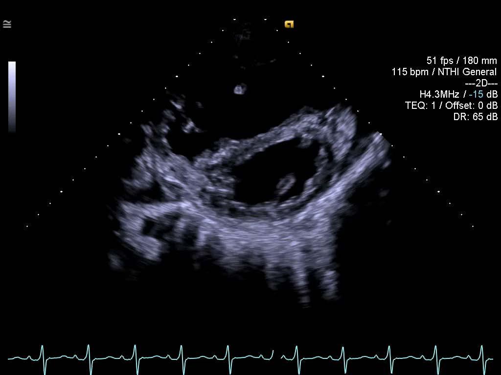
12 RV LINEAR DIMENSIONS RV Focused Apical 4 chamber View Advantages: easily obtained and a marker of RV dilatation. Limitation: highly dependent on probe rotation which can result in an underestimation of RV width. End diastole diameter > 41 mm (base) and > 35 mm (mid level)=rv dilatation > 83 mm (longitudinal) = RV enlargement Recommendations for Cardiac Chamber Quantification by Echo in Adults: update from ASE/EACI. JASE 2015;28:1-39
13 RV LINEAR DIMENSIONS RV Focused Apical 4 chamber View Recommendations: right-sided heart disease or PH should have measurements of RV basal, mid cavity, and longitudinal dimensions on a 4-chamber right ventricle-focused view. report the right ventricle as dilated despite measuring within the normal range, on the basis of a right ventricle appearing significantly larger than the left ventricle. Recommendations for Cardiac Chamber Quantification by Echo in Adults: update from ASE/EACI. JASE 2015;28:1-39
14 RIGTH VENTRICULAR OUTFLOW TRACT Proximal RVOT 30 mm Distal RVOT 35 mm 27mm End-diastole RVOT proximal PLAX: RV wall to IVS-aortic junction PSAX: RV wall to Aortic valve RVOT distal PSAX: just proximal to pulmonic valve Advantages: easily obtained from the left PSAX window. Limitation: Limited normative data, window for measurement not yet standardized, wall is often suboptimal. Recommendations for Cardiac Chamber Quantification by Echo in Adults: update from ASE/EACI. JASE 2015;28:1-39


15 RIGTH VENTRICULAR OUTFLOW TRACT Proximal RVOT 30 mm Distal RVOT 35 mm 27mm Recommendations: In congenital heart disease or arrhythmia potentially involving the RVOT, proximal and distal diameters of the RVOT should be measured from the PSAX or PLAX views. upper reference limit for the PSAX distal RVOT diameter is 27 mm PLAX proximal RVOT is 30mm PSAX proximal RVOT is 35mm Recommendations for Cardiac Chamber Quantification by Echo in Adults: update from ASE/EACI. JASE 2015;28:1-39
16 RIGHT VENTRICULAR SYSTOLIC FUNCTION QUANTITATIVE PARAMETERS (Global/Regional) With clinical utility and value based on studies are right ventricular index of myocardial performance (RIMP) tricuspid annular plane systolic excursion (TAPSE) 2D fractional area change (FAC) tricuspid annulus systolic velocity (S ) Need more data to demonstrate clinical utility: dp/dt 2D RV ejection fraction 3D RV ejection fraction RV longitudinal strain and strain rate Rudski et al. Guidelines for the echo assessment of the Right Heart in Adult. JASE 2010;23:
17 RIGHT VENTRICULAR SYSTOLIC FUNCTION Global Function Pulsed Doppler RIMP Tissue Doppler RIMP Global Systolic Function Fractional Area Change (RVFAC) 3D RVEF Longitudinal Systolic Function TAPSE Pulse Tissue Doppler S wave (Tricuspid annulus systolic velocity) Global Longitudinal Strain (GLS) Recommendations for Cardiac Chamber Quantification by Echo in Adults: update from ASE/EACI. JASE 2015;28:1-39
18 RV SYSTOLIC FUNCTION: Right Ventricular Index of Myocardial Performance (RIMP),or Myocardial Performance Index (MPI) MPI is defined as the ratio of isovolumic time divided by ET, or [(IVRT + IVCT)/ET] Tricuspid inflow Pulsed Doppler --4CV RVOT Pulsed Doppler --PSAX MPI = (TCO - ET)/ET TCO =Tricuspid valve Closing to Opening time ET =Right Ventricular Ejection Time PULSED DOPPLER METHOD PULSED TISSUE DOPPLER METHOD Advantages: >Less affected by heart rate (both methods) >single beat recording, no need for R-R interval matching (Pulse Tissue Doppler Method) Limitations: >Unreliable when RA pressure is elevated (both) >R-R interval matching of 2 separate recordings (Pulse Doppler Method) Recommendations for Cardiac Chamber Quantification by Echo in Adults: update from ASE/EACI. JASE 2015;28:1-39
19 RV SYSTOLIC FUNCTION: Right Ventricular Index of Myocardial Performance (RIMP),or Myocardial Performance Index (MPI) PULSED DOPPLER METHOD Tricuspid inflow Pulsed Doppler --4CV RVOT Pulsed Doppler --PSAX MPI is defined as the ratio of isovolumic time divided by ET, or [(IVRT + IVCT)/ET] MPI = (TCO - ET)/ET TCO =Tricuspid valve Closing to Opening time ET =Right Ventricular Ejection Time PULSED TISSUE DOPPLER METHOD Recommendations: used for initial and serial measurements of RV function in complement with other quantitative and nonquantitative measures. upper reference limit for the right-sided MPI is 0.43 using the pulsed Doppler method and 0.54 using the pulsed tissue Doppler method. It should not be used as the sole quantitative method for evaluation of RV function and should not be used with irregular heart rates. Recommendations for Cardiac Chamber Quantification by Echo in Adults: update from ASE/EACI. JASE 2015;28:1-39
20 RV SYSTOLIC FUNCTION: Tricuspid Annular Plane Systolic Excursion (TAPSE) RV focused View M-mode the tricuspid lateral annulus. Excursion from end-diastole to peak systole Abnormal <17 mm Advantages: Established prognostic value Validated against radionuclide EF Limitations: Angle dependency Partial representation of RV Global Function Recommendations for Cardiac Chamber Quantification by Echo in Adults: update from ASE/EACI. JASE 2015;28:1-39 Kaul S,. Am Heart J 1984;107: Lopez-Candales A, et al. Postgrad Med J 2008;84:40-5. Miller D, Farah MG, Liner A, Fox K, Schluchter M, Hoit BD. J Am Soc Echocardiogr 2004;17:443-7.
21 RV SYSTOLIC FUNCTION: Tricuspid Annular Plane Systolic Excursion (TAPSE) Recommendation: TAPSE should be used routinely as a simple method of estimating RV function, with a lower reference value for impaired RV systolic function of 17 mm. Recommendations for Cardiac Chamber Quantification by Echo in Adults: update from ASE/EACI. JASE 2015;28:1-39
22 RV SYSTOLIC FUNCTION: 2D RV Fractional Area Change (RVFAC) Advantages Established prognostic value Reflects both longitudinal and radial RV contraction Correlates with RVEF by CMR RV focused apical 4 C view RV area end diastole and end systole RV FAC (%) = 100 x (EDA - ESA)/EDA Abnormal <35% Limitations Neglects contribution of RV outflow tract to overall systolic function Recommendations for Cardiac Chamber Quantification by Echo in Adults: update from ASE/EACI. JASE 2015;28:1-39
23 RV SYSTOLIC FUNCTION: 2D RV Fractional Area Change (RVFAC) Recommendations: one of the recommended methods of quantitatively estimating RV function, with a lower reference value for normal RV systolic function of 35%.
24 RV SYSTOLIC FUNCTION: Tricuspid Annulus Systolic Velocity (S ) Pulse wave DTI of the lateral tricuspid annulus Apical 4C view with parallel alignment of Doppler beam with RV free wall longitudinal excursion Systolic Velocity <9.5 cm/sec is abnormal Recommendations for Cardiac Chamber Quantification by Echo in Adults: update from ASE/EACI. JASE 2015;28:1-39
25 RV SYSTOLIC FUNCTION: Tricuspid Annulus Systolic Velocity (S ) Advantages easily measured, reliable and reproducible. correlates well with other measures of global RV systolic function. Validated against radionuclide EF Established prognostic value Limitations Angle dependent Not fully representative of RV global function after thoracotomy, pulmonary thromboendarterectomy or heart transplantation
26 RV SYSTOLIC FUNCTION: Tricuspid Annulus Systolic Velocity (S ) Recommendation: should be used in the assessment of RV function. S < 9.5 cm/s should raise the suspicion for abnormal RV function Recommendations for Cardiac Chamber Quantification by Echo in Adults: update from ASE/EACI. JASE 2015;28:1-39
27 RV SYSTOLIC FUNCTION: RV dp/dt Ascending limb of the TR continuouswave Doppler signal Mark 1 and 2 m/sec (4 and 16 mmhg) dp = 12 mmhg dt =time required for the TR jet to increase in velocity from 1 to 2 m/s.


28 RV SYSTOLIC FUNCTION: RV dp/dt Advangtage Advantage: simple technique with sound physiologic basis Limitations: Lack of data in normal subjects Load dependent Recommendations: RV dp/dt < 400 mm Hg/s is likely abnormal cannot be recommended for routine use can be considered in subjects with suspected RV dysfunction.
29 RV SYSTOLIC FUNCTION: 2D RV Ejection Fraction Geometric assumptions area-length methods- based on modified pyramidal or ellipsoidal models disk summation method determine a RV body volume, using predominantly the apical 4- chamber view. (EDV-ESV)/EDV lower reference limit RV EF is 44%. Recommendations: derived estimation of RV EF is not recommended, because of the heterogeneity of methods and the numerous geometric assumptions.
30 RV SYSTOLIC FUNCTION: 3D RV Ejection Fraction Advantages: No geometric assumptions Includes RV outflow tract contribution to overall function Correlates with RV EF by CMR Limitations: Depends on adequate image quality Requires offline analysis and experience Prognostic value not established


31 RV SYSTOLIC FUNCTION: 3D RV Ejection Fraction Recommendations: 3D echocardiography RV EF may be reported. lower reference limit of 45% reserve 3D methods for serial volume and EF determinations.
32 RV SYSTOLIC FUNCTION: RV Strain and Strain Rate Strain = percentage change in myocardial deformation Strain rate = rate of deformation of myocardium over time. Strain rate has been closely correlated with myocardial contractility in vitro and in vivo DTI-derived Strain Speckle tracking Echo (STE) derived strain angle independent Global Longitudinal Strain Jamal F, Bergerot C, Argaud L, Loufouat J, Ovize M. Longitudinal strain quantitates regional right ventricular contractile function. Am J Physiol Heart Circ Physiol 2003;285:H


33 RV Systolic Function: Right Ventricular Global Longitudinal Strain Speckle tracking Echocardiography Average strain RV Free wall segments Average strain RV Free wall and septal segments
34 RV SYSTOLIC FUNCTION: RV Global Longitudinal Strain and - 2d Speckle Tracking Advantages: relatively angle independent possesses an improved signal-to-noise ratio. provide regional function estimates, as well as a more global function. Disadvantages: lack of normative data and need additional validation. different algorithms in different platforms may result in different normal ranges.
35 RV SYSTOLIC FUNCTION: RV Strain and Strain Rate Recommendations: Because of the lack of reproducibility and the paucity of data, this technique is not recommended for routine clinical use. No reference limits can be recommended, because of the large degree of variability.
36 Recommendation for the evaluation of RV systolic function Visual assessment of RV systolic function gives an initial qualitative evaluation of RV systolic function but remains insufficient Simple and reproducible methods of assessing RV systolic function should be incorporated into the routine echocardiographic assessment. ( FAC, TAPSE, pulsed tissue Doppler S, and MPI). Combining more than one measure of RV function, such as S and MPI, may more reliably distinguish normal from abnormal function. Rudski et al. Guidelines for the echo assessment of the Right Heart in Adult. JASE 2010;23: Miller D, Farah MG, Liner A, Fox K et al. J Am Soc Echocardiogr 2004;17:443-7.
37 Recommendation for the evaluation of RV systolic function At least one of the above quantitative measures be incorporated into the routine echocardiographic examination and report when RV dysfunction is suspected when the clinical indication for the study relates to a condition that may affect the right ventricle. Techniques such as strain, and strain rate are not currently recommended as routine and are best reserved for specific clinical and research applications. Rudski et al. Guidelines for the echo assessment of the Right Heart in Adult. JASE 2010;23: Miller D, Farah MG, Liner A, Fox K et al. J Am Soc Echocardiogr 2004;17:443-7.
38 Case Smoker Easy fatigability No orthopnea Bipedal edema

39 RV Linear Dimensions
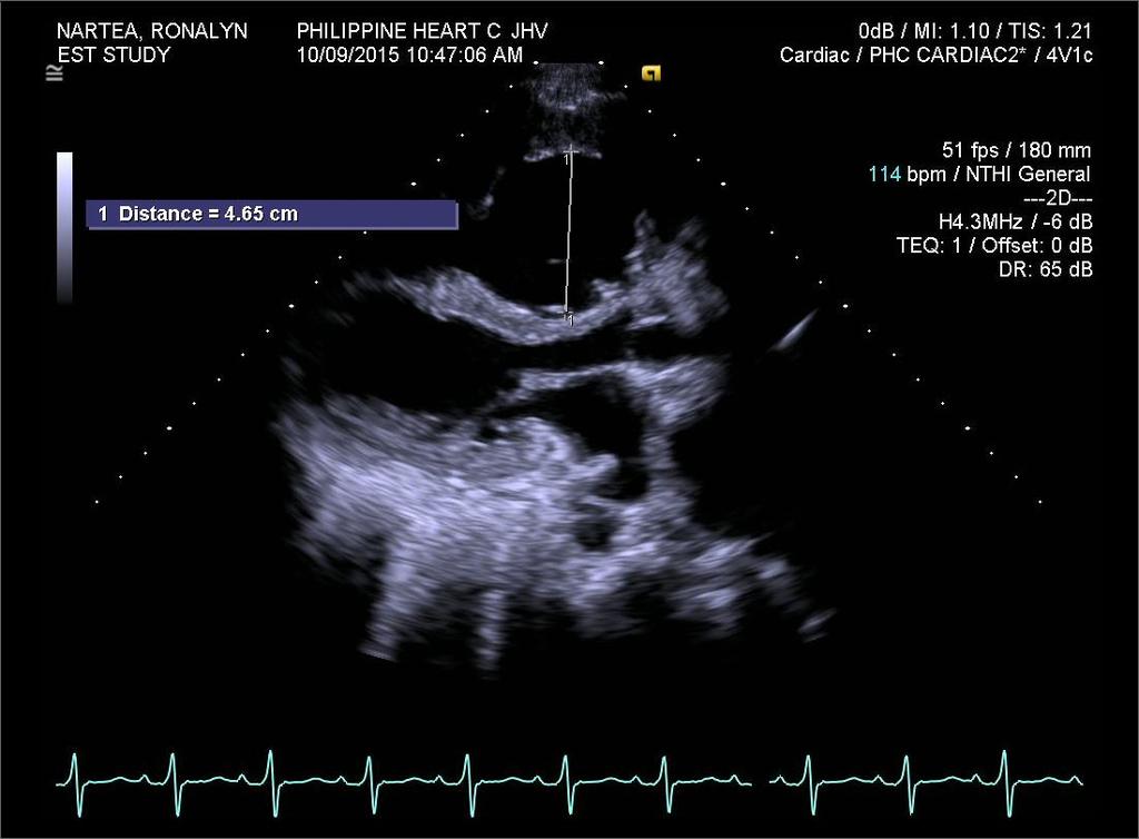
40 RV LINEAR DIMENSIONS
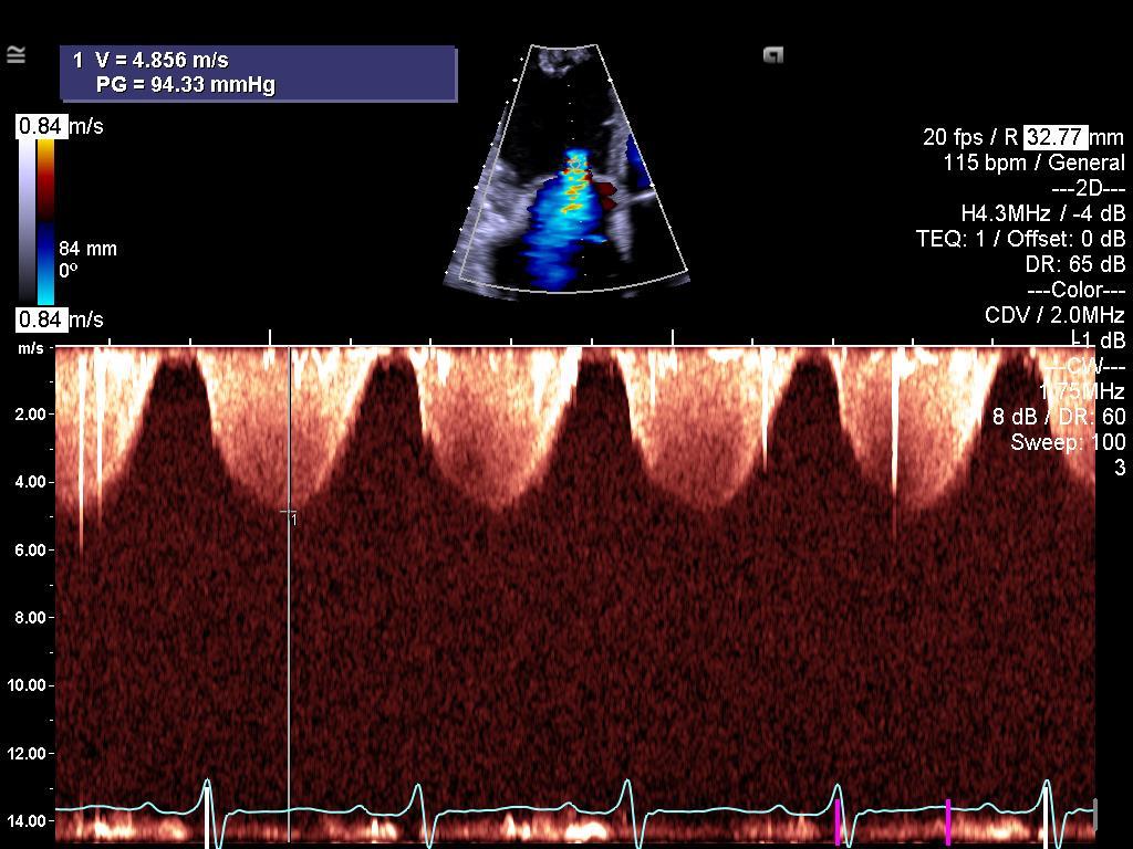
41 94 mm Hg


42 RV FRACTIONAL AREA CHANGE FAC 14 % (<35%)

43 TAPSE 14.8 mm (<17 mm)
 7.6 cm/sec (<9.5 cm/sec) 0.115 m/s (")
44 Tricuspid Annulus Systolic Velocity (S ) 7.6 cm/sec (<9.5 cm/sec) m/s (
/199msec =0.60 (>0.")
45 RIMP (Pulse Tissue Doppler Method) (TCO ET) / ET =(320msec 199msec)/199msec =0.60 (>0.54)

46 RV dp/dt dp = 16-4 = 12mmHg dt = 36msec dp/dt = mmhg/msec = 333mmHg/sec (<400mmHg/sec)
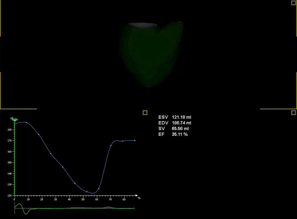
47 3D RVEF

48 RV GLOBAL LONGITUDINAL STRAIN -3.8%

49 Case Smoker Easy fatigability No orthopnea Bipedal edema DILATED HYPERTROPHIED RV RIGHT VENTRICULAR SYSTOLIC DYSFUNCTION SEVERE PULMONARY HYPERTENSION
50
Incorporating the New Echo Guidelines Into Everyday Practice
 Incorporating the New Echo Guidelines Into Everyday Practice Clinical Case RIGHT VENTRICULAR FAILURE Gustavo Restrepo MD President Elect Interamerican Society of Cardiology Director Fellowship Training
Quantification of Cardiac Chamber Size
 2017 KSE 2017-11-25 Quantification of Cardiac Chamber Size Division of Cardiology Keimyung University Dongsan Medical Center In-Cheol Kim M.D., Ph.D. LV size and function Internal linear dimensions PLX
Right Heart Evaluation ASE Guidelines Review. Chris Mann RDCS, RCS, FASE Faculty, Echocardiography Pitt Community College Greenville, NC
 Right Heart Evaluation ASE Guidelines Review Chris Mann RDCS, RCS, FASE Faculty, Echocardiography Pitt Community College Greenville, NC Objectives Briefly review right atrial and right ventricular anatomy
Quantitation of right ventricular dimensions and function
 SCCS Basics of cardiac assessment Quantitation of right ventricular dimensions and function Tomasz Kukulski, MD PhD Dept of Cardiology, Congenital Heart Disease and Electrotherapy Silesian Medical University
LV FUNCTION ASSESSMENT: WHAT IS BEYOND EJECTION FRACTION
 LV FUNCTION ASSESSMENT: WHAT IS BEYOND EJECTION FRACTION Jamilah S AlRahimi Assistant Professor, KSU-HS Consultant Noninvasive Cardiology KFCC, MNGHA-WR Introduction LV function assessment in Heart Failure:
Chamber Quantitation Guidelines - Update II
 Chamber Quantitation Guidelines - Update II Right Heart Measurements Steven A. Goldstein MD FACC FASE Professor of Medicine Georgetown University Medical Center MedStar Heart Institute Washington Hospital
10/7/2013. Systolic Function How to Measure, How Accurate is Echo, Role of Contrast. Thanks to our Course Director: Neil J.
 Systolic Function How to Measure, How Accurate is Echo, Role of Contrast Neil J. Weissman, MD MedStar Health Research Institute & Professor of Medicine Georgetown University Washington, D.C. No Disclosures
ECHOCARDIOGRAPHY DATA REPORT FORM
 Patient ID Patient Study ID AVM - - Date of form completion / / 20 Initials of person completing the form mm dd yyyy Study period Preoperative Postoperative Operative 6-month f/u 1-year f/u 2-year f/u
Pulmonary Hypertension. Echocardiography: Pearls & Pitfalls
 Pulmonary Hypertension Echocardiography: Pearls & Pitfalls Αθανάσιος Γ. Κουτσάκης Ειδικευόμενος Καρδιολογίας Α Καρδιολογική Κλινική ΑΠΘ Σεμινάρια Ομάδων Εργασίας Ελληνικής Καρδιολογικής Εταιρείας Ιωάννινα,
좌심실수축기능평가 Cardiac Function
 Basic Echo Review Course 좌심실수축기능평가 Cardiac Function Seonghoon Choi Cardiology Hallym university LV systolic function Systolic function 좌심실수축기능 - 심근의수축으로심실에서혈액을대동맥으로박출하는기능 실제임상에서 LV function 의의미 1Diagnosis
Ιπποκράτειες μέρες καρδιολογίας Θεσσαλονίκη, 9-10 Μαρτίου Φωτεινή Α. Λαζαρίδου Επιμελήτρια Α Γενικό Νοσοκομείο Αγιος Παύλος, Θεσσαλονίκη
 Ιπποκράτειες μέρες καρδιολογίας Θεσσαλονίκη, 9-10 Μαρτίου 2018 Φωτεινή Α. Λαζαρίδου Επιμελήτρια Α Γενικό Νοσοκομείο Αγιος Παύλος, Θεσσαλονίκη RV shape Triangular shape in frontal plane crescent shape in
ASCeXAM / ReASCE. Practice Board Exam Questions Monday Morning
 ASCeXAM / ReASCE Practice Board Exam Questions Monday Morning Ultrasound Physics Artifacts Doppler Physics Imaging, Knobology, and Artifacts Echocardiographic Evaluation of the RV Tricuspid and Pulmonary
Assessment of LV systolic function
 Tutorial 5 - Assessment of LV systolic function Assessment of LV systolic function A knowledge of the LV systolic function is crucial in the undertanding of and management of unstable hemodynamics or a
Adel Hasanin Ahmed 1
 Adel Hasanin Ahmed 1 PERICARDIAL DISEASE The pericardial effusion ends anteriorly to the descending aorta and is best visualised in the PLAX. PSAX is actually very useful sometimes for looking at posterior
COMPLEX CONGENITAL HEART DISEASE: WHEN IS IT TOO LATE TO INTERVENE?
 COMPLEX CONGENITAL HEART DISEASE: WHEN IS IT TOO LATE TO INTERVENE? Aurora S. Gamponia, MD, FPPS, FPCC, FPSE OBJECTIVES Identify complex congenital heart disease at high risk or too late for intervention
Right Ventricle Steven J. Lester MD, FACC, FRCP(C), FASE Mayo Clinic, Arizona
, FASE Mayo Clinic, Arizona") Right Ventricle Steven J. Lester MD, FACC, FRCP(C), FASE Mayo Clinic, Arizona 1. In which scenario will applying the simplified Bernoulli equation to the peak tricuspid regurgitation velocity and adding
Right Ventricle Steven J. Lester MD, FACC, FRCP(C), FASE Mayo Clinic, Arizona 1. In which scenario will applying the simplified Bernoulli equation to the peak tricuspid regurgitation velocity and adding
Cardiac Chamber Quantification by Echocardiography
 Cardiac Chamber Quantification by Echocardiography Maryam Bokhamseen, RCS, RCDS, EACVI Echotechnologist ǁ, Non invasive Cardiac Laboratory King Abdulaziz Cardiac Center. Outline: Introduction. Background
Evaluation of the Right Ventricle and Risk Stratification for Sudden Cardiac Death
 Evaluation of the Right Ventricle and Risk Stratification for Sudden Cardiac Death Presenters: Sabrina Phillips, MD FACC FASE Director, Adult Congenital Heart Disease Services The University of Oklahoma
Hemodynamic Assessment. Assessment of Systolic Function Doppler Hemodynamics
 Hemodynamic Assessment Matt M. Umland, RDCS, FASE Aurora Medical Group Milwaukee, WI Assessment of Systolic Function Doppler Hemodynamics Stroke Volume Cardiac Output Cardiac Index Tei Index/Index of myocardial
Stephen G. Worthley. Cardiovascular Research Centre, Royal Adelaide Hospital and University of Adelaide, Adelaide, South Australia 5000, Australia
 RIGHT VENTRICULAR SPECKLE TRACKING STRAIN HAS A CLOSER CORRELATION WITH RIGHT VENTRICULAR EJECTION FRACTION THAN OTHER ECHOCARDIOGRAPHIC INDICES OF RIGHT VENTRICULAR FUNCTION: A COMPARISON WITH CARDIAC
Chamber Quantitation Guidelines II Right Heart Measurements
 Chamber Quantitation Guidelines II Right Heart Measurements Steven A. Goldstein MD FACC FASE Director, Noninvasive Cardiology MedStar Heart Institute Washington Hospital Center Sunday, October 9, 2016
Chamber Quantitation Guidelines: What is New?
 Chamber Quantitation Guidelines: What is New? Roberto M Lang, MD J AM Soc Echocardiogr 2005; 18:1440-1463 1 Approximately 10,000 citations iase in itune Cardiac Chamber Quantification: What is New? Database
Evaluation of Left Ventricular Diastolic Dysfunction by Doppler and 2D Speckle-tracking Imaging in Patients with Primary Pulmonary Hypertension
 ESC Congress 2011.No 85975 Evaluation of Left Ventricular Diastolic Dysfunction by Doppler and 2D Speckle-tracking Imaging in Patients with Primary Pulmonary Hypertension Second Department of Internal
Fig.1 Normal appearance of RV in SAX:
 Tutorial 7 - Assessment of the right heart Assessment of the Right heart The right heart assessment clinically and echocardiographically is not a very important part of mainstream cardiology. In the ICU,
Right Ventricular Strain in Normal Healthy Adult Filipinos: A Retrospective, Cross- Sectional Pilot Study
 Right Ventricular Strain in Normal Healthy Adult Filipinos: A Retrospective, Cross- Sectional Pilot Study By Julius Caesar D. de Vera, MD Jonnah Fatima B. Pelat, MD Introduction Right ventricle contributes
Echocardiographic assessment of the right ventricle in paediatric pulmonary hypertension.
 Echocardiographic assessment of the right ventricle in paediatric pulmonary hypertension. Mark K. Friedberg, MD No disclosures Outline RV response to increased afterload Echo assessment of RV function
British Society of Echocardiography
 British Society of Echocardiography Affiliated to the British Cardiac Society A Minimum Dataset for a Standard Adult Transthoracic Echocardiogram From the British Society of Echocardiography Education
Appendix II: ECHOCARDIOGRAPHY ANALYSIS
 Appendix II: ECHOCARDIOGRAPHY ANALYSIS Two-Dimensional (2D) imaging was performed using the Vivid 7 Advantage cardiovascular ultrasound system (GE Medical Systems, Milwaukee) with a frame rate of 400 frames
The background of the Cardiac Sonographer Network News masthead is a diagnostic image:
 Number 5 Welcome Number 5 Welcome to the newsletter created just for you: sonographers who perform pediatric echocardiograms in primarily adult echo labs. Each issue features tips on echocardiography of
Diastolic Heart Function: Applying the New Guidelines Case Studies
 Diastolic Heart Function: Applying the New Guidelines Case Studies Mitral Regurgitation The New ASE William Guidelines: A. Zoghbi Role MD, of FASE, 2D/3D MACCand CMR Professor and Chairman, Department
Adel Hasanin Ahmed 1 LV MORPHOLOGY
 Adel Hasanin Ahmed 1 LV MORPHOLOGY The left ventricular wall comprises three layers- middle circumferential layer and superficial and deep longitudinal layers: 1. Subepicardial longitudinal layer (25%
Evalua&on)of)Le-)Ventricular)Diastolic) Dysfunc&on)by)Echocardiography:) Role)of)Ejec&on)Frac&on)
of)Le-)Ventricular)Diastolic) Dysfunc&on)by)Echocardiography:) Role)of)Ejec&on)Frac&on)") Evalua&on)of)Le-)Ventricular)Diastolic) Dysfunc&on)by)Echocardiography:) Role)of)Ejec&on)Frac&on) N.Koutsogiannis) Department)of)Cardiology) University)Hospital)of)Patras)! I have no conflicts of interest
Evalua&on)of)Le-)Ventricular)Diastolic) Dysfunc&on)by)Echocardiography:) Role)of)Ejec&on)Frac&on) N.Koutsogiannis) Department)of)Cardiology) University)Hospital)of)Patras)! I have no conflicts of interest
NEW GUIDELINES MAJOR ECHOCARDIOGRAPHIC CRITERIA FOR ARVC MINOR ECHOCARDIOGRAPHIC CRITERIA FOR ARVC
 NEW GUIDELINES The Echocardiographic Assessment of the Right Ventricle with particular reference to Arrhythmogenic Right Ventricular Cardiomyopathy A Protocol of the British Society of Echocardiography
Φωτεινή Α. Λαζαρίδου Επιμελήτρια Α Γενικό Νοσοκομείο Αγιος Παύλος, Θεσσαλονίκη
 17 ο Πανελλήνιο Καρδιολογικό Συνέδριο ΚΕΒΕ Θεσσαλονίκη, 24-26 Μαϊου 2018 Στρογγυλό τραπέζι: Κλινικά προβλήματα στην πνευμονική υπέρταση Φωτεινή Α. Λαζαρίδου Επιμελήτρια Α Γενικό Νοσοκομείο Αγιος Παύλος,
Strain and Strain Rate Imaging How, Why and When?
 Strain and Strain Rate Imaging How, Why and When? João L. Cavalcante, MD Advanced Cardiac Imaging Fellow Cleveland Clinic Foundation Disclosures: No conflicts of interest Movement vs Deformation Movement
Little is known about the degree and time course of
 Differential Changes in Regional Right Ventricular Function Before and After a Bilateral Lung Transplantation: An Ultrasonic Strain and Strain Rate Study Virginija Dambrauskaite, MD, Lieven Herbots, MD,
Nancy Goldman Cutler, MD Beaumont Children s Hospital Royal Oak, Mi
 Nancy Goldman Cutler, MD Beaumont Children s Hospital Royal Oak, Mi Identify increased LV wall thickness (WT) Understand increased WT in athletes Understand hypertrophic cardiomyopathy (HCM) Enhance understanding
PART II ECHOCARDIOGRAPHY LABORATORY OPERATIONS ADULT TRANSTHORACIC ECHOCARDIOGRAPHY TESTING
 PART II ECHOCARDIOGRAPHY LABORATORY OPERATIONS ADULT TRANSTHORACIC ECHOCARDIOGRAPHY TESTING STANDARD - Primary Instrumentation 1.1 Cardiac Ultrasound Systems SECTION 1 Instrumentation Ultrasound instruments
Martin G. Keane, MD, FASE Temple University School of Medicine
 Martin G. Keane, MD, FASE Temple University School of Medicine Measurement of end-diastolic LV internal diameter (LVIDd) made by properly-oriented M-Mode techniques in the Parasternal Long Axis View (PLAX):
Assessing the Impact on the Right Ventricle
 Advances in Tricuspid Regurgitation Congress of the European Society of Cardiology (ESC) Munich, August 25-29, 2012 Assessing the Impact on the Right Ventricle Stephan Rosenkranz, MD Clinic III for Internal
2/2/2011. Strain and Strain Rate Imaging How, Why and When? Movement vs Deformation. Doppler Myocardial Velocities. Movement. Deformation.
 Strain and Strain Rate Imaging How, Why and When? João L. Cavalcante, MD Advanced Cardiac Imaging Fellow Cleveland Clinic Foundation Disclosures: No conflicts of interest Movement vs Deformation Movement
Echocardiography: Guidelines for Valve Quantification
 Echocardiography: Guidelines for Echocardiography: Guidelines for Chamber Quantification British Society of Echocardiography Education Committee Richard Steeds (Chair), Gill Wharton (Lead Author), Jane
Tissue Doppler and Strain Imaging. Steven J. Lester MD, FRCP(C), FACC, FASE
, FACC, FASE") Tissue Doppler and Strain Imaging Steven J. Lester MD, FRCP(C), FACC, FASE Relevant Financial Relationship(s) None Off Label Usage None a. Turn the wall filters on and turn down the receiver gain. b. Turn
Tissue Doppler and Strain Imaging Steven J. Lester MD, FRCP(C), FACC, FASE Relevant Financial Relationship(s) None Off Label Usage None a. Turn the wall filters on and turn down the receiver gain. b. Turn
Basic Assessment of Left Ventricular Systolic Function
 WINFOCUS BASIC ECHO (WBE) Basic Assessment of Left Ventricular Systolic Function Ritesh Dhar, MD Director, Echocardiography Lab and Staff Cardiologist Intermountain Medical Center Murray, Utah Outline
Echo assessment of the failing heart
 Echo assessment of the failing heart Mark K. Friedberg, MD The Labatt Family Heart Center The Hospital for Sick Children Toronto, Ontario, Canada Cardiac function- definitions Cardiovascular function:
Adult Echocardiography Examination Content Outline
 Adult Echocardiography Examination Content Outline (Outline Summary) # Domain Subdomain Percentage 1 2 3 4 5 Anatomy and Physiology Pathology Clinical Care and Safety Measurement Techniques, Maneuvers,
Pulmonary Hypertension: Echocardiographic Evaluation of Pulmonary Hypertension and Right Ventricular Function. Irmina Gradus-Pizlo, MD
 Pulmonary Hypertension: Echocardiographic Evaluation of Pulmonary Hypertension and Right Ventricular Function Irmina Gradus-Pizlo, MD Disclosures: Nothing to disclose Overview Is pulmonary hypertension
COMPREHENSIVE EVALUATION OF FETAL HEART R. GOWDAMARAJAN MD
 COMPREHENSIVE EVALUATION OF FETAL HEART R. GOWDAMARAJAN MD Disclosure No Relevant Financial Relationships with Commercial Interests Fetal Echo: How to do it? Timing of Study -optimally between 22-24 weeks
How to Assess and Treat Obstructive Lesions
 How to Assess and Treat Obstructive Lesions Erwin Oechslin, MD, FESC, FRCPC, Director, Congenital Cardiac Centre for Adults Peter Munk Cardiac Centre University Health Network/Toronto General Hospital
The Doppler Examination. Katie Twomley, MD Wake Forest Baptist Health - Lexington
 The Doppler Examination Katie Twomley, MD Wake Forest Baptist Health - Lexington OUTLINE Principles/Physics Use in valvular assessment Aortic stenosis (continuity equation) Aortic regurgitation (pressure
Certificate in Clinician Performed Ultrasound (CCPU) Syllabus. Rapid Cardiac Echo (RCE)
 Syllabus. Rapid Cardiac Echo (RCE)") Certificate in Clinician Performed Ultrasound (CCPU) Syllabus Rapid Cardiac Echo (RCE) Purpose: Rapid Cardiac Echocardiography (RCE) This unit is designed to cover the theoretical and practical curriculum
Certificate in Clinician Performed Ultrasound (CCPU) Syllabus Rapid Cardiac Echo (RCE) Purpose: Rapid Cardiac Echocardiography (RCE) This unit is designed to cover the theoretical and practical curriculum
LV geometric and functional changes in VHD: How to assess? Mi-Seung Shin M.D., Ph.D. Gachon University Gil Hospital
 LV geometric and functional changes in VHD: How to assess? Mi-Seung Shin M.D., Ph.D. Gachon University Gil Hospital LV inflow across MV LV LV outflow across AV LV LV geometric changes Pressure overload
Transthoracic echocardiography in the evaluation of pediatric pulmonary hypertension and ventricular dysfunction
 REVIEW ARTICLE Transthoracic echocardiography in the evaluation of pediatric pulmonary hypertension and ventricular dysfunction Martin Koestenberger, 1 Mark K. Friedberg, 2 Eirik Nestaas, 3 Ina Michel-Behnke,
Prof. JL Zamorano Hospital Universitario Ramón y Cajal
 Prof. JL Zamorano Hospital Universitario Ramón y Cajal Should we forget TR? Nath J et al. Impact of tricuspid regurgitation on long-term survival. J Am Coll Cardiol. 2004; 43:405-409 Why is it difficult
Value of echocardiography in chronic dyspnea
 Value of echocardiography in chronic dyspnea Jahrestagung Schweizerische Gesellschaft für /Schweizerische Gesellschaft für Pneumologie B. Kaufmann 16.06.2016 Chronic dyspnea Shortness of breath lasting
DISCLOSURE. Myocardial Mechanics. Relevant Financial Relationship(s) Off Label Usage
 Off Label Usage") 7th Annual Team Echocardiography: The Heart of Cardiovascular Medicine Tissue Doppler, Strain, Speckle: What? How? Christopher J Kramer RDCS Aurora Medical Group Advanced Cardiovascular Services, Aurora
7th Annual Team Echocardiography: The Heart of Cardiovascular Medicine Tissue Doppler, Strain, Speckle: What? How? Christopher J Kramer RDCS Aurora Medical Group Advanced Cardiovascular Services, Aurora
Diastolic Function: What the Sonographer Needs to Know. Echocardiographic Assessment of Diastolic Function: Basic Concepts 2/8/2012
 Diastolic Function: What the Sonographer Needs to Know Pat Bailey, RDCS, FASE Technical Director Beaumont Health System Echocardiographic Assessment of Diastolic Function: Basic Concepts Practical Hints
Pediatric Echocardiographic Normal values. SIEC Firenze Febbraio 2016
 Pediatric Echocardiographic Normal values Massimiliano Cantinotti MD Fondazione Toscana G. Monasterio and Institute of Clinical Physiology (CNR) Massa and Pisa SIEC Firenze 18-20 Febbraio 2016 Background
How To Perform Strain Imaging; Step By Step Approach. Maryam Bo Khamseen Echotechnoligist II EACVI, ARDMS, RCS King Abdulaziz Cardiac Center- Riyadh
 How To Perform Strain Imaging; Step By Step Approach Maryam Bo Khamseen Echotechnoligist II EACVI, ARDMS, RCS King Abdulaziz Cardiac Center- Riyadh Outlines: Introduction Describe the basic of myocardium
Altered left ventricular geometry and torsional mechanics in high altitude-induced pulmonary hypertension:
 Altered left ventricular geometry and torsional mechanics in high altitude-induced pulmonary hypertension: a 3-D echocardiographic study B.W. De Boeck,* S. Kiencke, C. Dehnert, K. Auinger, # M. Maggiorini,
Aortic Stenosis: Spectrum of Disease, Low Flow/Low Gradient and Variants
 Aortic Stenosis: Spectrum of Disease, Low Flow/Low Gradient and Variants Martin G. Keane, MD, FASE Professor of Medicine Lewis Katz School of Medicine at Temple University Basic root structure Parasternal
Velocity, strain and strain rate: Doppler and Non-Doppler methods. Thoraxcentre, Erasmus MC,Rotterdam
 Velocity, strain and strain rate: Doppler and Non-Doppler methods J Roelandt J. Roelandt Thoraxcentre, Erasmus MC,Rotterdam Basics of tissue Doppler imaging Instantaneous annular velocity profiles IVCT
Strain Imaging: Myocardial Mechanics Simplified and Applied
 9/28/217 Strain Imaging: Myocardial Mechanics Simplified and Applied John Gorcsan III, MD Professor of Medicine Director of Clinical Research Division of Cardiology VECTORS OF CONTRACTION Shortening Thickening
Myocardial Strain Imaging in Cardiac Diseases and Cardiomyopathies.
 Myocardial Strain Imaging in Cardiac Diseases and Cardiomyopathies. Session: Cardiomyopathy Tarun Pandey MD, FRCR. Associate Professor University of Arkansas for Medical Sciences Disclosures No relevant
Vevo 2100 System Cardio Measurements. Dieter Fuchs, PhD FUJIFILM VisualSonics, Inc.
 Vevo 2100 System Cardio Measurements Dieter Fuchs, PhD FUJIFILM VisualSonics, Inc. dfuchs@visualsonics.com Instructions This document is a guideline on how to assess cardiac function in rodents imaged
Back to Basics: Common Errors In Quantitation In Everyday Practice
 Back to Basics: Common Errors In Quantitation In Everyday Practice Deborah Agler, ACS, RDCS, FASE October 9, 2017 ASE: Echo Florida Rebecca T. Hahn, MD Director of Interventional Echocardiography Professor
Right ventricular adaptation in endurance athletes. António Freitas. No conflict of interest
 The role of echocardiography in sports cardiology Right ventricular adaptation in endurance athletes. António Freitas Cardiology Department - Fernando Fonseca Hospital Lisbon Sports Medicine Centre - Lisbon
DOPPLER HEMODYNAMICS (1) QUANTIFICATION OF PRESSURE GRADIENTS and INTRACARDIAC PRESSURES
 QUANTIFICATION OF PRESSURE GRADIENTS and INTRACARDIAC PRESSURES") THORAXCENTRE DOPPLER HEMODYNAMICS (1) QUANTIFICATION OF PRESSURE GRADIENTS and INTRACARDIAC PRESSURES J. Roelandt DOPPLER HEMODYNAMICS Intracardiac pressures and pressure gradients Volumetric measurement
THORAXCENTRE DOPPLER HEMODYNAMICS (1) QUANTIFICATION OF PRESSURE GRADIENTS and INTRACARDIAC PRESSURES J. Roelandt DOPPLER HEMODYNAMICS Intracardiac pressures and pressure gradients Volumetric measurement
Global left ventricular circumferential strain is a marker for both systolic and diastolic myocardial function
 Global left ventricular circumferential strain is a marker for both systolic and diastolic myocardial function Toshinari Onishi 1, Samir K. Saha 2, Daniel Ludwig 1, Erik B. Schelbert 1, David Schwartzman
Conflict of Interests
 The Left Ventricle: How Should We Quantify Its Size and Function; Is It Time for 3D in Everyone? Roberto M Lang, MD Conflict of Interests Philips Medical Imaging Research Grants Speakers bureau Advisory
Tissue Doppler Imaging in Congenital Heart Disease
 Tissue Doppler Imaging in Congenital Heart Disease L. Youngmin Eun, M.D. Department of Pediatrics, Division of Pediatric Cardiology, Kwandong University College of Medicine The potential advantage of ultrasound
Evaluation of Systolic Function of the Left Ventricle
 Evaluation of Systolic Function of the Left Ventricle Roxy Senior MD DM FRCP FESC FACC and Vinay Kumar Bhatia PhD MRCP Department of Cardiovascular Medicine, Northwick Park Hospital and Institute for Medical
Echocardiographic Evaluation of the Cardiomyopathies. Stephanie Coulter, MD, FACC, FASE April, 2016
 Echocardiographic Evaluation of the Cardiomyopathies Stephanie Coulter, MD, FACC, FASE April, 2016 Cardiomyopathies (CMP) primary disease intrinsic to cardiac muscle Dilated CMP Hypertrophic CMP Infiltrative
B-Mode measurements protocols:
 Application Note How to Perform the Most Commonly Used Measurements from the Cardiac Measurements Package associated with Calculations of Cardiac Function using the Vevo Lab Objective The Vevo LAB offline
LUST trial. Echocardiography USER S MANUAL
 LUST trial Echocardiography USER S MANUAL Rosa Sicari, Luna Gargani Ins1tute of Clinical Physiology Na1onal Council of Research, Pisa, Italy Parameters required (1) Aortic root Measurement of aortic root
New 3D Quantification of Mitral Regurgitation Severity. Judy Hung, MD Cardiac Ultrasound Laboratory Massachusetts General Hospital Boston, MA
 New 3D Quantification of Mitral Regurgitation Severity Judy Hung, MD Cardiac Ultrasound Laboratory Massachusetts General Hospital Boston, MA No Financial Disclosures No off label discussion of devices
MITRAL STENOSIS. Joanne Cusack
 MITRAL STENOSIS Joanne Cusack BSE Breakdown Recognition of rheumatic mitral stenosis Qualitative description of valve and sub-valve calcification and fibrosis Measurement of orifice area by planimetry
Assessment of right ventricular contraction by speckle tracking echocardiography in pulmonary hypertension patients.
 Biomedical Research 2017; 28 (1): 173-177 ISSN 0970-938X www.biomedres.info Assessment of right ventricular contraction by speckle tracking echocardiography in pulmonary hypertension patients. Yudong Peng,
Strain/Untwisting/Diastolic Suction
 What Is Diastole and How to Assess It? Strain/Untwisting/Diastolic Suction James D. Thomas, M.D., F.A.C.C. Cardiovascular Imaging Center Department of Cardiology Cleveland Clinic Foundation Cleveland,
Echocardiography. Guidelines for Valve and Chamber Quantification. In partnership with
 Echocardiography Guidelines for Valve and Chamber Quantification In partnership with Explanatory note & references These guidelines have been developed by the Education Committee of the British Society
Normal TTE Examination, Doppler Echocardiography and Normal Antegrade Flow Patterns
 Normal TTE Examination, Doppler Echocardiography and Normal Antegrade Flow Patterns Pravin Patil, MD FACC FASE Associate Professor of Medicine Director, Cardiovascular Disease Training Program Lewis Katz
Review Article Transthoracic Echocardiography in Children and Young Adults with Congenital Heart Disease
 International Scholarly Research Network ISRN Pediatrics Volume 2012, Article ID 753481, 15 pages doi:10.5402/2012/753481 Review Article Transthoracic Echocardiography in Children and Young Adults with
Fetal cardiac function: what to use and does it make a difference?
 17 th International Conference on Prenatal Diagnosis and Therapy Lisbon, June 2013 Fetal cardiac function: what to use and does it make a difference? Fàtima Crispi Department of Maternal-Fetal Medicine,
ECHO HAWAII. My home. Pulmonary Hypertension and Pulmonary Embolism: Role of Echo U.S.A. Japan. Hawaii Island 1/9/2018
 Pulmonary Hypertension and Pulmonary Embolism: Role of Echo ECHO HAWAII January 15 19, 2018 Kenya Kusunose, MD, PhD, FASE Tokushima University Hospital Japan My home Japan U.S.A Hawaii Island 1 Economy
L ecocardiografia nello Scompenso Cardiaco Acuto e cronico: vecchi dogmi e nuovi trends.
 V SESSIONE SCOMPENSO CARDIACO 2015 Genova, 13-14 Novembre 2015 L ecocardiografia nello Scompenso Cardiaco Acuto e cronico: vecchi dogmi e nuovi trends. Gian Paolo Bezante, MD, FACC UOC Clinica di Malattie
Evaluation of the Right Ventricle in Candidates for Right Ventricular Assist Device Implantation.
 Evaluation of the Right Ventricle in Candidates for Right Ventricular Assist Device Implantation. Evaluation of RVAD Function. Ioannis A Paraskevaidis Attikon University Hospital Historical Perspective
5 Working With Measurements
 5 Working With Measurements Measurement Overview Measurements accompanying ultrasound images supplement other clinical procedures available to the attending physician. Accuracy of the measurements is determined
VECTORS OF CONTRACTION
 1/3/216 Strain, Strain Rate, and Torsion: Myocardial Mechanics Simplified and Applied VECTORS OF CONTRACTION John Gorcsan, MD University of Pittsburgh, Pittsburgh, PA Shortening Thickening Twisting No
Right Heart Hemodynamics: Echo-Cath Discrepancies
 Department of cardiac, thoracic and vascular sciences University of Padua, School of Medicine Padua, Italy Right Heart Hemodynamics: Echo-Cath Discrepancies Luigi P. Badano, MD, PhD, FESC, FACC **Dr. Badano
Squeeze, Squeeze, Squeeze: The Importance of Right Ventricular Function and PH
 Squeeze, Squeeze, Squeeze: The Importance of Right Ventricular Function and PH Javier Jimenez MD PhD FACC Director, Advanced Heart Failure and Pulmonary Hypertension Miami Cardiac & Vascular Institute
Index. K Knobology, TTE artifact, image resolution, ultrasound, 14
 A Acute aortic regurgitation (AR), 124 128 Acute aortic syndrome (AAS) classic aortic dissection diagnosis, 251 263 evolutive patterns, 253 255 pathology, 250 251 classifications, 247 248 incomplete aortic
THE RIGHT VENTRICLE IN PULMONARY HYPERTENSION R. DRAGU
 THE RIGHT VENTRICLE IN PULMONARY HYPERTENSION R. DRAGU Cardiology Dept. Rambam Health Care Campus Rappaport Faculty of Medicine Technion, Israel Why the Right Ventricle? Pulmonary hypertension (PH) Right
Giovanni Di Salvo MD, PhD, FESC Second University of Naples Monaldi Hospital
 Giovanni Di Salvo MD, PhD, FESC Second University of Naples Monaldi Hospital VSD is one of the most common congenital cardiac abnormalities in the newborn. It can occur as an isolated finding or in combination
The Patient with Atrial Fibrilation
 Assessment of Diastolic Function The Patient with Atrial Fibrilation Assoc. Prof. Adriana Ilieşiu, FESC University of Medicine Carol Davila Bucharest, Romania Associated Conditions with Atrial Fibrillation
Basic Approach to the Echocardiographic Evaluation of Ventricular Diastolic Function
 Basic Approach to the Echocardiographic Evaluation of Ventricular Diastolic Function J A F E R A L I, M D U N I V E R S I T Y H O S P I T A L S C A S E M E D I C A L C E N T E R S T A F F C A R D I O T
The importance of left atrium in LV diastolic function
 II Baltic Heart Failure Meeting and Congress of Latvian Society of Cardiology The importance of left atrium in LV diastolic function Dr. Artem Kalinin Eastern Clinical University Hospital Riga 30.09.2010.
Comprehensive Echo Assessment of Aortic Stenosis
 Comprehensive Echo Assessment of Aortic Stenosis Smonporn Boonyaratavej, MD, MSc King Chulalongkorn Memorial Hospital Bangkok, Thailand Management of Valvular AS Medical and interventional approaches to
Imaging Guide Echocardiography
 Imaging Guide Guide to Small Animal Echocardiography using the Vevo Imaging Systems System Compatibility: This guide contains instructions and suggestions for work on the Vevo2100, VevoLAZR, Vevo 3100
AIMI-HF PROCEDURE MANUAL TECHNICAL GUIDE FOR ECHOCARDIOGRAPHY. MHI Core Laboratory E. O Meara - J.C. Tardif J. Vincent, G. Grenier, C.
 AIMI-HF PROCEDURE MANUAL TECHNICAL GUIDE FOR ECHOCARDIOGRAPHY MHI Core Laboratory E. O Meara - J.C. Tardif J. Vincent, G. Grenier, C. Roy February 2016 Montreal Heart Institute HF Research Aude Turgeon,
Three-dimensional Wall Motion Tracking:
 Three-dimensional Wall Motion Tracking: A Novel Echocardiographic Method for the Assessment of Ventricular Volumes, Strain and Dyssynchrony Jeffrey C. Hill, BS, RDCS, FASE Jennifer L. Kane, RCS Gerard