IAEA. Department of Technical Cooperation. And. Nuclear Medicine Section RAS 6/063
|
|
|
- Charles Rogers
- 6 years ago
- Views:
Transcription
1 IAEA Department of Technical Cooperation And Nuclear Medicine Section RAS 6/063 Strengthening the Application of Nuclear Medicine in the Management of Cardiovascular Diseases
2 Cardiac Imaging CT and MR Prof Lin Tun Tun Head of Department of Radiology University of Medicine (1) Yangon General Hospital
3 CARDIAC CT
4 Advances in CT Technology Increased image quality Improvements in hardware and software such as refined image reconstruction methods. lower radiation exposure of cardiac CT especially for coronary CTA
5 INDICATIONS FOR CARDIAC CT Chest pain with intermediate pretest probability of CAD Acute coronary syndrome with intermediate pretest probability of CAD (no ECG changes and negative serial enzymes negative) Evaluation of bypass grafts and coronary anatomy Evaluation of complex congenital heart disease (anomalies of coronary circulation, great vessels, and cardiac chambers and valves) Evaluation of cardiac masses or pericardial conditions Evaluation of pulmonary vein anatomy before radiofrequency ablation, coronary vein Evaluation of suspected aortic dissection, aortic aneurysm, or pulmonary embolism.
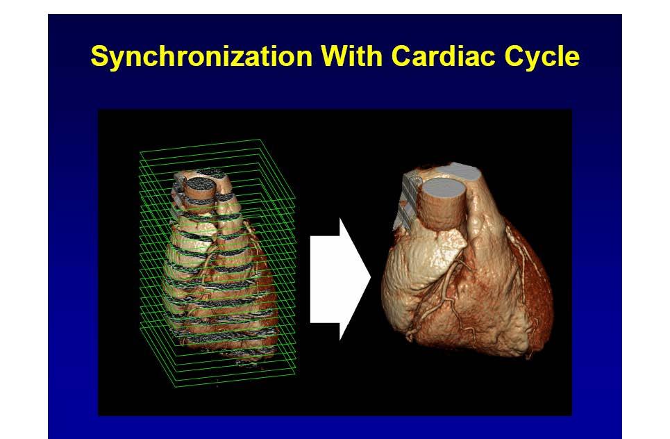
6 History EBCT mid to 3mm slice thickness 4 MSCT mm s thickness (30 heart beats minimum over all image-acquisition time) MSCT to 0.75 mm (4-8 heart beats)
7

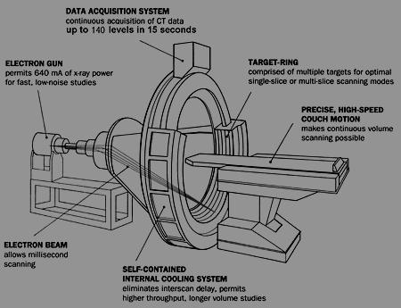
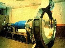
8 EBCT- Electron Beam CT
9
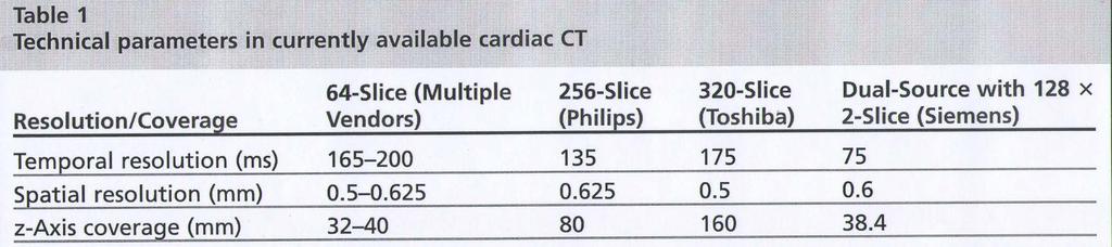
10 Evolution of CT - 40 years st CT Slip Ring Spiral Twin ub sec ½ sec 64 DSCT 256 / 640/ CT detector MSCT /FPD DSCT/DECT 0.33sec/ritation Multidetector, Multisource CT New detector technologies Data Handling, Dose management

11 Dual source CT
12 2005- Dual Source CT 2 X-ray sources and 2 detectors Faster than every beating heart DSCT became available around the year 2005, with 2 64 simultaneously acquired slices and a rotation time of 33 milliseconds and More recently, with simultaneously acquired slices and a gantry rotation time of 0.28 seconds. Capable of imaging full cardiac detail As much as 50 percent less radiation exposure compared to traditional CT scans

13 256 slice CT Breath holding time is reduced. The entire heart can be covered in a single rotation.
14

15 An 18-year-old man who presented with chest pain. Prospective ECG-gated axial images of the heart were obtained. Asc aorta, ascending aorta; Desc aorta, descending aorta; LA, left atrium; LAA, left atrial appendage; LAD, left anterior descending; LCX, left circumflex; LIMA, left internal mammary artery; LPV, left pulmonary vein; RA, right atrium; RCA, right coronary artery; RI, ramus intermedius; RIMA, right internal mammary artery; RPV, right pulmonary vein; RVOT, right ventricular outflow tract.
16

17 MIP rendering Technique Normal coronary arteries

18 MIP normal left MCA, LAD and several diagonal branches

19 MIP EBCT Normal RCA

20 Curved MPR The vessel can be seen in long length


21 2 reconstructions of the same image: VR does not well distinguished calcification from the lumen MIP is more transparent and overlapping vessels become more problematic


22 MIP LIMA : small diameter <1.5 mm and clips make interpretation difficult. Multiple views may necessary.


23 By-pass surgery 10 yrs ago: All 4 vessels are patent
 A 320-slice CT, (B) high-pitch spiral acquisition with")
24 Coronary CTA images acquired with the most recent CT hardware. (A) A 320-slice CT, (B) high-pitch spiral acquisition with dual-source CT.
 in a patient with a body weight of 93 kg. Conventional filtered back projection (top) shows considerable image noise.")
25 Reduction of image noise through iterative reconstruction. A coronary CT angiogram was acquired using 80 kv tube voltage (effective dose, 0.32 msv) in a patient with a body weight of 93 kg. Conventional filtered back projection (top) shows considerable image noise. Through iterative reconstruction (below), image noise can be reduced substantially.
26 Dual-Energy CT The subtraction of x-ray data obtained at different energy levels may, therefore, substantially increase contrast between these materials and all surrounding structures. Dual-energy CT can be achieved by simultaneous acquisition with 2 tubes and 2 detectors (DSCT) by extremely rapid switching of the emitted x-rays between different energy levels or by the design of detectors, which combine different elements with varying sensitivity to discrete kilovolt levels. Potential clinical applications in cardiovascular imaging include, for example, the improved delineation of vascular calcium (and its separation from the contrast enhanced lumen) and improved visualization of differences in myocardial contrast enhancement when myocardial perfusion is studied during exercise or at rest.
27 Clinical Applications Coronary Caicium Coronary CTA LV Function Evaluation of CABG Evaluation Stent Assessment of Myocardial Viability
28 Coronary CT Angiography Conventional coronary angiography is currently the gold standard and routine procedure for evaluating the extent of stenosis. Recent studies suggest that 64-slice coronary CT angiography (CTA) is highly accurate for exclusion of significant coronary artery stenosis (50% luminal narrowing), with a sensitivity range of 79% to 99% and specificity between 95% and 97%. The high negative predictive value associated with 64-slice scanners (98% 99%) in comparison with invasive coronary angiography enables the exclusion of obstructive CAD following a normal CTA with a high degree of certainty, obviating invasive cardiac catheterization, particularly in patients with low to intermediate risk of relevant stenoses.

29 Calcium Scoring: CAC score 1872 Significant CAC in all coronary arteries
 Calcium scoring by Agatston method 5 1217 in a 56-year-old woman presenting with chest")
30 (A) Calcium scoring by Agatston method 5 0 in a 43-year-old woman presenting with chest pain. (B) Calcium scoring by Agatston method in a 56-year-old woman presenting with chest pain
31 Calcium scoring Calcium Score Presence of Coronary Artery Disease (CAD) 0 No evidence of CAD 1 10 Minimal evidence of CAD Mild evidence of CAD Moderate evidence of CAD >400 Extensive evidence of CAD

32 Coronary Artery Anatomy Anomalous coronary artery Origin of left circumflex coronary artery from the right coronary ostium and course distal to the aortic root toward the left coronary groove

33 VR Technique: Need to evaluate the images fro multiple views as any given artery cannot be fully visualized from any angle

34 Coronary Artery Anatomy A 46-year-old woman who presented with chest pain. Curved MPR image of the RCA and LM with branches. Acute marg, acute marginal branch; D1, diagonal branch; LAD, left ant erior descending; LCX, left circumflex; LM, left main; OM, obtuse marginal branch; RCA, right coronary artery.
35
36

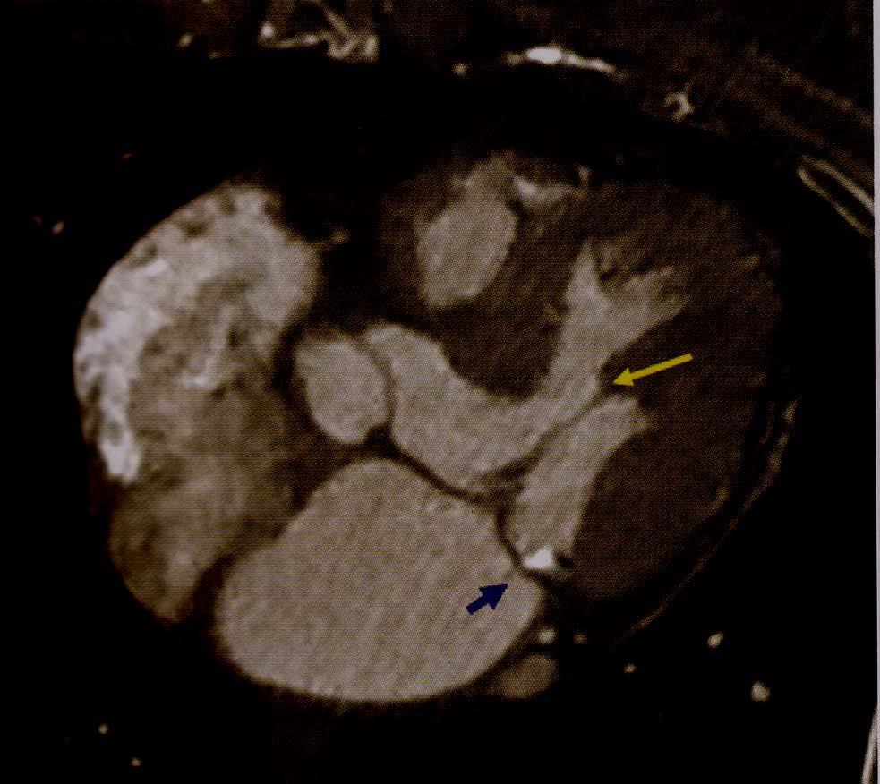
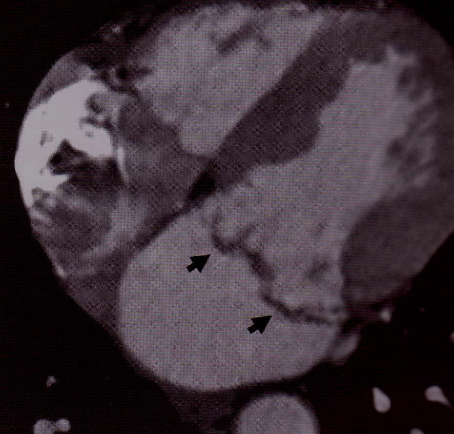
37 Multidetector CT images show acute thrombotic occlusion of the left circumflex artery
.")
38 . A 57-year-old male patient with CT-verified vulnerable plaque who subsequently developed ACS. CTA revealed a 50% stenotic lesion with positive remodeling, low-attenuation plaque, napkin ring sign, and spotty calcification within the proximal left anterior descending artery (A). Emergency coronary angiography revealed a 90% stenotic lesion at the site corresponding to the CT-verified vulnerable plaque in the proximal left anterior descending artery (B).
39 EVALUATION OF CABG Conventional angiography remains the gold standard in evaluating the patency of coronary bypass grafts; however, in recent years improvements in MDCT technology have enabled accurate and noninvasive visualization of grafts. Recent literature on 64-slice CT reports sensitivity and specificity ranges of 93.3% to 100% and 91.4% to 100%, respectively, in assessing CABG occlusion and significant stenosis (50%). The sensitivity, specificity, positive and negative predictive values, and accuracy for detecting graft stenosis were 93%, 98%, 93%,98%, and 97%, respectively. For graft occlusion the comparative values were 96.4%, 98.1%, 96.4%, 98.1%, and 97.6%, respectively.


40 MIP showing patent LIMA graft
41 Evaluation of Stents to visualize and accurately assess stent patency, restenosis, or neointimal hyperplasia But is limited in its ability to evaluate stent patency in coronary arteries. The diagnostic accuracy of MDCT in evaluating stent patency depends on various factors, including stent diameter, material, and design. Metallic struts in stents can create a blooming artifact, which can result in the appearance of a thicker strut and underestimation of lumen diameter. In addition, characterization of contrast enhancement patterns is vital in the analysis of stent patency.

42 VR - Stent in mid-left anterior descending artery

43 EBCT--- MIP the patent stent

44 MIP 64 MSCT Stent patenecy cannot be reliable as scan artifact from stent struts

45 Contrast is well visualised inside the stent allowing assessment of stent patency. Coronary stents are considered occluded when no contrast is visible inside the stent lumen, with decreased or loss of distal runoff, indicating significant restenosis. Visualization of contrast in the vessel distal to the stent alone does not necessarily indicate patency, because this may be the result of retrograde filling of the vessels


46 Ultra-high resolution Images from 64 MSCT of a stent mounted on a vessel model


47 64 MSCT Images of 3 stents which appear patent


48 Patent stent in the distal right coronary artery in vivo
49 Left Ventricular Function Accurate assessment of left ventricular (LV) function has both prognostic and therapeutic value in patients with coronary disease. Retrospective contrast-enhanced ECG-gated MDCT enables the acquisition of data within a cardiac cycle, which can be reformatted and used to determine LV volume and global function. Although MDCT is not the primary modality for functional analysis, it provides additional useful information in patients undergoing MDCT coronary angiography for detection of coronary artery obstruction, without the need for further imaging studies or additional radiation exposure. Cine magnetic resonance (MR) imaging is currently regarded as the gold standard for the assessment of global and regional cardiac function.

50 Images at ED and ES phases allowing EF and volume calculation
51 Assessment of Myocardial Viability Coronary revascularization by percutaneous coronary intervention (PCI) in patients with acute myocardial infarction may improve survival with the identification and restoration of viable myocardium. The evaluation of myocardial viability using contrast-enhanced MR imaging has been well established. However, recent preliminary studies have demonstrated the potential of cardiac MDCT in assessing myocardial perfusion as an alternative technique. Habis and colleagues performed cardiac CT imaging immediately after coronary angiography without contrast reinjection and demonstrated a promising technique in early assessment of myocardial viability.


52 Dual Energy CT 75-year-old man with stable angina. reversible perfusion defects of anteroseptal wall of left ventricle; these findings suggest ischemia. CT images show good correlation with SPECT images:


53 Dual Energy CT 75-year-old man with stable angina. reversible perfusion defects of anteroseptal wall of left ventricle; these findings suggest ischemia. CT images show good correlation with SPECT images:
 (arrow, A), and patent stent in RCA (arrow, B).")
54 A 45-year-old man with recent infarction of inferior left ventricle wall and stent in, undergoing dual-energy CT and SPECT before coronary artery bypass grafting. A and B, Coronary CT angiography images show long segment of occlusion in proximal LAD (LAD) (arrow, A), and patent stent in RCA (arrow, B). C and D, On SPECT horizontal stress (C) and rest (D) long-axis images, there is reversible perfusion defect of anteroseptal wall (arrows, C). E and F, Corresponding views for stress (E) and rest (F) dual-energy CT show reversible perfusion defect of anteroseptal wall (arrows, E). G, Volume-rendered dual-energy CT image shows perfusion defect of anterior left ventricle wall within context of entire thorax.
55 Venous Mapping for Atrial Fibrillation Ectopic arrhythmic foci originating within the pulmonary veins are a cause of both paroxysmal and persistent atrial fibrillation. Radiofrequency catheter ablation (RFCA) of the distal pulmonary veins and posterior left atrium is effective as a treatment for paroxysmal atrial fibrillation in patients with refractory atrial fibrillation or resistant to pharmacologic therapy or cardioversion. Multidetector CT of the pulmonary veins provides important anatomic information including the number, location, size, and orientation of pulmonary veins and their ostial branches noninvasively. Preprocedural mapping has been shown to decrease radiofrequency ablation procedure time.
56 Radiation Dose A combination of prospective triggering with low voltage settings is an effective measure for reducing the ED of coronary CTA to values of 2-4 msv independent of scanner system. Further dose reduction to nearly 1 msv can be achieved with high-pitch prospectively triggered coronary CTA.
57 CT Dose Reduction Prospective ECG Gating Retrospective ECG Gating Tube current modulation Iterative Reconstruction
58 Assessment of Cardiac Structure and Function


59 Pedunculated LV Apical thrombus left atrial appendage thrombus


60 Apical thrombus Left atrial myxoma

61 Bicuspid aortic valve


62 ASD Mitral Valve Prolapse
63 Mitral Calcifications Mitral Valve Prolapse


64 Pericardial calcification Myocardial (subendocardial Infarction)

65 Multiple pulmonary emboli
66 Cardiac CT in Emergency Department CAD Pulmonary Embolism Aortic Dissection
67 Limitations and pitfalls of coronary CTA feasible in patients with a stable heart rhythm, able to breath-hold for 20 seconds. The image quality is degraded by irregular heart rhythms; atrial fibrillation is a contraindication for MSCT imaging Well known pitfalls of MSCT coronary angiography include motion artefacts and severe coronary calcification. One of the most serious problems is that the presence of severe calcification in the coronary arteries reduces diagnostic accuracy, and it may even be impossible to assess some coronary segments if extremely severe calcification is present.
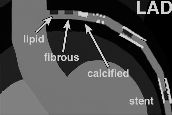
68 Evaluation of plaque Morphology by CTA Plaques characterized by positive remodeling, low attenuation, and napkin ring circular enhancement on contrast-enhanced coronary CTA have been regarded as rupture-prone vulnerable plaques, which account for about 60% of all vulnerable lesions and may be precursors of plaque rupture. Recently it demonstrated that the distribution and prevalence of computed tomography (CT)- verified vulnerable plaques were approximately similar to that of thin-cap fibroatheroma (TCFA) reported in autopsy cases, Coronary artery calcium score (CACS) alone may not be as useful for exclusion of a risk of acute coronary syndrome (ACS). Coronary CTA needs to demonstrate incremental prognostic value beyond the Framingham Risk Score (FRS) and other prognostic markers. The clinical usefulness of CT-based detection of vulnerable plaque remains to be demonstrated. CT-derived plaque features were demonstrated to predict the occurrence of slow-flow complications during percutaneous coronary interventions (PCIs), which opens a further potential field for coronary CTA.

69 Evaluation of plaque characteristics using new plaque analyzing software (Plaque Labeling Method). Aatypical chest pain, underwent coronary CTA revealed 25% stenotic lesion at the left main trunk. One year later, revealed a progressive 75% stenotic lesion at the left main trunk. Plaque characteristics - area and plaque area and plaque characteristics (red, necrotic core; blue, fibrous and/or fibrofatty; yellow, calcification) could be evaluated. The results showed that the necrotic core area (red area) progressed remarkably after 1 year.
70 Future CTA Single breath hold to reduce respiratory motion Higher spatial resolution Reduced slice thickness Increased no of detectors Reduced overall-imaging time Reductions in contrast requirement Rapidity and ease with coronary CTA- CT will be the preferred method for most applications in the absence of renal insufficiency or contrast allergy Better workstations for ease of use and diagnostic capabilities Multisector reconstruction Reduced radiation exposure
71 CARDIAC MR
72 MRI Multi-parametric cardiac magnetic resonance (CMR) (stress perfusion, rest perfusion, and late gadolinium enhancement) has highr sensitivity and negative predictive value (NPV) for the diagnosis of CAD. Delayed enhancement CMR (DEMR), one of the most common examinations for tissue characterization both in ischemic and non-ischemic myocardial diseases, has become the gold standard for visualization and quantification of infarcted myocardium and scar tissues as well as for the detection of infiltrative diseases of the heart.
73 Magnetic Resonance Myocardial Perfusion Imaging CMR has evolved significantly in the past decade CMR is able to assess multiple aspects of cardiovascular pathology in a single examination, including myocardial and coronary artery anatomy, ventricular function, myocardial perfusion, and viability. First-pass contrast-enhanced MR imaging has emerged as an excellent alternative imaging modality for the assessment of myocardial perfusion. Because perfusion abnormalities proceed systolic dysfunction, there is no surprise that direct perfusion imaging has higher sensitivity than indirect imaging (eg, wall motion dysfunction) for detection of ischemia, a concept described as ischemic cascade.
74 The ischemic cascade refers to the temporal sequence of events that develop over time when there is a progressive imbalance between myocardial oxygen demand and supply with abnormal myocardial perfusion being the first detectable event.

75 Potential mechanisms of hyperenhancement. In the acute and chronic infarct settings, hyperenhancement is hypothesized to directly result from consequences of myocyte cell death, with associated increase in extracellular volume of distribution for gadolinium-based contrast agents.

76 CMR images (upper panel ) and corresponding histologic slices (lower panel ) showing the exact match between the hyperenhanced area and the necrotic region defined by histology in the setting of acute myocardial infarction. Adapted from Wagner A, MahrholdtH, Holly TA, et al. Contrast-enhanced MR imaging and routine SPECT imaging for detection of subendocardial myocardial infarcts: an imaging study. Lancet 2003, in press; with permission.)
77 Multicomponent CMR stress testing protocol The multicomponent approach to CMR stress testing includes the following: 1. Cine MRI for the assessment of cardiac morphology and regional and global systolic function at baseline 2. Stress perfusion MRI to visualize regions of myocardial hypoperfusion during vasodilation (eg, with adenosine infusion) 3. Rest perfusion MRI to aid in distinguishing true perfusion defects from image artifacts 4. DE-MRI for the determination of myocardial infarction (MI)
 immediately adjacent to the delayed-enhancement images (DE-MRI).")
78 Images from a typical patient scan. Cine and delayed-enhancement images are acquired at six to eight short axis locations and at two to three long-axis locations during repeated breath holds. Images are interpreted with the cine images (cine-mri) immediately adjacent to the delayed-enhancement images (DE-MRI). In this patient example, DE-MRI demonstrates a myocardial infarction involving the inferior wall and inferoseptum of the left ventricle.
79 MR Viability Imaging Procedure Time Insert peripheral IV Place patient in scanner Obtain scout images Obtain cine images Inject gadolinium ( mmol/kg) Wait 10 minutes Obtain delayed enhancement images (segmented GRE with inversion prepulse )
80 A) Typical first-pass perfusion protocol for adenosine stress perfusion CMR. (B) Typical firstpass perfusion protocol for dipyridamole stress perfusion CMR. (C) Typical first-pass perfusion protocol for regadenoson stress perfusion CMR. (Modified from Gerber BL, Raman SV, Nayak K, et al. Myocardial first-pass perfusion cardiovascular magnetic resonance: history, theory, and current state of the art. J Cardiovasc Magn Reson 2008;10:18.)

81 Diagram illustrating the difference between the DE observed in ischemic and nonischemic diseases. Ischemic disease presents a vascular territory distribution and characteristically involves the endocardium. In non-ischemic conditions, the abnormal DE may be midwall, patchy, epicardial, or global subendocardial
 and horizontal long axis (B) demonstrates subendocardial enhancement of the interventricular septum with some focal areas of")
82 MI in a 50-year-old man. Viability study in short axis (A) and horizontal long axis (B) demonstrates subendocardial enhancement of the interventricular septum with some focal areas of transmural infarction. Based on the MRI findings, surgical revascularization was performed with significant improvement of the left ventricular function.
 and left ventricular dilation consistent with transmural infarction with true left")
83 Viability MR in a 55-year-old male patient with history of CAD and percutaneous intervention considered for revascularization surgery. Contrast-enhanced MR demonstrates extensive DE (arrows) and left ventricular dilation consistent with transmural infarction with true left ventricular aneurysm.
84 CMR Identification of specific patterns of myocardial DE that occur in a variety of non-ischemic diseases, such as myocarditis, sarcoidosis, hypertrophic cardiomyopathy, and amyloidosis, has further extended the clinical applications of contrast-enhanced CMR imaging.
 and horizontal long axis (B) demonstrate abnormal late gadolinium")
85 Dilated cardiomyopathy in a 60-year-old man. Delayed images after contrast injection in short axis (A) and horizontal long axis (B) demonstrate abnormal late gadolinium enhancement in the midventricular wall, with a nonvascular distribution (arrows).

86 Hypertrophic cardiomyopathy in a 53-year-old male. Irregular areas of contrast enhancement are appreciated in this short axis delayed images after contrast injection (arrows).
87 MRCA Coronary artery magnetic resonance (MR) angiography is useful for identifying coronary artery anomalies, in particular in younger individuals, without exposure to ionizing radiation or iodinated contrast medium. Only limited multicenter MRCA experience is available and there are still no data on the prognostic value of coronary MR imaging. Technical challenges that need to be addressed are further improvements in motion suppression and abbreviated scanning times aimed at improving spatial resolution and patient comfort. The development of new and specific contrast agents, high-field MR imaging with improved spatial resolution, and continued progress in MR imaging methods development will undoubtedly lead to further progress.

88 MR Coronary Angiogram MR coronary angiography in a young female athlete with exercise-induced chest pain. Free breathing 3-D noncontrast turbo field-echo images demonstrate normal origin and course of the proximal right and left coronary arteries (arrows).
89 Future Cardiovascular MRI MR cardiovascular imaging For Greater definition of tissue characteristics Perfusion Valvular function Lack of X-ray radiation Lack of need for contrast media Against Limited temporal and spatial resolution partial volume artifacts due to slice thickness limitations reliance on multiple breath holds Poor visualization of left main coronary artery
90 FUTURE DEVELOPMENTS Undoubtedly, both hardware and software for cardiac imaging will continue to be improved. Higher spatial and temporal resolution requires a substantially higher radiation dose to avoid excessive image noise. Close collaboration between clinicians, imaging researchers, hardware engineers, and software developers will, hence, be necessary to guide the further development to make full use of its potential.
91 Thank You!
Cardiac Computed Tomography
 Cardiac Computed Tomography Authored and approved by Koen Nieman Stephan Achenbach Francesca Pugliese Bernard Cosyns Patrizio Lancellotti Anastasia Kitsiou Contents CARDIAC COMPUTED TOMOGRAPHY Page 1.
Coronary Artery Imaging. Suvipaporn Siripornpitak, MD Inter-hospital Conference : Rajavithi Hospital
 Coronary Artery Imaging Suvipaporn Siripornpitak, MD Inter-hospital Conference : Rajavithi Hospital Larger array : cover scan area Detector size : spatial resolution Rotation speed : scan time Retrospective
Cardiac Imaging Tests
 Cardiac Imaging Tests http://www.medpagetoday.com/upload/2010/11/15/23347.jpg Standard imaging tests include echocardiography, chest x-ray, CT, MRI, and various radionuclide techniques. Standard CT and
Disclosures. GETTING TO THE HEART OF THE MATTER WITH MULTIMODALITY CARDIAC IMAGING Organ Review Meeting 25 September. Overview
 GETTING TO THE HEART OF THE MATTER WITH MULTIMODALITY CARDIAC IMAGING Organ Review Meeting 25 September Disclosures None relevant to this presentation Mini Pakkal Assistant Professor of Radiology University
Use of Nuclear Cardiology in Myocardial Viability Assessment and Introduction to PET and PET/CT for Advanced Users
 Use of Nuclear Cardiology in Myocardial Viability Assessment and Introduction to PET and PET/CT for Advanced Users February 1 5, 2011 University of Santo Tomas Hospital Angelo King A-V Auditorium Manila,
Dr Felix Keng. Imaging of the heart is technically difficult because: Role of Cardiac MSCT. Current: Cardiac Motion Respiratory Motion
 Siemens Philips Dr Felix Keng GE Toshiba Role of Cardiac MSCT Current: Structural / congenital heart imaging Extra-cardiac / Great vessel imaging Volumes and ejection fractions (cine + gating) Calcium
Calcium Scoring and Cardiac CT
 Calcium Scoring and Cardiac CT John C. Finley, MD, FACC, FASE Medical Director, CT Department; Alaska Heart and Vascular Institute February 9, 2018 1. Calcium Scoring 2. CT Coronary Angiography 3. Use
I have no financial disclosures
 Manpreet Singh MD I have no financial disclosures Exercise Treadmill Bicycle Functional capacity assessment Well validated prognostic value Ischemic assessment ECG changes ST segments Arrhythmias Hemodynamic
ADVANCED CARDIOVASCULAR IMAGING. Medical Knowledge. Goals and Objectives PF EF MF LF Aspirational
 Medical Knowledge Goals and Objectives PF EF MF LF Aspirational Know the basic principles of magnetic resonance imaging (MRI) including the role of the magnetic fields and gradient coil systems, generation
The Value of Stress MRI in Evaluation of Myocardial Ischemia
 The Value of Stress MRI in Evaluation of Myocardial Ischemia Dr. Saeed Al Sayari, MBBS, EBCR, MBA Department of Radiology and Nuclear Medicine Mafraq Hospital, Abu Dhabi United Arab Emirates Introduction
Index. radiologic.theclinics.com. Note: Page numbers of article titles are in boldface type.
 Index Note: Page numbers of article titles are in boldface type. A ALCAPA. See Anomalous left coronary artery from the pulmonary artery. Angiosarcoma computed tomographic assessment of, 809 811 Anomalous
Imaging congestive heart failure: role of coronary computed tomography angiography (CCTA)
") Imaging congestive heart failure: role of coronary computed tomography angiography (CCTA) Gianluca Pontone, MD, PhD, FESC, FSCCT Director of MR Unit Deputy Director of Cardiovascul CT Unit Clinical Cardiology
Imaging congestive heart failure: role of coronary computed tomography angiography (CCTA) Gianluca Pontone, MD, PhD, FESC, FSCCT Director of MR Unit Deputy Director of Cardiovascul CT Unit Clinical Cardiology
Low-dose prospective ECG-triggering dual-source CT angiography in infants and children with complex congenital heart disease: first experience
 Low-dose prospective ECG-triggering dual-source CT angiography in infants and children with complex congenital heart disease: first experience Ximing Wang, M.D., Zhaoping Cheng, M.D., Dawei Wu, M.D., Lebin
EAE Teaching Course. Magnetic Resonance Imaging. Competitive or Complementary? Sofia, Bulgaria, 5-7 April F.E. Rademakers
 EAE Teaching Course Magnetic Resonance Imaging Competitive or Complementary? Sofia, Bulgaria, 5-7 April 2012 F.E. Rademakers Complementary? Of Course N Engl J Med 2012;366:54-63 Clinical relevance Treatment
SPECT-CT: Τι πρέπει να γνωρίζει ο Καρδιολόγος
 SPECT-CT: Τι πρέπει να γνωρίζει ο Καρδιολόγος Δρ Αναστασία Κίτσιου Διευθύντρια, Καρδιολογική Κλινική, Σισμανόγλειο ΓΝΑ Chair, Education Committee, Section on Nuclear Cardiology & Cardiac CT, EACVI, ESC
General Cardiovascular Magnetic Resonance Imaging
 2 General Cardiovascular Magnetic Resonance Imaging 19 Peter G. Danias, Cardiovascular MRI: 150 Multiple-Choice Questions and Answers Humana Press 2008 20 Cardiovascular MRI: 150 Multiple-Choice Questions
Advanced Imaging MRI and CTA
 Advanced Imaging MRI and CTA Who and why may benefit. Matthew W. Martinez, M.D. FACC Lehigh Valley Health Network Director, Cardiovascular Imaging Learning Objectives Review basics of CMR and CTA Review
Pearls & Pitfalls in nuclear cardiology
 Pearls & Pitfalls in nuclear cardiology Maythinee Chantadisai, MD., NM physician Division of Nuclear Medicine, Department of radiology, KCMH Principle of myocardial perfusion imaging (MPI) Radiotracer
Ultrasound. Computed tomography. Case studies. Utility of IQon Spectral CT in. cardiac imaging
 Ultrasound Computed tomography Case studies Utility of IQon Spectral CT in cardiac imaging Cardiac imaging is a challenging procedure where it is necessary to image a motion-free heart. This requires a
J. Schwitter, MD, FESC Section of Cardiology
 J. Schwitter, MD, FESC Section of Cardiology CMR Center of the CHUV University Hospital Lausanne - CHUV Switzerland Centre de RM Cardiaque J. Schwitter, MD, FESC Section of Cardiology CMR Center of the
Cardiac Imaging. Kimberly Delcour, DO, FACC. Mahi Ashwath, MD, FACC, FASE. Director, Cardiac CT. Director, Cardiac MRI
 Cardiac Imaging Kimberly Delcour, DO, FACC Director, Cardiac CT Mahi Ashwath, MD, FACC, FASE Director, Cardiac MRI Cardiac Imaging Discuss the clinical applications of and indications for: Cardiac CT Nuclear
Multisclice CT in combination with functional imaging for CAD. Temporal Resolution. Spatial Resolution. Temporal resolution = ½ of the rotation time
 Multisclice CT in combination with functional imaging for CAD Prof. Juhani Knuuti, MD, FESC Turku University Hospital and University of Turku Turku, Finland MSCT and functional imaging for CAD Practical
CARDIAC MRI. Cardiovascular Disease. Cardiovascular Disease. Cardiovascular Disease. Overview
 CARDIAC MRI Dr Yang Faridah A. Aziz Department of Biomedical Imaging University of Malaya Medical Centre Cardiovascular Disease Diseases of the circulatory system, also called cardiovascular disease (CVD),
Recent developments in cardiac CT
 REVIEW Recent developments in cardiac CT With the introduction of 64-multidetector row CT, coronary CT angiography has become a clinical tool, owing to improved image quality and reduced breath-hold time,
ROLE OF MULTISLICE COMPUTED TOMOGRAPHY IN CARDIAC IMAGING
 ROLE OF MULTISLICE COMPUTED TOMOGRAPHY IN CARDIAC IMAGING Non-invasive coronary angiography along with multidetector computed tomography or magnetic resonance imaging is attracting increasing interest
CARDIAC AND CORONARY ARTERY ANATOMY NO DISCLOSURES. Axial Anatomy of Heart. Axial Anatomy of Heart. Axial Anatomy of Heart
 CARDIAC AND CORONARY ARTERY ANATOMY NO DISCLOSURES NASCI MEETING, ORLANDO FLORIDA 2009 KOSTAKI G. BIS, MD, FACR DEPARTMENT OF RADIOLOGY WILLIAM BEAUMONT HOSPITAL Royal Oak, Michigan OBJECTIVES CARDIAC
Cardiac CT Angiography
 Cardiac CT Angiography Dr James Chafey, Radiologist Why do we need a better test for C.A.D? 1. CAD is the leading cause of death in the US CAD 31% Cancer 23% Stroke 7% 2. The prevalence of atherosclerosis
TITLE: Multi-Slice Computed Tomography Coronary Angiography for Coronary Artery Disease: A Review of the Clinical Effectiveness and Guidelines
 TITLE: Multi-Slice Computed Tomography Coronary Angiography for Coronary Artery Disease: A Review of the Clinical Effectiveness and Guidelines DATE: 25 February 2009 CONTEXT AND POLICY ISSUES: Coronary
Horizon Scanning Technology Summary. Magnetic resonance angiography (MRA) imaging for the detection of coronary artery disease
 imaging for the detection of coronary artery disease") Horizon Scanning Technology Summary National Horizon Scanning Centre Magnetic resonance angiography (MRA) imaging for the detection of coronary artery disease April 2007 This technology summary is based
Horizon Scanning Technology Summary National Horizon Scanning Centre Magnetic resonance angiography (MRA) imaging for the detection of coronary artery disease April 2007 This technology summary is based
Ischemic heart disease
 Ischemic heart disease Introduction In > 90% of cases: the cause is: reduced coronary blood flow secondary to: obstructive atherosclerotic vascular disease so most of the time it is called: coronary artery
Radiologic Assessment of Myocardial Viability
 November 2001 Radiologic Assessment of Myocardial Viability Joshua Moss, Harvard Medical School Year III Patient EF 66yo female with a 3-year history of intermittent chest pain previously relieved by sublingual
MRI ACS-ben. Tamás Simor MD, PhD, Med Hab. University of Pécs, Heart Institute
 MRI ACS-ben Tamás Simor MD, PhD, Med Hab Time Course of Changes in Infarct Size, Viable Myocardium, and LV Mass After Reperfused and Nonreperfused MI Blue lines denote reperfused myocardial infarction
Disclosure Information
 Coronary CTA Pearls and Pitfalls Ricardo C. Cury, MD, FSCCT, FAHA, FACC Chairman of Radiology Radiology Associates of South Florida Director of Cardiac Imaging Miami Cardiac and Vascular Institute Past-President
Fundamentals, Techniques, Pitfalls, and Limitations of MDCT Interpretation and Measurement
 Fundamentals, Techniques, Pitfalls, and Limitations of MDCT Interpretation and Measurement 3 rd Annual Imaging & Physiology Summit November 20-21, 21, 2009 Seoul, Korea Wm. Guy Weigold, MD, FACC Cardiovascular
Improved Noninvasive Assessment of Coronary Artery Bypass Grafts With 64-Slice Computed Tomographic Angiography in an Unselected Patient Population
 Journal of the American College of Cardiology Vol. 49, No. 9, 2007 2007 by the American College of Cardiology Foundation ISSN 0735-1097/07/$32.00 Published by Elsevier Inc. doi:10.1016/j.jacc.2006.10.066
Chapter 45. Utility of Computed Tomographic Coronary Angiography Post Coronary Revascularization BACKGROUND CORONARY ARTERY BYPASS GRAFTING
 Chapter 45 Utility of Computed Tomographic Coronary Angiography Post Coronary Revascularization SANKAR NEELAKANTAN SANJAYA VISWAMITRA SRIKANTH SOLA BACKGROUND Coronary artery disease (CAD) is the leading
Cardiac MRI: Cardiomyopathy
 Cardiac MRI: Cardiomyopathy Laura E. Heyneman, MD I do not have any relevant financial relationships with any commercial interests Cardiac MRI: Cardiomyopathy Laura E. Heyneman, MD Duke University Medical
Chapter 5 Section 1.1. Diagnostic Radiology (Diagnostic Imaging)
") Radiology Chapter 5 Section 1.1 Issue Date: March 7, 1986 Authority: 32 CFR 199.4(a), (b)(2)(x), (c)(2)(viii), (e)(14) and 32 CFR 199.6(d)(2) 1.0 CPT 1 PROCEDURE CODES 70010-72292, 73000-76499, 77071-77084,
Radiology Chapter 5 Section 1.1 Issue Date: March 7, 1986 Authority: 32 CFR 199.4(a), (b)(2)(x), (c)(2)(viii), (e)(14) and 32 CFR 199.6(d)(2) 1.0 CPT 1 PROCEDURE CODES 70010-72292, 73000-76499, 77071-77084,
THE ROLE OF HIGH END MULTI DETECTOR CT IN CORONARY IMAGING ESSAY
 THE ROLE OF HIGH END MULTI DETECTOR CT IN CORONARY IMAGING ESSAY Submitted for partial fulfillment of Master degree in Radiodiagnosis By Ahmed Yehia Ahmed (M.B.B.Ch., Cairo University) Supervisors Prof.
Computed Tomography of the Coronary Arteries
 Cardiology Update DAVOS 2011 Computed Tomography of the Coronary Arteries Anders Persson M.D., Ph.D Director, Assoc. Professor Center for Medical Image Science and Visualization Linköping University SWEDEN
Case 47 Clinical Presentation
 93 Case 47 C Clinical Presentation 45-year-old man presents with chest pain and new onset of a murmur. Echocardiography shows severe aortic insufficiency. 94 RadCases Cardiac Imaging Imaging Findings C
Improvement of Image Quality with ß-Blocker Premedication on ECG-Gated 16-MDCT Coronary Angiography
 16-MDCT Coronary Angiography Shim et al. 16-MDCT Coronary Angiography Sung Shine Shim 1 Yookyung Kim Soo Mee Lim Received December 1, 2003; accepted after revision June 1, 2004. 1 All authors: Department
Imaging of the Heart Todd Tessendorf MD FACC
 Imaging of the Heart Todd Tessendorf MD FACC Outline Imaging Modalities for Structural Heart Disease ECHO, MRI Imaging Modalities for Ischemic Heart Disease SPECT, PET, CCTA Show lots of pretty pictures
Noncoronary Cardiac MDCT
 Noncoronary Cardiac MDCT David A. Bluemke, M.D., Ph.D. Professor, of Radiology and Medicine Johns Hopkins University School of Medicine Baltimore, Maryland Toshiba Disclosures Grant support Noncoronary
Sung A Chang Department of Internal Medicine, Division of Cardiology, Sungkyunkwan University School of Medicine, Samsung Medical Center
 CMR Perfusion and Viability A STICH Out of Time? Sung A Chang Department of Internal Medicine, Division of Cardiology, Sungkyunkwan University School of Medicine, Samsung Medical Center Can Imaging Improve
Is computed tomography angiography really useful in. of coronary artery disease?
 Is computed tomography angiography really useful in screening patients with high risk of coronary artery disease? Myeong-Ki Hong, M.D. Ph D Professor of Medicine Division of Cardiology, Severance Cardiovascular
b. To facilitate the management decision of a patient with an equivocal stress test.
 National Imaging Associates, Inc. Clinical guidelines EBCT HEART CT & HEART CT CONGENITAL CCTA CPT4 Codes: 75571 EBCT 75572, 75573 Heart CT & Heart CT Congenital 75574 - CCTA LCD ID Number: L33559 J K
What every radiologist should know about cardiac CT: A case-based pictorial review
 What every radiologist should know about cardiac CT: A case-based pictorial review Poster No.: C-0555 Congress: ECR 2010 Type: Educational Exhibit Topic: Cardiac Authors: C. M. Capuñay, P. Carrascosa,
Non Invasive Diagnostic Modalities for Coronary Artery Disease. Dr. Amitesh Aggarwal
 Non Invasive Diagnostic Modalities for Coronary Artery Disease Dr. Amitesh Aggarwal Ebers papyrus, ca. 1555 BCE If thou examine a man for illness in his cardia, and he has pains in his arms, in his breasts
Why Cardiac MRI? Presented by:
 Why Cardiac MRI? Presented by: Lisa G. Carkner, MD, FACC 1 Disclosures I have no financial disclosures Objectives Review basic principles of Cardiac MRI. What patient characteristics do I need to consider
SYMPOSIA. Coronary CTA. Indications, Patient Selection, and Clinical Implications
 SYMPOSIA Indications, Patient Selection, and Clinical Implications Christian Thilo, MD,* Mark Auler, MD,* Peter Zwerner, MD,w Philip Costello, MD,* and U. Joseph Schoepf, MD* Abstract: Recent technical
Introduction. Cardiac Imaging Modalities MRI. Overview. MRI (Continued) MRI (Continued) Arnaud Bistoquet 12/19/03
 MRI (Continued) Arnaud Bistoquet 12/19/03") Introduction Cardiac Imaging Modalities Arnaud Bistoquet 12/19/03 Coronary heart disease: the vessels that supply oxygen-carrying blood to the heart, become narrowed and unable to carry a normal amount
Introduction Cardiac Imaging Modalities Arnaud Bistoquet 12/19/03 Coronary heart disease: the vessels that supply oxygen-carrying blood to the heart, become narrowed and unable to carry a normal amount
The use of Cardiac CT and MRI in Clinical Practice
 The use of Cardiac CT and MRI in Clinical Practice Matthew W. Martinez, MD Assistant Professor of Medicine LVPG - Lehigh Valley Heart Specialists Lehigh Valley Health Network Oct. 3, 2009 DISCLOSURE Relevant
Fellows on this rotation are expected to attend nuclear conferences and multimodality imaging conference.
 Rotation: Imaging 1 Imaging 1 provides COCATS Level 1 experience for nuclear cardiology (including SPECT and PET) and cardiac CT. Fellows will administer, process, and read cardiac nuclear studies with
Coronary Artery Anomalies from Birth to Adulthood; the Role of CT Coronary Angiography in Sudden Cardiac Death Screening
 Coronary Artery Anomalies from Birth to Adulthood; the Role of CT Coronary Angiography in Sudden Cardiac Death Screening E O Dwyer 1, C O Brien 1, B Loo 1, A Snow Hogan 1, O Buckley1 2, B 1. Department
MSRS 6473 Vascular Noninvasive Imaging Procedures
 MSRS 6473 Vascular Noninvasive Imaging Procedures Rex T. Christensen MHA RT (R) (MR) (CT) (ARRT) CIIP Basic Physics Equipment Cardiac Positioning Perfusion Pathology MRI 1 Animal Magnetism MRI Basic Physics
Cardiac computed tomography: indications, applications, limitations, and training requirements
 European Heart Journal (2008) 29, 531 556 doi:10.1093/eurheartj/ehm544 SPECIAL ARTICLE Cardiac computed tomography: indications, applications, limitations, and training requirements Report of a Writing
Covered Indications. Evaluation of chest pain syndrome uninterpretable or equivocal stress test (exercise, perfusion, or stress echo)
") BCBS Plans Covered Indications Policy No. 230, Cardiac Computed Tomography, Cardiac Computed Tomography Angiography (CPT 75574, 75573,75572) Last reviewed January 2017 Cardiac Computed Tomography (CCT),
BCBS Plans Covered Indications Policy No. 230, Cardiac Computed Tomography, Cardiac Computed Tomography Angiography (CPT 75574, 75573,75572) Last reviewed January 2017 Cardiac Computed Tomography (CCT),
Chapter 5 Section 1.1
 Radiology Chapter 5 Section 1.1 Issue Date: March 7, 1986 Authority: 32 CFR 199.4(a), (b)(2)(x), (c)(2)(viii), (e)(14) and 32 CFR 199.6(d)(2) Copyright: CPT only 2006 American Medical Association (or such
2004;77:800 4 MSCT OF CORONARY ARTERY BYPASS GRAFTS. Results. CABG With Adequate Diagnostic Quality
 Isotropic Half-Millimeter Angiography of Coronary Artery Bypass Grafts With 16-Slice Computed Tomography Marc Dewey, MD, Alexander Lembcke, MD, Christian Enzweiler, MD, Bernd Hamm, MD, and Patrik Rogalla,
Review of Cardiac Imaging Modalities in the Renal Patient. George Youssef
 Review of Cardiac Imaging Modalities in the Renal Patient George Youssef ECHO Left ventricular hypertrophy (LVH) assessment Diastolic dysfunction Stress ECHO Cardiac CT angiography Echocardiography - positives
Current Indications for Cardiac MRI: What You See is What You Get?
 Current Indications for Cardiac MRI: What You See is What You Get? Javier Ganame, MD, PhD, FASE No disclosures Cardiology Update, Niagara, Sept 24th, 2016 The Ideal Diagnostic Technique Easy to apply Accurate
Cardiac MRI in ACHD What We. ACHD Patients
 Cardiac MRI in ACHD What We Have Learned to Apply to ACHD Patients Faris Al Mousily, MBChB, FAAC, FACC Consultant, Pediatric Cardiology, KFSH&RC/Jeddah Adjunct Faculty, Division of Pediatric Cardiology
Detailed Order Request Checklists for Cardiology
 Next Generation Solutions Detailed Order Request Checklists for Cardiology 8600 West Bryn Mawr Avenue South Tower Suite 800 Chicago, IL 60631 www.aimspecialtyhealth.com Appropriate.Safe.Affordable 2018
Solving the Dilemma of Ostial Stenting: A Case Series Illustrating the Flash Ostial System
 Volume 1, Issue 1 Case Report Solving the Dilemma of Ostial Stenting: A Case Series Illustrating the Flash Ostial System Robert F. Riley * and Bill Lombardi University of Washington Medical Center, Division
Chapter 43 Noninvasive Coronary Plaque Imaging
 hapter 43 Noninvasive oronary Plaque Imaging NIRUDH KOHLI The goal of coronary imaging is to define the extent of luminal narrowing as well as composition of an atherosclerotic plaque to facilitate appropriate
Cardiac MRI: Clinical Application to Disease
 Cardiac MRI: Clinical Application to Disease Jessi Smith, MD Cardiothoracic imaging, Indiana University Slides courtesy of Stacy Rissing, MD Outline Imaging planes Disease findings Pulse sequences used
Cardiac CT and MRI. Ashraf Hamdan, MD. Sheba Medical Center. Sheba Medical Center Tel Hashomer. Leviev Heart Center
 Cardiac CT and MRI Ashraf Hamdan, MD Sheba Medical Center Sheba Medical Center Tel Hashomer Leviev Heart Center Spatial resolution Resolution 4x4mm If the object incidentally placed in one pixel high image
Dual Energy CT of the Heart: Perfusion and Beyond
 Dual Energy CT of the Heart: Perfusion and Beyond U. Joseph Schoepf, MD, FAHA, FSCBT MR, FSCCT Professor of Radiology, Medicine, and Pediatrics Director of Cardiovascular Imaging Disclosures Consultant
Validation of CT Perfusion Imaging Against Invasive Angiography and FFR on a 320-MDCT Scanner
 Validation of CT Perfusion Imaging Against Invasive Angiography and FFR on a 320-MDCT Scanner Zhen Qian, Gustavo Vasquez, Sarah Rinehart, Parag Joshi, Eric Krivitsky, Anna Kalynych, Dimitri Karmpaliotis,
Case Report Preoperative Assessment of Anomalous Right Coronary Artery Arising from the Main Pulmonary Artery
 Case Reports in Medicine Volume 2011, Article ID 642126, 4 pages doi:10.1155/2011/642126 Case Report Preoperative Assessment of Anomalous Right Coronary Artery Arising from the Main Pulmonary Artery Marshall
Rotation: Imaging 2. Nuclear Cardiology (in Imaging 1 and 2)
") Rotation: Imaging 2 Imaging 2 provides addition nuclear cardiology experience and COCATS Level 1 cardiac MRI experience. Fellows administer, process, and read VHVI cardiac nuclear studies with cardiology
Rotation: Imaging 2 Imaging 2 provides addition nuclear cardiology experience and COCATS Level 1 cardiac MRI experience. Fellows administer, process, and read VHVI cardiac nuclear studies with cardiology
Correlation of Cardiac CTA to Conventional Cardiac Angiography in Diagnosing Coronary Artery Stenosis in a Community Based Center
 Correlation of Cardiac CTA to Conventional Cardiac Angiography in Diagnosing Coronary Artery Stenosis in a Community Based Center Mathieu Sabbagh, R3 Michigan State University Radiology Garden City Hospital
, David Stultz, MD. Cardiac CT. David Stultz, MD Cardiology Fellow, PGY 6 March 28, 2006
 Cardiac CT David Stultz, MD Cardiology Fellow, PGY 6 March 28, 2006 Courtesy Tom Kracus Courtesy Kettering Tom Medical Kracus Cente Kettering Medical Center 2003-2006, David Stultz, MD Courtesy Tom Kracus
Cardiac Stress MRI: Detection of Ischemia. Disclosures: Dobutamine Stress MR. April 28, 2018
 Cardiac MRI: Detection of Ischemia Cardiac MRI in Today s Clinical Practice Foundations of Cardiovascular Magnetic Resonance Daniel C. Lee, MD, MSc Assistant Professor of Medicine and Radiology Co-Director,
Indications of Coronary Angiography Dr. Shaheer K. George, M.D Faculty of Medicine, Mansoura University 2014
 Indications of Coronary Angiography Dr. Shaheer K. George, M.D Faculty of Medicine, Mansoura University 2014 Indications for cardiac catheterization Before a decision to perform an invasive procedure such
An Epidemic of Heart Disease The Silent Killer. Professor Mike Kirby FRCP University of Hertfordshire Institute of Diabetes for Older People
 An Epidemic of Heart Disease The Silent Killer Professor Mike Kirby FRCP University of Hertfordshire Institute of Diabetes for Older People Initial clinical presentation of CAD Angina MI Atypical CP Death
The role of Magnetic Resonance Imaging in the diagnosis of viability & Coronary Artery Disease
 The role of Magnetic Resonance Imaging in the diagnosis of viability & Coronary Artery Disease G.P. Spanos, MSc, Phd Head of CardioVascular Imaging Tomographia Diagnostic Center Cardiovascular magnetic
Gated blood pool ventriculography: Is there still a role in myocardial viability?
 Gated blood pool ventriculography: Is there still a role in myocardial viability? Oliver C. Alix, MD Adult Clinical and Nuclear Cardiology St. Luke s Medical Centre - Global City Case Presentation A 62-year-old
Isolated congenital coronary anomalies: Evaluation by multislice-ct or MRI
 Isolated congenital coronary anomalies: Evaluation by multislice-ct or MRI B.K. Velthuis, Dept. of Radiology UMC Utrecht, the Netherlands ESC 2010 Coronary artery anomalies CAA Uncommon 0.3-5% normal population
Case 1. Case 2. Case 3
 Case 1 The correct answer is D. Occasionally, the Brugada syndrome can present similar morphologies to A and also change depending on the lead position but in the Brugada pattern the r is wider and ST
CT or PET/CT for coronary artery disease
 CT or PET/CT for coronary artery disease Rotterdam 2012 Juhani Knuuti, MD, PhD, FESC Turku PET Centre University of Turku Turku, Finland Juhani.knuuti@utu.fi Turku PET Centre University of Turku Åbo Akademi
Case based learning: CMR in Heart Failure
 Case based learning: CMR in Heart Failure Milind Y Desai, MD FACC FAHA FESC Associate Professor of Medicine Heart and Vascular Institute, Cleveland Clinic Cleveland, OH Disclosures: none Use of Gadolinium
Utility of CT angiography for pre-operative evaluation of robotic-assisted minimally invasive mitral valve surgery.
 Utility of CT angiography for pre-operative evaluation of robotic-assisted minimally invasive mitral valve surgery. Poster No.: C-2214 Congress: ECR 2014 Type: Educational Exhibit Authors: M. Muthuvelu,
Hybrid cardiac imaging Advantages, limitations, clinical scenarios and perspectives for the future
 Hybrid cardiac imaging Advantages, limitations, clinical scenarios and perspectives for the future Prof. Juhani Knuuti, MD, FESC Turku, Finland Disclosure: Juhani Knuuti, M.D. Juhani Knuuti, M.D. has financial
A Noninvasive Assessment of CAD
 : A Noninvasive Assessment of CAD In this article, Dr. Heilbron and Dr. Forster look at the noninvasive assessment of coronary artery disease (CAD), by means of coronary computed tomography angiography
Perfusion, Viability, Edema and Hemorrhage: How it Can (and Should) Change Clinical Practice. Rohan Dharmakumar, Ph.D.
 Change Clinical Practice. Rohan Dharmakumar, Ph.D.") Perfusion, Viability, Edema and Hemorrhage: How it Can (and Should) Change Clinical Practice Rohan Dharmakumar, Ph.D. Director, Translational Cardiac Imaging Research Associate Director, Biomedical Imaging
Perfusion, Viability, Edema and Hemorrhage: How it Can (and Should) Change Clinical Practice Rohan Dharmakumar, Ph.D. Director, Translational Cardiac Imaging Research Associate Director, Biomedical Imaging
The Final 10-Year Follow-up Results from the Bari Randomized Trial J Am Coll Cardiol (2007) 49;1600-6
 49;1600-6") The Final 10-Year Follow-up Results from the Bari Randomized Trial J Am Coll Cardiol (2007) 49;1600-6 n&list_uids=17433949 64-Multislice Detector Computed Tomography Coronary Angiography as Potential Alternative
The Final 10-Year Follow-up Results from the Bari Randomized Trial J Am Coll Cardiol (2007) 49;1600-6 n&list_uids=17433949 64-Multislice Detector Computed Tomography Coronary Angiography as Potential Alternative
New Cardiovascular Devices and Interventions: Non-Contrast MRI for TAVR Abhishek Chaturvedi Assistant Professor. Cardiothoracic Radiology
 New Cardiovascular Devices and Interventions: Non-Contrast MRI for TAVR Abhishek Chaturvedi Assistant Professor Cardiothoracic Radiology Disclosure I have no disclosure pertinent to this presentation.
MR Assessment of Myocardial Viability
 MR Assessment of Myocardial Viability Definition of Viability Clinical Metabolism: Presence of glucose uptake Perfusion / Perfusion reserve Morphology: Wall thickness, wall thickening Contractility: Recovery
CT Perfusion. U. Joseph Schoepf, MD, FAHA, FSCBT MR, FSCCT Professor of Radiology, Medicine, and Pediatrics Director of Cardiovascular Imaging
 CT Perfusion U. Joseph Schoepf, MD, FAHA, FSCBT MR, FSCCT Professor of Radiology, Medicine, and Pediatrics Director of Cardiovascular Imaging Disclosures Consultant for / research support from Bayer Bracco
9/8/2009 < 1 1,2 3,4 5,6 7,8 9,10 11,12 13,14 15,16 17,18 > 18. Tetralogy of Fallot. Complex Congenital Heart Disease.
 Current Indications for Pediatric CTA S Bruce Greenberg Professor of Radiology Arkansas Children s Hospital University of Arkansas for Medical Sciences greenbergsbruce@uams.edu 45 40 35 30 25 20 15 10
Charles S. White, M.D. Director, Thoracic Imaging Diagnostic Radiology University of Maryland School of Medicine
 Charles S. White, M.D. Director, Thoracic Imaging Diagnostic Radiology University of Maryland School of Medicine INTRODUCTION Cardiac MRI and CT were first introduced in the 1980 s although cardiac CT
Case based interactive discussion Encourage debate Cover common conditions seen in MRI Give you the good and the bad of what we do ESC Guidelines and
 Dr Dan Sado Honorary Senior Lecturer in Cardiology, Kings College London Consultant in Cardiology and CMR Lead, Kings College Hospital South London Cardiology SpR Imaging Training Lead Case based interactive
Common Codes for ICD-10
 Common Codes for ICD-10 Specialty: Cardiology *Always utilize more specific codes first. ABNORMALITIES OF HEART RHYTHM ICD-9-CM Codes: 427.81, 427.89, 785.0, 785.1, 785.3 R00.0 Tachycardia, unspecified
Cardiac CT - Coronary Calcium Basics Workshop II (Basic)
") Cardiac CT - Coronary Calcium Basics Workshop II (Basic) J. Jeffrey Carr, MD, MSCE Dept. of Radiology & Public Health Sciences Wake Forest University School of Medicine Winston-Salem, NC USA No significant
Cardiac CT - Coronary Calcium Basics Workshop II (Basic) J. Jeffrey Carr, MD, MSCE Dept. of Radiology & Public Health Sciences Wake Forest University School of Medicine Winston-Salem, NC USA No significant
Low-dose and High-resolution Cardiac Imaging with Revolution CT
 GE Healthcare Case study Low-dose and High-resolution Cardiac Imaging with Revolution CT Prof. Philipp A. Kaufmann, M.D. Ronny R. Buechel, M.D. Fran Mikulicic, M.D. Dominik C. Benz, M.D. University of
Pushing the limits of cardiac CT. Steven Dymarkowski Radiology / Medical Imaging Research Centre
 Pushing the limits of cardiac CT Steven Dymarkowski Radiology / Medical Imaging Research Centre 5 X 2013 Introduction Rapid technological advances and new clinical applications in cardiovascular imaging
Cardiac Radiology In-Training Test Questions for Diagnostic Radiology Residents
 Cardiac Radiology In-Training Test Questions for Diagnostic Radiology Residents March, 2013 Sponsored by: Commission on Education Committee on Residency Training in Diagnostic Radiology 2013 by American
Cardiac MRI: Clinical Application to Disease
 Cardiac MRI: Clinical Application to Disease Stacy Rissing, MD! Cardiothoracic imaging, Indiana University! Outline Imaging planes Disease findings Pulse sequences used for each indication Pathophysiology