Electrical and Mechanical Properties of the Frog Heart. Carmen Lee. Nina Cung, Yuen Ki Ip, Lauren Nolen. Section 6 Phung Thai
|
|
|
- Edward Hensley
- 6 years ago
- Views:
Transcription
1 Electrical and Mechanical Properties of the Frog Heart Carmen Lee Nina Cung, Yuen Ki Ip, Lauren Nolen Section 6 Phung Thai May 16, 2012
2 Introduction: The heart is one of three components that make up the circulatory system, a system that is made up of two distinctive loops: systemic and pulmonary; the other two components are blood vessels and blood. The heart is located at the center of the chest cavity and acts as the mediator between the pulmonary circulation (mainly made up of the lungs) and the systemic circulation (the body but excluding the lungs). The human heart is a dual pump that is made up of four main components: left and right atrium and left and right ventricle. The atria make up the upper portion of the heart while the ventricles make up the lower portion. When people speak of the right side of the heart, they are referring to the collection of the right atrium and the right ventricle. Both are involved in returning blood from the systemic circulation to the pulmonary circulation. When people speak of the left side of the heart, they are referring to the collection of the left atrium and left ventricle. The left atrium and left ventricle are engaged in pumping blood from the pulmonary circulation into the systemic circulation. A more detailed description of the mechanism of blood flow will be discussed in the next paragraph. The heart, made up of striated cardiac muscle, is the pump that governs the flow of blood, being an important player in maintaining body homeostasis. The main purpose of the heart pumping blood throughout the body is to supply oxygen to and at the same time, get rid of carbon dioxide (waste) from body tissues. Deoxygenated and CO2-rich blood from the systemic circulation returns back to the right atrium via the venae cavae: inferior vena cava and superior vena cava. This blood then flows to the right ventricle and ultimately enters the lungs. At the lungs, the deoxygenated blood replenished with oxygen and the CO2 is rid. The now oxygenated and CO2-free blood returns back to the heart through the left atrium. The blood flows from the left atrium and into the left ventricle where it is pumped into the systemic
3 circulation and distributed to different parts of the body through the aorta. The above described process is how oxygen is transported to all areas of the body. The heart is able to do the above mentioned and pump blood because of the contractions it undergoes when pacemaker cells are depolarized. The pacemaker cells in humans are located at the sinoatrial node (SA node), atrioventricular node (AV node), bundle of His (atrioventricular bundle), and Purkinje fibers. The main, or fastest, pacemaker cell is at the SA node and it is these fast pacemaker cells that initiate action potentials to stimulate muscle contractions. In this exercise, we are studying the frog heart with the goal of better understanding the human heart. The frog heart is actually very similar to the human heart but they do have some major differences. Unlike the human heart, a frog heart has 2 atria and only 1 ventricle. The frog heart does not have a vena cava nor does it have an aorta. Instead, it has a sinus venosus and a conus arteriosus, analogous to the vena cava and aorta, respectively. Hence, blood returns from the systemic circulation returns to the right atrium via the sinus venosus instead and the blood leaves from the frog heart and enters the systemic circulation via the conus arteriosus. Since there is only one ventricle, one would be concerned that the oxygenated and the deoxygenated blood would mix together but in reality, the frog heart has trabecuale, something the human heart does not have. Trabeculae are spongy folds inside the ventricular wall and they along with the spiral valve, located inside the conus arteriosus, help to ensure that the 2 types of blood do not mix. One of the two general goals of this exercise was to examine the basic properties of the heart. We examined the electrical and mechanical activities of the heart as well as the heart s responses to agonists associated with sympathetic and parasympathetic input. For observing the electrical activity of the heart, we expected to see that the electrical trace to look like an ECG
4 where the sharp peak corresponds to the QRS. The portion of the trace prior to the peak is expected to correspond to the P wave while the portion of the trace following the peak corresponds to the T wave. For observing the mechanical trace, we expected to see that there are two kinds of peaks (or humps) that alternate from one to the other. The high peaks correspond to ventricular contractions while the low peaks correspond to atrial contractions. As for the responses to parasympathetic and sympathetic input, we expect to see, respectively, a decrease in contractile force and an increase in contractile as a result of the type of input. High voltage is used to stimulate the vagus nerve, a nerve that is part of the parasympathetic nervous system, and so a decrease in contractile force and rate is expected. That is because the stimulation of the vagus nerve activates the parasympathetic system. An increase in contractile and heart rate is expected after adding a dose of epinephrine because epinephrine binds to the B-1 adrenergic receptors, which are part of the sympathethic nervous system. That is because the binding epinephrine with B-1 adrenergic receptors causes an increase in adenylyl cyclase. That in turn causes an increase in CAMP. CAMP activates PKA and active PKA leads to an increase in intracellular Ca2+ concentration. The more intracellular Ca2+ there is, the stronger the contraction. Therefore, the addition of epinephrine is associated with an increase in contractile force. The other goal was to observe the responses to direct ventricular stimulation. In the instance of an extrasystolic contraction, we expected to see that an extra high peak contraction is seen right after the ventricular contraction, forming a sort of M shape on the mechanical trace. We expect to see that the extra peak to be lower than the ventricular contraction because of less filling time and hence, less filling; The Frank-Starling Law states that the more filling, the stronger the contraction and vice versa. A compensatory pause, the time between the
5 extrasystolic contraction and the normal contraction, allows for more filling time for the next normal ventricular contraction. As a result, we expect that contractile force for the next normal muscle contraction to be stronger than the contractile force before and during extrasystole because of the Frank-Starling Law, which states that more filling equals stronger contraction. Materials and Methods: The subject of this lab is the study and understanding of the heart. We are to examine the electrical, mechanical and different responsive properties of heart. Details about the materials/methods for this particular exercise can be found in Exercise 5 (pages 43-53) in the NPB101L Physiology Lab Manual (2 nd edition) by Erwin Bautista and Julia Korber. We first opened up the frog so to expose the heart and hooked up all the appropriate connections so that the recording machine is able to record heart activity. After the setup, the first part we did was recording the heart activity so to examine the electrical and mechanical traces of the heart. Then, we moved onto examining the properties of extrasystolic contractions. We stimulated the heart during late diastole and early diastole and recorded our observations. Following the part about extrasystolic contractions was vagal stimulation. We first increased the voltage and caused bradycardia, slowing of the heart, and we increased the voltage even more to get cardiac arrest, stoppage of the heart. The heart beat returned after a short period of time and that demonstrated the concept of vagal escape where the next fastest pacemaker cell takes over after the one at the SA node is affected and does not function properly. The last part of the exercise we did was adding epinephrine to the heart and observing the effects it had on the heart s activity. We recorded data for 6 minutes in total for this portion of the exercise. Of the above mentioned protocols, my group and I really only exposed the frog heart. We had problems with the connections as well as the frog heart itself since the wiring of the frog heart ventricle was too
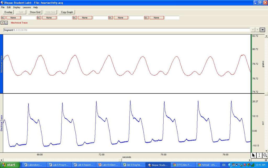
6 close and was about to rip the heart at any moment. As a result, we joined another group to complete the rest of the lab, following the instructions as listed in the lab manual. Results: Part 3: Electrical & Mechanical Activity of the Heart After opening up the frog and exposing the heart, we set up our apparatus so to record normal frog heart activity. From the mechanical and electrical trace of heart activity, the information in table 1 was obtained. As expected, ventricular contraction is much greater than atrial contraction. The latency is determined to be 120msec, meaning that it takes 120msec after stimulation before a contraction is seen. In the human reflex lab, the latency for the H wave was msec and so a latency of 120msec for heart contraction is very reasonable and expected. Also, the average heart rate of the heart was recorded as BPM. Table 1: General Data on Heart Activity Heart Data (averages) Atrial Contraction Force Ventricular Contraction Force Heart Rate Latency grams grams BPM 120 msec Part 4: Extrasystolic Contractions We determined the point of late diastole on the heart activity mechanical trace and stimulated the heart at that point in hope to elicit an extrasystole. We did this multiple times while slowly increasing the stimulus voltage until an extrasystole contraction was observed. This threshold stimulus voltage, 2.8V in this case, is the minimum voltage that can elicit an
7 extrasystolic contraction during late diastole. We then increased the stimulus voltage to 2x the threshold, 5.6V in this case, and elicited an extrasystolic contraction during late diastole as well. Lastly, we increased the voltage to determine the threshold voltage that would elicit an extrasystole at early diastole. The threshold for that turned out to be 8.0V. The observed trend is that a much lower threshold is needed to elicit an extrasystole at late diastole than at early diastole. A threshold that is almost 3x that of the threshold during late diastole was needed to elicit an extrasystole. The observed results are, for the most part, consistent with the expected. The slight difference would be regarding to the extrasystole elicited during early diastole. During early diastole, a much greater threshold voltage is needed before a contraction is seen. The threshold voltage during early diastole is more than 5.2 V greater than the threshold voltage during late diastole. Such an elevated threshold during early diastole is due to the refractory period that directly follows ventricular contraction. Without the high voltage, there would not be an extrasystole contraction. Table 2: Extrasystole Data: Stimulus voltages for different thresholds threshold during late diastole 2x threshold during late diastole threshold during early diastole Stimulus Voltage 2.8 V 5.6 V 8.0V Table 3: Contractile Force before, during, and after extrasystole (late diastole) Contractile Force Before extrasystole 2.47 grams During extrasystole 2.20 grams After extrasystole grams
8 Table 4: Contractile Force before, during, and after extrasystole (early diastole) Contractile Force Before extrasystole During extrasystole After extrasystole grams grams grams Figure 1. Comparison of Contractile Force Regarding Different Phases of Extrasystolic Contraction (stimulated at late diastole) Contractile Force (grams) Contractile Force Extrasystolic Contraction Phase Figure 1 A plot comparing the contraction force 1) before extrasystole 2) during extrasystole and 3) after extrasystole when extrasystole was stimulated at late diastole. Contraction force of the heart is measured at different instances. The observed trend is that the contraction forces before and after extrasystole is notably greater than during extrasystole. Contraction force after extrasystole is the greatest of the three, slightly greater than before extrasystole.
9 Figure 2. Comparison of Contractile Force Regarding Different Phases of Extrasystolic Contraction (stimulated at early diastole) Contractile Force (grams) Contractile Force Extrasystolic Contraction Phase Figure 2 A plot comparing the contraction force 1) before extrasystole 2) during extrasystole and 3) after extrasystole when extrasystole was stimulated at early diastole. Contraction force of the heart is measured at different instances. The observed trend is that the contraction forces before and after extrasystole is notably greater than during extrasystole. However, contraction force after extrasystole is the less than that of before extrasystole. The compensatory pause is the time period between an extrasystolic contraction and the next normal ventricular contraction. From the data collected, we determined the compensatory pause for late diastole, 2x late diastole, and early diastole. We simply looked at the mechanical trace of the heart, located the extrasystole and next normal ventricular contraction and determined the difference in time between the two points. We expected to see that the compensatory pauses be the same for the two instances of late diastole. However, the observed is that the compensatory pause for 2x late diastole is longer than that of late diastole. That could be due to the fact that the contraction was stimulated earlier for the 2x late diastole, resulting in a longer waiting time. The compensatory pause for early diastole is expected to be higher or rather, highest because the contraction is stimulated the earliest during early diastole. That means that
10 there is a longer wait time before the next normal ventricular contraction. The observed trend, does comply with that there is an increase in compensatory pause. Table 5. Comparison of Compensatory Pauses Compensatory Pause (msec) Late Diastole 2x Late Diastole Early Diastole 131 msec msec msec Figure 3. Comparison of Compensatory Pauses 200 Time (msec) Time Compensatory Pauses Figure 3 A plot comparing compensatory pauses at 1) late diastole, 2) 2x late diastole, and 3) early diastole. Compensatory pauses determined at each of the three types of extrasystolic stimulation. The observed is that there is an increase in compensatory pause with an increase in voltage. The compensatory pause at early diastole should be the longest one because extrasystolic contraction stimulated at late diastole has the longest filling time due to early stimulation.
11 Part 5: Vagal Stimulation- BRADYCARDIA As part of the vagal stimulation portion of this exercise, we performed bradycardia on the heart. We stimulated the vagus nerve and the voltage was raised up until the first sign of the heart slowing down. The voltage at which the first sign of a slow heart is displayed is the threshold stimulus voltage for bradycardia. After bradycardia, the recording machine continued to record for 30secs more. We then used our recorded data and compared the heart rate and contractile force before and after bradycardia. The observed trends for heart rate and contractile force are opposite of one another. It is seen that heart rate prior to bradycardia is 1.265BPM greater than that of after bradycardia. However, it is seen that the contractile force after bradycardia is 0.042grams greater than that of before bradycardia. Table 6: Bradycardia HR and Contractile Force before and after bradycardia Heart Rate Contractile Force Before Bradycardia BPM grams After Bradycardia BPM grams
12 Figure 4. Heart Rate Comparison before and after bradycardia 39.5 Heart Rate (BPM) Heart Rate Phase of Bradycardia Figure 4 A plot comparing the heart rate 1)before and 2)after bradycardia. Frog heart stimulated at 1.0V and that resulted in bradycardia. The heart rate before and after bradycardia were recorded. The observed trend is that the heart rate for before bradycardia is greater than after bradycardia, slowing of the heart. Figure 5. Contractile Force Comparison before and after bradycardia Contractile Force (grams) Contractile Force phase of bradycardia Figure 5 A plot comparing the contraction force 1)before and 2)after bradycardia. Frog heart stimulated at 1.0V and that resulted in bradycardia. Contraction force before and after
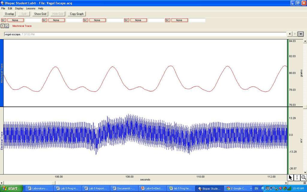
13 bradycardia were recorded. The observed trend is that contraction force is greater after bradycardia as compared to before. Part 5: Vagal Stimulation- VAGAL ESCAPE Vagal Escape was part of the later portion of the vagal stimulation section. Vagal escape is where the heart stops beating but after some time, resumes to essentially normal beating. We first stimulated the vagus nerve and gradually increased the voltage until the heart stopped beating; 2.4V was the stimulus voltage that corresponded to cardiac arrest. Heart activity was recorded throughout this process. Heart activity was recorded for 60 seconds after a heart beat returned. One of the two observed trends is that heart rate prior to vagal escape is significantly greater than the heart rate recorded after vagal escape. There is a BPM difference between the two heart rates. This relationship is consistent to the expected. The other observed trend is that heart contractile force is greater after vagal escape. This is also consistent with the expected. Table 7: HR and Contraction Force before and after vagal escape Heart Rate Before Vagal Escape BPM Contraction Amplitude grams After Vagal Escape BPM grams
14 Figure 6. Heart Rate Comparison before and after vagal escape Heart Rate (BPM) phase of Vagal Escape Heart Rate Figure 6 A plot comparing heart rate 1) before and 2) after vagal escape, the return of the heart beating after cardiac arrest. Frog s vagus nerve was stimulated at a high voltage of 2.4V and that resulted in cardiac arrest. Heart rate was recorded for before and after vagal escape. The observed trend is that the heart rate before vagal escape is greater than the heart rate after vagal escape. Figure 7. Comparison of Contractile Force before and after vagal escape contractile force (grams) phase of vagal escape Contractile Force Figure 7 A plot comparing contractile force 1) before and 2) after vagal escape. The observed trend is that contraction force before vagal escape (when the heart was in bradycardia and about
15 to enter cardiac arrest) is less than the contraction force after vagal escape (when the heart began beating again). Figure 8. Cardiac Arrest/Vagal Escape Progression Tension (grams) Time (second) Figure 8 A plot showing the change in contraction force during which the heart was in cardiac arrest. X-axis represents the time that passed after the heart stopped. Y-axis represents muscle tension. Time = 0 represents the beginning of cardiac arrest. Contraction force was measured at 30 second intervals. The observed is that contraction force decreases during cardiac arrest, as shown with a dramatic dip on the curve, and contraction force gradually increases after the heart beat returned. Part 6: The Effects of Epinephrine Contractile force was recorded throughout vagal escape. We allowed the heart to beat normally and then we stimulated the vagus nerve and that led to bradycardia. When we increased the voltage, the heart arrested, resulting in a complete stoppage. In figure 6 below, time 0sec corresponds with the heart being in late bradycardia and time 12sec corresponds to
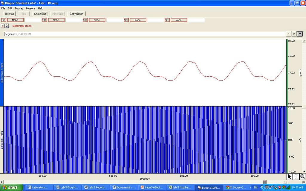
16 when the heart came to a complete stop. At both those times, the tension was recorded to be around 1gram. The dip after 12sec illustrates the decrease in tension to essentially zero. Starting at around 60sec, the curve begins to rise again, indicating that a heart beat is returning. At 120sec, the contractile force drastically increases from the grams at 90sec to grams. By 150sec, the contractile force reaches grams. From the point when the heart arrested at 12sec to at 150sec, there is a 2.4grams difference in contractile force. Overall, the observed trend is that contractile force, over time, increases and heart activity resumes to normality after the heart has been arrested. The contractile force even exceeds the contractile force prior to cardiac arrest. We added a few drops of epinephrine to the ventricle of the frog heart so to observe the response the heart has to sympathetic input. After adding epinephrine, we collected data fro 6 min. With the data collected, we compared the heart rate and contractile force before and after epinephrine application. As shown in the two figures below, it is observed that both heart rate and contractile force are greater after epinephrine application. This observation agrees with what is expected. An increase in heart rate and contractile force are supposed to result with the sympathetic input. Table 8: HR and Contraction Force before and after epinephrine Heart Rate Contraction Amplitude Before Epinephrine BPM grams After Epinephrine BPM grams
17 Figure 9. Heart Rate Comparison before and after epineprhrine addition Heart Rate (BPM) Heart Rate Phase of epinephrine addition Figure 9 A plot comparing heart rate 1) before and 2) after the addition of epinephrine to the heart. Between 4 and 6 drops of epinephrine were added to the frog s ventricle. Data was collected for a total of 6 minutes. The heart rate for both cases were recorded and compared. The observed trend is that a higher heart rate is seen after the addition of epinephrine. In contrast, a relatively low heart rate is seen before the addition of epinephrine. Figure 10. Contraction Force comparison before and after epinephrine addition contraction force (grams) phase of epinephrine addition Contraction Force
18 Figure 10 A plot comparing contraction force 1) before and 2) after the addition of epinephrine. Between 4 and 6 drops of epinephrine were added to the frog s ventricle. Data was collected for a total of 6 minutes. The observed trend is that contraction force is greater after the addition of epinephrine. Discussion: In the heart, there are autorhythmic or pacemaker cells that regulate the beating of the heart. Pacemaker cells only make up a very minor portion of cardiac cells because they are only responsible for initiating and conducting action potentials needed for heart contraction. Pacemaker cells don t have resting potentials. Instead, they have what is known as pacemaker potential. According to Sherwood, a pacemaker potential is an autorhythmic cell membrane s slow drift to threshold. Three different ion movements, together, are responsible for pacemaker potential: an increase in Na+ influx, decrease in K+ efflux, and increase in Ca2+ influx. Nat+ first enter through special voltage gated channels. Then, K+ channels start to close, resulting in a decrease in K+ efflux. Lastly, there is an increase in Ca2+ influx due to the opening of T-type Ca2+ channels. All three ion movements ultimately contribute to the membrane reaching threshold for an action potential (Sherwood p ). There are four locations in which pacemaker cells can be found and these locations are 1) SA node, 2) AV node, 3) Bundle of His (atrioventricular bundle), and 4) Purkinje fibers. The SA node is located near the superior vena cava in the right atrial wall. The AV node is located at the bottom of the right atrium. The bundle of His, right and left, extends from the AV node to the interventricular septum. The Purkinge fibers go from the bundle of His and extend all over the ventricular myocardium. All these pacemakers have different rates of depolarization to threshold and the fastest pacemaker cells are located in the SA node. The pacemaker cells at the other
19 locations have to be activated by the SA action potentials before they are able to reach threshold, at a slower rate, on their own. With that said, the pacemaker cells at the SA node are the primary regulators of the autorhythmic activity of the heart (Sherwood p ) The excitation-contraction coupling process for heart (cardiac muscle) contraction is very similar to that of skeletal muscle contraction. A stimulus causes a ventricular action potential in cardiac contractile cell. That action potential travels down T tubules and opens up L-type Ca2+ channels, leading to an influx of Ca2+. This influx of Ca2+, in turn, causes the release of a great amount of Ca2+ from the sarcoplasmic reticulum. The end result of the multiple influx of Ca2+ is high cytosolic Ca2+. With sufficient cytosolic Ca2+, there is Ca2+ to bind to Troponin C so that it can no longer bind to Tropomyosin. Without Troponin C attached to it, Tropomyosin can no longer attach to and block the myosin binding site. The myosin binding site is now vacant for myosin to attach and when myosin attaches, cross-bridge cycling occurs. The resulting power strokes of cross-bridge cycling causes sliding of thick and thin filaments. Cardiac muscle contraction is then the end result (Sherwood p.315). The main difference between skeletal muscle contraction and cardiac muscle contraction is that Ca2+ influx during phase 2 of ventricular action potential is required for cardiac contraction. As mentioned earlier, there is not always enough cytosolic Ca2+ in cardiac muscle and so in instances when there is a lack of cytosolic Ca2+, Troponin C will still be bound to Tropomyosin, allowing Tropomyosin to continue blocking the myosin binding site. Myosin needs to bind to the myosin binding site to initiate cross-bridge cycling and so with the site blocked, cross-bridge cycling is not possible and that means that there won t be cardiac muscle contraction (Sherwood p.315).
20 The ventricular action potential is the mechanism behind the excitation of the heart. There are multiple phases to this mechanism and each phase is symbolic of different ion activity. In phase 0, there is a steep increase in membrane potential and that is due to the influx of mainly Na+ and Ca2+ because Na+ and Ca2+ channels opened up. As a result, depolarization occurs in phase 0. Phase 1 is where a quick repolarization, as indicated by the curve sloping down, takes place and that is due to the inactivation of Na+ channels. Phase 2 is where the curve plateaus as a result of more Ca2+ influx. The Ca2+ in phase 2 are coming through L type Ca2+ channels. In phase 3, there is more repolarization but this due to Ca2+ channel inactivation and K+ efflux as a result of K+ channel activation. Phase 4 is the last phase and that is where resting membrane potential is resumed (Sherwood p ) The heart acts as the organ pump that is responsible for pumping blood to all over the body. In doing so, the heart is supplying oxygen and eliminating waste, like CO2, from body tissues. Blood from the body flows back to the heart and by then, the blood is deoxygenated (since oxygen has been delivered to tissues) and CO2-rich (since CO2 has been picked up, as waste, from the tissues). The blood enters the right atrium through the venae cavae. So to oxygenate the blood again, the blood must enter the pulmonary system and get oxygenated in the lungs. The blood leaves the right atrium and enters the right ventricle where it then enters the lungs to get rid of the CO2 and be supplied with O2. The oxygenated blood enters the left atria and then left ventricle and ultimately goes into the system circulation by exiting through the aorta. Our ultimate goal for this exercise is to better understand the properties of the human heart although we are using the heart of a frog, amphibians that are able to supply oxygen and nutrients to the cardiac muscle from blood due to the anatomy of the heart: 2 atria and 1 ventricle,
21 for the experiment. The heart is a complicated and extremely essential part of the body. It is made up of cardiac muscle. Cardiac muscle, like skeletal muscle, is striated, meaning that the muscle is made up of alternating thick and thin filaments. Cardiac muscles are made up of cardiac muscle fibers and these cardiac muscle fibers are connected to each other by intercalated discs. The two types of membrane junctions that are inside an intercalated disc are desmosomes, those responsible for mechanical connections, and gap junctions, those responsible for electrical connections (Sherwood p. 308). Part 3: Electrical & Mechanical Activity of the Heart In this exercise, we studied the properties of the frog heart, which is fairly similar to the human heart and the first thing we did after dissecting the frog and exposing its heart is exploring the normal activity of the heart. It is expected to see that ventricular contraction be greater than atrial contraction. As shown in Table 1, atrial contractile force is significantly less than that of ventricular contractile force. That is because the left ventricle is actually the part of the heart that pumps blood to the rest of the body. A larger contraction is needed to achieve that, hence the gram difference between atrial and ventricular contraction force. Also, the ventricles are larger than the atria and so the ventricles are capable of larger contractions. The latency for heart contraction is expected to be greater than that of human reflexes as explored in lab 3. The latency for heart contraction is recorded as 120msec while the latency for a H wave is msec. The latency for heart contraction was observed to be longer and that s because there are more processes that the body has to go through, as compared to a reflex, before a contraction can occur.
22 Part 4: Extrasystolic Contractions Extrasystolic contractions are premature ventricular contractions. They appear right after a ventricular contraction when stimulated at either late or early diastole. In Table 2, note the threshold stimulus voltage for extrasystolic contraction at late and early diastole. The threshold for early diastole is significantly greater than the threshold during late diastole. That is because of the difference in the refractory period that each was stimulated in. During early diastole, the muscle is in the effective/absolute refractory period. That is the time period where no action potential can occur. In the case of late diastole, the muscle is in the relative refractory period. Unlike with effective refractory period, an action potential can occur. As seen in Table 4, the contractile force for extrasystole in early diastole is less than that of late diastole. Technically speaking, there should not even be a contraction seen in effective refractory period. The values for late diastole are greater than those at there is more filling time for late diastole and less filling time for early diastole. As expected, a stronger contraction corresponds to a greater filling of the ventricle. This is known as the Frank-Starling Law of the Heart (Sherwood p. 328). Often times, the extrasystolic contraction is of lesser contractile force because there is less filling time. The ventricular contraction after the extrasystolic contraction and the compensatory pause should be greater than that of before and during extrasystole. That is because there is more filling time and more filling time equals increased end diastolic volume which ultimately leads to stronger contractions. The data collected during late diastole, as seen in figure 1, complies with the expected. The data collected during early diastole, as seen in figure 2, show that contraction after extrasystolic contraction is of lesser intensity than before extrasystolic. That is probably due to the continual twitching of the frog heart after extrasystole and before the next normal
23 contraction. The multiple twitches interrupted the elongated filling time and so no only did we not see an increase in contractile force in post extrasystole, we saw a decrease instead. Like mentioned earlier, the heart goes into 2 types of refractory period after ventricular contraction. Effective refractory period is the one directly after ventricular contraction while relative refractory period is the one after effective refractory period. During the effective refractory period, no action potentials can occur and the purpose of that is to prevent the heart from going into tetany (Sherwood p. 316). The cardiac muscle, in this sense, is really different from that of skeletal muscle, because tetany is muscle is promoted and seen as the ultimate contraction goal. There are no protective factors, like the effective refractory period, that protects the muscle from reaching tetany. The effective refractory period is what causes the compensatory pause when extrasystole is induced. In addition to effective refractory period, cardiac muscles also have relative refractory period and the purpose of a relative refractory period is simply to have a longer filling time and a stronger contraction. In table 5 and figure 3, a comparison between the compensatory pauses from late diastole, 2x late diastole, and early diastole was conducted. It is expected to see that the compensatory pauses at late diastole be about the same because the muscle is stimulated at the time period and so the filling time between the extrasystolic contraction and next ventricular contraction should be the same. As evidently seen in figure 3, the compensatory pauses for the two late diastole instances are not the same. That 13.2 msec difference could be that the extrasystolic contractions were not stimulated at the same exact time and so slight difference in compensatory pause resulted. The compensatory pause for early diastole is observed to be longer than the other two and that is consistent with the expected because stimulated an extrasystolic contraction earlier on allows for a longer filling time, hence, a longer compensatory period.
24 Part 5: Vagal Stimulation- BRADYCARDIA During bradycardia, the heart is slowed as a result the increased voltage that stimulated the vagus nerve, a nerve that s part of the parasympathetic system. It is defined as heart rate < 60bpm. Some of the symptoms that point toward bradycardia are fatigue, lethargy, nausea, dizziness, confusion, pre/syncope, and shortness of breath. (Ahsley et. al, ch 8 Bradycardia). Our heart rates are ~40bpm, well below 60bpm, and so can be classified as bradycardia. It is expected that the heart rate before bradycardia be greater than after bradycardia because bradycardia is the concept of slowing of the heart. The heart rate after bradycardia should be lower because bradycardia is part of the parasympathetic nervous system and the mechanism governing it is the chain of reaction that starts with the release of Ach. The parasympathetic system releases Ach and Ach binds to muscarinic Ach receptors. Pacemaker cell activity in the SA node is slowed as a result of Ach binding to the receptor. That leads to a decrease in HR. The observed HR data, as seen in figure 4, agrees with the expected. The same expected results go with contractile force. Contractile force after bradycardia should be lower because of parasympathetic activity. As described above, the SA node pacemaker activity is affected but in addition, the AV node pacemaker activity is also affected in the way that Ach makes it hard to spread action potential. In other words, that makes a contraction harder to elicit. The observed data actually goes against the expected; figure 5 shows that contractile force after bradycardia is greater than before. The difference between the two contractile forces is only grams, not very much at all and so a simple yet plausible explanation would be that human errors while analyzing the data caused for such disagreeing results.
25 Since bradycardia is part of parasympathetic activity that affects heart activity, any sort of sympathetic activity would have a counteract effect and inhibit bradycardia. A study shows that galanin, a cardiac sympathetic co-transmitter has an inhibiting effect on bradycardia. Galanin reduces acetylcholine release and as described earlier, Ach binds to muscarinic Ach receptors so to slow the pacemaker cell activity at the SA node. When galanin is present, there will be a decrease in Ach due to decrease Ach release and the chain of parasympathetic reactions that follow is made impossible. The end result of that would be no effect on SA node pacemaker cell activity and so no bradycardia (Herring et. al p.1) Part 5: Vagal Stimulation- VAGAL ESCAPE Vagal Escape is where the heart stops beating and after a period of time, the [less intense] heart beat returns. It is the result of prolonged vagal stimulation in which bradycardia and then cardiac arrest is the end result. In the process, the SA node pacemaker activity is affected and so that s why we see the stopping of the heart. The return of the heart beat is when the next fastest pacemaker cells take over. This concept of taking over the role of the SA node is supported Wallace and Daggett. They say that the pacemaker cells at different areas other than the SA node usually take over the pacemaker role. The atrium, AV node, or ventricles have been suggested as sites of pacemaker activity when the SA node is no longer available for rhythmic regulation (Wallace et. al p.93). In our preparation, I do think that there is tonic vagal inhibition of the heart because in the beginning, the heart seemed to beat at a more normal pace. As more activities were done because after stimulating the heart for a while, our frog heart seemed to beat slower, showing signs of bradycardia and perhaps progressing to cardiac arrest.
26 When regarding heart rate and contractile force, it is expected that both will be lower for after vagal escape and that is because of slower pacemaker cells taking over the SA node. From figure 6, we can see that HR after vagal escape is lower. That agrees with the expected result, meaning that parasympathetic activity affected heart activity. In figure 7, opposing results are shown for contractile force before and after vagal escape. Contractile force is supposed to be less after vagal escape but the observed shows that contractile force after vagal escape is greater than before vagal escape. This odd result is probably due to the fact that the data for the vagal escape section is not very consistent or accurate. In my case, the contractile force before vagal escape actually corresponds more to that of bradycardia because normal heart activity was not recorded. If normal heart activity was recorded, the contractile force after vagal escape would indeed be lower because normal activity is regulated by the SA node pacemakers (fastest) while after-vagal-escape activity is regulated by other slower pacemakers, hence, weaker contraction. Figure 8 is an illustration of how the vagal escape works. At the far left side of the curve, the tension is relatively high and as time passes by, the tension begins to drop, signifying bradycardia. At time= 0 was when the heart stopped. Tension drastically dropped as indicated by the sharp dip in the curve. The duration of cardiac arrest was calculated to be seconds. At ~60sec, or 1min, after the heart stopped, a heart beat returns and that s when the next fastest pacemaker cells take over. Tension continues to gradually increase. Part 6: The Effects of Epinephrine Epinephrine is released by the sympathetic nervous system. Epi binds to B-1 adrenergic receptors and that increases the heart rate. With that said, it is expected that an increase in heart rate be seen after applying epi. In figure 9, we see, as expected, that an increase in HR did result
27 after epi addition due to increase in sympathetic activity. The application of epi also increases Ca2+ influx. As discussed in the cardiac muscle excitation-contraction coupling section, Ca2+ influx is responsible for generating heart contractions. The more cytosolic Ca2+ there is, the greater the heart contractions because of more cross-bridging. With that in mind, it is expected that after the application of epi, there will increase the contractile intensity. Like with HR, contractile intensity also agrees with the expected result. Norepinephrine, like epinephrine, is part of the sympathetic nervous system and so the application of it will also lead to increase HR and contractile force. According to a study, the stimulation of adenosine A1 receptors has effects that protect the heart. These receptors are able to protect the heart through the inhibition of norepinpehrine (Schutte et. al, p. 1). As said before, norepinephrine increases sympathetic activity and so adenosine A1 receptors act like buffers for the heart. Conclusion: In conclusion, through this exercise, we explored the properties of the heart and found that there are many factors that can affect heart contraction. Factors like degree of ventricular filling, compensatory pause, parasympathetic activity, sympathetic activity, cytosolic Ca2+ concentration, types of pacemaker cells, etc. all contribute in determining whether there will be a contraction and if so, how strong it might be. Because the heart is a complicated organ, the mechanisms associated with it are too. We found that a higher voltage is needed to stimulate an extrasystolic contraction during early diastole because of the effective refractory period. We also found that the compensatory pause is the longest for when during early diastole because the early extrasystole stimulation allowed for more filling time. Very importantly, we got a better
28 understanding of how parasympathetic and sympathetic activities affect heart rate and contractile force. Parasympathetic activity includes stimulation of the vagus nerve and release of Ach, neurotransmitter that will bind to muscarinic receptors to decrease HR and contractile force. Sympathetic activity includes the release of epinephrine, or norepinephrine, by sympathetic nerves and the binding of the either neurotransmitter to B1-adrenergic receptors so to increase HR and contractile force. The findings from this laboratory exercise are beneficial in researching for cures or preventions for heart diseases and other illnesses relating to the heart. With heart disease accounting for the most deaths in the U.S., better understanding of the heart and is always amongst the goals researchers strive for.
29 References 1. Ashley EA, Niebauer J. Cardiology Explained. Chapter 8, Section Bradycardia. London: Remedica; Herring, N., Cranley, J., Lokale, MN., Li, D., Shanks, J., Alston, EN., Girard, BM., Carter, E., Parsons, RL., Habecker, BA., Paterson, DJ The Cardiac Sympathetic Co-transmitter Galanin Reduces Acetylcholine Release and Vagal Bradycardia: Implications for Neural Control of Cardiac Excitability. Journal of Molecular and Cellular Cardiology 52(3): Schutte, F., Burgdorf, C., Richardt, G., Kurz, T Adenosine A1 Receptor- Mediated Inhibition of Myocardial norepinephrine Release Involves Neither Phospholipase C nor Protein Kinase C but Does Involve Adenylyl Cyclase. Canadian Journal of Physiology and Pharmacology. 84(5): , /y Sherwood, L. Human Physiology: From Cells to Systems, 7 th edition. Canada. 5. Wallace, A.G. & Daggett, W. M Pacemaker Activity During Vagal Escape Rhythms. Circulation Research. 15: doi: /01. RES
30
31
32
33
THE CARDIOVASCULAR SYSTEM. Heart 2
 THE CARDIOVASCULAR SYSTEM Heart 2 PROPERTIES OF CARDIAC MUSCLE Cardiac muscle Striated Short Wide Branched Interconnected Skeletal muscle Striated Long Narrow Cylindrical PROPERTIES OF CARDIAC MUSCLE Intercalated
Chapter 13 The Cardiovascular System: Cardiac Function
 Chapter 13 The Cardiovascular System: Cardiac Function Overview of the Cardiovascular System The Path of Blood Flow through the Heart and Vasculature Anatomy of the Heart Electrical Activity of the Heart
QUIZ/TEST REVIEW NOTES SECTION 1 CARDIAC MYOCYTE PHYSIOLOGY [CARDIOLOGY]
![QUIZ/TEST REVIEW NOTES SECTION 1 CARDIAC MYOCYTE PHYSIOLOGY [CARDIOLOGY]](/thumbs/96/126998162.jpg "QUIZ/TEST REVIEW NOTES SECTION 1 CARDIAC MYOCYTE PHYSIOLOGY [CARDIOLOGY]") QUIZ/TEST REVIEW NOTES SECTION 1 CARDIAC MYOCYTE PHYSIOLOGY [CARDIOLOGY] Learning Objectives: Describe the ionic basis of action potentials in cardiac contractile and autorhythmic cells Explain the relationship
QUIZ/TEST REVIEW NOTES SECTION 1 CARDIAC MYOCYTE PHYSIOLOGY [CARDIOLOGY] Learning Objectives: Describe the ionic basis of action potentials in cardiac contractile and autorhythmic cells Explain the relationship
Cardiovascular system
 BIO 301 Human Physiology Cardiovascular system The Cardiovascular System: consists of the heart plus all the blood vessels transports blood to all parts of the body in two 'circulations': pulmonary (lungs)
Cardiovascular System
 Cardiovascular System The Heart Cardiovascular System The Heart Overview What does the heart do? By timed muscular contractions creates pressure gradients blood moves then from high pressure to low pressure
Collin County Community College. ! BIOL Anatomy & Physiology! WEEK 5. The Heart
 Collin County Community College! BIOL. 2402 Anatomy & Physiology! WEEK 5 The Heart 1 (1578-1657) A groundbreaking work in the history of medicine, English physician William Harvey s Anatomical Essay on
The Cardiovascular System
 Chapter 18 Part A The Cardiovascular System 1/19/16 1 Annie Leibovitz/Contact Press Images Similarities of Cardiac and Skeletal Muscle RMP Ion concentration Deploarization Action Potential Repolarization
Introduction. Circulation
 Introduction Circulation 1- Systemic (general) circulation 2- Pulmonary circulation carries oxygenated blood to all parts of the body carries deoxygenated blood to the lungs From Lt. ventricle aorta From
Where are the normal pacemaker and the backup pacemakers of the heart located?
 CASE 9 A 68-year-old woman presents to the emergency center with shortness of breath, light-headedness, and chest pain described as being like an elephant sitting on her chest. She is diagnosed with a
The conduction system
 The conduction system In today s lecture we will discuss the conducting system of the heart. If we placed the heart in a special solution that contains Ca+ it will keep on contracting, keep in mind that
Cardiovascular System: The Heart
 Cardiovascular System: The Heart I. Anatomy of the Heart (See lab handout for terms list) A. Describe the size, shape and location of the heart B. Describe the structure and function of the pericardium
Electrical Conduction
 Sinoatrial (SA) node Electrical Conduction Sets the pace of the heartbeat at 70 bpm AV node (50 bpm) and Purkinje fibers (25 40 bpm) can act as pacemakers under some conditions Internodal pathway from
PART I. Disorders of the Heart Rhythm: Basic Principles
 PART I Disorders of the Heart Rhythm: Basic Principles FET01.indd 1 1/11/06 9:53:05 AM FET01.indd 2 1/11/06 9:53:06 AM CHAPTER 1 The Cardiac Electrical System The heart spontaneously generates electrical
CARDIOVASCULAR SYSTEM
 CARDIOVASCULAR SYSTEM Overview Heart and Vessels 2 Major Divisions Pulmonary Circuit Systemic Circuit Closed and Continuous Loop Location Aorta Superior vena cava Right lung Pulmonary trunk Base of heart
The Heart. Size, Form, and Location of the Heart. 1. Blunt, rounded point; most inferior part of the heart.
 12 The Heart FOCUS: The heart is composed of cardiac muscle cells, which are elongated, branching cells that appear striated. Cardiac muscle cells behave as a single electrical unit, and the highly coordinated
Chapter 20: Cardiovascular System: The Heart
 Chapter 20: Cardiovascular System: The Heart I. Functions of the Heart A. List and describe the four functions of the heart: 1. 2. 3. 4. II. Size, Shape, and Location of the Heart A. Size and Shape 1.
11/10/2014. Muscular pump Two atria Two ventricles. In mediastinum of thoracic cavity 2/3 of heart's mass lies left of midline of sternum
 It beats over 100,000 times a day to pump over 1,800 gallons of blood per day through over 60,000 miles of blood vessels. During the average lifetime, the heart pumps nearly 3 billion times, delivering
Conduction System of the Heart 4. Faisal I. Mohammed, MD, PhD
 Conduction System of the Heart 4 Faisal I. Mohammed, MD, PhD 1 Objectives List the parts that comprise the conduction system Explain the mechanism of slow response action potential (pacemaker potential)
Collin County Community College
 Collin County Community College BIOL. 2402 Anatomy & Physiology WEEK 5 The Heart 1 The Heart Beat and the EKG 2 1 The Heart Beat and the EKG P-wave = Atrial depolarization QRS-wave = Ventricular depolarization
Cardiac muscle is different from other types of muscle in that cardiac muscle
 6 E X E R C I S E Cardiovascular Physiology O B J E C T I V E S 1. To define autorhythmicity, sinoatrial node, pacemaker cells, and vagus nerves 2. To understand the effects of the sympathetic and parasympathetic
ELECTROCARDIOGRAPHY (ECG)
") ELECTROCARDIOGRAPHY (ECG) The heart is a muscular organ, which pumps blood through the blood vessels of the circulatory system. Blood provides the body with oxygen and nutrients, as well as assists in
ELECTROCARDIOGRAPHY (ECG) The heart is a muscular organ, which pumps blood through the blood vessels of the circulatory system. Blood provides the body with oxygen and nutrients, as well as assists in
Effects of Temperature, Stretch, and Various Drug Treatments on the
 Nicole Rodi Bio 235: Animal Physiology Heart Muscle Lab Report 10/24/2014 Effects of Temperature, Stretch, and Various Drug Treatments on the Cardiac Muscle Activity of Rana pipiens Abstract Mechanical
Conduction system of the heart
 Conduction system of the heart -For skeletal muscle to contract, it has to be innervated by spinal nerves (there must be a neuromuscular junction). *The heart is innervated by autonomic nervous system
Chapter 18 - Heart. I. Heart Anatomy: size of your fist; located in mediastinum (medial cavity)
") Chapter 18 - Heart I. Heart Anatomy: size of your fist; located in mediastinum (medial cavity) A. Coverings: heart enclosed in double walled sac called the pericardium 1. Fibrous pericardium: dense connective
Chapter 18 - Heart I. Heart Anatomy: size of your fist; located in mediastinum (medial cavity) A. Coverings: heart enclosed in double walled sac called the pericardium 1. Fibrous pericardium: dense connective
Cardiac physiology. b. myocardium -- cardiac muscle and fibrous skeleton of heart
 I. Heart anatomy -- general gross. A. Size/orientation - base/apex B. Coverings D. Chambers 1. parietal pericardium 2. visceral pericardium 3. Layers of heart wall a. epicardium Cardiac physiology b. myocardium
(D) (E) (F) 6. The extrasystolic beat would produce (A) increased pulse pressure because contractility. is increased. increased
 (E) (F) 6. The extrasystolic beat would produce (A) increased pulse pressure because contractility. is increased. increased") Review Test 1. A 53-year-old woman is found, by arteriography, to have 5% narrowing of her left renal artery. What is the expected change in blood flow through the stenotic artery? Decrease to 1 2 Decrease
Review Test 1. A 53-year-old woman is found, by arteriography, to have 5% narrowing of her left renal artery. What is the expected change in blood flow through the stenotic artery? Decrease to 1 2 Decrease
Lab 2. The Intrinsic Cardiac Conduction System. 1/23/2016 MDufilho 1
 Lab 2 he Intrinsic Cardiac Conduction System 1/23/2016 MDufilho 1 Figure 18.13 Intrinsic cardiac conduction system and action potential succession during one heartbeat. Superior vena cava ight atrium 1
Outline. Electrical Activity of the Human Heart. What is the Heart? The Heart as a Pump. Anatomy of the Heart. The Hard Work
 Electrical Activity of the Human Heart Oguz Poroy, PhD Assistant Professor Department of Biomedical Engineering The University of Iowa Outline Basic Facts about the Heart Heart Chambers and Heart s The
Cardiovascular Physiology
 Cardiovascular Physiology The mammalian heart is a pump that pushes blood around the body and is made of four chambers: right and left atria and right and left ventricles. The two atria act as collecting
Investigation of human cardiovascular physiology is very interesting, but many
 6 E X E R C I S E Frog Cardiovascular Physiology O B J E C T I V E S 1. To list the properties of cardiac muscle as automaticity and rhythmicity, and to define each. 2. To explain the statement, Cardiac
Conduction System of the Heart. Faisal I. Mohammed, MD, PhD
 Conduction System of the Heart Faisal I. Mohammed, MD, PhD 1 Objectives l List the parts that comprise the conduction system l Explain the mechanism of slow response action potential (pacemaker potential)
10/23/2017. Muscular pump Two atria Two ventricles. In mediastinum of thoracic cavity 2/3 of heart's mass lies left of midline of sternum
 It beats over 100,000 times a day to pump over 1,800 gallons of blood per day through over 60,000 miles of blood vessels. During the average lifetime, the heart pumps nearly 3 billion times, delivering
37 1 The Circulatory System
 H T H E E A R T 37 1 The Circulatory System The circulatory system and respiratory system work together to supply cells with the nutrients and oxygen they need to stay alive. a) The respiratory system:
*Generating blood pressure *Routing blood: separates. *Ensuring one-way blood. *Regulating blood supply *Changes in contraction
 *Generating blood pressure *Routing blood: separates pulmonary and systemic circulations *Ensuring one-way blood flow: valves *Regulating blood supply *Changes in contraction rate and force match blood
Practice Exercises for the Cardiovascular System
 Practice Exercises for the Cardiovascular System On the diagram below, color the oxygen-rich blood red and the oxygen-poor blood blue. Label the parts: Continued on the next page... Label the parts on
AnS SI 214 Practice Exam 2 Nervous, Muscle, Cardiovascular
 AnS SI 214 Practice Exam 2 Nervous, Muscle, Cardiovascular Select the best answer choice in the questions below. 1) On the electrocardiogram, repolarization of the atria is represented by the: A) P wave
Cardiovascular System Notes: Physiology of the Heart
 Cardiovascular System Notes: Physiology of the Heart Interesting Heart Fact Capillaries are so small it takes ten of them to equal the thickness of a human hair. Review What are the 3 parts of the cardiovascular
The cardiovascular system is composed of the heart and blood vessels that carry blood to and from the body s organs. There are 2 major circuits:
 1 The cardiovascular system is composed of the heart and blood vessels that carry blood to and from the body s organs. There are 2 major circuits: pulmonary and systemic. The pulmonary goes out to the
The HEART. What is it???? Pericardium. Heart Facts. This muscle never stops working It works when you are asleep
 This muscle never stops working It works when you are asleep The HEART It works when you eat It really works when you exercise. What is it???? Located between the lungs in the mid thoracic region Apex
The Heart. Happy Friday! #takeoutyournotes #testnotgradedyet
 The Heart Happy Friday! #takeoutyournotes #testnotgradedyet Introduction Cardiovascular system distributes blood Pump (heart) Distribution areas (capillaries) Heart has 4 compartments 2 receive blood (atria)
The Cardiovascular System
 Essentials of Human Anatomy & Physiology Elaine N. Marieb Seventh Edition Chapter 11 The Cardiovascular System Slides 11.1 11.19 Lecture Slides in PowerPoint by Jerry L. Cook The Cardiovascular System
Chapter 20b Cardiac Physiology
 Chapter 20b Cardiac Physiology Heart Valve Mechanics The heart valve openand close because of pressure gradients. When pressure on one side is greater than the other, it pushes the valve open. For example,
The Function of an ECG in Diagnosing Heart Conditions. A useful guide to the function of the heart s electrical system for patients receiving an ECG
 The Function of an ECG in Diagnosing Heart Conditions A useful guide to the function of the heart s electrical system for patients receiving an ECG Written by Erhan Selvi July 28, 2014 Audience and Scope
BIPN100 F15 Human Physiology I (Kristan) Problem set #5 p. 1
 Problem set #5 p. 1") BIPN100 F15 Human Physiology I (Kristan) Problem set #5 p. 1 1. Dantrolene has the same effect on smooth muscles as it has on skeletal muscle: it relaxes them by blocking the release of Ca ++ from the
BIPN100 F15 Human Physiology I (Kristan) Problem set #5 p. 1 1. Dantrolene has the same effect on smooth muscles as it has on skeletal muscle: it relaxes them by blocking the release of Ca ++ from the
Ch 19: Cardiovascular System - The Heart -
 Ch 19: Cardiovascular System - The Heart - Give a detailed description of the superficial and internal anatomy of the heart, including the pericardium, the myocardium, and the cardiac muscle. Trace the
The Electrocardiogram
 The Electrocardiogram Chapters 11 and 13 AUTUMN WEDAN AND NATASHA MCDOUGAL The Normal Electrocardiogram P-wave Generated when the atria depolarizes QRS-Complex Ventricles depolarizing before a contraction
Chapter 20 (2) The Heart
 The Heart") Chapter 20 (2) The Heart ----------------------------------------------------------------------------------------------------------------------------------------- Describe the component and function of
Chapter 20 (2) The Heart ----------------------------------------------------------------------------------------------------------------------------------------- Describe the component and function of
Chapter 12: Cardiovascular Physiology System Overview
 Chapter 12: Cardiovascular Physiology System Overview Components of the cardiovascular system: Heart Vascular system Blood Figure 12-1 Plasma includes water, ions, proteins, nutrients, hormones, wastes,
During exercise the heart rate is 190 bpm and the stroke volume is 115 ml/beat. What is the cardiac output?
 The Cardiovascular System Part III: Heart Outline of class lecture After studying part I of this chapter you should be able to: 1. Be able to calculate cardiac output (CO) be able to define heart rate
Physiology sheet #2. The heart composed of 3 layers that line its lumen and cover it from out side, these layers are :
 Physiology sheet #2 * We will talk in this lecture about cardiac muscle physiology, the mechanism and the energy sources of their contraction and intracellular calcium homeostasis. # Slide 4 : The heart
The Cardiovascular System
 The Cardiovascular System The Cardiovascular System A closed system of the heart and blood vessels The heart pumps blood Blood vessels allow blood to circulate to all parts of the body The function of
Chapter 9, Part 2. Cardiocirculatory Adjustments to Exercise
 Chapter 9, Part 2 Cardiocirculatory Adjustments to Exercise Electrical Activity of the Heart Contraction of the heart depends on electrical stimulation of the myocardium Impulse is initiated in the right
Lab #3: Electrocardiogram (ECG / EKG)
") Lab #3: Electrocardiogram (ECG / EKG) An introduction to the recording and analysis of cardiac activity Introduction The beating of the heart is triggered by an electrical signal from the pacemaker. The
Lab #3: Electrocardiogram (ECG / EKG) An introduction to the recording and analysis of cardiac activity Introduction The beating of the heart is triggered by an electrical signal from the pacemaker. The
Unit 10 ~ Learning Guide
 Unit 10 ~ Learning Guide Name: INSTRUCTIONS Complete the following notes and questions as you work through the related lessons. You are required to have this package completed BEFORE you write your unit
Anatomy Review: The Heart Graphics are used with permission of A.D.A.M. Software, Inc. and Benjamin/Cummings Publishing Co.
 Anatomy Review: The Heart Graphics are used with permission of A.D.A.M. Software, Inc. and Benjamin/Cummings Publishing Co. Anatomy Views Label the diagrams of the heart below: Interactive Physiology Study
Cardiac Properties MCQ
 Cardiac Properties MCQ Abdel Moniem Ibrahim Ahmed, MD Professor of Cardiovascular Physiology Cairo University 2007 1- Cardiac Valves: a- Prevent backflow of blood from the ventricles to the atria during
d) Cardiovascular System Higher Human Biology
 Cardiovascular System Higher Human Biology") d) Cardiovascular System Higher Human Biology What can your remember about the heart and blood vessels? What is the Cardiovascular System? The cardiovascular system, also known as the circulatory system,
d) Cardiovascular System Higher Human Biology What can your remember about the heart and blood vessels? What is the Cardiovascular System? The cardiovascular system, also known as the circulatory system,
Heart. Structure Physiology of blood pressure and heartbeat
 Heart Structure Physiology of blood pressure and heartbeat Location and Anatomy Location and Anatomy Pericardial cavity: surrounds, isolates, and anchors heart Parietal pericardium lined with serous membrane
Approximately the size of your fist Location. Pericardial physiology
 Heart Anatomy Approximately the size of your fist Location Superior surface of diaphragm Left of the midline Anterior to the vertebral column, posterior to the sternum Wednesday, March 28, 2012 Muscle
Rhythmical Excitation of the Heart
 Rhythmical Excitation of the Heart KALEB HOOD AND JIMMY JOHNSON Special Excitory and Conductive System of the Heart Sinus Node (or sinoatrial node or S-A): A small node with almost no contractile muscle,
Principles of Biomedical Systems & Devices. Lecture 8: Cardiovascular Dynamics Dr. Maria Tahamont
 Principles of Biomedical Systems & Devices Lecture 8: Cardiovascular Dynamics Dr. Maria Tahamont Review of Cardiac Anatomy Four chambers Two atria-receive blood from the vena cave and pulmonary veins Two
The Cardiovascular System (Heart)
") The Cardiovascular System The Cardiovascular System (Heart) A closed system of the heart and blood vessels The heart pumps blood Blood vessels allow blood to circulate to all parts of the body The function
The Cardiovascular System The Cardiovascular System (Heart) A closed system of the heart and blood vessels The heart pumps blood Blood vessels allow blood to circulate to all parts of the body The function
Human Anatomy, First Edition
 Human Anatomy, First Edition McKinley & O'Loughlin Chapter 22 : Heart 1 Functions of the Heart Center of the cardiovascular system, the heart. Connects to blood vessels that transport blood between the
Department of medical physiology 7 th week and 8 th week
 Department of medical physiology 7 th week and 8 th week Semester: winter Study program: Dental medicine Lecture: RNDr. Soňa Grešová, PhD. Department of medical physiology Faculty of Medicine PJŠU Cardiovascular
TEST BANK FOR ECGS MADE EASY 5TH EDITION BY AEHLERT
 Link download full: http://testbankair.com/download/test-bank-for-ecgs-made-easy-5thedition-by-aehlert/ TEST BANK FOR ECGS MADE EASY 5TH EDITION BY AEHLERT Chapter 5 TRUE/FALSE 1. The AV junction consists
Cardiovascular System
 Cardiovascular System Purpose Transport oxygen and nutrients Take waste products away from tissues & organs Things we learned Blood pressure: the force of blood pushing against the walls of blood vessels
CIRCULATION. Cardiovascular & lymphatic systems Functions. Transport Defense / immunity Homeostasis
 CIRCULATION CIRCULATION Cardiovascular & lymphatic systems Functions Transport Defense / immunity Homeostasis 2 Types of Circulatory Systems Open circulatory system Contains vascular elements Mixing of
Cardiovascular Physiology. Heart Physiology. Introduction. The heart. Electrophysiology of the heart
 Cardiovascular Physiology Heart Physiology Introduction The cardiovascular system consists of the heart and two vascular systems, the systemic and pulmonary circulations. The heart pumps blood through
Arrhythmias. 1. beat too slowly (sinus bradycardia). Like in heart block
. Like in heart block") Arrhythmias It is a simple-dysfunction caused by abnormalities in impulse formation and conduction in the myocardium. The heart is designed in such a way that allows it to generate from the SA node electrical
Arrhythmias It is a simple-dysfunction caused by abnormalities in impulse formation and conduction in the myocardium. The heart is designed in such a way that allows it to generate from the SA node electrical
Conduction System of the Heart
 Conduction System of the Heart -Conduction system is very important; without it we will not have impulses or action potentials and there will be NO contraction of the heart muscle. -It consists of modified
The Circulatory System. The Heart, Blood Vessels, Blood Types
 The Circulatory System The Heart, Blood Vessels, Blood Types The Closed Circulatory System Humans have a closed circulatory system, typical of all vertebrates, in which blood is confined to vessels and
A. Incorrect! The left ventricle receives oxygenated blood from the lungs via the left atrium.
 Anatomy and Physiology - Problem Drill 16: The Cardiovascular System No. 1 of 10 Instruction: (1) Read the problem statement and answer choices carefully (2) Work the problems on paper as needed (3) Pick
Principles of Anatomy and Physiology
 Principles of Anatomy and Physiology 14 th Edition CHAPTER 20 The Cardiovascular System: The Heart Introduction The purpose of the chapter is to: 1. Learn about the components of the cardiovascular system
Section 5.1 The heart and heart disease
 Section 5.1 The heart and heart disease Mammals are too large to rely on diffusion. They need a circulatory system to move substances around the body. Blood moves down pressure gradients, from high to
EKG Abnormalities. Adapted from:
 EKG Abnormalities Adapted from: http://www.bem.fi/book/19/19.htm Some key terms: Arrhythmia-an abnormal rhythm or sequence of events in the EKG Flutter-rapid depolarizations (and therefore contractions)
TEACH Lesson Plan Manual for Herlihy s The Human Body in Health and Illness 5 th edition
 TEACH Lesson Plan Manual for Herlihy s The Human Body in Health and Illness 5 th edition Chapter 17 Function of the Heart Lesson 17.1 Function of the Heart 1. Define cardiac cycle with respect to systole
4. The two inferior chambers of the heart are known as the atria. the superior and inferior vena cava, which empty into the left atrium.
 Answer each statement true or false. If the statement is false, change the underlined word to make it true. 1. The heart is located approximately between the second and fifth ribs and posterior to the
The Heart. C h a p t e r. PowerPoint Lecture Slides prepared by Jason LaPres Lone Star College - North Harris
 C h a p t e r 20 The Heart PowerPoint Lecture Slides prepared by Jason LaPres Lone Star College - North Harris Copyright 2009 Pearson Education, Inc., publishing as Pearson Benjamin Cummings Introduction
Electrocardiography Abnormalities (Arrhythmias) 7. Faisal I. Mohammed, MD, PhD
 7. Faisal I. Mohammed, MD, PhD") Electrocardiography Abnormalities (Arrhythmias) 7 Faisal I. Mohammed, MD, PhD 1 Causes of Cardiac Arrythmias Abnormal rhythmicity of the pacemaker Shift of pacemaker from sinus node Blocks at different
Electrocardiography Abnormalities (Arrhythmias) 7 Faisal I. Mohammed, MD, PhD 1 Causes of Cardiac Arrythmias Abnormal rhythmicity of the pacemaker Shift of pacemaker from sinus node Blocks at different
CARDIAC PHYSIOLOGY. Amelyn U. Ramos-Rafael,M.D. Functional Anatomy of the Heart
 CARDIAC PHYSIOLOGY Amelyn U. Ramos-Rafael,M.D. Functional Anatomy of the Heart 1 Functional Anatomy of The Heart The Atria relatively thin walled The Ventricles ventricular walls thicker than atrial walls
Cardiac Cycle. Each heartbeat is called a cardiac cycle. First the two atria contract at the same time.
 The Heartbeat Cardiac Cycle Each heartbeat is called a cardiac cycle. First the two atria contract at the same time. Next the two ventricles contract at the same time. Then all the chambers relax. http://www.youtube.com/watch?v=frd3k6lkhws
The Heart. The Heart A muscular double pump. The Pulmonary and Systemic Circuits
 C H A P T E R 19 The Heart The Heart A muscular double pump circuit takes blood to and from the lungs Systemic circuit vessels transport blood to and from body tissues Atria receive blood from the pulmonary
Lab 4: Introduction to Physiological Measurements - Cardiovascular
 Lab 4: Introduction to Physiological Measurements - Cardiovascular INTRODUCTION: This lab will demonstrate cardiovascular measurements by creating an ECG with instruments used in previous labs. Students
Pearson's Comprehensive Medical Assisting Administrative and Clinical Competencies
 Pearson's Comprehensive Medical Assisting Administrative and Clinical Competencies THIRD EDITION CHAPTER 27 The Cardiovascular System Lesson 1: Overview of the Cardiovascular System Lesson Objectives Upon
The Cardiovascular System
 11 PART A The Cardiovascular System PowerPoint Lecture Slide Presentation by Jerry L. Cook, Sam Houston University ESSENTIALS OF HUMAN ANATOMY & PHYSIOLOGY EIGHTH EDITION ELAINE N. MARIEB The Cardiovascular
MR Advance Techniques. Cardiac Imaging. Class IV
 MR Advance Techniques Cardiac Imaging Class IV Heart The heart is a muscular organ responsible for pumping blood through the blood vessels by repeated, rhythmic contractions. Layers of the heart Endocardium
IP: Regulation of Cardiac Output
 ANP 1105D Winter 2013 Assignment 9: The Heart, part 2: Chap... Assignment 9: The Heart, part 2: Chapter 18 Signed in as Alex Sokolowski Help Close Resources Due: 11:59pm on Monday, March 25, 2013 Note:
10. Thick deposits of lipids on the walls of blood vessels, called, can lead to serious circulatory issues. A. aneurysm B. atherosclerosis C.
 Heart Student: 1. carry blood away from the heart. A. Arteries B. Veins C. Capillaries 2. What is the leading cause of heart attack and stroke in North America? A. alcohol B. smoking C. arteriosclerosis
Chapter 20! The Heart!
 Chapter 20! The Heart! SECTION 20-1! The heart is a four-chambered organ, supplied by the coronary circulation, that pumps oxygen-poor blood to the lungs and oxygen-rich blood to the rest of the body!
Electrocardiography Biomedical Engineering Kaj-Åge Henneberg
 Electrocardiography 31650 Biomedical Engineering Kaj-Åge Henneberg Electrocardiography Plan Function of cardiovascular system Electrical activation of the heart Recording the ECG Arrhythmia Heart Rate
Functions of the Heart
 Cardiovascular System The Heart What is the Cardiovascular System? Blood circulated in Arteries, veins, and capillaries by the Pumping action of the heart Functions of the Heart Generating blood pressure
BIOL 219 Spring Chapters 14&15 Cardiovascular System
 1 BIOL 219 Spring 2013 Chapters 14&15 Cardiovascular System Outline: Components of the CV system Heart anatomy Layers of the heart wall Pericardium Heart chambers, valves, blood vessels, septum Atrioventricular
2. right heart = pulmonary pump takes blood to lungs to pick up oxygen and get rid of carbon dioxide
 A. location in thorax, in inferior mediastinum posterior to sternum medial to lungs superior to diaphragm anterior to vertebrae orientation - oblique apex points down and to the left 2/3 of mass on left
The Circulatory System. Lesson Overview. Lesson Overview The Circulatory System
 33.1 THINK ABOUT IT More than one-third of the 1.2 million Americans who suffer a heart attack each year die. This grim evidence shows that the heart and the circulatory system it powers are vital to life.
IB TOPIC 6.2 THE BLOOD SYSTEM
 IB TOPIC 6.2 THE BLOOD SYSTEM THE BLOOD SYSTEM TERMS TO KNOW circulation ventricle artery vein 6.2.U1 - Arteries convey blood at high pressure from the ventricles to the tissues of the body Circulation
Heart. Heart 2-Tunica media: middle layer (media ='middle') muscle fibers (smooth or cardiac).
 muscle fibers (smooth or cardiac).") t. innermost lumenal General Circulatory system heart and blood vessels walls have 3 layers (inside to outside) 1-Tunica interna: aka tunica intima layer--lumenal layer epithelium--endothelium simple squamous
t. innermost lumenal General Circulatory system heart and blood vessels walls have 3 layers (inside to outside) 1-Tunica interna: aka tunica intima layer--lumenal layer epithelium--endothelium simple squamous
Anatomy of the Heart
 Biology 212: Anatomy and Physiology II Anatomy of the Heart References: Saladin, KS: Anatomy and Physiology, The Unity of Form and Function 8 th (2018). Required reading before beginning this lab: Chapter
Class XI Chapter 18 Body Fluids and Circulation Biology
 Question 1: Name the components of the formed elements in the blood and mention one major function of each of them. The component elements in the blood are: (1) Erythrocytes: They are the most abundant
Biology 212: Anatomy and Physiology II. Lab #5: Physiology of the Cardiovascular System For Labs Associated With Dr. Thompson s Lectures
 Biology 212: Anatomy and Physiology II Lab #5: Physiology of the Cardiovascular System For Labs Associated With Dr. Thompson s Lectures References: Saladin, KS: Anatomy and Physiology, The Unity of Form
Circulatory System Function Move circulatory fluid (blood) around body Gas Transport Nutrient Transport Excretory Product Transport
 around body Gas Transport Nutrient Transport Excretory Product Transport") Lecture 37 Introduction to Circulation BY DR QAZI IMTIAZ RASOOL OBJECTIVES Functions of the Heart Generating blood pressure Routing blood: separates pulmonary and systemic circulations Ensuring one-way
Lecture 37 Introduction to Circulation BY DR QAZI IMTIAZ RASOOL OBJECTIVES Functions of the Heart Generating blood pressure Routing blood: separates pulmonary and systemic circulations Ensuring one-way