2yrs 2-6yrs >6yrs BMS 0% 22% 42% DES 29% 41% Nakazawa et al. J Am Coll Cardiol 2011;57:
|
|
|
- Caitlin Hopkins
- 6 years ago
- Views:
Transcription
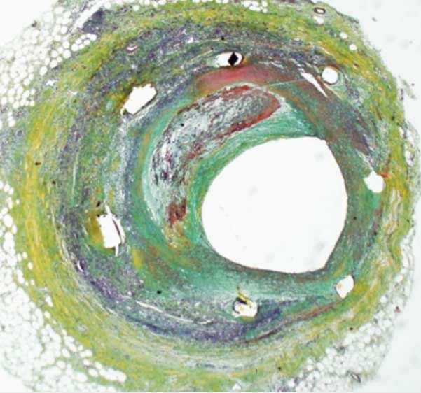
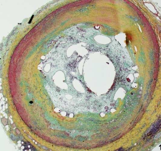
1 Pathology of In-stent Neoatherosclerosis in BMS and DES 197 BMS, 103 SES, and 106 PES with implant duration >30 days The incidence of neoatherosclerosis was significantly greater in DES (31%) than BMS (16%; p < 0.001). Median stent duration with neoatherosclerosis was shorter in DES than BMS (420 days v 2,160 days, p < 0.001). 2yrs 2-6yrs >6yrs BMS 0% 22% 42% DES 29% 41% 7 BMS and 3 DES had TCFA or plaque rupture occurring with shorter implant durations for DES (1.5±0.4 years) compared to BMS (6.1± years). Nakazawa et al. J Am Coll Cardiol 2011;57:
2 Percentage of Patients With Atherosclerotic Changes in DES Versus BMS in Relation to Duration of Implant at Autopsy Nakazawa et al. J Am Coll Cardiol Img 2009;2:625-8
3 In-stent Morphologies and OFDI Appearance Histology Collagen-rich neointima Proteoglycan-rich neointima Fibrin deposition OFDI description bright and homogenous tissue, smooth luminal surface relatively dark tissues, smooth luminal surface relatively light around luminal area following by attenuation, heterogenous tissues with irregular surface Signal Analysis Intensity Attenuation Strong Gentle Medium Gentle Medium Moderate Lipid core dark area without clear borders, more cholesterol crystal content likely to be darker* Weak Steep Foamy Macrophage accumulation Organized thrombus thin bright layer and following shadow Very Strong Steep dark area independent of surrounding tissues Weak Angiogenesis One or numerous black holes NA NA Calcified neointima a dark mass delineated with clear borders Weak Gentle Sudden Virmani et al. J Am Coll Cardiol Img 2009;2:625-8

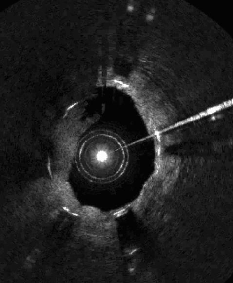
4 Disruption of neointima In-stent calcification Neoangiogenesis
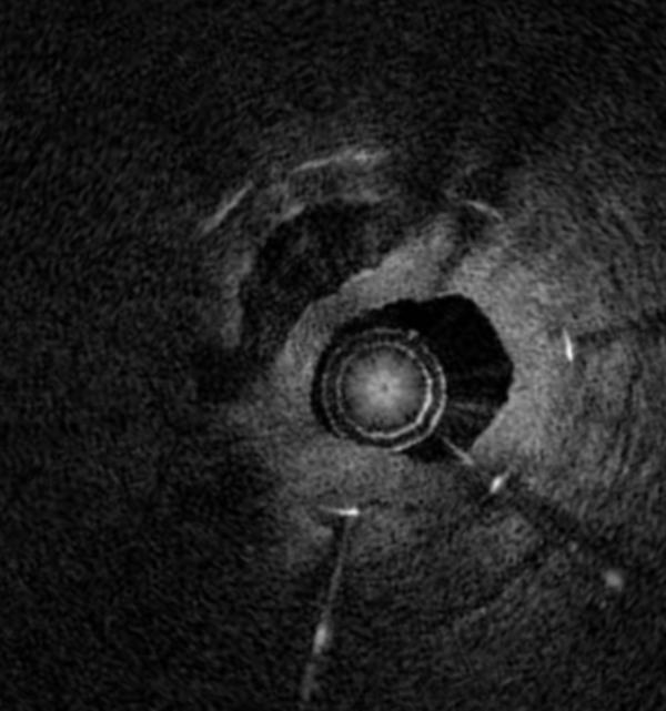
5 Organized thrombus Ca++ Ca++
6 TCFA in neointima


7 Macrophage infiltration on luminal surface
8 OCT and In-stent Neoatherosclerosis after BMS - I <6months >5years # Lipid laden intimal 0 67% Intimal disruption 0 38% Thrombus 5% 52% Intraintimal neovasacularization 0% 62% Takano et al. J Am Coll Cardiol 2009;55:26-33 In 39 pts (60 BMS) who underwent OCT imaging 6.5±1.3ys after BMS implantation, lipid-rich neointima was found in 20 stents (33.3%) in 16 pts (41%) with an average fibrous cap thickness of 56.7±5.8µ. Six pts had plaque disruption and 6 patients had mural thrombus. Hou et al. Heart. 2010;96:
9 OCT and In-stent Neoatherosclerosis after BMS - II >5 years <1 year P-value # Homogeneous neointima* 39.5±28.5% 94.2±11.5% < Heterogeneous neointima* 60.5%±28.5% 5.8±11.5% < Microvessels* Peri-stent 25.6±18.6% 6.8±8.6% < Neointima 13.1±12.8% 0 < Disrupted neointima 18.6% Intraluminal material 20.9% 2.6% 0.02 With shadowing 16.2% Without shadowing 4.7% 2.6% 1.0 *of sections throughout the stent Habara et al. Circ Cardiovasc Interv 2011;4:232-8
10 Normal Intima and Atherosclerotic Intima Thrombus, Intimal Disruption, and Neovascularization Takano et al. J Am Coll Cardiol 2010;55:26-32
11 In-stent Neoatherosclerosis after DES (n=50, median follow-up of 32 months) 52% lesions had at least one in-stent TCFA-like neointima 58% had at least one in-stent neointimal rupture. Patients presenting with unstable angina showed Thinner fibrous cap (55µ vs. 100µ, p=0.006) Higher incidence of TCFA-like neointima (75% vs. 37%, p=0.008) Higher incidence of neointimal rupture (75% vs. 47%, p=0.044) Higher incidence of thrombi (80% vs. 43%, p=0.010) and red thrombi (30% vs. 3%, p=0.012) Kang et al. Circulation 2011;123:
12 (composite of TCFA-like neointima, neointimal rupture, and red thrombus)

13 Late in-stent neoatherosclerosis in DES Microvessel TCFA-like neointima Calcium Neointimal rupture

14 Red thrombus White thrombus
15 VH Composition of Neointima at Various Follow-Up Times in 117 ISR Lesions Combining 47 BMS and 70 DES >36Mo (n=26) 52.2* 5.6* 27.2* 15.0* 24-36Mo (n=15) 54.9* 7.1 # 25.8* 12.2* 12-24Mo (n=12) # 6-12Mo (n=42) <6Mo (n=22) (%) *p<0.01 and # p<0.05, vs. lesions at follow-up time <6 months Kang et al. Am J Cardiol. 2010;106:1561-5
16 VH Composition of Neointima at Various Follow-Up Times in 70 DES Restenosis Lesions >36Mo (n=0) 24-36Mo (n=10) 53.3* 6.2 # 26.4* 13.1* 12-24Mo (n=11) # 6-12Mo (n=33) <6Mo (n=16) (%) *p<0.01 and #p<0.05, vs. lesions at follow-up time <6 months


17 Various Neointimal VH Composition at the Maximal %IH Sites 6-mo Taxus %NC 8% %DC 2% 9-mo Taxus %NC 28% %DC 8% 22-mo Taxus %NC 39% %DC 20% 48-mo BMS %NC 40% %DC 25% 57-mo BMS %NC 57% %DC 15%
18 VH-IVUS comparison of neointimal hyperplasia within 23 DES vs 15 BMS Follow-up was similar between DES (38 mos) vs. BMS (40 mos). % NC volume was significantly greater in DES than BMS: 19.5% vs. 12.1% (p=0.006) significantly increased with time in BMS (p=0.007), but not in DES (p=0.24) was greater in DES than in BMS at at any given time point. VH-IVUS in-stent VH-TCFAs were detected only in DES: 34.8% vs. 0% (p=0.013) Wakabayashi et al. J Inv Cardiol 2011;23:262-8
19 Late DES Catch-Up Among IVUS Substudy Patients %IH volume 25 Cypher FIM (SR) Baseline Early* 2 years *defined as 4-9 months Cypher FIM (FR) ASPECT (low dose) ASPECT (high dose) TAXUS-II Double Dose Diabetes (single dose SES) Double Dose Diabetes (double dose SES) AMC (SES) AMC (PES)
20 IVUS analysis of 23 very late DES thrombosis cases at Asan Medical Center LSM was observed in 17 DES patients (73.9%) Disease progression with neointimal rupture within the stent was observed in 10 DES patients (43.5%) and reference segment plaque rupture in another 5 DES patients (21.7%) Lee et al. J Am Coll Cardiol 2010;55:

21 Proximal Distal mm
22 Correlation of IVUS Findings With Aspirates in 28 Patients with Very Late DES Thrombosis 28 pts with very late DES ST and 26 controls LSM in 73% of very late DES ST segments. Maximal LSM area measured 6.2±2.4mm 2, and length measured 9.4±9.5mm. LSM area exceeded 5.0mm 2 in 5 of 8 segments (63%) Controls WBCs p Eos p Spontaneous MI 291±94± 7±10± Early ST-BMS 146±117 1±1 Early ST-DES 73±117 1±2 Very late ST-BMS 84±50 2± Very late ST-DES 283±149 20±24 LSM area correlated with total eosinophil count (p=0.008) Cook et al. Circulation 2009;120:391-9
23 Comparison Among OCT, VH-IVUS, and Grayscale IVUS Neotintimal rupture OCT + OCT - Grayscale IVUS Grayscale IVUS TCFA VH-IVUS VH-IVUS Agreement=50% Agreement=78% Thrombus Grayscale IVUS Grayscale IVUS Agreement=44% Kang et al. Circulation 2011;123:
24 Conclusion There is emerging evidence -- from pathology, VH- IVUS, and especially OCT (but least with grayscale IVUS) -- of the development of in-stent neoatherosclerosis While not a universal finding, in-stent neoatherosclersis is more common in DES than in BMS and occurs earlier in DES than in BMS In-stent neoatherosclerosis is responsible for some cases of very late stent thrombosis as well as late catch-up (late in-stent restenosis).
as a Mechanism of Stent Failure
 In-Stent t Neoatherosclerosis e osc e os s as a Mechanism of Stent Failure Soo-Jin Kang MD., PhD. University of Ulsan College of Medicine, Heart Institute Asan Medical Center, Seoul, Korea Disclosure I
Insights in Thrombosis and In-Stent Restenosis
 Clinical Value of OCT Insights in Thrombosis and In-Stent Restenosis Fernando Alfonso MD, PhD, FESC Interventional Cardiology. Cardiovascular Institute. Clinico San Carlos University Hospital. Madrid.
Can IVUS Define Plaque Features that Impact Patient Care?
 Can IVUS Define Plaque Features that Impact Patient Care? A Pichard L Satler, K Kent, R Waksman, W Suddath, N Bernardo, N Weissman, M Angelo, D Harrington, J Lindsay, J Panza. Washington Hospital Center
Left main coronary artery (LMCA): The proximal segment
: The proximal segment") Anatomy and Pathology of Left main coronary artery G Nakazawa Tokai Univ. Kanagawa, Japan 1 Anatomy Difinition Left main coronary artery (LMCA): The proximal segment RCA AV LAD LM LCX of the left coronary
Anatomy and Pathology of Left main coronary artery G Nakazawa Tokai Univ. Kanagawa, Japan 1 Anatomy Difinition Left main coronary artery (LMCA): The proximal segment RCA AV LAD LM LCX of the left coronary
Imaging Atheroma The quest for the Vulnerable Plaque
 Imaging Atheroma The quest for the Vulnerable Plaque P.J. de Feijter 1. Department of Cardiology 2. Department of Radiology Coronary Heart Disease Remains the Leading Cause of Death in the U.S, Causing
OCT Findings: Lesson from Stable vs Unstable Plaques
 ANGIOPLASTY SUMMIT TCTAP 2010 Imaging Workshop OCT Findings: Lesson from Stable vs Unstable Plaques Giulio Guagliumi MD Ospedali Riuniti di Bergamo, Italy DISCLOSURE OF FINANCIAL INTERESTS Consultant Boston
OCT; Comparative Imaging Results with IVUS, VH and Angioscopy
 OCT; Comparative Imaging Results with IVUS, VH and Angioscopy Takashi Akasaka, M.D. Department of Cardiovascular Medicine Wakayama, Japan Comparison among coronary imaging techniques OCT IVUS MRI CAG Angioscopy
CLINICAL APPLICATIONS OF OPTICAL COHERENCE TOMOGRAPHY. Konstantina P. Bouki, FESC 2 nd Department of Cardiology General Hospital Of Nikea, Pireaus
 CLINICAL APPLICATIONS OF OPTICAL COHERENCE TOMOGRAPHY Konstantina P. Bouki, FESC 2 nd Department of Cardiology General Hospital Of Nikea, Pireaus OPTICAL COHERENCE TOMOGRAPHY (OCT) IVUS and OCT IVUS OCT
Imaging Overview for Vulnerable Plaque: Data from IVUS Trial and An Introduction to VH-IVUS Imgaging
 Imaging Overview for Vulnerable Plaque: Data from IVUS Trial and An Introduction to VH-IVUS Imgaging Gary S. Mintz,, MD Cardiovascular Research Foundation New York, NY Today, in reality, almost everything
IVUS Analysis. Myeong-Ki. Hong, MD, PhD. Cardiac Center, Asan Medical Center University of Ulsan College of Medicine, Seoul, Korea
 IVUS Analysis Myeong-Ki Hong, MD, PhD Cardiac Center, Asan Medical Center University of Ulsan College of Medicine, Seoul, Korea Intimal disease (plaque) is dense and will appear white Media is made of
Catch-up Phenomenon: Insights from Pathology
 Catch-up Phenomenon: Insights from Pathology Michael Joner, MD CVPath Institute Inc. Gaithersburg, MD USA Path Lessons learned from the BMS and DES (1 st Gen) era Neointimal Thickness [mm] In Stent Re
Invasive Coronary Imaging Modalities for Vulnerable Plaque Detection
 Invasive Coronary Imaging Modalities for Vulnerable Plaque Detection Gary S. Mintz, MD Cardiovascular Research Foundation New York, NY Greyscale IVUS studies have shown Plaque ruptures do not occur randomly
Pathology of Vulnerable Plaque Angioplasty Summit 2005 TCT Asia Pacific, Seoul, April 28-30, 2005
 Pathology of Vulnerable Plaque Angioplasty Summit 25 TCT Asia Pacific, Seoul, April 28-3, 25 Renu Virmani, MD CVPath, A Research Service of the International Registry of Pathology Gaithersburg, MD Plaque
Optical Coherence Tomography for Intracoronary Imaging
 Optical Coherence Tomography for Intracoronary Imaging Lorenz Räber Stephan Windecker Department of Cardiology Swiss Cardiovascular Center and Clinical Trials Unit Bern Bern University Hospital, Switzerland
Added Value of Invasive Coronary Imaging for Plaque Rupture and Erosion
 Assessment of Coronary Plaque Rupture and Erosion Added Value of Invasive Coronary Imaging for Plaque Rupture and Erosion Yukio Ozaki, MD, PhD, FACC, FESC Cardiology Dept., Fujita Health Univ. Toyoake,
Assessment of Vulnerable Plaque by IVUS and VH-IVUS
 Assessment of Vulnerable Plaque by IVUS and VH-IVUS Akiko Maehara, MD Director of Intravascular Imaging & Physiology Core Laboratories Associate Director of MRI/MDCT Core Laboratory Cardiovascular Research
Appearance of Lipid-Laden Intima and Neovascularization After Implantation of Bare-Metal Stents
 Journal of the American College of Cardiology Vol. 55, No. 1, 2010 2010 by the American College of Cardiology Foundation ISSN 0735-1097/10/$36.00 Published by Elsevier Inc. doi:10.1016/j.jacc.2009.08.032
Drug eluting stents (DES) have decreased
 have decreased") JACC: CARDIOVASCULAR IMAGING VOL. 5, NO. 11, 1 1 BY THE AMERICAN COLLEGE OF CARDIOLOGY FOUNDATION ISSN 1936-878X/$36. PUBLISHED BY ELSEVIER INC. http://dx.doi.org/1.116/j.jcmg.1.. BRIEF REPORT OCT-Verified
JACC: CARDIOVASCULAR IMAGING VOL. 5, NO. 11, 1 1 BY THE AMERICAN COLLEGE OF CARDIOLOGY FOUNDATION ISSN 1936-878X/$36. PUBLISHED BY ELSEVIER INC. http://dx.doi.org/1.116/j.jcmg.1.. BRIEF REPORT OCT-Verified
Plaque Characteristics in Coronary Artery Disease. Chourmouzios Arampatzis MD, PhD, FESC
 Plaque Characteristics in Coronary Artery Disease Chourmouzios Arampatzis MD, PhD, FESC Disclosure Statement of Financial Interest Regarding this Presentation NONE Atherosclerosis Model proposed by Stary
Pathology of Coronary Artery Disease
 Pathology of Coronary Artery Disease Seth J. Kligerman, MD Pathology of Coronary Artery Disease Seth Kligerman, MD Assistant Professor Medical Director of MRI University of Maryland Department of Radiology
High-risk vulnerable plaques. Kostis Raisakis G.Gennimatas General Hospital of Athens
 High-risk vulnerable plaques. Kostis Raisakis G.Gennimatas General Hospital of Athens Overview: 1 Definition-Pathology 2 3 Diagnostic Strategies Invasive Non Invasive Prognostic Value of Detection 4 Treatment
Optical coherence tomography patterns of in-stent restenosis: Comparison between bare-metal stent and drug-eluting stent
 Optical coherence tomography patterns of in-stent restenosis: Comparison between bare-metal stent and drug-eluting stent heart institute, Division of cardiology, Tokyo, Japan Yusuke Watanabe, Ryuta Asano,
Vulnerable Plaque Pathophysiology, Detection, and Intervention. VP: A Local Problem or Systemic Disease. Erling Falk, Denmark
 Vulnerable Plaque Pathophysiology, Detection, and Intervention VP: A Local Problem or Systemic Disease Erling Falk, Denmark Vulnerable Plaque Pathophysiology, Detection, and Intervention VP: A Local Problem
Review Article Pathologic Etiologies of Late and Very Late Stent Thrombosis following First-Generation Drug-Eluting Stent Placement
 ombosis Volume 2012, Article ID 608593, 16 pages doi:10.1155/2012/608593 Review Article Pathologic Etiologies of Late and Very Late Stent ombosis following First-Generation Drug-Eluting Stent Placement
IVUS Assessment of the Mechanism of In-stent Restenosis? Gary S. Mintz, MD Cardiovascular Research Foundation
 IVUS Assessment of the Mechanism of In-stent Restenosis? Gary S. Mintz, MD Cardiovascular Research Foundation SURE Trial: Restenosis in non-stented lesions Average of the two image slices with the smallest
Cardiovascular Research Foundation and Columbia University Medical Center, New York.
 Virtual Histology Intravascular Ultrasound Analysis of Non-culprit Attenuated Plaques Detected by Grayscale Intravascular Ultrasound in Patients with Acute Coronary Syndromes Xiaofan Wu, Akiko Maehara,
OCT Analysis in Patients With Very Late Stent Thrombosis
 JACC: CARDIOVASCULAR IMAGING VOL. 6, NO. 6, 2013 ª 2013 BY THE AMERICAN COLLEGE OF CARDIOLOGY FOUNDATION ISSN 1936-878X/$36.00 PUBLISHED BY ELSEVIER INC. http://dx.doi.org/10.1016/j.jcmg.2013.02.006 OCT
The PROSPECT Trial. A Natural History Study of Atherosclerosis Using Multimodality Intracoronary Imaging to Prospectively Identify Vulnerable Plaque
 The PROSPECT Trial Providing Regional Observations to Study Predictors of Events in the Coronary Tree A Natural History Study of Atherosclerosis Using Multimodality Intracoronary Imaging to Prospectively
Invasive Imaging (IVUS, VH-IVUS, and OCT): How I Implement into My
: How I Implement into My") Invasive Imaging (IVUS, VH-IVUS, and OCT): How I Implement into My Practice Gary S. Mintz, MD Cardiovascular Research Foundation Modalities FFR IVUS (with or without VH, imap, or IB-IVUS) OCT NIRS (with
Invasive Imaging (IVUS, VH-IVUS, and OCT): How I Implement into My Practice Gary S. Mintz, MD Cardiovascular Research Foundation Modalities FFR IVUS (with or without VH, imap, or IB-IVUS) OCT NIRS (with
Coronary Artery Thermography
 Coronary Artery Thermography The 10th Anniversary, Interventional Vascular Therapeutics Angioplasty Summit 2005 TCT Asia Pacific Christodoulos Stefanadis Professor of Cardiology Athens Medical School In
Characteristics of Neoatherosclerosis Within Implanted Coronary Stents in Patients with Acute Coronary Syndromes
 Journal of Cardiovascular Emergencies 2016;2(1):19-26 DOI: 10.1515/jce-2016-0004 JOURNAL OF CARDIOVASCULAR EMERGENCIES ORIGINAL RESEARCH Characteristics of Neoatherosclerosis Within Implanted Coronary
Prognostic Value of Gated Myocardial Perfusion SPECT
 Current Use of IVUS & FFR George D. Dangas, MD, PhD, FACC, FSCAI Professor of Medicine Mount Sinai School of Medicine Prognostic Value of Gated Myocardial Perfusion SPECT 0.6% / year, Cardiac Death and
What Does the Yellow Color of Angioscopy Mean? Why Yellow Plaque Is Always Vulnerable?
 Review Angioscopy 2017; 3: 9 18 What Does the Yellow Color of Angioscopy Mean? Why Yellow Plaque Is Always Vulnerable? Kyoichi Mizuno, MD, PhD, *1 and Masamichi Takano, MD, PhD 2 1 Mitsukoshi Health and
Que nos puede aportar el OCT intracoronario
 XXXI Jornadas SOLACI. 10ª Región CONOSUR LIIIº Congreso Chileno de Cardiología y Cirugía Cardiovascular Hotel Patagónico. Puerto Varas. Chile (30 Nov 1 Dic 2016) Que nos puede aportar el OCT intracoronario
Can We Identify Vulnerable Patients & Vulnerable Plaque?
 Can We Identify Vulnerable Patients & Vulnerable Plaque? We Know Enough to Treat High-Risk Lesions? Takashi Akasaka, MD, PhD Department of Cardiovascular Medicine, Japan Disclosure Statement of Financial
Assessment of plaque morphology by OCT in patients with ACS
 Assessment of plaque morphology by OCT in patients with ACS Takashi Akasaka, M.D. Department of Cardiovascular Medicine Wakayama, Japan Unstable plaque Intima Lipid core Plaque rupture and coronary events
Biology of in-stent restenosis and rational for debulking
 LINC 2016 Leipzig January 26-29 2016 Biology of in-stent restenosis and rational for debulking Jos C. van den Berg, MD PhD Ospedale Regionale di Lugano, sede Civico Lugano Switzerland ISR-complexity Osherov
Histopathology: Vascular pathology
 Histopathology: Vascular pathology These presentations are to help you identify basic histopathological features. They do not contain the additional factual information that you need to learn about these
Review Article Optical Coherence Tomography Imaging in Acute Coronary Syndromes
 SAGE-Hindawi Access to Research Cardiology Research and Practice Volume 2011, Article ID 312978, 7 pages doi:10.4061/2011/312978 Review Article Optical Coherence Tomography Imaging in Acute Coronary Syndromes
Chapter 43 Noninvasive Coronary Plaque Imaging
 hapter 43 Noninvasive oronary Plaque Imaging NIRUDH KOHLI The goal of coronary imaging is to define the extent of luminal narrowing as well as composition of an atherosclerotic plaque to facilitate appropriate
Multimodality Imaging Atlas of Coronary Atherosclerosis
 JCC: CRDIOVSCUR IMGING VO. 3, NO. 8, 2010 2010 BY THE MERICN COEGE OF CRDIOOGY FOUNDTION ISSN 0735-1097/$36.00 PUBISHED BY ESEVIER INC. DOI:10.1016/j.jcmg.2010.06.006 IMGING VIGNETTE Multimodality Imaging
Optical Coherence Tomography
 Optical Coherence Tomography Disclosure Information Demetrius Lopes MD The following relationships exist related to this presentation: University Grant/Research Support: Rush University Industry Grant
Sites of Atherosclerosis In order of Frequency
 Pathological Features of Peripheral Atherosclerosis: Implication for Device Development G Nakazawa Tokai Univ. Kanagawa, Japan 1 Sites of Atherosclerosis In order of Frequency carotid (3) (3) Coronary
1st Department of Cardiology, University of Athens, Hippokration Hospital, Athens, Greece
 Konstantinos Toutouzas, Maria Riga, Antonios Karanasos, Eleftherios Tsiamis, Andreas Synetos, Maria Drakopoulou, Chrysoula Patsa, Georgia Triantafyllou, Aris Androulakis, Christodoulos Stefanadis 1st Department
Pathophysiology of Cardiovascular System. Dr. Hemn Hassan Othman, PhD
 Pathophysiology of Cardiovascular System Dr. Hemn Hassan Othman, PhD hemn.othman@univsul.edu.iq What is the circulatory system? The circulatory system carries blood and dissolved substances to and from
OCT Technology: Differences between Biodegradable and Durable Polymers: Insights from the LEADERS Trial LEADERS OCT
 OCT Technology: Differences between Biodegradable and Durable Polymers: Insights from the LEADERS Trial LEADERS OCT Substudy Carlo Di Mario, MD Peter Barlis, MD Evelyn Regar, MD Peter Juni, MD Patrick
Pathology of the Vulnerable Plaque
 Journal of the American College of Cardiology Vol. 47, No. 8 Suppl C 2006 by the American College of Cardiology Foundation ISSN 0735-1097/06/$32.00 Published by Elsevier Inc. doi:10.1016/j.jacc.2005.10.065
Integrating IVUS, FFR, and Noninvasive Imaging to Optimize Outcomes. Gary S. Mintz, MD Cardiovascular Research Foundation
 Integrating IVUS, FFR, and Noninvasive Imaging to Optimize Outcomes Gary S. Mintz, MD Cardiovascular Research Foundation COURAGE Nuclear Substudy (n=314) Death/MI according the residual ischemia (SPECT)
Evaluation of stent placement and outcomes with optical coherence tomography
 REVIEW Evaluation of stent placement and outcomes with optical coherence tomography Optical coherence tomography (OCT) is an imaging modality based on fiberoptic technology. OCT imaging systems use optical
SMJ Singapore Medical Journal
 SMJ Singapore Medical Journal ONLINE FIRST PUBLICATION Online first papers have undergone full scientific review and copyediting, but have not been typeset or proofread. To cite this article, use the DOIs
Gary S. Mintz,, MD. IVUS Observations in Acute (vs Chronic) Coronary Artery Disease: Structure vs Function
 Coronary Artery Disease: Structure vs Function") Gary S. Mintz,, MD IVUS Observations in Acute (vs Chronic) Coronary Artery Disease: Structure vs Function Important IVUS Observations: Remodeling Originally used (first by Glagov) ) to explain atherosclerosis
Gary S. Mintz,, MD IVUS Observations in Acute (vs Chronic) Coronary Artery Disease: Structure vs Function Important IVUS Observations: Remodeling Originally used (first by Glagov) ) to explain atherosclerosis
Muzina Akhtar, Wei Liu. History of ISR visualization. Introduction
 Perspective Use of intravascular ultrasound vs. optical coherence tomography for mechanism and patterns of in-stent restenosis among bare metal stents and drug eluting stents Muzina Akhtar, Wei Liu Cardiology
Fielder XT: Initial and. Department of Cardiology, Asan Medical Center, Ulsan University of college of medicine
 Fielder XT: Initial and Professional Use for CTO Seung-Whan Lee, MD, PhD D t t f C di l A M di l C t Department of Cardiology, Asan Medical Center, Ulsan University of college of medicine Plastic-Jacket
Culprit Lesion Remodeling and Long-term (> 5years) Prognosis in Patients with Acute Coronary Syndrome
 Prognosis in Patients with Acute Coronary Syndrome") Culprit Lesion Remodeling and Long-term (> 5years) Prognosis in Patients with Acute Coronary Syndrome Hiroyuki Okura*, MD; Nobuya Matsushita**,MD Kenji Shimeno**, MD; Hiroyuki Yamaghishi**, MD Iku Toda**,
Culprit Lesion Remodeling and Long-term (> 5years) Prognosis in Patients with Acute Coronary Syndrome Hiroyuki Okura*, MD; Nobuya Matsushita**,MD Kenji Shimeno**, MD; Hiroyuki Yamaghishi**, MD Iku Toda**,
The PROSPECT Trial. A Natural History Study of Atherosclerosis Using Multimodality Intracoronary Imaging to Prospectively Identify Vulnerable Plaque
 The PROSPECT Trial Providing Regional Observations to Study Predictors of Events in the Coronary Tree A Natural History Study of Atherosclerosis Using Multimodality Intracoronary Imaging to Prospectively
Davide Capodanno, MD, PhD Associate Professor, University of Catania, Italy
 Restenosis - Capodanno CardioLucca, November 28, 2014 Slide 1 Autumn in Lucca V Simposio - 28 Novembre 2014 4.30PM-4.45PM L incubo del paziente e le incognite del cardiologo: la restenosi intrastent resta
Assessment of Coronary Plaque Vulnerability with Optical Coherence Tomography
 Review Article Acta Cardiol Sin 2014;30:1 9 Assessment of Coronary Plaque Vulnerability with Optical Coherence Tomography Shiro Uemura, Tsunenari Soeda, Yu Sugawara, Tomoya Ueda, Makoto Watanabe and Yoshihiko
Clinical Value of OCT. Guidance for Coronary Stenting. Giulio Guagliumi, MD
 Clinical Value of OCT Guidance for Coronary Stenting Giulio Guagliumi, MD 100 % Endovascular Imaging Indications of use 87.5 % 75 % 57.5 % 50 % 45 % 25 % 15 % 0 Lesion morphology Stent optimization Lesion
The Pathology of Neoatherosclerosis in Human Coronary Implants
 Journal of the American College of Cardiology Vol. 57, No. 11, 2011 2011 by the American College of Cardiology Foundation ISSN 0735-1097/$36.00 Published by Elsevier Inc. doi:10.1016/j.jacc.2011.01.011
BIOFREEDOM: Polymer free Biolimus A9 eluting
 TCTAP 2011 Seoul, April 27 29, 2011 BIOFREEDOM: Polymer free Biolimus A9 eluting Stents and Paclitaxel eluting stents Eberhard Grube MD, FACC, FSCAI Hospital Oswaldo Cruz - Dante Pazzanese, São Paulo,
Clinical Application of OCT in Stent Evaluation
 Imaging & Physiology Summit 2010 in Soul #1. Basics of Image Interpretation: IVUS/VH/OCT Clinical Application of OCT in Stent Evaluation Mitsuyasu Terashima, MD, PhD, FACC Stent implantation Stent Apposition
Tratamiento de la Reestenosis del Stent Farmacoactivo
 XXXI Jornadas SOLACI. 10ª Región CONOSUR LIIIº Congreso Chileno de Cardiología y Cirugía Cardiovascular Hotel Patagónico. Puerto Varas. Chile (30 Nov 1 Dic 2016) Tratamiento de la Reestenosis del Stent
Assessment of vulnerable plaque by OCT
 Assessment of vulnerable plaque by OCT Comparison with histology and possible clinical applications Takashi Akasaka, M.D. Department of Cardiovascular Medicine Wakayama, Japan Identification of vulnerable
CPIS So-Yeon Choi, MD., PhD. Department of Cardiology Ajou University School of MedicineSuwon, Korea
 So-Yeon Choi, MD., PhD. Department of Cardiology Ajou University School of MedicineSuwon, Korea Coronary Artery Imaging The ideal coronary imaging technology would be capable of identifying not only vessel
Advance Publication. Atherosclerotic Component of the Yellow Segment After Drug-Eluting Stent Implantation on Coronary Angioscopy
 Circulation Journal doi: 10.1253/circj.CJ-18-0671 ORIGINAL ARTICLE Imaging Atherosclerotic Component of the Yellow Segment After Drug-Eluting Stent Implantation on Coronary Angioscopy An Ex-Vivo Validation
TVA_C02.qxd 8/8/06 10:27 AM Page 19 PART 2. Pathology
 TVA_C2.qxd 8/8/6 :27 AM Page 19 2 PART 2 Pathology TVA_C2.qxd 8/8/6 :27 AM Page TVA_C2.qxd 8/8/6 :27 AM Page 21 2 CHAPTER 2 The pathology of vulnerable plaque Renu Virmani, Allen P Burke, James T Willerson,
Failure of positive. Recanalization and CTO formation. TCFA rupture with (fatal) thrombotic occlusion. TCFA Lipid pool
 thrombotic occlusion. TCFA Lipid pool") Vulnerable Plaque features on coronary CT Jin Ho Choi, MD, PhD Department of Internal Medicine, Emergency Medicine Samsung Medical Center, Sungkyunkwan University School of Medicine, Seoul, Korea IPS /
Vulnerable Plaque features on coronary CT Jin Ho Choi, MD, PhD Department of Internal Medicine, Emergency Medicine Samsung Medical Center, Sungkyunkwan University School of Medicine, Seoul, Korea IPS /
Clinical Characteristics of In-stent Neoatherosclerosis Causing Late Stent Failure
 Original Paper J Jpn Coron Assoc 2016; 22: 245-250 Clinical Characteristics of In-stent Neoatherosclerosis Causing Late Stent Failure Koji Isodono, Akiko Matsuo, Atsushi Kyodo, Yumika Tsuji, Akira Sakamoto,
A Novel Low Pressure Self Expanding Nitinol Coronary Stent (vprotect): Device Design and FIH Experience
: Device Design and FIH Experience") A Novel Low Pressure Self Expanding Nitinol Coronary Stent (vprotect): Device Design and FIH Experience Juan F. Granada, MD Medical Director, Skirball Center for Cardiovascular Research The Cardiovascular
A Novel Low Pressure Self Expanding Nitinol Coronary Stent (vprotect): Device Design and FIH Experience Juan F. Granada, MD Medical Director, Skirball Center for Cardiovascular Research The Cardiovascular
Usefulness of OCT during coronary intervention
 Usefulness of OCT during coronary intervention Takashi Akasaka, M.D. Department of Cardiovascular Medicine Wakayama, Japan Predictors at 12 Months of Stent Thrombosis and Target Lesion Revascularization
Neointimal coverage of bare-metal and sirolimuseluting stents evaluated with optical coherence tomography
 Neointimal coverage of bare-metal and sirolimuseluting stents evaluated with optical coherence tomography B X Chen, F Y Ma, W Luo, J H Ruan, W L Xie, X Z Zhao, S H Sun, X M Guo, F Wang, T Tian, X W Chu
Pathology of Second-Generation Everolimus-Eluting Stents Versus First-Generation Sirolimus- and Paclitaxel-Eluting Stents in Humans
 Pathology of Second-Generation Everolimus-Eluting Stents Versus First-Generation Sirolimus- and Paclitaxel-Eluting Stents in Humans Fumiyuki Otsuka, MD, PhD; Marc Vorpahl, MD; Masataka Nakano, MD; Jason
Bifurcation Stenting: IVUS and OCT Information
 Bifurcation Stenting: IVUS and OCT Information Yoshinobu Murasato MD, PhD (New Yukuhashi Hospital) On behalf of J-REVERSE investigators October 14-15, 2011, Lisbon Proximal stent deformation induced by
Cover Page. The handle holds various files of this Leiden University dissertation.
 Cover Page The handle http://hdl.handle.net/1887/64938 holds various files of this Leiden University dissertation. Author: Liu, S. Title: Optical coherence tomography for coronary artery disease : analysis
RESTENOSIS Facing up to the problem
 RESTENOSIS Facing up to the problem Petr Kala University Hospital Brno Czech Republic ESC 2011, Paris Disclosure Scientific Advisory Boards or Education presentations fee Abbott, Boston Scientific, Cordis
Optical Coherence Tomography (OCT): A New Imaging Tool During Carotid Artery Stenting
: A New Imaging Tool During Carotid Artery Stenting") Chapter 6 Optical Coherence Tomography (OCT): A New Imaging Tool During Carotid Artery Stenting Shinichi Yoshimura, Masanori Kawasaki, Kiyofumi Yamada, Arihiro Hattori, Kazuhiko Nishigaki, Shinya Minatoguchi
Chapter 6 Optical Coherence Tomography (OCT): A New Imaging Tool During Carotid Artery Stenting Shinichi Yoshimura, Masanori Kawasaki, Kiyofumi Yamada, Arihiro Hattori, Kazuhiko Nishigaki, Shinya Minatoguchi
Research Article Pathological Perspective of Drug-Eluting Stent Thrombosis
 Thrombosis Volume 2012, Article ID 219389, 8 pages doi:10.1155/2012/219389 Research Article Pathological Perspective of Drug-Eluting Stent Thrombosis Katsumi Inoue Department of Laboratory Medicine, Kokura
malapposition assessed by OCT
 Stent t coverage and malapposition assessed by OCT Myeong-Ki Hong, M.D. Ph D Professor of Medicine Division of Cardiology, Severance Cardiovascular Hospital Yonsei University College of Medicine, Seoul,
Stent Fracture and Longitudinal Compression on CT Angiography between the
 2014 ASCI Stent Fracture and Longitudinal Compression on CT Angiography between the First- and New-Generation Drug-Eluting Stent Mi Sun Chung, Dong Hyun Yang,Young-Hak Kim, Jae-Hyung Roh, Joon-Won Kang,
IN-STENT RESTENOSIS. K.Boerlage-van Dijk CarVasZ 2014
 IN-STENT RESTENOSIS K.Boerlage-van Dijk CarVasZ 2014 Definition ISR Angiographic: recurrent diameter stenosis >50% at the stent segment or edges (5-mm segments adjacent to stent) Mehran system morphological
Plaque Imaging: What It Can Tell Us. Kenneth Snyder, MD, PhD L Nelson Hopkins MD FACS Elad Levy MD MBA FAHA FACS Adnan Siddiqui MD PhD
 Plaque Imaging: What It Can Tell Us Kenneth Snyder, MD, PhD L Nelson Hopkins MD FACS Elad Levy MD MBA FAHA FACS Adnan Siddiqui MD PhD Buffalo Disclosure Information FINANCIAL DISCLOSURE: Research and consultant
Dr Rodney Itaki Lecturer Anatomical Pathology Discipline. University of Papua New Guinea School of Medicine & Health Sciences Division of Pathology
 Arterial Diseases Dr Rodney Itaki Lecturer Anatomical Pathology Discipline University of Papua New Guinea School of Medicine & Health Sciences Division of Pathology Disease Spectrum Arteriosclerosis Atherosclerosis
Neoatherosclerosis: overview of histopathologic findings and implications for intravascular imaging assessment
 European Heart Journal (2015) 36, 2147 2159 doi:10.1093/eurheartj/ehv205 REVIEW Imaging Neoatherosclerosis: overview of histopathologic findings and implications for intravascular imaging assessment Fumiyuki
Analysis of neointimal coverage after silolimus-eluting stent implantation using optical coherence tomography.
 Analysis of neointimal coverage after silolimus-eluting stent implantation using optical coherence tomography. Division of Cardiology, Department of Internal Medicine, Fasculty of Medicine, Kinki University,
Drug Eluting Stent DES Pathology Update What we know, what we do not know
 Drug Eluting Stent DES Pathology Update What we know, what we do not know 13 th Summit TCT Asia 25 th April 2008 Renu Virmani, MD. CVPath Institute Inc. Gaithersburg, MD Disclosure Statement of Financial
OCT in the Evaluation of Vascular Healing Following DES Implantation: Will It Be a Helpful Tool to Reduce Stent Thrombosis?
 OCT in the Evaluation of Vascular Healing Following DES Implantation: Will It Be a Helpful Tool to Reduce Stent Thrombosis? Juan F. Granada, MD Medical Director, Skirball Center for Cardiovascular Research
State of the Art. Advances in Cardiovascular Imaging. ESC Congres Stockholm September 1, 2010 Frank E. Rademakers, MD, PhD, FESC
 State of the Art Advances in Cardiovascular Imaging ESC Congres Stockholm September 1, 2010 Frank E. Rademakers, MD, PhD, FESC Coronary Artery Disease Content Patho Physiology Imaging requirements Economical
OCT GUIDED TREATMENT OF CALCIFIED LESIONS RICHARD SHLOFMITZ, MD CHAIRMAN OF DEPT. OF CARDIOLOGY ST. FRANCIS HOSPITAL ROSLYN, NEW YORK
 OCT GUIDED TREATMENT OF CALCIFIED LESIONS RICHARD SHLOFMITZ, MD CHAIRMAN OF DEPT. OF CARDIOLOGY ST. FRANCIS HOSPITAL ROSLYN, NEW YORK Disclosure Statement of Financial Interest Within the past 12 months,
Citation Journal of Cardiology Cases (2013), Ltd.; この論文は出版社版でありません 引用の際には出版社版をご確認ご利用ください This is not the publ
, Ltd.; この論文は出版社版でありません 引用の際には出版社版をご確認ご利用ください This is not the publ") Title Pathological analyses of very longimplantation in human coronary arte Author(s) Imai, Masao; Kimura, Takeshi; Tazak Erika; Inoue, Katsumi Citation Journal of Cardiology Cases (01), Issue Date 01-
Title Pathological analyses of very longimplantation in human coronary arte Author(s) Imai, Masao; Kimura, Takeshi; Tazak Erika; Inoue, Katsumi Citation Journal of Cardiology Cases (01), Issue Date 01-
Shockwave Intravascular Lithotripsy System treatment of calcified lesions: Intravascular OCT analysis
 Shockwave Intravascular Lithotripsy System treatment of calcified lesions: Intravascular OCT analysis Andrew Holden, MBChB, FRANZCR, EBIR Director of Interventional Radiology Auckland, New Zealand LINC
IVUS vs FFR Debate: IVUS-Guided PCI
 IVUS vs FFR Debate: IVUS-Guided PCI Gary S. Mintz, MD Cardiovascular Research Foundation New York, NY Disclosure Statement of Financial Interest Within the past 12 months, I have had a financial interest/arrangement
Index. B Bare metal stents (BMS) vs. DES, 172 OCT findings, 170, 172
 vs. DES, 172 OCT findings, 170, 172") Index A Absorbable metal stent (AMS), 189 Absorb BVS, 184 187 Acquired malapposition in DES, stent thrombosis. See also Incomplete stent apposition (ISA) coronary angiography, 155, 156 DAPT therapy, 155
Index A Absorbable metal stent (AMS), 189 Absorb BVS, 184 187 Acquired malapposition in DES, stent thrombosis. See also Incomplete stent apposition (ISA) coronary angiography, 155, 156 DAPT therapy, 155
Between Coronary Angiography and Fractional Flow Reserve
 Visual-Functional Mismatch Between Coronary Angiography and Fractional Flow Reserve Seung-Jung Park, MD., PhD. University of Ulsan, College of Medicine Asan Medical Center, Seoul, Korea Visual - Functional
Analysis of macrophage accumulation using optical coherence tomography one year after sirolimus, paclitaxel and zotarolimus-eluting stent
 Analysis of macrophage accumulation using optical coherence tomography one year after sirolimus, paclitaxel and zotarolimus-eluting stent implantation. Department of Cardiology, Ehime Prefectural Imabari
DES In-stent Restenosis
 DES In-stent Restenosis Roxana Mehran, MD Columbia University Medical Center The Cardiovascular Research Foundation DES Restenosis Mechanisms Predictors Morphological patterns Therapy approach Mechanisms
Stephen G. Ellis, M.D. Professor of Medicine Director Invasive Services Co-Director Cardiac Gene Bank
 From ABSORB Cohort A to ABSORB III and IV Randomized Trials Stephen G. Ellis, M.D. Professor of Medicine Director Invasive Services Co-Director Cardiac Gene Bank Disclosures Consultant, Abbott Vascular
PCI for In-Stent Restenosis. CardioVascular Research Foundation
 PCI for In-Stent Restenosis ISR of BMS Patterns of In-Stent Restenosis Pattern I : Focal Type IA: Articulation / Gap Type IB: Marginal Type IC: Focal body Type ID: Multifocal Pattern II,III,IV : Diffuse
Ambiguity in Detection of Necrosis in IVUS Plaque Characterization Algorithms and SDH as Alternative Solution
 Ambiguity in Detection of Necrosis in IVUS Plaque Characterization Algorithms and SDH as Alternative Solution Amin Katouzian, Ph.D., Debdoot Sheet, M.S., Abouzar Eslami, Ph.D., Athanasios Karamalis, M.Sc.,
In-stent Restenosis Diagnostic and Therapeutic Challenges. Kostis Raisakis General Hospital of Athens «G. Gennimatas»
 In-stent Restenosis Diagnostic and Therapeutic Challenges Kostis Raisakis General Hospital of Athens «G. Gennimatas» Introduction With POBA, rates of acute and chronic vessel occlusion at 30% to 60%, secondary
In-Stent Restenosis. Can we kill it?
 In-Stent Restenosis Can we kill it? However, In-stent Restenosis is the most serious problem (2-25%) More than 15, lesions will need treatment because of in-stent restenosis. Varying Prevalence Rates of