|
|
|
- Jemimah Byrd
- 6 years ago
- Views:
Transcription
1
2
3 Dear Friends Dear Colleagues, This International Symposium, already celebrating its XVIIth edition, is set to offer you the most relevant clinical evidence in Vascular and Endovascular fields, aiming, for the third consecutive year, to broadcast a series of selected live cases, a National pioneering approach driving this event. With our Faculty, we jointly created a teaching experience, proudly endorsed once again by the LINC and SITE initiatives and recognized by our Medical College and University. The full support and commitment of the Hospital Administration Council, Autonomous Surgical Management Unit and Vascular Surgical Department was essential to improve all the logistics and organization, allowing for our Hospital based initiative to be valued and shared by the vascular community. The changing pattern in vascular Medicine, with endovascular procedures being part of the actual portfolio treatment options in an increasing number of situations, incorporating new materials and techniques, is assumed in this Symposium as a platform in which the sponsoring companies can have their fundamental partnership role highlighted and properly included. Financial constraints and patient risk / benefit analysis oblige to proper assessment of the clinical decisions and therapeutic devices choice, taking into consideration the state of the art knowledge and the capabilities of our group. I have to mention that this event has been, for us, a crucial opportunity to interact with the most skilled surgeons, upgrade the Department expertise, treat our patients with better results and justify future reference Centre ambitions. It also has to be emphasized the post graduate learning intention of the Symposium in which the involvement of our residents must be truly underlined. From the most complex aortic cases treatment to PAD, carotid and venous disease management, all topics have been carefully selected for this interactive meeting. I am most grateful for your presence and participation, your kindness and availability to be part of it, and please consider this event as a joint solid project, to sound your voice and reflect your experiences. Enjoy the meeting and our beautiful city of Porto. You will certainly become part of our History. José Fernando Teixeira Symposium President
4
5 President Dr. José Fernando Teixeira Secretary General Prof. Doutor Sérgio Sampaio Honorary Presidents Prof. Doutor António Braga Dra. Fernanda Viana Prof. Doutor Roncon de Albuquerque Organizing committee Serviço de Angiologia e Cirurgia Vascular do Centro Hospitalar do São João EPE & Associação ANGIOVASC Prof. Doutor Roncon de Albuquerque Dr. José Fernando Teixeira Prof. Doutor Sérgio Sampaio Dr. Joel Sousa Dr. João Neves Dr. Luís Machado Dr. José Pedro Pinto Live cases and Handbook Dra. Ana Sofia Ferreira Dr. Luís Machado Dra. Marina Neto Dr. João Neves Dr. Ricardo Ferreira Dr. Joel Sousa Dr. José Pedro Pinto Short cases committe Dr. Joel Sousa Dr. João Neves Web Supervisor Dr. João Neves Logistics coordination Dr. José Almeida Lopes Dr. Pedro Henrique Almeida
6 XVII OF ANGIOLOGY AND VASCULAR SURGERY INTERNATIONAL SYMPOSIUM PARTICIPANTS Adelino Moreira Albuquerque de Matos Alexandra Canedo Alfredo Cerqueira Amílcar Mesquita Ana Sofia Ferreira Andrej Schmidt António Assunção António Braga António Simões Armando Lobato Armando Mansilha Barend Mees Carlos Vaquero Ciro Ferrer Dalila Rolim Daniel Menezes Duarte Medeiros Eurico Norton Fernandez Noya Fernanda Viana Fernando Ramos Francisco Aranha Frank Vermassen Gaspar Mestres Giancarlo Biamino Gonçalo Cabral Hervé Rousseau Ignacio Lojo Isabel Vilaça Javier Villaverde Rodríguez Joana de Carvalho João Almeida Pinto João Rocha Neves João Silva e Castro João Vasconcelos Joel Ferreira Sousa Jorge Almeida Jorge Costa Lima Jorge Tenreiro José Almeida Lopes José Carlos Vidoedo Porto Coimbra Vila Nova de Gaia Porto Guimarães Porto Leipzig Braga Porto Viseu São Paulo Porto Maastricht Valladolid Roma Porto Lisboa Lisboa Porto Santiago de Compostela Porto Porto Lisboa Brussels Barcelona Leipzig Lisboa Toulouse Santiago de Compostela Porto Santiago de Compostela Braga Penafiel Porto Lisboa Penafiel Porto Porto Porto Porto Porto Penafiel José Fernandes e Fernandes José Fernando Teixeira Laura Cappocia Leonor Vasconcelos Luis Filipe Machado Luís Loureiro Luís Mota Capitão Luis Silvestre Manuel Alonso Manuel Fonseca Maria José Ferreira Mariangela Demasi Marina Neto Mario Lachat Mário Macedo Mário Vieira Marta Moura Marzia Lugli Matas do Campo Michael Piorkowski Miguel Lobo Miguel Maia Nilo Mosquera Óscar Gonçalves P.M. Kasprzak Paulo Correia Paulo Dias Paulo Pinho Pedro Henrique Almeida Pedro Paz Dias Pereira Albino Ricardo Castro Ferreira Ricardo Vale Pereira Rocha e Silva Roncon de Albuquerque Rosa Moreno Rui Almeida Rui Machado Sérgio Eufrásio Sérgio Sampaio Vincent Riambau Lisboa Porto Roma Lisboa Porto Porto Lisboa Lisboa Oviedo Coimbra Almada Marselha Porto Zurich Lisboa Porto Porto Modena Barcelona Frankfurt Vila Nova de Gaia Penafiel Orense Coimbra Regensburg Porto Porto Porto Porto Porto Lisboa Porto Coimbra Porto Porto Madrid Porto Porto Évora Porto Barcelona
7 APRIL 1 ST MORNING Sessions 09H00 Type B Aortic Dissection Chairpersons: Rui Almeida, Paulo Pinho Moderators: Adelino Moreira, Gaspar Mestres, Duarte Medeiros, João Silva e Castro Best medical treatment and surveillance in chronic dissection Jorge Almeida TEVAR neurologic complications: prevention strategies Mariangela Demasi XVII OF ANGIOLOGY AND VASCULAR SURGERY INTERNATIONAL SYMPOSIUM Live Cases Operating Room - room 9 AAA + bilateral common iliac aneurysms EVAR + Bilateral IBD Nilo Mosquera Comment: Duarte Medeiros 10H30 10H30 11H00 11H00 Management of TEVAR neurologic complications: a starting approach Marina Neto Image evaluation in treatment planning and surveillance Hervé Rousseau COFFEE BREAK TEVAR indications: when and why, after the trials P.M. Kasprzak Open surgery indications: when and why, from our experience Barend Mees Fate of patent false lumen after Type B dissection Ciro Ferrer Operating Room room 10 Juxtarenal AAA Fenestrated Anaconda J. Fernandez Noya Comment: Javier Villaverde Angiosuite - 1st floor TAAA + iliac aneurysm TEVAR + EVAR + CHIMPS Armando Lobato Comment: Gaspar Mestres 12H30 12H30 13H00 13H00 14H30 Arch intervention The Nexus stentgraft applicability Mario Lachat Opening Session LUNCH tbc to be confirmed
8 APRIL 1 ST AFTERNOON Sessions 14H30 Juxta and suprarenal abdominal aortic aneurysms Chairpersons: António Braga, Daniel Menezes Moderators: Manuel Alonso, António Assunção, Gonçalo Cabral, José Carlos Vidoedo What are the choices to be made, if the choice is endo? P.M. Kasprzak Complex Aortic Aneurysms A Patient tailored approach. How to decide? The experience of the Lisbon Aortic Centre José Fernandes e Fernandes Endovascular vs open surgery in thoraco-abdominal aneurysms, a comparison in a matched population Ciro Ferrer XVII OF ANGIOLOGY AND VASCULAR SURGERY INTERNATIONAL SYMPOSIUM Live Cases Operating Room - room 9 AAA EVAR Incraft Nilo Mosquera Comment: Ricardo Vale Pereira Carotid stenosis Carotid Stent F. Vermassen Comment: Javier Villaverde 16H30 16H30 17H00 17H00 19H00 Parallel grafts: indications and results Armando Lobato COFFEE BREAK SITE SESSION Hostile neck aneurysms Chairpersons: Vincent Riambau, Mota-Capitão Moderators: P.M. Kasprzak, Rui Machado, Miguel Maia, Rocha e Silva Defining a hostile neck in 2016 Nilo Mosquera How to tackle a hostile neck in 2016 Gaspar Mestres Critical IMA and ectopic renal arteries: the use of parallel grafts to preserve them during EVAR Fernandez Noya Rationale of EndoAnchors in abdominal aortic aneurysms with short or angulated necks Vincent Riambau Operating Room room 10 (Backup room) Angiosuite - 1st floor SFA CTO Supera Michael Piorkowski Comment: Ciro Ferrer Popliteal CTO DEB - Lutonix Michael Piorkowski Comment: Ciro Ferrer
9 APRIL 2 ND MORNING Sessions 09H00 Carotid Disease Chairpersons: Matas do Campo, Roncon de Albuquerque Moderators: Pereira Albino, Armando Mansilha, João Vasconcelos, Marta Moura Symptomatic stenosis. ICSS late results what to conclude Frank Vermassen Asymptomatic lesions and contralateral occlusion Luís Silvestre How dangerous is a carotid plaque? P.M. Kasprzak XVII OF ANGIOLOGY AND VASCULAR SURGERY INTERNATIONAL SYMPOSIUM Live Cases Operating Room - room 9 Thoracic AA Scallop Bovine Trunk TEVAR Vincent Riambau Comment: Matas do Campo SFA Restenosis ELUVIA Stent Rui Machado Comment: Luís Loureiro 11H00 11H00 11H30 CEA within 48 hours in symptomatic patients decision making Laura Cappocia Filter and reversed flow embolic protection during carotid stenting comparison Carlos Vaquero COFFEE BREAK Operating Room room 10 Chronic iliac and infra-renal vena cava occlusion Venous stenting + IVUS Marzia Lugli Comment: João Almeida Pinto 11H30 LINC ENDORSED SESSION Peripheral Session Chairpersons: Giancarlo Biamino, Duarte Medeiros Moderators: Leonor Vasconcelos, Óscar Gonçalves, Ignacio Lojo, Miguel Lobo Peripheral redo endo surgery after open surgery Andrej Schmidt Vascular mimetic technology: concept, artery preparation and deployment technique Michael Piorkowski Angiosuite - 1st floor TAAA (3rd step) TEVAR + Chimneys Extension Mário Lachat Comment: Rosa Moreno 13H00 Treatment of SFA lesions: clinical evidence Giancarlo Biamino 13H00 14H30 LUNCH
10 APRIL 2 ND AFTERNOON Sessions XVII OF ANGIOLOGY AND VASCULAR SURGERY INTERNATIONAL SYMPOSIUM Live Cases 14H30 Short Cases Chairpersons: Alexandra Canedo, António Simões Moderators: Alfredo Cerqueira, Eurico Norton, Sérgio Eufrásio Circumstances predisposing the development of infectious complications In angiosurgical patients. Artemova Anastasia - Saint Petersburg Hemangioendotelioma epitelióide (HEE) da Veia Jugular Interna (VJI) Isabel Armas Porto Malformação arterio venosa pélvica e gravidez. A propósito de um caso clinico. Pereira Albino Lisboa Pelvic arteriovenous malformation and pregnancy. A particular clinical report Pereira Albino Lisboa Splenic Artery Aneurysm Two Cases of Endovascular Stent Graft Treatment Andreia Coelho Vila Nova de Gaia Operating Room - room 9 Iliac Lesion Covered stent Begraft Bentley Isabel Vilaça Comment: Jorge Tenreiro Operating Room - room 10 Pelvic Congestion Syndrome Embolization Ignacio Lojo Comment: Mário Macedo TEVAR with scallop - a taillored approach for hostile anatomies Joel Sousa Porto 15H30 Use of Excluder Iliac Branch device to preserve hypogastric artery. Alba Méndez Fernández Ourense Venous Disease Session Chairpersons: Albuquerque Matos, Pereira Albino Moderators: Paulo Correia, Maria José Ferreira, Amílcar Mesquita, Manuel Fonseca 15H30 Real world data in DVT anticoagulation: rivaroxaban and the Xalia Study Paulo Dias Rationale and applicability of IVUS in deep venous disease Marzia Lugli Peripheral venous aneurysms: indications and treatment options Carlos Vaquero Deep venous system reconstruction surgery: learning curve considerations Marzia Lugli 18H00 18H00 Venous Surgery in Portugal: National Trends Ricardo Castro Ferreira Closing Session
11 APRIL 1 ST SPEAKEARS LECTURES
12

13 Jorge Almeida Cardiologist Consultant Hospital Sao Joao Porto - Portugal Header of the Echo-Lab of the Surgical Thoracic Centre. Hospital Sao Joao Porto - Portugal Best medical treatment and surveillance in chronic dissection According to IRAD data, emergent surgical intervention for early complications was necessary within the acute setting of 14 days in 24% of patients with Type B dissection. So, most of the cases with type B AD do not complicate and can be treated medically by effective blood pressure and heart rate control, complemented by regular image evaluation aiming to identify patients at risk for late aortic events. Prophylactic intervention by TEVAR in non complicated type B dissection cases remains a controversial issue. Most of published studies refer to uncontrolled prospective or retrospective cohorts or case series, including complicated and non-complicated forms of type B AD. To date, only 1 randomized trial of TEVAR versus medical management for non-acute type B aortic dissection has been completed. In the INSTEAD trial, a total of 140 patients with sub-acute (<14 days) uncomplicated Type B AD were randomized for TEVAR or medical treatment: - At 2-years, endovascular repair did not demonstrate a significant difference in cumulative survival compared to best medical treatment for all-cause deaths [88.9±3.7% with TEVAR versus 95.6±2.5% with optimal medical therapy (P=0.15)] or aorta related deaths (p=0,44). TEVAR showed to be effective in aortic lumen remodeling (with true-lumen recovery and thoracic false lumen thrombosis occurring in 91.3% of patients with TEVAR versus 19.4% of those who received medical treatment (p<0.001) but at the expense of major procedural events. - At 5-years, the results are more favorable to TEVAR for aorta-specific mortality [6.9 versus 19.3%; P=0.045] and disease progression [27.0% versus 46.1% (P=0.04)]. However, no difference was found regarding total mortality (11.1% versus 19.3%; P=0.13). The recommended practice in the chronic phase of non-complicated Type B AD, is watchful medical treatment for heart rate and blood pressure control, complemented by periodic image evaluation (TEE, CT scan and/or MRI) for early detection of patients at risk. Symptom recurrence or progressive aortic dilatation (total aortic diameter 50 mm or total early aortic diameter increase > 4 mm), are clear indications for TEVAR or Surgery when an endovascular procedure is not feasible. Patients with proximal location of the entry tear, entry tear size > 10 mm or Marfan syndrome, are at higher risk for aortic dilatation and justify a close follow-up. Because of its lower incidence, the best practice in descending aorta intramural hematoma and penetrating aortic ulcer is less well established. According to expert consensus, intervention by TEVAR or surgery is recommended in the acute phase in patients with a complicated course: haemodynamic instability, persistent pain, signs of impending rupture and progressive periaortic haemorrhage in two successive imaging studies. In the chronic phase, medical treatment is necessary, with image control follow-up at 7 days, 3 and 6 months and annually thereafter. An aortic diameter >55 mm or a yearly increase 5 mm should be considered indications for open surgery or thoracic endovascular treatment, with the latter being preferred in the presence of suitable anatomy.
 in aortic abdominal aneurysm before endovascular treatment.")
14 Mariangela DE MASI Mariangela DE MASI (MD) studied medicine at Catholic University Sacred Heart of Rome (Italy) and obtained her diploma in Vascular Surgery in After her specialization she moved in Marseille in 2005 where she makes a research with a flow model for studied the neck angulation (NA) in aortic abdominal aneurysm before endovascular treatment. In 2006 she spends one year at University of California (UCSF) in the department of vascular surgery as a postdoctoral fellowship. Since 2007 she works at the Aix Marseille University in the department of Vascular Surgery and integrated the reference center for aortic disease (Centre Aorte Timone). She is a member of the SICVE, SCV and ESVES. TEVAR neurologic complications: prevention strategies INTRODUCTION: Aortic dissection is a rare but potentially life-threatening condition. Thoracic endovascular aortic repair (TEVAR) had significantly decreased surgical morbidity and mortality, duration of hospital stay, and provide excellent short- and mid-term outcomes (1, 2). However, stroke and spinal cord ischemia (SCI) are still a matter of concern. The goal of that paper is to review clinical and radiological markers to preoperatively identify patients who are at risk for stroke and SCI and to review prevention measures to minimize the risk of complications. SPINAL CORD ISCHEMIA Previous reports demonstrated that the incidence of SCI after TEVAR varied from 0.8% to 7.5%. (3, 4). SCI after endovascular stenting is multifactorial and can be attributed to sacrifice of arteries that supply the spinal cord (Adamkiewicz s artery, intercostal and lumbar arteries) and/or perioperative hypotension or embolic event (5). Identified risk factors of SCI are: Clinical including: age, gender, renal failure and presence of an abdominal aneurysm Anatomic: a) The length of aortic coverage is an independent predictive factor of SCI with 205 mm as a threshold for increased risk (3). But in aortic dissection it is difficult to assess the length of false lumen thrombosis provided by the endovascular treatment. b) Simultaneous or previous open infra renal aortic replacement (6) c) Coverage the subclavian or hypogastric arteries (7) Hemodynamic with prolonged hypotension during endovascular intervention (8). Pre-operative planning is a major step to avoid neurologic complications. Willis polygons anatomy and possibilities to avoid LSA or hypogastric artery coverage are included in the treatment strategies. Assessment of Adamkiewicz artery is not performed systematically. In case of a high-risk procedure for neurologic complications, prevention of SCI is required and promotes the increase of perfusion pressure of the spinal cord by two means: 1) CSF drainage has been used to lower the intrathecal pressure and subsequently increase spinal cord perfusion. In our conventional practice, the goal of CSF drainage is to maintain intrathecal pressure <10 mmhg by draining spinal uid within 48 hours. After that period of time, the drain could be clamped and neurologic symptoms have to be carefully recorded. If the patient remains asymptomatic for 24 hours with a clamped drain, the catheter could be removed. 2) Blood pressure increase is one of the first therapeutic maneuvers employed using volume resuscitation and vasopressor therapy with an mean arterial pressure objective> 90 mmhg.
15 STROKE Endovascular treatment of type B-AD is linked with a relatively lower incidence of stroke when compared with open repairs. IRAD investigators (9) reported stroke in 9.0% of patients treated with traditional surgery, whereas Waterford et al. reported a much lower rate of 3.2% after TEVAR in a systematic review (10). Stroke after TEVAR is multifactorial and could be due to: An embolic event from the aortic wall or carotid and vertebral-basilar arteries or air embolism (11), and or an Ischemic mechanism du to the coverage of the LSA. Coverage of the LSA without revascularization was associated with a much higher risk of arms and vertebrobasilar ischemia compared with patients who did not undergo LSA coverage (4, 12). These data were confirmed by a meta-analysis published recently: LSA coverage without revascularization may increase stroke rates compared to coverage with revascularization, and LSA revascularization may lower the rate of posterior stroke (10). Precautions against strokes should be taken perioperatively: Planning to reduce procedural time (4). Guide wire or stent graft delivery systems within the aortic arch must be manipulated carefully throughout the whole procedure. In the case of insufficient proximal landing zone, routine preoperative revascularization of LSA is suggested in patients who need elective TEVAR. CONCLUSION Stroke and SCI continue to be one of the most devastating complications after TEVAR. The key to minimizing their impact is identifying high-risk patients and adopting strategies to decrease their occurrence. The synergic effect of improvement in many different areas offer the best hope to further reducing the incidence of neurological complications. REFERENCES 1. Dake MD, Kato N, Mitchell RS, Semba CP, Razavi MK, Shimono T, et al. Endovascular stent-graft placement for the treatment of acute aortic dissection. N Engl J Med. 1999;340(20): Eggebrecht H, Nienaber CA, Neuhauser M, Baumgart D, Kische S, Schmermund A, et al. Endovascular stent-graft placement in aortic dissection: a meta-analysis. Eur Heart J. 2006;27(4): Amabile P, Grisoli D, Giorgi R, Bartoli JM, Piquet P. Incidence and determinants of spinal cord ischaemia in stent-graft repair of the thoracic aorta. Eur J Vasc Endovasc Surg. 2008;35(4): Buth J, Harris PL, Hobo R, van Eps R, Cuypers P, Duijm L, et al. Neurologic complications associated with endovascular repair of thoracic aortic pathology: Incidence and risk factors. a study from the European Collaborators on Stent/Graft Techniques for Aortic Aneurysm Repair (EUROSTAR) registry. J Vasc Surg. 2007;46(6): ; discussion Carroccio A, Marin ML, Ellozy S, Hollier LH. Pathophysiology of paraplegia following endovascular thoracic aortic aneurysm repair. J Card Surg. 2003;18(4): Baril DT, Carroccio A, Ellozy SH, Palchik E, Addis MD, Jacobs TS, et al. Endovascular thoracic aortic repair and previous or concomitant abdominal aortic repair: is the increased risk of spinal cord ischemia real? Ann Vasc Surg. 2006;20(2): Khoynezhad A, Donayre CE, Bui H, Kopchok GE, Walot I, White RA. Risk factors of neurologic deficit after thoracic aortic endografting. Ann Thorac Surg. 2007;83(2):S882-9; discussion S Chiesa R, Melissano G, Marrocco-Trischitta MM, Civilini E, Setacci F. Spinal cord ischemia after elective stent-graft repair of the thoracic aorta. J Vasc Surg. 2005;42(1): Trimarchi S, Nienaber CA, Rampoldi V, Myrmel T, Suzuki T, Bossone E, et al. Role and results of surgery in acute type B aortic dissection: insights from the International Registry of Acute Aortic Dissection (IRAD). Circulation. 2006;114(1 Suppl):I Waterford SD, Chou D, Bombien R, Uzun I, Shah A, Khoynezhad A. Left Subclavian Arterial Coverage and Stroke During Thoracic Aortic Endografting: A Systematic Review. Ann Thorac Surg. 2016;101(1): Bismuth J, Garami Z, Anaya-Ayala JE, Naoum JJ, El Sayed HF, Peden EK, et al. Transcranial Doppler findings during thoracic endovascular aortic repair. J Vasc Surg. 2011;54(2): Matsumura JS, Lee WA, Mitchell RS, Farber MA, Murad MH, Lumsden AB, et al. The Society for Vascular Surgery Practice Guidelines: management of the left subclavian artery with thoracic endovascular aortic repair. J Vasc Surg. 2009;50(5):
 by Faculty of Medicine, University of Porto (2010).")
16 Marina Dias-Neto Resident of Angiology and Vascular Surgery at São João Hospital Center (since 2012). Graduation in Basic Health Sciences and Integrated master s degree in Medicine (MD/MSc) by Faculty of Medicine, University of Porto (2010). PhD student in Cardiovascular Sciences at Faculty of Medicine, University of Porto (since 2013). Instructor of Physiology (since 2009) and of Angiology and Vascular Surgery (since 2015) of undergraduate students at Faculty of Medicine, University of Porto. Management of TEVAR neurologic complications: a starting approach Introduction: Spinal cord ischemia (SCI) after TEVAR remains a feared complication. In a recent meta-analysis (1) that included 5349 patients and 50 studies, the overall risk of paraplegia/paraparesis after TEVAR was estimated to be 3.9%. Implementation of protocols for prevention of SCI in open repair of thoracoabdominal aneurysms has been shown to reduce the incidence of SCI and paralysis. Concerning the endovascular approach, a recent study by Dias NV et al (2) showed that introduction of a protocol of SCI prevention resulted in a residual rate of SCI of 13.2% as opposed to 33.3%. The use of cerebrospinal fluid (CSF) drainage during TEVAR lacks consensus: some centers use drainage selectively in anatomically high-risk situations, while others perform drainage only in patients who become symptomatic during the postoperative period (1). Objective: To present a protocol of SCI prevention for TEVAR. Methods: MEDLINE search of articles that included data on institutional protocols of SCI prevention and the results of its application. Development of an algorithm based on the available scientific evidence. Results: The protocol of SCI prevention for TEVAR includes the following: definition of high-risk criteria for development of SCI in TEVAR that justify intraoperative prophylactic drainage of CSF, measures to be taken in patients considered at high risk but without intra or post-operative neurologic complications (asymptomatic), measures to be taken in case of intra or postoperative neurologic complications in patients with and without prophylactic drainage, cessation of drainage in the presence and absence of neurologic complications and neurologic monitoring. Discussion and Conclusion: The increasing number of TEVARs, along with more complex cases, implies a greater concern with potential complications, particularly the dreaded SCI. SCI protocols aims at reducing the incidence of neurologic complications and its application should be encouraged. Prospective institutional monitoring to evaluate benefits versus incidence of possible complications associated with CSF drainage should be cautiously engaged. References: 1. Rizvi AZ, Sullivan TM. Incidence, prevention, and management in spinal cord protection during TEVAR. J Vasc Surg Oct;52(4 Suppl):86S- 90S. 2. Dias NV, Sonesson B, Kristmundsson T, Holm H, Resch T. Short-term Outcome of Spinal Cord Ischemia after Endovascular Repair of Thoracoabdominal Aortic Aneurysms. Eur J Vasc Endovasc Surg Feb 10.

17 Hervé Rousseau Department of Radiology, CHU Rangueil, Toulouse, France Image evaluation in treatment planning and surveillance, for type B aortic dissection An ideal imaging modality will precisely, safely, and rapidly confirm suspected aortic pathology with quantitative information on aneurysm formation and progression, as well as on tear location, extent, and type of dissection including evaluation for imminent complications. A clear and efficient imaging strategy is required, according to the technology available at the institution and the ease of performing each test, especially after hours. Imaging Strategy Today, invasive angiography has been replaced by noninvasive imaging strategies with CT angiography (CTA), transeosophageal echocardiography (TEE) and MRI. Under emergency conditions, acute aortic syndromes can be imaged and confirmed at the bedside by TEE, particularly to identify type A aortic dissection, however, CTA become the diagnostic method of choice in most institutions. The obvious advantages of CTA include rapid image acquisition, the ability to view vessels from the lung apices to the groin, in multiple projections, in < 20 seconds. A significant drawback is a radiation dose, especially of concern in young patients often subject to serial cardiovascular imaging. Special Diagnostic Considerations: The report shall specify the precise level of the lesion, different diameters, the extension to the main thoracic, abdominal and iliofemoral axes, the presence of complications, as well as the indication for an invasive treatment and technical possibilities. 1. Localization and size of Intimal Tears: Usually, patients have both entry and reentry tears and, in addition, may have multiple intermediary tears. 2. Differentiation of the True and False Lumens: Compared to systolic forward flow in the true lumen, delayed or even reversed flow in the false lumen may be seen on CTA. However, the degree of contrast visualization of the false lumen is dependent on the extent of communication with the true lumen. The delayed or reversed flow is also dependent of the main entry tear level. 3. Blood Extravasation: Extravasation of blood in the pericardium, pleural space or mediastinum often signals an emergency knowing that dissection usually ruptures into these spaces. 4. Side Branch Involvement: Branch ischemia related to aortic dissection contributes significantly to the perioperative mortality of aortic dissections. Knowledge of the involvement of the aortic arch vessels, visceral arteries and iliofemoral arteries is also important for therapeutic planning. A classification of malperfusion, separating the so-called static mechanisms when the dissection flap propagate into the vessel and the dynamic lesion when the aortic flap does not enter the branch vessel but prolapses across the branch vessel origin, covering it like a curtain, must be used. The two mechanisms can coexist in a mixed type. Subsequently, complete avulsion with
18 residual intimal flap projecting into the branch, which may cause a stenosis. An image-based understanding of the different mechanisms responsible of malperfusion helps selecting the right endovascular techniques to treat the complications of aortic dissection. 5. Stent Graft insertion possibility: The analysis of the supraaortic and visceral extension, the level, size and number of the entry tears, the femoral access, as well as the aortic diameters and length of the lesion to be covered is crucial to select the best therapeutic options. Although conservative management remains indicated in uncomplicated Acute type B dissection, several predictors might be used to identify the patients at high risk for aortic growth. These patients might benefit from closer follow-up or early endovascular intervention. Specific predictors of early or late adverse events identified in multiple studies include: 1. Progressive dilatation of the aorta: 2. A maximal aortic diameter 40 mm during the acute phase 3. Large false lumen diameter ( 22 mm in the upper descending thoracic aorta). 4. Large primary entry tear (> 1 cm) at the level of the Isthmus. 5. An elliptical configuration of the true lumen with a circular formation of the false lumen. 6. Partially thrombosed false lumen appears to be higher than that in patients with a completely thrombosed or patent false lumen. Follow-Up Strategy after medical or surgically After discharge, follow-up by CTA or MRI is indicated depending on technique availability and patient characteristics such as age, renal function, and test tolerance, at 3, 6, 12 months and annually thereafter. MRI appears to be an excellent technique, avoiding exposure to ionizing radiation and the nephrotoxic contrast agent used for CTA and is less invasive than TEE. Morphologic and dynamic information may be useful for predicting aortic dissection evolution and identifying the subgroup of patients with a greater tendency to severe aortic enlargement. The risk of late aneurysmal degeneration of the aorta seems to be correlated to specific anatomic features of the aorta and branch vessels: a) an infrarenal extension b) a short stent graft coverage, c) patients with a large baseline thoracic FL diameter or a large abdominal aortic diameter (>37 mm), d) patients who are receiving long-term anticoagulant therapy for cardiac reasons e) residual thoracic FL perfusion f) the number of reentry tears below the stent graft, g) The number of visceral branches and intercostal artery arising from the FL leaving the FL pressurized. Patients with these anatomic features should have particularly close follow-up to assess the need for reintervention after TEVAR placement. Actually, consideration should be given to more aggressive endovascular intervention, when residual thoracic FL perfusion is present. But adherence to universal terms, definitions, and classifications is crucial. FL perfusion after TEVAR is generally related to 2 mechanisms: Endoleak and/or Reentry from the aorta or collaterals. Both of them may continue to perfuse and pressurize the FL, thereby conferring an ongoing risk of aortic enlargement and rupture, but the therapeutic strategies are different. CONCLUSIONS: In clinical practice and particularly in an emergency situation, the diagnostic method of aortic dissection depends primarily on the availability of high-performance equipment and the local organization of radio-medico-surgical resources. CTA is the imaging technique usually carried out in cases of suspected acute aortic dissection. This is chiefly due to the considerable around-theclock availability of CT scanners. The ability to detect extension to visceral arteries and malperfusion syndrome, to select the best endovascular technique, gives a major advantage to CTA. MRI seems preferable for long-term monitoring of aortic dissection, particularly in order to limit repeated radiation exposure.
19 P.M. Kasprzak Vascular and Endovascular Surgery, University Hospital Regensburg, Germany TEVAR indications for type B aortic dissection: when and why, after the trials Introduction: The treatment of type B aortic dissection has evolved during the past several decades. Since Dake et al and Nienaber et al described in 1999 the implantation of thoracic stent grafts for acute type B dissection, in our days, a large variety of indications for endovascular aortic repair of complicated and even uncomplicated type B aortic dissections has evolved. Development of the topic: Up to 20% of uncomplicated type B aortic dissections with best medical treatment develop chronic aortic expansion with the risk of false lumen rupture over time. Therefore, the application of TEVAR to seal the primary entry tear, thus preventing rupture, and to redirect and re-establish adequate true lumen flow, thereby treating dynamic aortic branch obstruction, has emerged as the preferred treatment modality in large false lumen and big size entry, although the real benefit of this early endovascular approach is still examined in clinical trials. The late results of the INSTEAD trial and data from the ADSORB trial have shown a benefit for TEVAR regarding false lumen thrombosis in uncomplicated type B aortic dissections. In complicated type B aortic dissections presenting with persistent thoracic pain, rapid aortic expansion, malperfusion of the renovisceral organs, spinal cord or peripheral ischemia an immediate endovascular sealing of the proximal entry using TEVAR can restore true lumen flow and visceral perfusion in most patients, sometimes requiring more specific secondary interventions according to the remaining malperfusion sites. Indications for elective endovascular interventions for type B chronic aortic dissection is given in patients with progression of the aortic aneurysm of more than 10 mm in one year or an aortic aneurysm size of more than 55 mm. Emergency cases are those with symptoms despite best medical treatment including persistent pain, mesenteric, renal, spinal cord or peripheral ischemia, ruptured aneurysm or aortic fistula. Over time patients with chronic aortic type B dissections will frequently develop post-dissection thoracoabdominal aortic aneurysm (TAAA) following chronic aortic expansion. Recently, we have published our experience with 31 patients, treated with fenestrated and branched-tevar for aneurysmal expansion of the false lumen with a technical success of 93.5% and a 30-day mortality of 9.6%. Nevertheless, treating post-dissection TAAA is associated with additional technical challenges, due to the narrow true lumen, perfusion of visceral arteries from the true and/or false lumen, a floating dissection membrane and a vulnerable aortic wall. Custom-made fenestrated stent grafts are preferably used in patients with small true diameter and target vessels arising from the aortic wall close to the implanted stent graft. Additional treatment modalities include distal occlusion and flow reduction of the false lumen using coils, plugs and stent graft modifications. Conculsion: TEVAR is the preferred treatment for acute complicated and some asymptomatic type B aortic dissection with immediate sealing of the proximal entry tear, positive aortic remodeling, false lumen thrombosis and improved survival. Post-dissection TAAA can be treated by fenestrated or branched-evar to redirect true lumen flow with renovisceral perfusion, depending on the patients aortic anatomy. The time-point for endovascular intervention in type B dissection is given by the patients symptoms, the progression of the disease or the estimated risk of complications. Future development of endovascular techniques will improve outcome and increase indications for endovascular treatment of type B aortic dissection with patient specific indications for an individualized aortic repair.
 and Paris, France (INSERM 689) General Surgery and Vascular Surgery training in Rotterdam, The Netherlands (Erasmus MC), 2005-2011 Consultant Vascular and")
20 Barend Mees Medical University in Leiden, the Netherlands (LUMC) and Rome, Italy (Universita La Sapienza) PhD in Vascular and Cell Biology: Vascular growth: Just say NO! 2011, Rotterdam, the Netherlands (ErasmusMC) and Paris, France (INSERM 689) General Surgery and Vascular Surgery training in Rotterdam, The Netherlands (Erasmus MC), Consultant Vascular and Endovascular Surgeon in Maastricht, The Netherlands (MUMC) 2014 present Open surgery indications: when and why, from our experience Introduction: 20% of TAAA are the result of chronicdissection Tendtobe more extensivethandegenerative TAAA Complete different spinalcordvasculature Proximal entry tear = inadequate landing zone forendovascularsolutions = debranching Side branches coming of trueandfalse lumen False lumen most often patent andperfused Rigid septum Frequent in connective tissue patient: young Proposed treatment algorithm: Treatment post Type B TAA(A) Open Surgery Endovascular Surgery Connective Tissue Disorder X TAA(A)>6 cm, endo anatomy, comorbidities X TAA(A)>6 cm, good surgical candidate X Previous Surgery, Frozen Chest X Failed Endovascular Surgery X
21 Ciro Ferrer Fellow in Vascular Surgery at San Camillo Forlanini Hospital in Rome Residency in Vascular Surgery at the University Vita-Salute San Raffaele of Milan. Fate of patent false lumen after Type B dissection Surgical intervention is generally recommended for Stanford type A aortic dissection, and medical management is recommended for non-complicated type B dissection. 1 Surgical repair of type B dissections is reserved for cases that are complicated by impending rupture, organ ischemia due to involvement of major branches of the aorta, propagation of the dissection, or refractory pain and hypertension. 2,3 However, a not negligible percentage of patients with type B dissection, die of aortic rupture during the chronic period despite medical or interventional management. 4,5 In fact, the reported 1-, 5-, and 10-year survival rates of patients with medically treated type B dissection are 95, 90, and 55%, respectively, similar to those with surgically treated type B dissection. 5-7 These data are inclusive for both patients with a thrombosed false lumen and patients with a patent false lumen. Juvonen et al. reported that the status of the false lumen is not associated with an increased risk of rupture,4 whereas other investigators report that patency of the false lumen is an important predictor of aortic rupture or an otherwise poor outcome However, there are few reports focusing on patency of the false lumen, particularly in type B dissection, in relation to long-term outcome, and the clinical significance of the patent false lumen has not been clearly defined. Thus, several clinically important questions have arisen: whether patency of the false lumen affects the outcome of type B patients, when the type of treatment should be determined, and how the patients who would benefit from surgical or endovascular treatment can be identified. A possible answer to these questions can be given by the staged endovascular approach, in which the progressive coverage of thoracic intimal tears is followed by the use of branched or fenestrated endograft in order to cover the visceral and abdominal tears and definitively exclude the false lumen. Increasing evidence is reporting encouraging results following endovascular treatment of post-dissection aortic aneurysms treated using branched or fenestrated endografts. 11,12 However, there is still a lot of work to be done before total endovascular repair can be effectively and widely implemented in post-dissection aneurysms. In fact patients with postdissection aneurysm are often younger than those suffering from atherosclerotic disease, and long-term durability of fenestrated and branched repair remains unclear so far. 13 References 1. Daily PO, Trueblood HW, Stinson EB, et al. Management of acute aorticdissections. AnnThoracSurg 1970;10: Anagnostopoulos CE, Prabhakar MJ, Kittle CF. Aorticdissections and dissectinganeurysms. Am J Cardiol 1972;30: Crawford ES, Svensson LG, Coselli JS, et al. Aorticdissection and dissectingaorticaneurysms. AnnSurg 1988;208: Juvonen T, Ergin MA, Galla JD, et al. Riskfactors for ruptureofchronictype B dissections. J ThoracCardiovascSurg 1999;117: Masuda Y, Yamada Z, Morooka N, Watanabe S, InagakiY.Prognosis of patients with medicallytreatedaorticdissections. Circulation 1991; 84 (Suppl. 3): Schor JS, Yerlioglu ME, Galla JD, et al. Selective management of acute type B aorticdissection: long-term follow-up. AnnThoracSurg 1996;61: Glower DD, Fann JI, Speier RH, et al. Comparison of medical and surgicaltherapy for uncomplicateddescendingaorticdissection. Circulation 1990;82 (Suppl. 4): Dinsmore RE, Willerson JT, Buckley MJ.Dissectinganeurysm of the aorta: aortographicfeaturesaffectingprognosis. Radiology 1972;105: Shimizu H, Yoshino H, Udagawa H, et al. Prognosis of aorticintramuralhemorrhagecompared with classicaorticdissection. Am J Cardiol 2000;85: Marui A, Mochizuki T, Mitsui N, et al. Toward the best treatment for uncomplicatedpatients with type B acute aorticdissection: a consideration for sound surgicalindication. Circulation 1999;100(Suppl. 2): Kitagawa A, Greenberg RK, Eagleton MJ, et al. Fenestrated and branchedendovascularaorticrepairforchronictype B aorticdissection with thoracoabdominalaneurysms. J VascSurg 2013;58:625e Spear R, Sobocinski J, Settembre N, et al. Earlyexperience of endovascularrepair of post-dissectionaneurysmsinvolving the thoraco-abdominal aorta and the arch. EurJ VascEndovascSurg pii: S (15) De Rango P. Whatwe can learn from experts and expertise with fenestrated and branchedstentgrafts: the croissant-doughnut concept for post-dissectionaneurysmrepair. Eur J VascEndovascSurg pii: S (15)

22 Mario Lachat Head of Vascular Surgery University Hospital Zurich Arch intervention The Nexus stentgraft applicability Background: To describe the applicability and short-term results of new aortic stentgraft. Nexus is a unique system designed specifically to face aortic arch challenges. Methods: Stentgraft system consisting in a) arch module including a branch for the brachiocephalic trunk and b) module for the ascending aorta was used as in 6 FIM (First In Human) and 9 compassionate patients. Arch module was introduced transfemorally and advanced over a brachio-femoral guide wire up to the level of the braciocephalic trunk bifurcation and deployed. Ascending module was advanced in correct position and deployed with help of rapid pacing. The procedure was completed by molding the landing zones and overlapping. All procedures were performed after extensive patient specific rehearsal. Results: All implantations could be completed as intended and there was no device-related event. Two patients died postoperative from pneumonia and myocardial infarction and one patient died 15 months postoperative of unknown reason. Stroke occurred postoperatively in 4 patients, but all recovered. One patient with extensive arch and descending aortic aneurysm developed postoperative spinal cord ischemia that was successfully reverted after cerebrospinal fluid drainage. During mean follow-up of 8 months ± 7 months one endoleak I/III was detected and there was no aneurysm enlargement. Conclusions: In this very first series of 15 selected study patients or compassionate cases with favorable anatomy, Nexus showed to be safe and promising concept with no device-related events during implantation and stable short term function.
23 P.M. Kasprzak Vascular and Endovascular Surgery, University Hospital Regensburg, Germany What are the choices to be made, if the choice is endo? Introduction: Thoracoabdominal aortic aneurysm (TAAA) repair still remains a challenging clinical pathology for vascular surgeons. Over several decades open TAAA repair became established as a primary treatment option with excellent results in specialized centers. However, patients with relevant comorbidities and advanced age had to be excluded from aortic repair. During the last decade endovascular aortic repair (EVAR) using fenestrated (FEVAR) or branched (BEVAR) endografts has become a valuable treatment option for patients with TAAA, with promising long-term results and increasing implantation rates, initially considered for high risk patients unfit for open surgery. Development: Endovascular treatment of thoracoabdominal aortic aneurysms requires an extensive preoperative evaluation of the aneurysm morphology focusing on the aortic segments considered as proximal and distal landing zones with determination of the origin, orientation and diameter of the renomesenteric arteries. Based on these planning data custom-made fenestrated or branched endografts (F/B-EVAR) are constructed requiring a fabrication time of 8-10 weeks. Fenestrated stent grafts are suitable for patients with juxtarenal aortic aneurysms with a small aortic diameter and a non-aneurysmal aortic segment at the level of target vessels. Since 2012 an off-the-shelf branched endograft, t-branch, became available for early treatment of symptomatic or ruptured TAAA s. These t-branch endografts can preferably be used in cases with an aortic diameter at the level of the renomesenterial axis of at least 26 mm. Other alternatives for treatment of TAAA include hybrid operations with open surgical visceral debranching and endovascular aneurysm exclusion or sandwich procedures using the chimney and periscope technique implanted between doubled stent graft layers. Possible complications after endovascular TAAA repair include stent graft migration, side branch dislocation, side branch stenosis or thrombosis and endoleaks localized at the landing zones, the side branch connecting stent grafts or from retrograde perfusion of intercostal, lumbar or mesenteric arteries. Patency rates of connecting stent grafts between the fenestrated or branched stent graft and the renomesenteric arteries are high, usually above 95 % and long-term follow up data support these findings. Recent studies did not show a difference in occlusion or reintervention rate for branch vessels mated with balloon-expandable or selfexpanding stent grafts. However, perioperative spinal cord ischemia (SCI) with paraplegia or paraparesis is still one of the most severe complications after TAAA repair, with an incidence of 0-20%. Several adjuncts have been used to reduce the risk of SCI including cerebrospinal fluid drainage, prevention of anemia and hypotension and preservation of subclavian and hypogastric arterial perfusion. We have recently reported the implementation of the concept of temporary aneurysm sac perfusion (TASP) as an adjunct for prevention of SC) showing promising results, with a reduced SCI rate of 5%. A new type of stent graft represents the so called 2-in-1 stent grafts, which can markedly reduce the diameter of the proximal landing zone and, thereby, reduce the length of the overstented proximal aorta. Especially in emergency cases, off-the-shelf branched stent grafts (t-branch) or the chimney technique are valuable alternatives. However, in the case of the chimney technique a proximal type Ia endoleak called gutter has to be taken into
24 account and probably will require secondary reinterventions for sealing. As for all endovascular procedures, long-term follow-up is mandatory after F/BEVAR or parallel graft techniques with evaluation for progression of the aortic disease probably requiring secondary endovascular reinterventions. Conculsion: Endovascular aortic repair for TAAA using F/B-EVAR has become a minimal invasive and valuable treatment option. During extended thoracoabdominal aortic repair SCI is still a relevant complication and staged interventions with temporary aneurysm sac perfusion (TASP) might provide the time for restauration of spinal cord perfusion. Off- the-shelf branched stent grafts are now available for immediate BEVAR in symptomatic or ruptured TAAA s. Further development of endovascular techniques with steerable catheters, navigation techniques, CTA overlay and robotic technology might improve the treatment of extended thoracoabdominal aortic aneurysms.
 both thoracic and abdominal segments of the aorta, ii) thoraco-abdominal aorta and ostia of the visceral vessels and iii) when")
25 José Fernandes e Fernandes Department of Vascular Surgery, Hospital Santa Maria-CHLN, Faculty of Medicine University of Lisbon Complex Aortic Aneurysms A Patient tailored approach. Complex aortic aneurysms can be defined as aneurysms involving i) both thoracic and abdominal segments of the aorta, ii) thoraco-abdominal aorta and ostia of the visceral vessels and iii) when aneurysmal repair requires a major vascular procedure in other arterial segments, cerebrovascular or visceral. Its etiology is known: degenerative atherosclerosis, aortic dissection, Takayasu s disease, degenerative mediopathies like Marfan s, Loyes-Dietz and Ehler-Danlos syndromes. Often these patients have severe co-morbidities due to age, concomitant cardiac, pulmonary and renal dysfunction that increase their treatment risk. Open repair (OR) in major reference centers has been the gold standard for their management, but new endovascular procedures with the use of fenestrated and branched endografts and combined hybrid procedures have challenged established practices with reduced operative risk and expanding treatment to high-risk patients for open repair. We have adopted the concept of 3C s as rule for their management: Complex patient, Cooperation (institutional), multidisciplinary and multiprofessional Centralization of management combining cardiac and vascular surgery expertise, dedicated anesthesiology, ITU support and nursing perfusionist teams, blood management and hematology support plus per-operative adjuvants for optimal management such as TDC (transcranial Doppler), Neuromonitoring (somato-sensory evoked potentials) during open repair, spinal cord fluid drainage and post-operative dedicated intervention such as dialysis, prolonged ventilation, cardiopulmonary support. Those are the requirements that are essential to offer all treatment alternatives and tailored them to the patient s condition, etiology and extent of the disease and those are the basis and rationale for the implementation of the Aortic Center. Endovascular repair (ER) often requires the use of fenestrated/branched prosthesis with careful planning to optimize landing zones and preserve all functional visceral vessels. The aim of this presentation is to review our experience from 2012 to December 2015 of a joint and dedicated approach to these very difficult patients and our experience has been developed with international cooperation for both endo and open repair. 696 aortic aneurysms were treated, 394 by vascular surgery, 286 by cardiac surgery and 16 types II/III TAA being a combined effort. 31 arch aneurysms were treated by hybrid procedures (arch debranching plus thoracic endoprosthesis) - group A - and 121 patients with complex thoraco-abdominal and JR/PR/SR aneurysms were treated group B -and are the basis for the present analysis, including selection, techniques and results. In Group A, 23 patients were treated electively with 9% hospital mortality (1 had also a massive stroke) and 8 were treated because of contained ruptures with hospital mortality of 25%. None of the surviving patients experienced paraplegia or other central neurological deficit. Group B included 49 patients with TAA, 36 treated by open repair (OR) and 13 by endovascular techniques (ER). Hospital stay was 20 days median for the OR group, hospital mortality 11% with OR and 11 days and 0 hospital mortality in EV. Spinal cord ischemia occurred in 5.5% (2/36) 1 paraplegia and 1 paraparesis; in the EV group 2 patients (2/13) experienced neurological signs completely reversed by increased spinal drainage and rise of mean arterial pressure > 90mmHg. No persistent neurological deficit occurred. Deterioration of renal function occurred in the early post-operative period in 44% but only 2 (5.5%) in the OR required permanent dialysis; in the ER group transient renal dysfunction occurred in 46% and 0 required permanent dialysis. 49 patients with JR/PR/SR elective aortic aneurysms were treated 27 by OR and 22 by ER. In OR hospital stay (median) was 12 vs 5 days for EV, in-hospital mortality was 11% (3/27) for OR and 0 for ER.
26 Transient renal dysfunction was similar but in OR 1 patient (3.7%) required permanent dialysis and 0 in the ER; 5 (3.7%) patients in OR group required increased ventilation >72hrs but 0 in the ER group. Our experience reflects an organized approach to these complex aortic aneurysms, is based upon multiple institutional cooperation, embodied in the Aortic Center, provided a tailored approach using both open and endovascular repairs. The results show reduced mortality rates in the endovascular treated group in the TAA and J/S/P renal aneurysms, no definitive spinal cord ischemia and reduced incidence of persistent renal failure requiring dialysis in the endo group. For arch aneurysms the hybrid approach provided expansion of treatment to higher risk patients for OR, both in elective and patients with contained ruptures.

27 Ciro Ferrer Fellow in Vascular Surgery at San Camillo Forlanini Hospital in Rome Residency in Vascular Surgery at the University Vita-Salute San Raffaele of Milan. Endovascular vs open surgery in thoraco-abdominal aneurysms, a comparison in a matched population The possibility to manage aortic aneurysms by endovascular means has been one of the major innovations of the past 20 years in vascular surgery. Currently endovascular repair has become the predominant treatment option for thoracic (TAA) and abdominal aortic aneurysms (AAA) that comply with morphological feasibility criteria.1-4 Open Surgery (OS) still remains the gold standard in case of complex aortic aneurysms involving visceral vessels, nevertheless there are relatively few vascular surgeons undertaking open surgery for thoraco-abdominal aortic aneurysm (TAAA) offering patients low mortality and morbidity risk exposure.5-7 Similarly, there are only few centers involved in endovascular repair (ER) who report encouraging results with branched and fenestrated stentgrafts in TAAA.8 Reliable unbiased data comparing open and endovascular technique for complex aortic aneurysms involving thoraco-abdominal aorta are lacking. We recently reviewed the outcomes of all TAAA patients undergoing repair at three Italian vascular centers between January 2007 and December 2014, stratifying them according to treatment by ER or OS and comparing the outcomes using propensity score matching (1:1). Covariates included age, sex, aneurysm extent, hypertension, coronary disease, chronic pulmonary disease, diabetes, and renal function. The primary endpoint were mortality and paraplegia. Secondary endpoints included any spinal cord ischemia (SCI), renal and respiratory insufficiency and a composite of these complications or death at 30 days. All-cause survival and reintervention-freedom were also compared in the two groups. Out of 341 patients, 84 (25%) underwent ER and 257 underwent OS (75%). After propensity score matching (65 patients per group), no significant differences were observed in rates of 30-day mortality (7.7% in ER and 6.2% in OS; p=1), and paraplegia (9.2% and 10.8%; p=1). Any SCI, renal and respiratory insufficiency were 12.3% and 20% (p=0.34), 9.2% and 12.3% (p=0.78), and 0% and 12.3% (p=0.006), in ER and OS respectively. The incidence of composite endpoint was significantly lower in ER patients (18.5% in ER vs. 36.0% in OS; p=0.03). According to Kaplan Meier estimates, all-cause survival at 24 months was 82.8% in ER and 84.9% in OS with rates unchanged at 42 months (p=0.9). Reintervention-freedom rates were 91.0% vs. 89.7% at 24 months and 80.0% vs. 79.9% at 42 months, in ER vs. OS, respectively (p=0.3). In conclusion, a propensity score analysis in patients with TAAA undergoing repair suggests an early benefit from ER compared to OS with regards to composite endpoint due to reduced respiratory 30-day complications, while no significant differences were found in SCI and renal insufficiency at 30 days, and survival and reintervention rates at mid-term.
28 References. 1. Demers P, Miller DC, Mitchell RS, Kee ST, Sze D, Razavi MK, Dake MD. Midterm results of endovascular repair of descending thoracic aortic aneurysms with first-generation stent grafts. J Thorac Cardiovasc Surg. 2004;127(3): Cheng D, Martin J, Shennib H, Dunning J, Muneretto C, Schueler S, Von Segesser L, Sergeant P, Turina M. Endovascular aortic repair versus open surgical repair for descending thoracic aortic disease a systematic review and meta-analysis of comparative studies. J Am Coll Cardiol. 2010;55(10): Giles KA, Pomposelli F, Hamdan A, Wyers M, Jhaveri A, Schermerhorn ML. Decrease in total aneurysm-related deaths in the era of endovascular aneurysm repair. J Vasc Surg 2009;49: Schwarze ML, Shen Y, Hemmerich J, Dale W. Age-related trends 1 in utilization and outcome of open and endovascular repair for abdominal aortic aneurysm in the United States, J Vasc Surg 2009;50: Jacobs MJ, Mommertz G, Koeppel TA, Langer S, Nijenhuis RJ, Mess WH, Schurink GW. Surgical repair of thoracoabdominal aortic aneurysms. J Cardiovasc Surg (Torino). 2007;48: Coselli JS, Bozinovski J, LeMaire SA. Open surgical repair of 2286 thoracoabdominal aortic aneurysms. Ann Thorac Surg. 2007;83:S862-4; discussion S Kazen UP, Blohmé L, Olsson C, Hultgren R. Open Repair of Aneurysms of the Thoracoabdominal Aorta. Thorac Cardiovasc Surg Sep 24. [Epub ahead of print]. 8. Greenberg R, Eagleton M, Mastracci T. Branched endografts for thoracoabdominal aneurysms. J Thorac Cardiovasc Surg. 2010;140(6 Suppl):S171-8.

29 Armando Lobato Vascular and Endovascular Surgery. Vascular and Endovascular Surgery Institute of São Paulo Parallel grafts: indications and results
: Qualify Number 641/9122 Fellowship: Angiology and Vascular Surgery.")
30 Nilo Mosquera Medicine Doctor, Course School of Medicine and Surgery, USC (Santiago de Compostela University) Ph Dr Courses: Surgery and Anesthesiology Advances Course. Schoof of Medicine and Surgery, USC Research sufficiency titulation reached in September USC. FELLOWSHIP MIR examination 2000 (Year 2001): Qualify Number 641/9122 Fellowship: Angiology and Vascular Surgery General Surgery Department: Angiology and Vascular Surgery Department: CHUS (Clinical and Universitary Hospital of Santiago de Compostela) Non Invasive Vascular Ultrasound Diagnostic titulation: Spanish Society for Vascular Surgery (SEACV): October CURRENT POSITION AEA: Angiologist and Vascular Surgeon Endovascular Therapy Area Director. Angiology and vascular Surgery Department. Complexo Hospitalario Universitario de Ourense. (CHUO) Defining a hostile neck in 2016
 since 2011.")
31 Gaspar Mestres Vascular surgeon of the Vascular Surgery Division Cardiovascular Diseases Institute, Hospital Clinic, University of Barcelona, Spain. Clinical Professor of the Faculty of Medicine (University of Barcelona) since Residency in Angiology and Vascular Surgery in Vall d Hebron University Hospital (Autonomous University of Barcelona How to tackle a hostile neck in 2016 Introduction Since Parodi in 1991 and Volodos in demonstrated the feasibility of aortic aneurysm exclusion with endografts (EVAR), this technique has been used exponentially and widespread. Different devices, configurations, delivery systems and implantation techniques have also been developed in order to increase indications and improve long-term results. Endovascular surgery, compared to open surgery, has been able to improve short-term results (30 day morbidity and mortality, in-hospital transfusions, quality of life) 3-5. But these results have not been maintained in the long-term follow-up due to increased complications and reinterventions in the endovascular group 3-5. The most frequent complication after EVAR are the endoleaks: 13 cases per 100 patients/year for the early endografts 6. Indeed, this has been one of the most investigated focuses in this field: how to increase proximal fixation and reduce type Ia endoleaks. The instructions for use (IFU) of every endograft detail the conditions to implant the device, and a minimum aortic neck length, appropriate anatomy without sever angulation, thrombus or calcification, assuring proximal fixation sealing zone, is one of the more limiting factors in EVAR use 7. Actually, hostile anatomy is usually defined as the presence of one or all of the following characteristics: neck length <15 mm, diameter >28 mm, and neck angulation >60, proximal neck circumferential thrombus or calcification (>50%), or tapered/conical neck Unquestionably, the most important factor in ensuring a durable repair with a conventional stent graft is the anatomy of the proximal aortic neck, and anatomy studies found that nearly 35% of men and 60% of women remain ineligible for EVAR solely based on anatomical requirements. Indeed, some groups treat patients with non-favorable anatomies (outside the IFU), but reaching worst results and increased taxes of endoleaks and reinterventions 7. Actually, a review of the collected data of EVAR revealed that 58% of EVAR in United States were done outside of the IFU11, and numerous studies have demonstrated that patients with short proximal necks are at significantly higher risk for device-related complications. In the EUROSTAR registry, patients with aortic necks <10 mm had a fourfold greater risk of proximal endoleak through 30 days of follow-up compared to those with necks >15 mm. 6 Development of the topic The specific techniques to tackle hostile necks depend on the specific neck anatomy. However, the first lesson to be learned is to follow the IFU to avoid future complications, and search the best device or technique for each problem. The current endovascular therapeutic armamentarium offers us a huge variety of therapeutic options, which should be known by the endovascular therapist, but working outside the IFU should be considered in very selected and real unfit cases.

32 Image 1 and 2: Hostile proximal neck AAA, with severe angulation. Neck was straightened with the stiff guidewire, and an infrarenal fixation endograft was used. 1. Short proximal neck The first step to assure the proximal aortic neck length is an accurate CT analysis with accurate center-lumen line study and previewed endograft behavior. Most endografts recommend a minimum of 10 to 15mm length, but it should be carefully documented. In short necks (between 10 to 15mm), some groups trend to increase oversizing to 20-25% and use suprarenal fixation endografts, but this is a not-evidence based behavior. It is also common to use suprarenal fixation endografts to treat these cases. However, in a study comparing the midterm performance of suprarenal and infrarenal fixations endografts, it was seen that there were no differences in the rates of migration, AAA sac stability, and other associated complications 12. The use of proximal adjunctive bare stents (i.e. Palmaz stent) has also been used to improve proximal sealing. However, probably the best recommendation is to use endografts with accurate proximal deployment, like the use of tip-capture (i.e., Endurant, Zenith, Incraft devices), or the use of repositionable devices (i.e., Gore Excluder C3, Anaconda) To treat patients with shorter proximal necks (<10mm) to the visceral vessels (or even thoracoabdominal aneurysms), fenestrated and branched endografts offer excellent results 13, and should be the first considered option for these cases. However, these devices are not available in a short-time period, are expensive, time-consuming and technical demanding interventions. To avoid these disadvantages, different techniques have raised: custom made fenestrations (not recommendable when no experience), use of off-the-shelf stent grafts with standardized fenestrations, use of endostaples (described in next chapter) or the use of parallel stent technique (usually known as chimney or snorkel technique)9, based on the use of conventional infrarenal endografts and visceral stentgrafts, with intentional coverage of visceral ostium with the endograft. The chimney technique is a clear outside the IFU condition, but it led us to treat patients in faster, easier and cheaper way, it is available even in emergent cases or in intraoperative complications (inadvertent coverage of visceral vessels), and the most important point, it has shown very good results in terms of aneurysm exclusion, proximal endoleaks and visceral patency 14. It is usually performed in infrarenal aneurysms with short necks, preferably using one chimney (when there is a high distance between both renal arteries ostium), but it has been used with up to 4 chimneys (both renals, SMA and CT) without an increase of complications. In short and conical necks, the use of endovascular sealing devices (EVAS, the Nellix-Endologix device) can also be helpful, decreasing type II endoleaks, simplifying the procedure in specific cases, and it can also be used with proximal chimneys. However, longer
33 experience should be published with these devices. Other devices with specific proximal fixation (Ovation) use a proximal sealing ring that creates no chronic outward force and insulates the neck from blood pressure, resulting in a supposed no neck dilatation over time, and which is supposed to get better adaptation to calcified and irregular necks. Nevertheless, longer experiences are needed to assure these assumptions. 2- Angulated proximal neck Aortic neck angle should be examined in the suprarenal and infrarenal zone, where endografts and proximal bare stents will land. Determining this angle with CT center-lumen line analysis can improve planning and X-ray arm positioning, improving endograft deployment. In addition, the use of stiff or extra-stiff guidewires can straighten these angulations and improve endograft deployment. Long angulated necks, mainly when associated to suprarenal angulation, can be treated with flexible infrarenal fixation endografts (i.e., Aorfix, Excluder, Anaconda). Actually, the Aorfix device has been approved to treat necks with up to 90º angulation, due to its high flexibility. The use of suprararenal fixation endografts can also aid the apposition between the stentgraft and the aortic wall, when lower suprarenal angles. In these cases, the adjunctive use of prophylactic bare metal stents (i.e., Palmaz) can improve the proximal fixation and angle straightening. Of course, the use of endostaples can be helpful to avoid migrations and to improve endograft-aortic wall apposition (described in next chapter). 3- Thrombus and calcification Most IFUS recommend against EVAR when significant thrombus or calcification in the neck (>50%), as it can be related to higher migration rate due to poor endograft attachment 15. However, there is no evidence on this topic, because active and suprarenalfixation can avoid these migrations 15, thrombus in the neck may have a protective effect against long-term complications, and actually more aneurysms sac regression has been related to not sever aortic neck calcification Nevertheless, when significant thrombus and calcification are present, the same techniques used for short necks should be considered. 4- Neck diameter Aortic neck diameter is one of the unachievable issues during EVAR. The maximum aortic neck diameter that can be treated with EVAR is 32mm (with a 36mm device), and bigger diameters should be treated with non-conventional devices (fenestrated, branched or chimney-evar). Some publications describe the use of thoracic endografts (more than 36mm) to seal big aortic necks, but this is not a recommendable therapeutic option. Conclusions The presence of a hostile proximal neck (short, angulated, calcified, with thrombus) is one of the most common difficulty during EVAR. Some specific techniques and devices to tackle these cases can be used. However, the first lesson to be learned is to follow the IFU to avoid future complications, perform very accurate preoperative planning to avoid complications and to better diagnose the difficulties, and search the best device or technique for each problem. The current endovascular therapeutic armamentarium offers us a huge variety of therapeutic options, which should be known by the endovascular therapist, but working outside the IFU should be considered in selected and real unfit cases.
34 References 1. Parodi JC, Palmaz JC, Barone HD. Transfemoral intraluminal graft implantation for abdominal aortic aneurysms. Ann Vasc Surg 1991; 5: Volodos NL, Karpovich IP, Troyan VI, Kalashnikova YuV, Shekhanin VE, Ternyuk NE, Neoneta AS, Ustinov NI, Yakovenko LF. Clinical experience of the use of self-fixing synthetic prostheses for remote endoprosthetics of the thoracic and the abdominal aorta and iliac arteries through the femoral artery and as intraoperative endoprosthesis for aorta reconstruction. Vasa Suppl. 1991;33: Prinssen M, Verhoeven EL, Buth J, Cuypers PW, van Sambeek MR, Balm R, Buskens E, Grobbee DE, Blankensteijn JD; Dutch Randomized Endovascular Aneurysm Management (DREAM)Trial Group. A randomized trial comparing conventional and endovascular repair of abdominal aortic aneurysms. N Engl J Med. 2004;351: Blakensteijn JD, De Jong S, Prinssen M, et al: Two-year outcomes after conventional or endovascular repair of abdominal aortic aneurysms. N Engl J Med 2005;352: United Kingdom EVAR Trial Investigators, Greenhalgh RM, Brown LC, Powell JT, Thompson SG, Epstein D, Sculpher MJ. Endovascular versus open repair of abdominal aortic aneurysm. N Engl J Med. 2010;362: Leurs LJ, Buth J, Laheiji RJ, for the EUROSTAR Collaborators: Long-term results of endovascular abdominal aortic aneurysm treatment with the first generation of commercially available stent grafts. Arch Surg 142:33-41, Abbruzzese TA, Kwolek CJ, Brewster DC, Chung TK, Kang J, Conrad MF, LaMuraglia GM, Cambria RP. Outcomes following endovascular abdominal aortic aneurysm repair (EVAR): an anatomic and device-specific analysis. J Vasc Surg. 2008;48: Choke E, Munneke G, Morgan R, et al. Outcomes of endovascular abdominal aortic aneurysm repair in patients with hostile neck anatomy. Cardiovasc Intervent Radiol. 2006;29: Aburahma AF, Campbell JE, Mousa AY, et al. Clinical outcomes for hostile versus favorable aortic neck anatomy in endovascular aortic aneurysm repair using modular devices. J Vasc Surg. 2011;54: Stather PW, Sayers RD, Cheah A, et al. Outcomes of endovascular aneurysm repair in patients with hostile neck anatomy. Eur J Vasc Endovasc Surg. 2012;44: Schanzer A, Greenberg RK, Hevelone N, et al. Predictors of abdominal aortic aneurysm sac enlargement after endovascular repair. Circulation. 2011;123: Hager ES, Cho JS, Makaroun MS, et al. Endografts with suprarenal fixation do not perform better than those with infrarenal fixation in the treatment of patients with short straight proximal aortic necks. J Vasc Surg. 2012;55: Ou J, Chan YC, Cheng SW. A Systematic Review of Fenestrated Endovascular Repair for Juxtarenal and Short-Neck Aortic Aneurysm: Evidence So Far. Ann Vasc Surg. 2015;29: Donas KP, Lee JT, Lachat M, Torsello G, Veith FJ; PERICLES investigators. Collected world experience about the performance of the snorkel/ chimney endovascular technique in the treatment of complex aortic pathologies: the PERICLES registry. Ann Surg. 2015;262: Bastos Goncalves F, Verhagen HJ, Chinsakchai K, et al. The influence of neck thrombus on clinical outcome and aneurysm morphology after endovascular aneurysm repair. J Vasc Surg. 2012;56: Kaladji A, Cardon A, Abouliatim I, et al. Preoperative predictive factors of aneurysmal regression using the reporting standards for endovascular aortic aneurysm repair. J Vasc Surg. 2012;55: Wyss TR, Dick F, Brown LC, Greenhalgh RM. The influence of thrombus, calcification, angulation, and tortuosity of attachment sites on the time to the first graft-related complication after endovascular aneurysm repair. J Vasc Surg. 2011;54:

35 Jorge Fernández Noya Vascular Surgeon at Angiology and Vascular Surgery department. UniversityClinical Hospital. Santiago de Compostela. Associate Professor at University of Santiago de Compostela Critical IMA and ectopic renal arteries: the use of parallel grafts to preserve them during EVAR Introduction Historically, the importance of renal parenchymal preservation during open aneurysm surgery has been emphasized because postoperative renal insufficiency is markedly associated with worse outcomes, including mortality. The Society of Vascular Surgery Consensus statement for the treatment of abdominal aortic aneurysm (AAA) recommends for preservation and reimplantation of a sizable (3 mm) ARA or those that supply one-third or more of the renal parenchyma, while recognizing that only low-quality evidence exists to support any one strategy in the management of ARAs in both open and endovascular aortic aneurysm repair. In the EVAR era we have been concerned about the IMA due to the type II endoleaks but in some cases the revascularization of the IMA can be recomended. In the cases that we decide to preserve the ectopic renal arteries or critical IMAs we have different options from hybrid procedures to fenestrated or branched EVAR and a more recent option the Use of Parallel grafts. Aim To show the experience with this technique, parallel grafts for preserving critical IMA or ectopic renal arteries, because although is limited, in the small number of cases published the outcomes seem to be really good. Conclusion In some AAA selected cases with bilateral hipogastric occlusion the IMA chimney endovascular technique can be considered a option to decrease the risk for bowel ischemia. Parallel graft techniques can be also considered for preserving relevant coexistent ARAs (>4mm) if we suspect that with occlusion the patient will develop important renal function impairment.

36 Vincent Riambau ESVS Past President President SITE President Endovascular Foundation Director of VR Vascular Centre at TEKNON MEDICAL CENTER. Barcelona Professor and Chief of Vascular Surgery Division at CardioVascular Institute, Hospital Clinic, University of Barcelona Rationale of EndoAnchors in abdominal aortic aneurysms with short or angulated necks Hostile neck represents a challenge for EVAR durability. Type I endoleaks 4.5x more likely at 1-year after endograft implantation in hostile proximal aortic neck anatomy. Aneurysm-related mortality risk 9x greater in hostile neck anatomy. Features like length, angulation, diameter, thrombus formation, calcification, conical shape are included in hostile neck definitions and considered in the most part of the Instructions for Use (IFU) for any single endograft manufacturer. Nevertheless, the clinical experience demonstrates that even following the IFUs we would need to be aware that for good long term outcomes, we should be even more stricts than the current well known IFUs. Recent studies, like ANCHOR, demonstrated that necks wider than 26 mm or length shorter than 17 mm are independent predictor factors for type Ia endoleaks. Endoanchor thecnology is new adjuvant approach that may effectively prevent migrations and type I endoleaks in EVAR and TEVAR treatments on the long run and should improve their durability. In addition of that, endoanchors can be useful, in very selected cases, to fix secondary type I endoleaks. Clinical experience has been systematically collected and assessed in the multinational ANCHOR registry that is still on going. We will describe the most interesting features of the endoanchor system as well as some personal tricks and tips related with the technique. Finally we ll present a very promising up to date interim analysis of ANCHOR study with over 600 cases.
37 APRIL 2 ND SPEAKEARS LECTURES
38
 12/1/1991")
39 Frank Vermassen Professional Career Medical studies at the Ghent University - graduated as M.D. in 1984, maxima cum laude Surgical training at the Ghent University Hospital, dept. of general, thoracic and cardiovascular surgery Fellow at the department of cardio-thoracic and vascular surgery at the Sint-Antonius Hospital, Nieuwe gein (the Netherlands) 12/1/1991 Recognition as specialist (consultant) in surgery 22/05/1991 Doctor in biomedical sciences (PhD): maxima cum laude - Ghent University Title: Venous allografts for vascular reconstructions Associate head of clinic at the department of general, thoracic and cardiovasculair surgery, Ghent University Hospital Head of clinic at the department of general, thoracic and cardiovasculair surgery, Ghent University Hospital Since 1997 Recognition as head of training in vascular surgery Since 2001 Chief of the department of thoracic and vascular surgery, Ghent University Hospital Since 10/2000 Professor at Ghent University Since 09/2011 Chairman of the Division of Metabolic and Cardiovascular Diseases at Ghent university Hospital Symptomatic stenosis. ICSS late results what to conclude
 in approximately 10% of patients.")
40 Luís Silvestre Vascular Surgeon, Hospital de Santa Maria (Centro Hospitalar Lisboa Norte) Asymptomatic lesions and contralateral occlusion Carotid artery stenosis is associated with contralateral carotid occlusion (CCO) in approximately 10% of patients. These patients have a reduced collateral cerebral circulation and an overall more advanced state of vascular disease and have been historically considered at high risk for carotid endarterectomy (CEA), with higher rates of morbidity and mortality than the general population with carotid stenosis. However, results from randomized controlled trials on carotid surgery, concerning the subgroups with contralateral occlusion, have shown conflicting results. A subgroup analysis of the North American Symptomatic Carotid Endarterectomy Trial (NASCET), in which 43 patients with a CCO were followed and compared with 616 patients with contralateral patent carotid artery, revealed that in both the medically and surgically treated groups, patients with CCO were more than twice as likely to have an ipsilateral stroke by 2 years than patients with patent contralateral arteries. On the contrary, post hoc analysis of the Asymptomatic Carotid Atherosclerosis Study (ACAS), concluded that endarterectomy in asymptomatic patients with CCO provides no long-term benefit (and may be harmful) in preventing stroke and death. The ACAS study established that the long-term risk of ipsilateral stroke in neurologically asymptomatic patients with a >60% carotid stenosis was reduced by CEA. The estimated 5-year risk for ipsilateral stroke was 11% for the medical arm and 5.1% for the surgical group, with a 53% relative risk reduction with surgery. Among the 163 participants with CCO, 77 of whom were randomized to medical management and 86 to surgical therapy, the 5-year event rate was 3.5% for medical management and 5.5% for surgical management, with a 2% increase in absolute risk with surgery. The authors speculated that collateral circulation could explain such difference, arguing that since NASCET patients were symptomatic, they likely had poor collateral circulation, whereas asymptomatic ACAS patients had better collateral circulation, protecting them from stroke over time. An alternative but more likely explanation of such intriguing results is that ACAS patients with contralateral occlusion have had overestimation of the carotid stenosis in the ipsilateral artery because of the artificially elevated peak systolic velocity observed in the presence of CCO. They would actually have had moderate (<60%) carotid stenosis in the artery being treated, explaining the lack of any benefit with revascularization and the benign long-term outcomes with medical management. Recently published data from the Society for Vascular Surgery Vascular Registry provided a large analysis of the real world results. This study included CEA procedures, comprising 666 patients with CCO and with a patent contralateral internal carotid artery. An increased risk of stroke was found in patients with CCO compared with patients with a patent contralateral carotid artery (3.1% vs 1.1%, respectively; P <.0001). This difference persisted even after comparing homogeneous groups of patients by symptomatic or asymptomatic symptom status. Those results are consistent with two distinct meta-analysis published recently. Carotid artery stenting (CAS) has been suggested as an alternative treatment modality for carotid stenosis with CCO because of the high risk of these patients for CEA. Data from the Society for Vascular Surgery Vascular Registry, which included 6826 CAS procedures, comprising 1128 in patients with CCO, showed no difference in the risk of stroke between patients with occluded and patent contralateral carotid artery (2.1% vs 2.3%, respectively; P=0.83). Thus, contralateral carotid occlusion does not increase the risk of stroke in patients submitted to CAS and, among this subgroup, CAS and CEA seem to have equivalent results.

41 P.M. Kasprzak Vascular and Endovascular Surgery, University Hospital Regensburg How dangerous is a carotid plaque? Introduction: The best therapy of carotid artery disease is an ongoing discussion after several studies over the last years. Based on symptoms of carotid plaques and/or degree of carotid stenosis there are advocacies of medical, operative or interventional treatment from different medical specialities. Nevertheless after a 7-year process 20 medical societies agreed to a S3 guideline of extracranial carotid stenosis at the end of A general consensus to classify carotid stenosis was found two years before in Ultrasound criterias were transferred from local (ECST) to distal (NASCET) degree of stenosis to obtain valid data for comparison and evaluation of studies worldwide. On the other hand the estimated risk of carotid plaques remains controversial but plaque morphology has been arousing great interest. Material and method: PubMed ( analysis showed nearly 2000 publications over the past ten years. Imaging modalities like high-resolution CT scan, MRI and ultrasound especially contrast enhanced ultrasound (CEUS) has been applied to characterize carotid plaques composition and differentiate between benign or malignant plaque. Histopathology, immunohistochemistry after endarterectomy and biomarkers such as vascular endothelial growth factor (VEGF) and metalloproteins (MMP) in tissue and blood samples are used to estimate the risk of cerebrovascular events. Results: Echolucency with a standardized grayscale median (GSM) has been a well-known risk factor for embolization since Biasi published 2004 the ICAROS (Imaging in Carotid Angioplasty and Risk of Stroke) study. Hence continuously improving the detection of intraplaque hemorrhage and neovascularization is crucial. In contrast to potential nephrotoxic contrast agents in CT or MRI CEUS allows the imaging and measurement of neovessels in ultrasound in a very simple way. All recently published studies focused on CEUS preferably in correlation to histology. In a review Staub et al has clearly shown the importance of CEUS with the main advantage of a blood-pool agent (SonoVue, Bracco) to identify carotid intraplaque neovasculariszation and vasa vasorum. Furthermore patient s age, gender, diabetes mellitus and renal insufficiency are associated with plaque phenotype and ischemic attacks Atheromatous plaques in seniors and men contain less smooth muscle cells (SMC), more local inflammation (e.g. macrophages) and increased biomarkers such as MMP s accelerating cardiovascular complications. A previous ocular ischemic event has fewer vulnerable plaque features than cerebral ones. Therefore local atherosclerotic plaque can be predictive for further cardiovascular outcome. Conclusions Intraplaque neovascularisation and composition is the key point to identify carotid vulnerable plaques. As mentioned before CEUS is advantageous in comparison to high resolution CT and MRI to estimate the risk of stroke. Patient specific parameters like age, gender, renal insufficiency, diabetes mellitus and blood tests (VEGF, MMPs) as an interesting but unspecific adjunct can be helpful to assess overall cardiovascular outcome. Ultrasonic molecular imaging with highly specific targeted contrast agents might be a promising perspective for the future. In summary to confirm these and similar results to draw clinical decisions a randomized trial will be necessary not only to define an asymptomatic plaque at risk but to control the effect of drugs like statins as a stabilizing factor. Not only different therapeutic regimens may be influenced by the plaque morphology but as well the indication for carotid surgery or stent.

42 Laura Cappocia Vascular and Endovascular Surgery Division, Department of Surgery Paride Stefanini, Policlinico Umberto I, Sapienza University of Rome, Italy From 2011 Assistant Professor in Vascular Surgery at Sapienza University of Rome, Italy CEA within 48 hours in symptomatic patients decision making Introduction Current guidelines on symptomatic carotid treatment still recommend carotid endarterectomy (CEA) to be performed within 14 days following the first neurological event. Nevertheless, some authors have demonstrated the feasibility and safety of CEA performed in the very early period following the first neurological event, with excellent results in terms of prevention of brain embolism recurrence, and neurological status improvement. Carotid endarterectomy in symptomatic patients carries a risk of intraoperative brain embolism that is considered increased when compared to asymptomatic patients but that can be minimized with some intraoperative techniques (i.e. early clamping of internal carotid artery). But above all, risk of reperfusion of brain zones with loss of autoregulation, hyperperfusion, hemorrhagic conversion of acute infarct, and increased perilesional oedema is feared to the utmost when performing CEA in a recently damaged brain, thus frequently preventing physicians from indicate early CEA as a safe and feasible treatment. Those risks in the first days must be counterbalanced by the risk of recurrent carotid embolism following the first event, that is reported in recent literature up to 11.5% at 14 days1, so that comparing risks and benefits of invasive manoeuvres can be really challenging and should be often based on a case-by-case evaluation. It is therefore of the utmost importance to define those patients that can be safely submitted to very early CEA in order to offer them the greater chances of neurological improvement and to minimize the risk of embolism recurrence. Development of the topic When evaluating risks and benefits of very early CEA in symptomatic patients, conflicting reports can be found in literature. Data on CEA within 48 hours following the first neurological event from the SwedVasc Registry published in 2012 reported a risk of embolism recurrence to be down to 2% at 2 days, while the risk of neurological events following 48-hour CEA was up to 11.5% in their series2,3. On the contrary, the Austrian experience recently published has demonstrated a very low risk of neurological event in patients submitted to CEA within 48 hours from symptoms onset, confirming a risk of embolism recurrence much higher in those same patients submitted to medical treatment alone, and in line with previous report on very early symptomatic patients4. To explain those conflicting data a careful look into methods and results is mandatory. Inclusion criteria for evaluating recurrence risk vary significantly in different reports so that also definition of recurrence is affected. Moreover, inclusion of patients to CEA is crucial so that a strict protocol is mandatory to effectively include only those (and all those) who can really benefit from early intervention. When evaluating results, a careful look should be employed when classifying neurological events following CEA. As stated above, the real adjunctive risk in patients submitted to CEA after a very recent carotid-related neurologic event, is related to the possibility of the transformation of the ischemic brain area into an haemorrhage, or of the increase of the perilesional oedema. Analysing results from the Swedish series, percentages of those kinds of complications are extremely low (<1%) in all time-group patients, thus confirming that the real risk that must be feared of increasing brain damage rather than curing it is extremely low in experienced stroke centres. Some authors have reported the presence of a brain ischemic area to be the only predictor of adverse outcome in recently symptomatic carotid patients, and this is why current guidelines recommend early CEA to be performed in TIA patients or patients with a minor stroke. However, it is well known that brain response to ischemic insult is related to ischemia volume and localization, but also to ischemic penumbra volume and collaterals, so that characterization of the brain lesion can help to identify those patients that can be safely submitted to very early CEA, thus possibly including also patients

43 presenting with a mild and moderate neurological deficit. Therefore, it is of the utmost importance to define a protocol in order to include to urgent invasive treatment all patients adequately studied by advanced brain imaging. In 2000 we started in Rome, Italy, a close collaboration with neurologists in order to improve results in stroke patients, by offering a round-the-clock service of urgent carotid ultrasound diagnostic service and carotid invasive treatment. All strokes classified as carotid-related were included in a work-up protocol (Systematic Advanced Features Evaluation SAFE CEA protocol; Table I) in order to enrol patients for expedite CEA. Carotid and brain imaging were employed in an emergency setting, together with a grading scale for neurological deficit (National Institute of Health Stroke Scale NIHSS) to be employed at different time-points during the diagnostic and therapeutic work-up. Up to today the main points of our protocol remain the brain infarction area/volume evaluation by appropriate diagnostic imaging, and the prompt diagnosis of stroke etiology. The previous is nowadays well recognized as mandatory in stroke treatment by recently published guidelines, based on recent results from trials on cerebral intrarterial thrombectomy. The latter can only be achieved by a multidisciplinary work that has always be the cornerstone of our stroke center. Neurologically stable and unstable patients with TIA, mild, and mild-to-moderate stroke have been treated over time, and we have been able to maximize neurological improvements in those patients, keeping brain complications below 3%. In our series NIHSS score decrease of more than 4 points was achieved in 45% of patients, with better results in patients presenting the worst neurological status on admission (NIHSS 8 on admission improvement of more than 4 points in 77% after CEA, NIHSS 4-7 on admission improvement of more than 4 points in 27% after CEA)5. Despite the refining of guidelines over time, still the need for prompt diagnosis of carotid disease as possibly causing stroke, reported in literature in 15-20% of cases, is still lacking in the very early phases of stroke treatment. The possibility of recognizing the role of carotid disease and of treating it invasively can represent the next frontier of stroke treatment. Conclusions Despite conflicting data in literature embolism recurrence risk in carotid plaques is highly unpredictable. In some experiences the only predictor of adverse outcome is the presence of a brain ischemic lesion and there is emerging evidence from most recent stroke trials that advanced imaging can help to tailor the best treatment strategy in different stroke patients. Nevertheless, prompt diagnosis of the stroke etiology/cause (i.e. carotid embolism) might be the turning point of tailored treatment, so that carotid specialists should be involved in stroke patients treatment in the acute phase. The as soon as possible strategy is still winning in stroke treatment, that nowadays is more and more a multidisciplinary work. Table I. Systematic Advanced Features Evaluation SAFE CEA protocol NIHSS: National Institute of Health Stroke Scale; ICA: internal carotid artery
44 REFERENCES 1. Johansson E, Cuadrado-Godia E, Hayden D, Bjellerup J, Ois A, Roquer J, Wester P, Kelly PJ. Recurrent stroke in symptomatic carotid stenosis awaiting revascularization: A pooled analysis. Neurology Feb 9;86(6): Strömberg S, Gelin J, Osterberg T, Bergström GM, Karlström L, Osterberg K; Swedish Vascular Registry (Swedvasc) Steering Committee. Very urgent carotid endarterectomy confers increased procedural risk. Stroke May;43(5): Strömberg S, Nordanstig A, Bentzel T, Österberg K, Bergström GM. Risk of early recurrent stroke in symptomatic carotid stenosis. Eur J Vasc Endovasc Surg Feb;49(2): Rantner B, Schmidauer C, Knoflach M, Fraedrich G. Very urgent carotid endarterectomy does not increase the procedural risk. Eur J Vasc Endovasc Surg Feb;49(2): Capoccia L, Sbarigia E, Speziale F, Toni D, Fiorani P. Urgent carotid endarterectomy to prevent recurrence and improve neurologic outcome in mildto-moderate acute neurologic events. J Vasc Surg Mar;53(3):622-7.
45 Carlos Vaquero-Puerta Full Professor of the University of Valladolid Head of Service of Angiology and Vascular Surgery of the University Hospital of Valladolid. Director of Department of Surgery. University of Valladolid. Director of Laboratory of Surgical Research and Experimental Techniques. University of Valladolid President of Chapter of Endovascular Surgery Filter and reversed flow embolic protection during carotid stenting comparison Introduction A carotid stenosis is responsible for about 30% of strokes occurring. Whenever a clinical onset carotid stenosis, as well as nondisabling stroke (INI), transient ischemic attack (TIA) and even as a disabling stroke (stroke), we considered which would rapidly the optimum time for surgical repair and what treatment should be used. The CEA is now widely accepted and its usefulness demonstrated in both symptomatic patients (NASCET and ECST) and asymptomatic (ACAS and ACST). According to the NASCET and ACAS results provides a risk reduction of 5.4% and 5.9% respectively and the rate of stroke and symptomatic patients exitus 0 to 11.1% and 0-5.5% in asymptomatic. Actually there is a great controversy on the indication in the time in order to carry out the surgery on a symptomatic carotid (CVA, TIA, INI). The aim of this revision is to determine that good moment of agreement the most recent scientific literature. Carotid angioplasty and stenting (CAS) for the treatment of severe carotid obstructive disease is becoming more widely performed, and is now widely accepted as a less invasive technique that provides an attractive alternative for many patients, particularly those with significant co-morbidities. The risk of divers complications and special neurological complications and brain embolism remains the major drawback to this procedure of Carotid Angioplasty and Stenting (CAS). This study was a nonblinded, retrospective analysis of data obtained from September 2002 to present in the CAS femoral access group and the CAS neck access group. Material and methods A comparative study between 432 patients, 231 to which has implanted carotid stent by means of transcervical access of the carotid, with carotid reversal flow and 201 with implantation of stent by femoral route with cerebral protection is made. 22 cases have been making without protection. The criteria of inclusion of patients in both groups have been similar. Aspects like local and systemic complications have been valued, insolvent attempts of positioning of stent, results. The period of pursuit has been of 1 month to six year. Results The results in almost all cases have been improvement in patients with reduction of clinical symptoms. It has often been palliative especially in progressive and advanced neoplasic problems. In all cases, and if it has had time to mint, they have established pathways collateral circulation.
 PARAMETERS GROUPS STATISTICAL SIGNIFICANT FEMORAL ACCESS (rate) NECK ACCESS (rate) DEATH 0.18% 0.18% N.S. MAJOR STROKE 1.45% 0.34% P<0.05 MINOR STROKE/TIA 2.53% 1.25% P<0.")
46 Fig. 1. Angiography image of a carotid stentgraft Table I The accidents you will tilt cerebral in postoperating the immediate one was superior in the transfemoral group that in the transcervical (6.04% against 4.15%) PARAMETERS GROUPS STATISTICAL SIGNIFICANT FEMORAL ACCESS (rate) NECK ACCESS (rate) DEATH 0.18% 0.18% N.S. MAJOR STROKE 1.45% 0.34% P<0.05 MINOR STROKE/TIA 2.53% 1.25% P<0.05 RESTENOSES. YEAR 3.57% 3.85% N.S. 30 DAYS TRANSIENT ISCHEMIC ATTACK (TIA)/stroke/death) 2.16% 2.56% N.S. Discussion It is necessary good indications, good patients, lesion selection, correct techniques, brain protection devices, good choice of the stent, good team, pharmacological adjuncts and correct indications. The results indicate that the transcervical route offers better results in relation to insolvent attempts of positioning, local and general complications that the transfemoral but long term route stent of both groups presents/displays resemblances behaviors. Complication rates range from 0.9% to 9.3% following the experience of different centres. It is of fundamental importance that physicians be able to recognise and manage the various possible complications of carotid angioplasty and stenting.
47 References 1. Bosiers M, Deloose K, Verbist J, Peeters P- What practical factors guide the choice of stent and protection device during carotid angioplasty? Eur J Vasc Endovasc Surg 2008; 5, Brizuela JA,González JA, Revilla A, Merino B, Arce N, Vieira AC, Vaquero C. Avaliaçao por ecodoppler da progressao da doença aterosclerótica em pacientes com endarterectomia carotidea contralateral. Rev Por Cardio-toracia e Vascular 2011; 18,2: Coward LJ, Featherstone RL, Brown MM. Safety and efficacy of endovascular treatment of carotid artery stenosis compared with carotid.endarterectomy: a Cochrane systematic review of the randomized evidence.stroke 2005;36: Flach ZH, Ouhlous M, Hendriks JM, van Sambeek MR, Veenland JF, van Dijk LC, van der Lugt A. Diffusion-weighted imaging to compare different cerebral protection devices in carotid artery stenting. EuroIntervention. 2007;3(2): Harada K, Kakumoto K, Morioka J, Saito T, Fukuyama K. Combination of flow reversal and distal filter for cerebral protection during carotid artery stenting. Ann Vasc Surg. 2014;28(3): Hoffmann A, Taschner C, Engelter S, Lyrer P, Rem J, Radu E, et al. Carotid artery stenting versus carotid endarterectomy. A prospective, randomised trial with long term follow up (BACASS). Schweiz Arch Neurol Psychiatr 2006; Ling F, Jiao LQ. Preliminary report of Trial of Endarterectomy versus Stenting for the Treatment of Carotid Atherosclerotic Stenosis in China (TESCAS-C). Chin J Cerebrovas Dis 2006;3: Luebke.t, AleksicM, Brunkwall J, Meta-analysis of randomized trials comparing carotid endarterectomy and endovascular treatment Eur J Vasc Endovascular Surg 20 07:34, Mas JL, Chatellier G, Beyssen B, Branchereau A, Moulin T, Becquemin JP, Larrue V et al. Endarterectomy versus stenting in patients with symptomatic severe carotid stenosis. N Engl J Med 2006;355: Murad MH, Flynn DN, Elamin MB, Guyatt GH, Hobson RW 2nd, Erwin PJ, et al. Endarterectomy vs stenting for carotid artery stenosis: a systematic review and meta-analysis. J Vasc Surg. 2008;48: Parodi JC, Schönholz C, Ferreira LM, Mendaro E, Ohki T. Seat belt and air bag technique for cerebral protection during carotid stenting. J Endovasc Ther. 2002;9(1): Ramos MJ, González-Fajardo JA, Vaquero C, Vallina-Vitoreto M, Vicente M, Vaquero F, Alvarez A, Alvarez J. Asymtomatic carotid stenosis in patients with intermittent claudication: epidemiological study J Cardiovas Surg (impact factor 1.352) 2011;52:1-2

48 Andrej Schmidt Professional Career 01/2013 Priv.-Doz. University of Leipzig 09/2010 MPG-GCP-Training Since 09/2014 Universitätsklinikum Leipzig AöR, Department of Interventional Angiology Chief senior physician 2006 to 2014 Park Hospital Leipzig, Department of Angiology Chief senior physician 2001 to 2006 Heart Center, University of Leipzig, Department of Cardiology/Angiology Senior physician 1998 to 2001 Heart Center, University of Erlangen, Department of Cardiology/ Angiology 1995 to1998 University of Dresden Carl Gustav Carus, Department of Angiology Peripheral redo endo surgery after open surgery Aguardo envio de texto

49 Michael Piorkowski Consultant and senior interventionalist for angiology and cardiology 03/2014 Speciality board for Angiology 04/2012 Speciality board for Internal Medicine and Cardiology 04/ /2014 Center for Vascular Medicine, Department of Internal Medicine I / Angiology and Cardiology, Park Hospital Leipzig 01/ /2010 Internal Clinic II / Nephrology, Hematology and Oncology, Carl-Thiem-Clinic Cottbus 01/ /2008 Clinic for Cardiology, Heart-Center Cottbus Vascular mimetic technology: concept, artery preparation and deployment technique Endovascular therapy have gained wide acceptance as a treatment option in patients with peripheral artery occlusive disease. The development of nitinol stents, dedicated crossing devices, dedicated ballons, atherectomy and thrombectomy devices have markley improved outcome of patients with critical limb ischemia and stable claudication. Furthermore individual tailored strategies for each patient and lesion are the key for a successful treatment. Especially long and calcified lesions still remain a challenge for the interventionalist. A recommendation for systematic stenting of such complex lesions has been shown to be superior to ballon angioplasty and provisional stenting. But still normal nitinol stents have their limitations regarding the requirments of biomechanical forces in the femoropopliteal arteries. Beyond radial force and kink resistance, the occurrences of stent fractures remain an important concern. The introduction of the interwoven Supera Peripheral Stent System helped to overcome these limitations. In large registry trials for the superficial femoral and the popliteal artery 12 month patency rates between 80% and 90% could be demonstrated according to the length and localization of the lesion. Despite this encouraging patency rates especially in complex long and calcified lesions, outcome of the patients is also determined by the experience of the interventionalist. There are many pitfalls during the procedure especially in terms of choosing length and diameter of the stent, vessel preparation and stent invagination as well as stent elongation. Furthermore the occurrence of stent thrombosis and in-stent restenosis require different treatment strategies compared to normal nitinol stents. Especially in undersized diameters the stent behaves not like a normal nitinol or ballons expandable stents. The interventional treatment of such unsolvable situations is limited to highly experienced centers with a variety of tools for endovascular procedures. In this talk we will demonstrate the concept, the vessel preparation as well as some tips and tricks to avoid pitfalls of the implantation of the Supera Peripheral Stent System.

50 Giancarlo Biamino Professor of Internal Medicine, Cardiology, Angiology Study of medicine at the Freie Universität of Berlin Medical state examination Doctorate to Residency to Scientific assistant Institute of Physiology of the FU Berlin to Guest assistant at the Physiological Institute of the University Göteborg (Sweden) PhD, Habilitation in Physiology and clinical Physiology Approval as internist by the Ärztekammer Berlin PhD, Habilitation of internal medicine to Head physician at the Heart Center in Munich/ Bavaria Nomination to the extraordinary professor at the FU Berlin Nomination as full professor for Internal Medicine and Cardio- Pulmology Permanent Head-deputy of the Dept. of Internal Medicine and Cardio-Pulmology, FU Berlin March 1984 and Sept./ Oct Guest professor at the Mid American Heart Institution in Kansas City, USA Spring 1986-Nov Medical Director of the CVLA-project at the Laser-Medizin- Zentrum Berlin (LMZ) to Head of the Dept. Laser Angioplasty, Universitätsklinikum Rudolf Virchow/ Wedding Zentralinstitut für Röntgendiagnostik to Director of the Dept. for Clinical and Interventional Angiology Virchow Klinikum, Medizinische Fakultät der Humbold-Universität zu Berlin to Head of Cardiovascular Unit, Villa Cherubini, Florence, Italy to Head of Dept. for Vascular Intervention Center for Cardiology and Vascular Intervention Prof. Mathey/ Prof. Schofer & Partner Hamburg, Germany to Director of the Dept. for Clinical and Interventional Angiology, Heart Center Leipzig, Germany From Febraury 2006 To March 2011 Chairman Gruppo Villa Maria Endovascular, Cortignola, Italy From 2004 to 2013 Clinical and Scientific Consulting Clinca Montevergine, Dept of Cardiovascular Med., Mercogliano (AV); Italy Treatment of SFA lesions: clinical evidence Having the vision of a potential endovascular treatment (EVT) of PAOD A.Grünzig proposed in 1971 a ballon based dilatation of the obstructed artery. Already in 77 he was publishing in a booklet his results related to the desobliteration of chronic total occlusions of the SFA. In the preface of that bocklet Prof.W. Siegenthaler was writing: I am convinced that percutaneous transluminal angioplasty represents a real expansion of our therapeutic possibilities...the advantage of this easy, safe, and cost reducing technique is evident...we have to evidentiate its value on the basis of credible scientific data, analyzing indication, acute and long term results. The Realization of this vision in the daily practice and the Extrapolation to recommendations was a long very hart way and is still not concluded. Anyway, the clinical penetration of EVT Is in the meantime an unstoppable process and we could change the paradigm in the treatment of PAOD, however we have still to clearly DEFINE the limits avoiding a potential discrimination of useful technologies, just because of their misuse.
51 Moving towards extreme revascularization is nowadays routine to try to recanalize long complex and calcified SFA lesions. Using new dedicated guide-wires, reentry-systems and particularly retrograde approaches it is possible to pass more of 95% of total occlusions. The problem remains the high rate of restenosis using OPB, ranging between 30 and 80% after one year. The expected BREAKTHROUGH implanting NITINOL STENTS was only partially fulfilled. In fact In-stent-restenoses occur with an incidence of 18-40% after one year. The recurrence rate after POB treatment is very high: 49.9% in class I (focal), 53.3% in class II (diffuse),84.8% in class III (occlusive). This problem has a high clinical relevance (> In-Stent-Restenoses / y. estimated in USA for SFA) and at the moment we do not have a clear answer, may be drug coated balloons? How can we further improve the results of femoral stenting? It is mandatory to improve the mechanical stent characteristics. The results of different groups are indicating that using the Supera stent excellent results with a primary patency rate of 76% after 24m can be achieved also in complex SFA lesions. Now the question is: Do we need ulterior long term concepts for the treatment of infrainguinal lesions? The potential answer is the proposition of drug coated balloons: 1.Anti-proliferative therapy while leaving nothing behind,.2 Broad anatomical applicability,3. Avoid stent fracture and ISR burden, 4.Preserve future options,5. Matches patient s quality of life expectancy. The results of numerous CRS are clearly indicating the superiority of DCB in comparison to PTA. DCB changed the paradigm of the SFA treatment radically, at least for non calcified lesions, showing a patency rate of 79% after two years and a TLR of 2,5% at one year. In conclusion: Compelling Evidence supports the use of DCB as front-line therapy for femoro-popliteal revascularization Highest Primary Patency and lowest TLR Sustained QoL, functional benefit and walking improvement up to 2-year Interventional treatment of complex and long SFA lesions remains to be a challenge Optimized stent designs with high flexibility, durability and radial strength are mandatory Likely the future for long lesions will be a combination of antiproliferative local drug delivery and dedicated stent platforms either as DES or DEB/Stent combinations

52 Paulo Dias Angiology and Vascular Surgery Department, São João Hospital, Porto Real world data in DVT anticoagulation: rivaroxaban and the Xalia Study Hierarchies of evidence should be replaced by accepting indeed embracing a diversity of approaches. Sir Michael Rawlins Chairman of the National Institute of Health and Care Excellence (NICE), 2008 Anticoagulation is still the mainstay of venous thromboembolism treatment and it is strongly recommended in all guidelines addressing this subject. Until recently, the standard of care was parenteral anticoagulation agents, such as low-molecular-weight heparin, followed by vitamin K antagonists, with a target international normalised ratio of For several decades, vitamin K antagonists, such as warfarin or acenocoumarol, were the only oral anticoagulants available for longterm VTE treatment. Its drawbacks, limitations and potential hazard side effects were well known and felt. Therefore, new oral anticoagulants were long time desired, for both clinicians and patients. And the new agents were expected to have a predictable pharmacology (with few food or drugs interactions and targeting a single coagulation factor), a wide therapeutic window (with no requirement for routine coagulation monitoring, that can be used at fixed doses) and improved benefit-risk profiles. Resuming those characteristics, rivaroxaban, apixaban, dabigatran and edoxaban were finally developed and have been shown to be effective in phase 3 randomised trials for acute and long-term treatment of venous thromboembolism. In 2011 rivaroxaban was the first direct oral anticoagulant to be approved by the European Medicines Agency (EMA) for the treatment of acute symptomatic deep-vein thrombosis and secondary prevention of recurrent venous thromboembolism. This approval followed the RCT phase 3 EINSTEIN DVT study that showed rivaroxaban as effective as LMWH and vitamin K antagonists, with similar frequency of bleeding. The importance of clinical trials and level 1 evidence in the approval of a new drug or technology is undeniable. The major advantage of a trial over an observational study is the ability to demonstrate causality. In particular, randomly assigning the intervention can eliminate the influence of confounding variables and that is the hallmark of a new drug market introduction. However, clinical trials and phase 3 studies address narrow clinical questions, have selective inclusion criteria and the reproducibility of their findings needs to be assessed in broader patient populations that are seen in routine clinical practice. Therefore real-world evidence can offer a complementary resource. It allows treatments to be better tailored, tracked and understood. In fact, such kind of data provides significant insight into how a drug performs or is used in real-world medical settings.
53 The XA inhibition with rivaroxaban for Long-term and Initial Anticoagulation in venous thromboembolism (XALIA) study was done to assess the safety and effectiveness of rivaroxaban for DVT treatment in patients typically seen and managed in routine practice, and to meet a regulatory request during the assessment procedure for marketing authorization from the EMA. It was a multicentre, international, prospective, non-interventional study of patients with deep-vein thrombosis, done in hospitals and community care centres in 21 countries, including Portugal. The study compared rivaroxaban with standard anticoagulation therapy. Eligible patients were adults (aged 18 years) with an objectively confirmed diagnosis of deep-vein thrombosis, and an indication to receive anticoagulation treatment for at least 3 months. Following approval of rivaroxaban for the pulmonary embolism indication, patients with deep-vein thrombosis and concomitant pulmonary embolism were also eligible; however, those with isolated pulmonary embolism were not included. Type, dose, and duration of therapy for each patient were at the physician s discretion. The primary effectiveness and safety outcomes were major bleeding, recurrent venous thromboembolism and all-cause mortality. Propensity score-adjusted analyses were done to account for potential imbalances between groups. The Xalia study was published online in 7th December 2015, in The Lancet Haematology. The article reference is listed below and will be reviewed. We aim to appraise the real-world data provided by this study. Ageno, Walter et al. Safety and effectiveness of oral rivaroxaban versus standard anticoagulation for the treatment of symptomatic deep-vein thrombosis (XALIA): an international, prospective, non-interventional study. The Lancet Haematol. 2016; 3: The EINSTEIN Investigators. Oral rivaroxaban for symptomatic venous thromboembolism. N Engl J Med. 2010; 363: Kearon C, Akl E, Comerota AJ, Prandoni P, Bounameaux H, Goldhaber SZ, Nelson ME, Wells PS, Gould MK, Dantali F, Crowther M, Kahn SR: Antithrombotic therapy and prevention of thrombosis. ACCP evidence based clinical practice guidelines (9th ed). Chest 141(2 Suppl):e419S 494S, Nicolaides AN, Fareed J, Kakkar AK, Comerota AJ, Goldhaber SZ, Hull R, Myers K, Samama M, Fletcher J, Kalodiki E, Bergqvist D, Bonnar J, Caprini JA, Carter C, Conard J, Eklof B, Elalamy I, Gerotziafas G, Geroulakos G, Giannoukas A, Greer I, Griffin M, Kakkos S, Lassen MR, Lowe GD, Markel A, Prandoni P, Raskob G, Spyropoulos AC, Turpie AG, Walenga JM, Warwick D: Prevention and treatment of venous thromboembolism: International Consensus Statement. Int Angiol Apr; 32(2): Ageno, W, Mantovani, LG, Haas, S et al. XALIA: rationale and design of a non-interventional study of rivaroxaban compared with standard therapy for initial and long-term anticoagulation in deep vein thrombosis. Thromb J. 2014; 12: 16
54 Marzia Lugli Deputy Chief of Vascular Surgery in the Department of Cardiac-Thoracic-Vascular Surgery at Hesperia Hospital Modena, Italy. She performed more than 6000 vascular surgery operation concerning arterial surgery (open or endovascular) and venous surgery (superficial and deep venous surgery open or endovascular). Her equipe has been nominate among the ten Centres of world excellence in deep venous surgery. In particular Dr. Lugli has acquired extensive experience in venous diseases, with high level of competence in performing endovascular procedures for diagnosis, treatment of acute and chronic diseases, stenting implant. She directed the first Hands-On Courses in surgical and endovascular surgery on deep venous system. President, Moderator, Scientific Secretary or Invited Speaker in more than 150 International and National Congresses and Courses. Author of 60 published papers, co-author in 13 edited textbooks, author of 70 medical films. Chair of the Research, Education, Publications Committee of European Venous Forum Society. Peer-reviewer for 3 Journal dealing on vascular surgery. Member of 5 Scientific Societies. Rationale and applicability of IVUS in deep venous disease


55 Carlos Vaquero-Puerta Full Professor of the University of Valladolid Head of Service of Angiology and Vascular Surgery of the University Hospital of Valladolid. Director of Department of Surgery. University of Valladolid. Director of Laboratory of Surgical Research and Experimental Techniques. University of Valladolid President of Chapter of Endovascular Surgery Peripheral venous aneurysms: indications and treatment options Introduction Venous aneurysm can be defined as a persistent isolated dilatation of twice the normal vein diameter or three times in its normal size. Venous aneurysms have been described in quite of all the major veins. Isolated venous aneurysms are very rare. In clinical practice it is predominantly seen in the course of a random diagnosis to find the cause of pulmonary embolism or venous thrombosis. Venous aneurysms have been reported in the intra and extracranial veins, in the extremities, in the superior vena cava, and in the visceral-portal veins and common iliac system. Venous aneurysms are usually uncommon but due to an increased use of duplex scans there is also an increase in the number of diagnoses of venous aneurysms even if patients are still asymptomatic. Material and methods We evaluated patients with venous aneurysms of extremities. Aneurysms were located in the lower extremities and the upper extremities and were centrally localized. We analyzed the clinical features including sex, age, duration between the onset and the time of the diagnosis, the anatomical location, and the accompanying subjective symptoms. All the patients presented with signs and symptoms of venous thrombosis were examined by computed tomography angiographic scan for venous images, color duplex scan of extremities. The precise prevalence is yet unknown. Results The natural history of the venous aneurysm remains poorly defined. Upper extremities vascular aneurysms are usually asymptomatic and are most frequently treated for aesthetic reasons, while deep venous lower extremities aneurysms may be associated with thromboembolism and then surgery should be the recommended approach. The choice of therapeutic procedure in our experience is dependent on each individual clinic as well as the size and morphology (fusiform or saccular) of the aneurysm. Fig. 1. AngioTC image of subclavia venous aneurysm Fig. 2. Dilatation of pelvic veins. May-thurner Syndrome
56 The results in almost all cases have been improvement in patients with reduction of clinical symptoms. It has often been palliative especially in progressive and advanced neoplasic problems. In all cases, and if it has had time to mint, they have established pathways collateral circulation Discussion Conservative therapy is initially used for venous aneurysms. Sclerotherapy therapy, or surgical resection is considered with endovascular treatment, in combination with the use of compressive garments. Surgical resection is indicated for completely resectable lesions and is appropriate for large lesions in terms of cosmetic benefit. However, partial resection may result in excessive bleeding or postoperative recurrence The therapy for venous aneurysms should be decided based on the degree of disability in daily living, adjacent tissue damage, and cosmetic concerns after appropriate differential diagnostic investigations and biopsy References 1. Barbosa M, Carpintero LA. Gonzalez Fajardo, Vaquero C. Degeneración quística adventicial de la vena poplítea. Angiología 2015; 67,5: Baydoun A, Sabeh MK, Abdul Rahman R, Park SJ, Parikh SA. Saphenous vein graft aneurysm presenting as abdominal pain. Am J Cardiol. 2015;115(11): García-Gimeno M, Rodríguez-Camarero S, Tagarro-Villalba S, Ramalle-Gomara E, Ajona García JA, González Arranz MA, López García D, González- González E, Vaquero Puerta C. Reflux patterns and risk factors of primary varicose veins clinical severity. Phlebology. 2013:28: Johnstone JK, Fleming MD, Gloviczki P, Stone W, Kalra M, Oderich GS, Duncan AA, De Martino RR, Bower TC. Surgical treatment of popliteal venous aneurysms. Ann Vasc Surg. 2015;29(6): Laurenzi A, Ettorre GM, Lionetti R, Meniconi RL, Colasanti M, Vennarecci G. Portal vein aneurysm: What to know. Dig Liver Dis ;47(11): Montero-Baker MF, Branco BC, Leon LL Jr, Labropoulos N, Echeverria A, Mills JL Sr. Management of inferior vena cava aneurysm. J Cardiovasc Surg (Torino). 2015;56(5): Norimatsu T, Aramoto H. Surgical repair for popliteal venous aneurysm causing severe or recurrent pulmonary hromboembolism: three case reports. Ann Vasc Dis. 2015;8(1): Park JS, Kim JY, Kim M, Park SC, Lee KY, Won YS. Ruptured aneurysm of the external iliac vein. J Vasc Surg Venous Lymphat Disord. 2016;4(1): Piccolo C 3rd, Madden N, Famularo M, Domer G, Mannella W. Partial Aneurysmectomy of Venous Aneurysms in Arteriovenous Dialysis Fistulas. Vasc Endovascular Surg. 2015;49(5-6): Thompson PC, Ullery BW, Fleischmann D, Chandra V. Novel Approach to a Giant External Iliac Vein Aneurysm Secondary to Posttraumatic Femoral Arteriovenous Fistula. Vasc Endovascular Surg. 2015;49(5-6):148-51
 and venous surgery (superficial and deep venous surgery open or endovascular).")
57 Marzia Lugli Deputy Chief of Vascular Surgery in the Department of Cardiac-Thoracic-Vascular Surgery at Hesperia Hospital Modena, Italy. She performed more than 6000 vascular surgery operation concerning arterial surgery (open or endovascular) and venous surgery (superficial and deep venous surgery open or endovascular). Her equipe has been nominate among the ten Centres of world excellence in deep venous surgery. In particular Dr. Lugli has acquired extensive experience in venous diseases, with high level of competence in performing endovascular procedures for diagnosis, treatment of acute and chronic diseases, stenting implant. She directed the first Hands-On Courses in surgical and endovascular surgery on deep venous system. President, Moderator, Scientific Secretary or Invited Speaker in more than 150 International and National Congresses and Courses. Author of 60 published papers, co-author in 13 edited textbooks, author of 70 medical films. Chair of the Research, Education, Publications Committee of European Venous Forum Society. Peer-reviewer for 3 Journal dealing on vascular surgery. Member of 5 Scientific Societies. Deep venous system reconstruction surgery: learning curve considerations
 surgery is the most common vascular procedure in Europe. It is a technique frequently performed by surgeons without vascular specialization.")
58 Ricardo Castro Ferreira Invited Teacher of Cardiovascular Morphofisiology at Faculty of Medicine University of Porto Vascular Surgery Trainee at São João Hospital Center Venous Surgery in Portugal: National Trends INTRODUCTION Varicose vein (VV) surgery is the most common vascular procedure in Europe. It is a technique frequently performed by surgeons without vascular specialization. We aim to describe the frequencies and compare the results of VV surgery in Portugal, performed by vascular or general surgeons. METHODS Every patient submitted to VV surgery between 2004 and 2014, whose information was contained within in the national administrative database of healthcare was included in the study. In a random sample of 275 patients (145 from 6 departments of vascular surgery and 130 of 7 departments of general surgery) a phone enquiry was performed. RESULTS Close to patients were identified, 48% were operated by general surgery, 39% by vascular surgeons and in 13% it was not possible to identify the specialty. Nineteen deaths were registered (no differences between groups). In the general surgery group, 15% of patients were hospitalized for 3 or more nights compared to 3% in the vascular group (p<0.001). The evaluation of the 275 phone enquires revealed that patients operated by vascular surgeons have less residual varices (p<0.001), are more satisfied with the surgical scars (p<0.001), are less time absent from work (27 vs 41 days, p<0,001) and return faster to routine exercise (41 vs 60 days, p<0.001). In the vascular group, 90% of the patients would again be operated in the future vs 79% in the general group (p<0.001). The ratio of short vs long stripping of the great saphenous vein was 57/43 in the vascular group and 15/85 in the general group (p<0,001). Short stripping was associated with less post-operative pain (p<0.001) and a faster return to routine exercise (41 vs 54 days, p=0.019). A significant and similar improvement in the quality of live assessed by the CIVIQ-14 score was observed in both groups after surgery. The majority (97%) of the inquired patients had a venous ultrasonography performed prior to surgery, but only a subset of patients in the vascular group (15%) had their VV marked with an ultrasound executed in the moment of the surgery. When that occurred, less residual varices (p<0.001) and higher general satisfaction with the surgery were reported (p=0,031). CONCLUSION In the past 10 years, the majority of VV surgery in Portugal has been performed by general surgeons. This study highlights important advantages when it is performed by vascular surgeons. Should this be enough to promote a praxis change?
59 LIVE CASES
60
61 LIVE CASES OVERVIEW FRIDAY APRIL 1 ST MORNING OR room 9 (25 Kw Ziehm + Stille) OR room 10 (25 kw Ziehm + Stille) Angio Suite (100 Kw Philips) 09h (will start later in the morning) FRIDAY APRIL 1 ST AFTERNOON 14h00 2 AAA + bilateral common iliac aneurysm EVAR + Bilateral IBD Cook + Bard Nilo Mosquera FR + LM + TS Comment: Duarte Medeiros OR room 9 (25 Kw Ziehm + Stille) AAA EVAR Incraft Cordis Juxtarenal AAA Fenestrated Anaconda Cardiva + Bard J. Fernandez Noya PPD + MV + JN Comment: Javier Villaverde TAAA + iliac aneurysm TEVAR + EVAR + CHIMPS Medtronic + Gore Armando Lobato PD + RF Comment: Gaspar Mestres Nilo Mosquera LM + JS + ASF Comment: Ricardo Vale Pereira 16h00 3 Carotid stenosis 6 SFA CTO Carotid Stent Abbott Supera Abbott F. Vermassen JS + JRN Michael Piorkowsky RF Comment: Javier Villaverde Comment: Ciro Ferrer 7 Popliteal CTO DEB - Lutonix Bard Michael Piorkowsky JS Comment: Ciro Ferrer
62 SATURDAY APRIL 2 ND MORNING OR room 9 (25 Kw Ziehm + Stille) OR room 10 (25 kw Ziehm + Stille) Angio Suite (100 Kw Philips) 09h Thoracic AA Scallop Bovine Trunk TEVAR Bolton Vincent Riambau DR + AS Chronic iliac and infrarenal vena cava occlusion Venous stenting + IVUS Optimed + Volcano Marzia Lugli JCV + LM + JPP TAAA (3rd step) TEVAR + Chimneys Extension Gore Mário Lachat PPD + MV + JS 9 Comment: Matas do Campo SFA Restenosis Comment: João Almeida Pinto Comment: Rosa Moreno ELUVIA Stent Boston Scientific Rui Machado DR + AS Comment: Luís Loureiro SATURDAY APRIL 2 ND AFTERNOON 14h00 10 OR room 9 (25 Kw Ziehm + Stille) SFA Occlusion Viabahn Gore F. Fernandez Noya MN Comment: Joana de Carvalho 16h00 11 Iliac Lesion Covered stent Begraft Bentley Isabel Vilaça Comment: Jorge Tenreiro 13 Pelvic Congestion Syndrome Embolization Medtronic + Terumo Ignacio Lojo DR + JS + MN Comment: Mário Macedo
 Male, 69 years old Comorbidities: chronic kidney disease, former smoker, arterial hypertension, dyslipidemia Diagnosis: infrarenal")

63 CASE 1 Infrarenal AAA and bilateral common iliac aneurysms (N. Mosquera) Male, 69 years old Comorbidities: chronic kidney disease, former smoker, arterial hypertension, dyslipidemia Diagnosis: infrarenal AAA + bilateral common iliac aneurysm Procedure: EVAR + bilateral IBD Cook /Bard Procedure Steps: Bilateral percutaneous femoral access and Proglide placement Bilateral IBD COOK device deployment and internal iliac catheterization Life Stream stent BARD as bridging to an internal iliac branch Infra-renal AAA correction with ZENITH COOK Percutaneous femoral closure
 Male, 71 years old Comorbidities: arterial")


64 CASE 2 AAA (N. Mosquera) Male, 71 years old Comorbidities: arterial hypertension, smoker, coronary heart disease Diagnosis: Infrarenal AAA Procedure: AAA exclusion with Incraft Cordis Procedure Steps: Bilateral percutaneous femoral access and Proglide placement INCRAFT CORDIS endoprosthesis deployment Femoral access closure

65 CASE 3 Carotid stenosis (F.E. Vermassen) Male, 57 years old Comorbidities: smoker, DAP submitted to femoro-femoral bypass, oropharynx carcinoma submitted to chemo and radiotherapy (without recurrence after 5 years). Current medication: aspirin, atorvastatine, ogastro, lisinopril, fludex Carotid Doppler: left internal carotid artery stenosis of 70%; retrograde flux in the left vertebral artery Diagnose: asymptomatic stenosis of the left internal carotid artery Procedure: Stenting (Abbott ) Procedure Steps: Retrograde femoral access Diagnostic angiogram Catheterization of left internal carotid artery with embolic protection device Stenting Control angiogram
 Male, 70 years old Comorbidities: smoker, hypertension, dyslipidemia, cerebrovascular disease, coronary heart disease (previous MI, normal systolic function), renal failure, cerebral aneurysm")
 Procedure: fevar with Anaconda Cardiva with three fenestrations Bard Procedure Steps: Bilateral groin incisions Right 20F sheath Angiogram Endoprosthesis positioning and deployment through right")
66 CASE 4 Justarenal AAA (J. Noya) Male, 70 years old Comorbidities: smoker, hypertension, dyslipidemia, cerebrovascular disease, coronary heart disease (previous MI, normal systolic function), renal failure, cerebral aneurysm repair in 2002 Current medication: sinvastatine, bisoprolol, lostartan/hct, fenobibrate Diagnose: justarenal AAA (7cm, tortuosity index >1.6) Procedure: fevar with Anaconda Cardiva with three fenestrations Bard Procedure Steps: Bilateral groin incisions Right 20F sheath Angiogram Endoprosthesis positioning and deployment through right common femoral artery Contralateral catheterization using magnet wire Terumo Catheterization of the visceral branches through left sheath (upward sequence) Balloon expandable covered stent deployment and flare (downward sequence) Left limb deployment Right limb deployment
 Male, 72 years old Comorbidities: smoker, diabetes, hypertension, dyslipidemia, depression Current")
, justa-renal abdominal aortic aneurysms (57mm) and right common iliac artery")

67 CASE 5 TAA and Common Iliac aneurysms (A. Lobato) Male, 72 years old Comorbidities: smoker, diabetes, hypertension, dyslipidemia, depression Current medication: Captopril, Aspirin, Serenal, Prvastine, Sertraline, Zomarist Diagnose: Descending thoracic aorta aneurysm (58mm), justa-renal abdominal aortic aneurysms (57mm) and right common iliac artery aneurysm (26mm) Procedure: TEVAR + EVAR + CHIMPS Procedure Steps: TEVAR Two chimneys right and left renal artery Viabahn - Gore EVAR deployment of endoprosthesis Medtronic below the superior mesenteric artery Sandwich technique for endorevascularization of the right internal iliac artery
 Male, 86 years old Comorbidities: past smoker, diabetes, hypertension, obesity, left femoropopliteal DVT (2005), intestinal adenocarcinoma (2005),")

68 CASE 6 SFA long CTO (M. Piokorwsky) Male, 86 years old Comorbidities: past smoker, diabetes, hypertension, obesity, left femoropopliteal DVT (2005), intestinal adenocarcinoma (2005), surgery due to proatate hypertrophy. Current medication: aspirin, glicalzide, amlodipine, latanoprost, losartan/hct, tiotropium Physical exam: not palpable left popliteal and bilateral distal pulses; recurrent left leg ulcer ABI: R0.69/L0.46 Diagnosis: PAD (Rutherford grade 5) Procedure: Supera Abbott Procedure Steps: Left retrograde contralateral percutaneous femoral access Cross over Sheath introduction Intimal/subintimal recanalization Predilatation SUPERA implantation

69 CASE 7 Popliteal artery CTO (M. Piokorwsky) Male, 61 years old Comorbidities: past smoker, COPD Current medication: aspirin, statin and salmeterol Diagnosis: PAD (Rutherford grade 5) ABI: R0.58/L1.15; distal right ankle pressure of 60mmHg Procedure: PTA with DEB Lutonix Bard Procedure Steps: Right anterograde percutaneous femoral access Intimal/subintimal recanalization PTA with DEB
 Male, 45 years old Comorbidities: no prior medical conditions Diagnose: Thoracic aortic aneurysm (50mm); left subclavian debranching on March 2016 Procedure: TEVAR with Scallop bovine trunk")
70 CASE 8 Thoracic aortic aneurysm (V. Riambau) Male, 45 years old Comorbidities: no prior medical conditions Diagnose: Thoracic aortic aneurysm (50mm); left subclavian debranching on March 2016 Procedure: TEVAR with Scallop bovine trunk Bolton Procedure Steps: Positioning of a custom made Relay of dimensions 34/24x130mm with proximal scallop 15mm wide and 18mm long to maintain the patency of the LCCA This strategy considers proximal landing zone just downstream LCCA (therefore covering LSA). Proximal landing zone according to Ishimaru classification: 2
 Male, 73 years old Comorbidities: hypertension, dyslipidemia, DAP")


71 CASE 9 SFA restenosis (R. Machado) Male, 73 years old Comorbidities: hypertension, dyslipidemia, DAP with prior angioplasty of the SFA Current medication: enalapril/hct, trimetazidine, atorvastatin, aspirin ABI: D1.00/E0.29 Diagnose: PAD (Rutherford grade 5), restenosis Procedure: DES with ELUVIA Stent Boston Procedure Steps: Left anterograde percutaneous femoral access Intimal/subintimal recanalization Distal PTA Treatment of the SFA with DES
 Male, 55 years old Comorbidities: smoker, dyslipidaemia, psoriasis Physical exam: not palpable popliteal and distal")

72 CASE 10 SFA lesions (J. Noya) Male, 55 years old Comorbidities: smoker, dyslipidaemia, psoriasis Physical exam: not palpable popliteal and distal pulses ABI: R0.50/L0.50, right ankle pressure of 48mmHg and 78mmHg in, respectively, anterior tibial and posterior tibial arteries Diagnosis: DAP (Ruderford grade 5) Procedure: Viabahn Procedure Steps: Left retrograde contralateral percutaneous femoral access Cross over Sheath introduction Intimal/subintimal recanalization Viabahn deployment


73 CASE 11 Right iliac artery lesions (TBA) Female, 58 years old Comorbidities: smoker, hypertension, cervix carcinoma submitted to radio and chemotherapy Current medication: aspirin, atorvastatin, olmesartan, zolnor Physical exam: not palpable pulses in right lower limb ABI: R0.51/L0.86, ankle pressure of 70mmHg in the right limb AngioCT: right external iliac artery stenosis and infragenicular lesions Diagnose: DAP (Rutherford grade 3) Procedure : right external iliac stent Bentley Begraft Procedure Steps: Left retrograde contralateral percutaneous femoral access Cross over Sheath introduction Intimal external iliac artery catheterization Stenting with Bentley Begraft

, collaterals of the anterior abdominal wall and")
 Procedure Steps: Bilateral common femoral vein and right jugular")
74 CASE 12 Obstructive post thrombotic syndrome (M. Lugli) Female, 42 years old History of DVT in 2009, with occlusion of the vena cava and the two iliac veins. No history of thrombophilia. Present state: Bilateral venous claudication. Lumbar pain, bilateral swelling despite compression therapy, varicose veins. CT and MRI: No identification of the inferior vena cava on its infrarenal path, nor the iliac veins. Extensive venous collateral circulation, including through the lumbar veins (with drainage for hemiazygos and azygos), collaterals of the anterior abdominal wall and pelvic varicose veins. Diagnose: Obstructive post thrombotic syndrome Procedure: Deep venous stenting Optimed + IVUS (Volcano ) Procedure Steps: Bilateral common femoral vein and right jugular vein access Wire crossage Phlebography, IVUS (VOLCANO) Predilatation (Atlas Balloon BARD) Implatation of dedicated iliac vein stents: IVC - Sinus XL (OPTIMED), Kissing iliac vein stents - Sinus XL Flex (OPTIMED) High-pressure post dilatation (Atlas Balloon BARD)
 Female, 58 years old Comorbidities: chronic pelvic pain, dyspareunia Diagnosis: Pelvic congestion syndrome, with ovarian and pelvic varicosities described in MRI Procedure: Embolization with")
75 CASE 13 Pelvic congestion syndrome (I. Lojo) Female, 58 years old Comorbidities: chronic pelvic pain, dyspareunia Diagnosis: Pelvic congestion syndrome, with ovarian and pelvic varicosities described in MRI Procedure: Embolization with Medtronic and Terumo coils Procedure Steps: Local anesthesia Basilica vein access in upper right limb 4F introducer Selective catheterization of Gonadal Vein Phlebography Distal Coils deployment by scaffolding technique Foam sclerotherapy with 2% Etoxisclerol Proximal Coils deployment Final phlebography with anterograde contrast
 Male, 60 years old Comorbidities: smoker, hypertension, obesity, coronary heart disease, COPD, ascending aortic aneurysm (48mm).")


76 CASE 14 Descendent thoracic aortic aneurysm (3rd step) (M. Lachat) Male, 60 years old Comorbidities: smoker, hypertension, obesity, coronary heart disease, COPD, ascending aortic aneurysm (48mm). History of ruptured abdominal aortic aneurysm (78mm) submitted to EVAR with two chimneys to renal arteries in December 2015 (1st step); visceral debranching of the hepatic and superficial mesenteric arteries from left iliac artery with hybrid vascular prosthesis (Vortec ) in March 2016 (2nd step) Diagnose: Descendent thoracic aortic aneurysm (74mm) Procedure: TEVAR Gore + CHIMPS Procedure Steps: Upper limb access and catheterization of previous chimneys to renal arteries Proximal extensions of previous chimneys with Viabahn TEVAR Gore covering TAA and visceral trunks (previously vascularized in the 2nd step)
77 MATERIALS
78
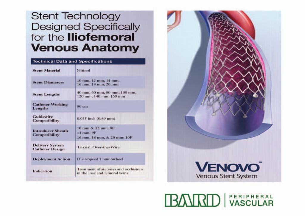
79
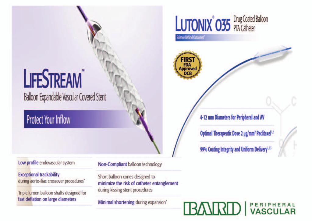
80

81
82
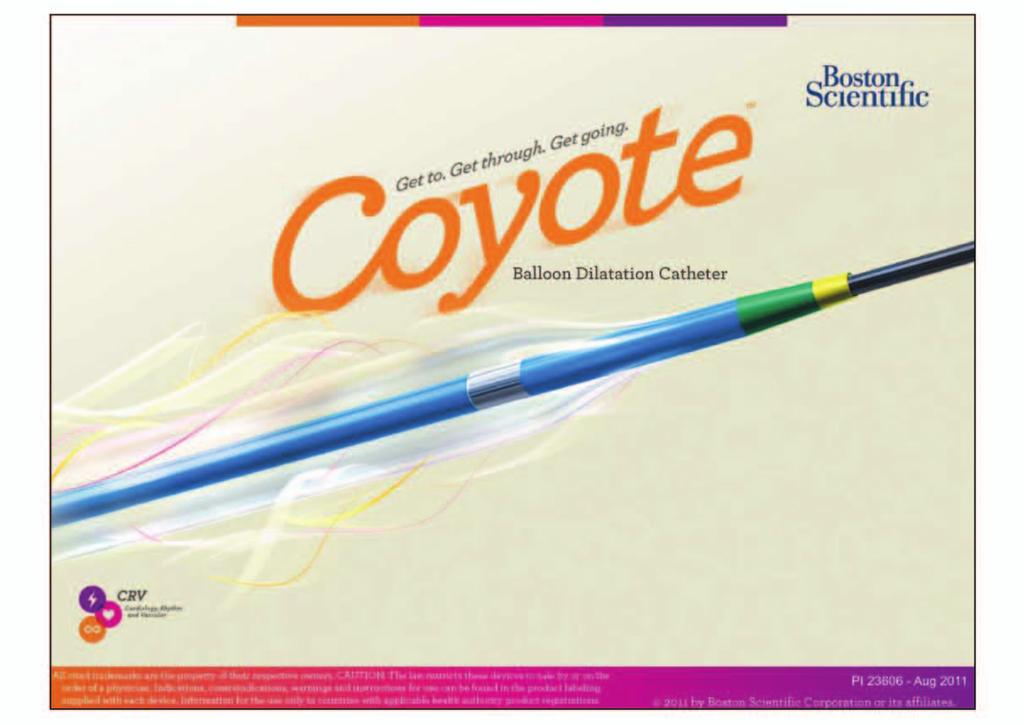
83
84
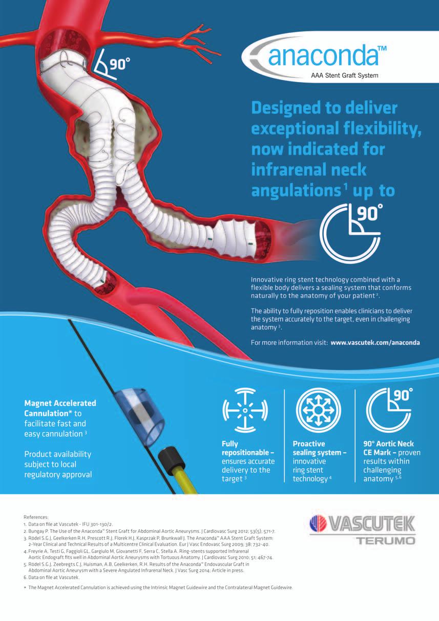
85
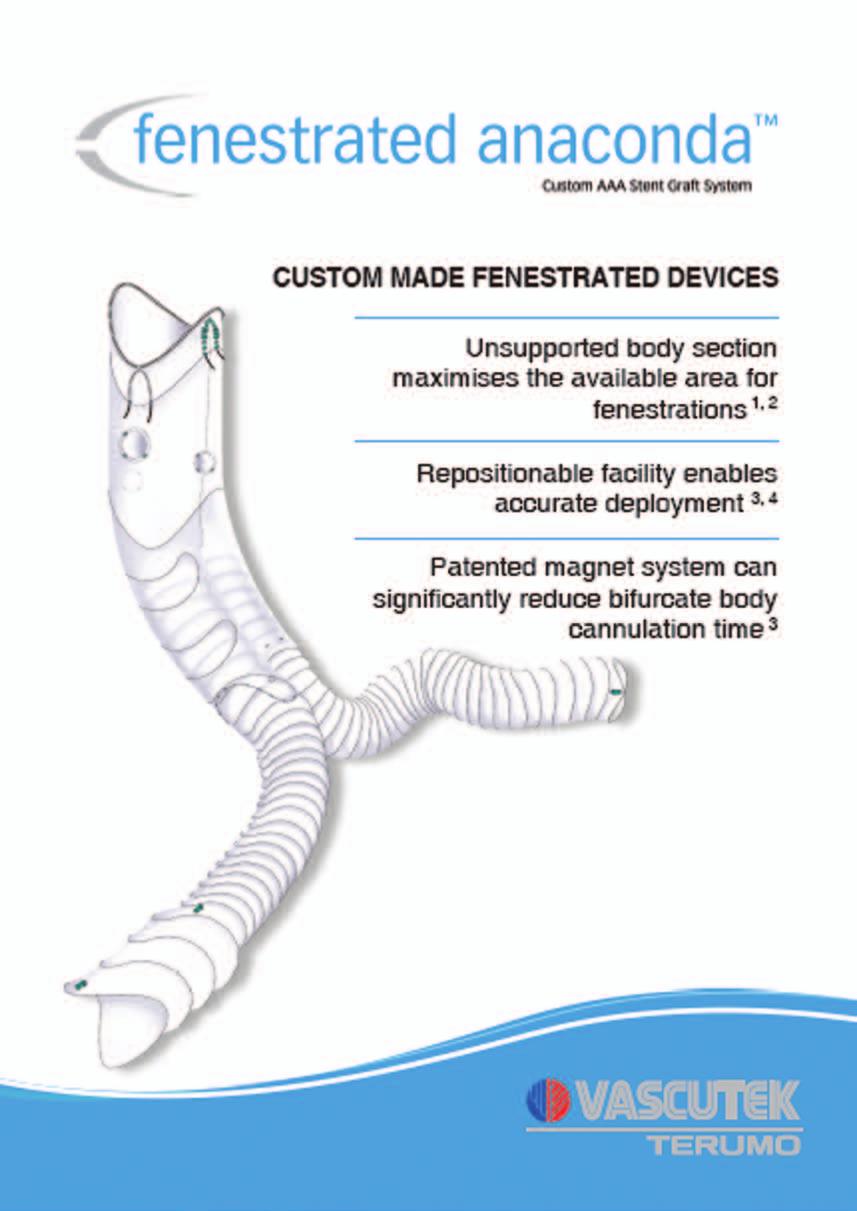
86
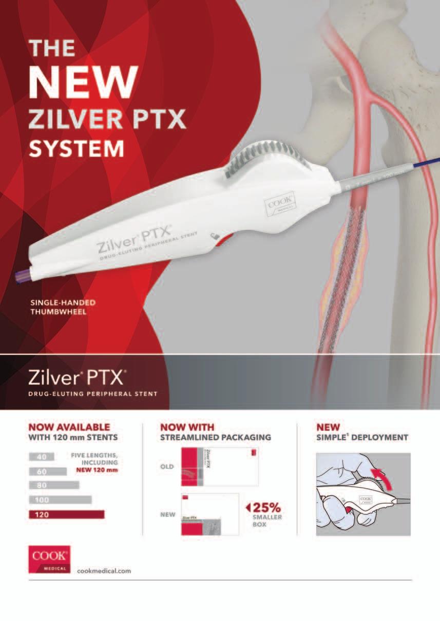
87
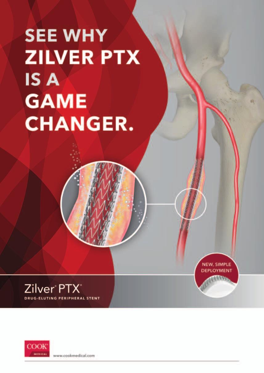
88
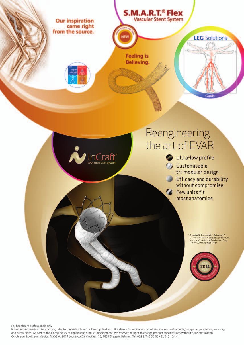
89
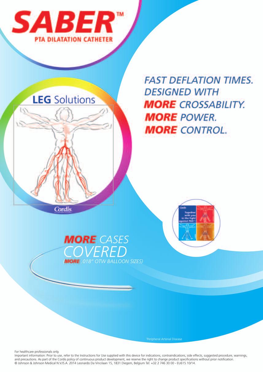
90

91

92
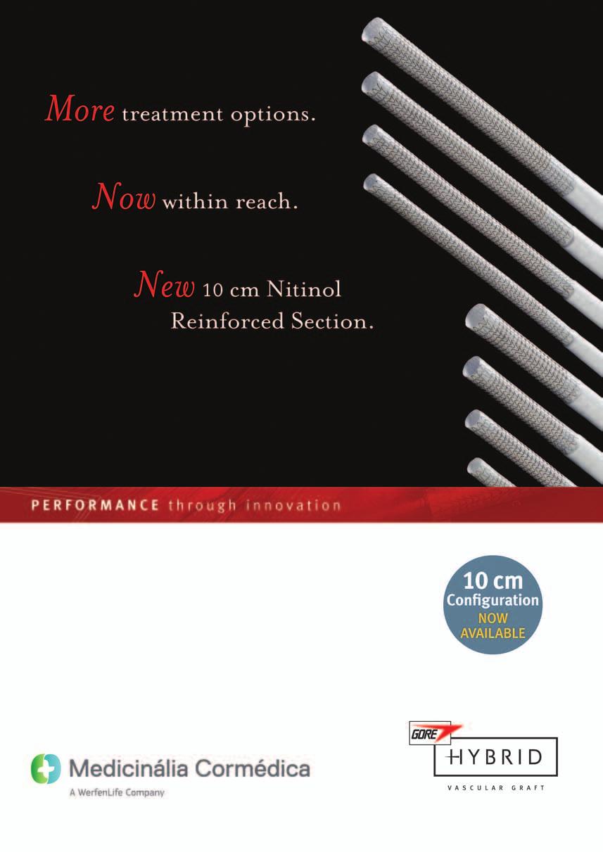
93
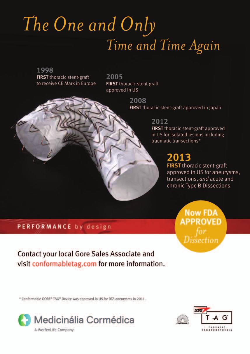
94
95
96

97
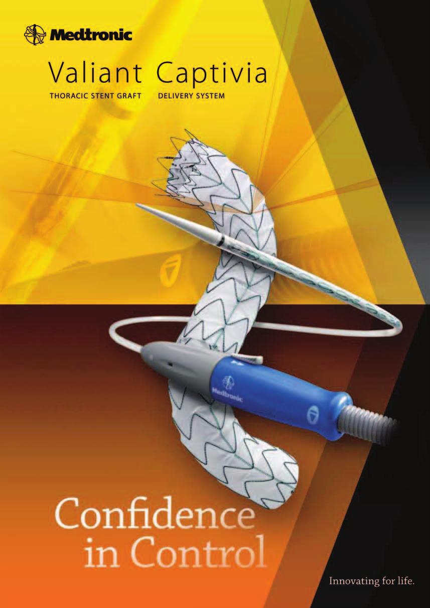
98
SYMPOSIUM PORTO OF APRIL INTERNATIONAL PALÁCIO HOTEL. Live Cases. New challenges, New solutions. Endorsed by.
 INTERNATIONAL SYMPOSIUM XVIIIOF ANGIOLOGY AND VASCULAR SURGERY New challenges, New solutions. 7-8 OF APRIL PORTO2017 PALÁCIO HOTEL Endorsed by www.portovascularsymposium.com XVIII International Symposium
President Dr. José Fernando Teixeira Vice - President Prof. Doutor Sérgio Sampaio
 President Dr. José Fernando Teixeira Vice - President Prof. Doutor Sérgio Sampaio Secretary General Dr. Pedro Henrique Almeida Honorary Presidents Prof. Doutor Roncon de Albuquerque Prof. Doutor António
President Dr. José Fernando Teixeira Vice - President Prof. Doutor Sérgio Sampaio
 President Dr. José Fernando Teixeira Vice - President Prof. Doutor Sérgio Sampaio Secretary General Dr. Pedro Henrique Almeida Honorary Presidents Prof. Doutor Roncon de Albuquerque Prof. Doutor António
Neurological Complications of TEVAR. Frank J Criado, MD. Union Memorial-MedStar Health Baltimore, MD USA
 ISES Online Neurological Complications of Frank J Criado, MD TEVAR Union Memorial-MedStar Health Baltimore, MD USA frank.criado@medstar.net Paraplegia Incidence is 0-4% after surgical Rx of TAAs confined
Challenges. 1. Sizing. 2. Proximal landing zone 3. Distal landing zone 4. Access vessels 5. Spinal cord ischemia 6. Endoleak
 Disclosure I have the following potential conflicts of interest to report: Consulting: Medtronic, Gore Employment in industry Stockholder of a healthcare company Owner of a healthcare company Other(s)
Acute dissections of the descending thoracic aorta (Debakey
 Endovascular Treatment of Acute Descending Thoracic Aortic Dissections Nimesh D. Desai, MD, PhD, and Joseph E. Bavaria, MD Acute dissections of the descending thoracic aorta (Debakey type III or Stanford
Abdominal and thoracic aneurysm repair
 Abdominal and thoracic aneurysm repair William A. Gray MD Director, Endovascular Intervention Cardiovascular Research Foundation Columbia University Medical Center Abdominal Aortic Aneurysm Endografts
CUSTOM-MADE SCALLOPED THORACIC ENDOGRAFTS IN DIFFERENT HOSTILE AORTIC ANATOMIES
 CUSTOM-MADE SCALLOPED THORACIC ENDOGRAFTS IN DIFFERENT HOSTILE AORTIC ANATOMIES A SERIES OF THREE CASE REPORTS Joel Sousa Department of Department of Angiology and Vascular Surgery Hospital S. João, Porto,
Open fenestration for complicated acute aortic B dissection
 Art of Operative Techniques Open fenestration for complicated acute aortic B dissection Santi Trimarchi 1, Sara Segreti 1, Viviana Grassi 1, Chiara Lomazzi 1, Marta Cova 1, Gabriele Piffaretti 2, Vincenzo
Combined Endovascular and Surgical Repair of Thoracoabdominal Aortic Pathology: Hybrid TEVAR
 Combined Endovascular and Surgical Repair of Thoracoabdominal Aortic Pathology: Hybrid TEVAR William J. Quinones-Baldrich MD Professor of Surgery Director UCLA Aortic Center UCLA Medical Center Los Angeles,
Conflicts of Interest. When and Why Complex EVAR in Tx of juxta/suprarenal AAA? Summary. Infrarenal EVAR for short necks 2y postop
 When and Why Complex EVAR in Tx of juxta/suprarenal AAA? Tim Resch MD Vascular Center Skane University Hospital Conflicts of Interest COOK Medical - Consulting, Speakers Bureau, IP, Research support Medtronic
No Disclosure. Aortic Dissection in Japan. This. The Challenge of Acute and Chronic Type B Aortic Dissections with Endovascular Aortic Repair
 No Disclosure The Challenge of Acute and Chronic Type B Aortic Dissections with Endovascular Aortic Repair Toru Kuratani Department of Cardiovascular Surgery Osaka University Graduate School of Medicine,
Toward Total Endovascular Therapy of the Aorta. Adam W. Beck, MD. Associate Professor of Surgery Division of Vascular Surgery and Endovascular Therapy
 Toward Total Endovascular Therapy of the Aorta Adam W. Beck, MD Associate Professor of Surgery Division of Vascular Surgery and Endovascular Therapy University of Alabama at Birmingham Disclosures Grant
Residual Dissection and False Lumen Aneurysm After TEVAR
 WHAT WOULD YOU DO? Residual Dissection and False Lumen Aneurysm After MODERATOR: MARK FARBER, MD PANEL: YAZAN DUWAYRI, MD; MATTHEW J. EAGLETON, MD; WILLIAM D. JORDAN Jr, MD; TILO KÖLBEL, MD, PhD; AND ERIC
Thoracoabdominal Aorta: Advances and Novel Therapies
 Thoracoabdominal Aorta: Advances and Novel Therapies Robert Meisner, MD FACS Sidney Kimmel Medical Center Assistant Professor of Surgery Vascular / Endovascular Surgeon at Lankenau Medical Center November
I-Hui Wu, M.D. Ph.D. Clinical Assistant Professor Cardiovascular Surgical Department National Taiwan University Hospital
 Comparisons of Aortic Remodeling and Outcomes after Endovascular Repair of Acute and Chronic Complicated Type B Aortic Dissections I-Hui Wu, M.D. Ph.D. Clinical Assistant Professor Cardiovascular Surgical
Percutaneous Approaches to Aortic Disease in 2018
 Percutaneous Approaches to Aortic Disease in 2018 Wendy Tsang, MD, SM Assistant Professor, University of Toronto Toronto General Hospital, University Health Network Case 78 year old F Lower CP and upper
Endovascular therapy for Ischemic versus Nonischemic complicated acute type B aortic dissection (catbad).
.") Endovascular therapy for Ischemic versus Nonischemic complicated acute type B aortic dissection (catbad). AS. Eleshra, MD 1, T. Kölbel, MD, PhD 1, F. Rohlffs, MD 1, N. Tsilimparis, MD, PhD 1,2 Ahmed Eleshra
Endovascular therapy for Ischemic versus Nonischemic complicated acute type B aortic dissection (catbad). AS. Eleshra, MD 1, T. Kölbel, MD, PhD 1, F. Rohlffs, MD 1, N. Tsilimparis, MD, PhD 1,2 Ahmed Eleshra
Acute Type B dissection. Closure of the infra diaphragmatic tear: how and when?
 Acute Type B dissection. Closure of the infra diaphragmatic tear: how and when? Prof. Olgierd Rowiński II Department of Clinical Radiology Medical University of Warsaw Disclosure Speaker name: Olgierd
Management of Acute Aortic Syndromes. M. Grabenwoger, MD Dept. of Cardiovascular Surgery Hospital Hietzing, Vienna, Austria
 Management of Acute Aortic Syndromes M. Grabenwoger, MD Dept. of Cardiovascular Surgery Hospital Hietzing, Vienna, Austria I have nothing to disclose. Acute Aortic Syndromes Acute Aortic Dissection Type
Development of a Branched LSA Endograft & Ascending Aorta Endograft
 Development of a Branched LSA Endograft & Ascending Aorta Endograft Frank R. Arko III, MD Sanger Heart & Vascular Institute Carolinas Medical Center Charlotte, North Carolina, USA Disclosures Proximal
FEVAR FIFTEEN YEARS OF EFFICIENCY E.DUCASSE MD PHD FEBVS CHU DE BORDEAUX
 FEVAR FIFTEEN YEARS OF EFFICIENCY E.DUCASSE MD PHD FEBVS CHU DE BORDEAUX 2018 A BIT OF HISTORY First use of F-EVAR : 1990s Park et al. J Vasc Interv Radiol. 1996;7:819-823. Faruqi et al. J Endovasc Surg.
How to manage the left subclavian and left vertebral artery during TEVAR
 How to manage the left subclavian and left vertebral artery during TEVAR Jürg Schmidli Chief of Vascular Surgery Inselspital Hamburg 2017 Dept Cardiovascular Surgery, Bern, Switzerland Disclosure No Disclosures
What is the benefit. of MEP s in BEVAR for TAAA. in preventing paraplegia?
 What is the benefit of MEP s in BEVAR for TAAA in preventing paraplegia? P M Kasprzak Department of Vascular Surgery, Endovascular Surgery University Hospital Regensburg, Germany Disclosures Dr. Kasprzak
Is there a way to predict the risk in uncomplicated Type B aortic dissections? FRANS MOLL University Medical Centre Utrecht - Netherlands
 Is there a way to predict the risk in uncomplicated Type B aortic dissections? FRANS MOLL University Medical Centre Utrecht - Netherlands Disclosures: - Consultant Philips Health Care - Best Doctors Overview
Paraplegia in endovascular repair of TAA and in TEVAR: Incidence, prevention and therapy. Johannes Lammer Medical University Vienna, Austria
 Paraplegia in endovascular repair of TAA and in TEVAR: Incidence, prevention and therapy Johannes Lammer Medical University Vienna, Austria Conflict of interests: none 68y, male, PAU in coral reef aorta,
Optimal repair of acute aortic dissection
 Optimal repair of acute aortic dissection Dept. of Vascular Surgery, The 2nd Xiang-Yale Hospital, Central-South University, China Hunan Major Vessels Diseases Clinical Center Chang Shu Email:changshu01@yahoo.com
COMPLICATIONS OF TEVAR
 COMPLICATIONS OF TEVAR P. Bergeron, A.Petrosyan, F.Markatis, T.Abdulamit, J.-C. Trastour IMAD CONGRESS 2010 Liège Belgium BACKGROUND Stentgrafting is a recognized treatment for TAA & TAD and has been proposed
Understanding the Predictors of Aneurysmal Degeneration in Type B Dissection
 Understanding the Predictors of Aneurysmal Degeneration in Type B Dissection A case example illustrating when early endovascular intervention may provide the best outcome. BY DITTMAR BÖCKLER, MD, PhD;
UC SF. Disclosures. Thoracic Endovascular Aortic Repair 4/24/2009. Management of Acute Dissections: Is There Still a Role for Open Surgery?
 UC SF Management of Acute Dissections: Is There Still a Role for Open Surgery? Darren B. Schneider, M.D. Assistant Professor of Surgery and Radiology Division of Vascular Surgery University of California
Indications for stent grafts in type B aortic dissection
 expert opinion I Akin C A Nienaber Indications for stent grafts in type B aortic dissection ibrahim akin christoph a nienaber Department of Medicine, Divisions of Cardiology, Pulmology and Intensive Care
Endovascular Treatment of the Aorta with Fenestrated and Branched Grafts
 Endovascular Treatment of the Aorta with Fenestrated and Branched Grafts Eric LG Verhoeven,MD, PhD, A. Katsargyris, MD Vascular and Endovascular Surgery, Paracelsus Medical University, Nuremberg, Germany
Redo treatment and open conversion after TEVAR
 Redo treatment and open conversion after TEVAR Roberto Chiesa Vascular Surgery, Vita-Salute University Scientific Institute San Raffaele Milan, Italy Number of procedures Off-Label indications for TEVAR
EVAR and TEVAR: Extending Their Use for Rupture and Traumatic Injury. Conflict of Interest. Hypotensive shock 5/5/2014. none
 EVAR and TEVAR: Extending Their Use for Rupture and Traumatic Injury Bruce H. Gray, DO MSVM FSCAI Professor of Surgery/Vascular Medicine USC SOM-Greenville Greenville, South Carolina none Conflict of Interest
Endoleaks after F-BEVAR How to Assess & Treat? Gustavo S. Oderich, MD Mayo Clinic Rochester, MN
 Endoleaks after F-BEVAR How to Assess & Treat? Gustavo S. Oderich, MD Mayo Clinic Rochester, MN FACULTY DISCLOSURE Gustavo S. Oderich MD Consulting, DSMB, CEC* Cook Medical Inc., WL Gore, Lombardi Honoraria
Jean M Panneton, MD Professor of Surgery Program Director Vascular Surgery Chief EVMS. Arch Pathology: The Endovascular Era is here
 Jean M Panneton, MD Professor of Surgery Program Director Vascular Surgery Chief EVMS Arch Pathology: The Endovascular Era is here Disclosures Consultant: Cook Medical, Bolton Medical, Medtronic Inc, Volcano,
Follow-up of Aortic Dissection: How, How Often, Which Consequences Euro Echo 2011
 Follow-up of Aortic Dissection: How, How Often, Which Consequences Euro Echo 2011 Susan E. Wiegers, MD, FASE Director of Clinical Echocardiography Hospital of the University of Pennsylvania Disclosure
Kopp R, Puippe G, Rancic Z, Hofmann M, Pecoraro F, Pfammatter T, Lachat M.. University Hospital Zurich, Switzerland
 Low risk of spinal cord ischemia after endovascular repair for suprarenal and thoracoabdominal aortic aneurysms using parallel stent graft implantation. Kopp R, Puippe G, Rancic Z, Hofmann M, Pecoraro
THORACOABDOMINAL AORTIC ANEURYSMS HYBRID REPAIR
 Update on Open and Endovascular Therapeutic Option for Aortic Repair CENTRE CARDIO-TORACIQUE DE MONACO Friday November 7 th, 2014 THORACOABDOMINAL AORTIC ANEURYSMS HYBRID REPAIR Roberto Chiesa Vascular
Malperfusion Syndromes Type B Aortic Dissection with Malperfusion
 Malperfusion Syndromes Type B Aortic Dissection with Malperfusion Jade S. Hiramoto, MD, MAS April 27, 2012 Associated with early mortality Occurs when there is end organ ischemia secondary to aortic branch
What is the best treatment for False Lumen growth after type B Dissection
 What is the best treatment for False Lumen growth after type B Dissection Nikolaos Tsilimparis, Fiona Rohlffs, Yuk Law, Sebastian Debus, Tilo Kölbel N. Tsilimparis, MD, PhD, FEBVS Deputy Head of German
Abdominal Aortic Aneurysms. A Surgeons Perspective Dr. Derek D. Muehrcke
 Abdominal Aortic Aneurysms A Surgeons Perspective Dr. Derek D. Muehrcke Aneurysm Definition The abnormal enlargement or bulging of an artery caused by an injury or weakness in the blood vessel wall A localized
Do the Data Support Endovascular Therapy for Descending Thoracic AD? Woong Chol Kang, M.D.
 Do the Data Support Endovascular Therapy for Descending Thoracic AD? Woong Chol Kang, M.D. Gil Hospital, Gachon University Incheon, Korea Classification of AD Acute vs. Chronic (2weeks) (IIIa, b) type
Experience of endovascular procedures on abdominal and thoracic aorta in CA region
 Experience of endovascular procedures on abdominal and thoracic aorta in CA region May 14-15, 2015, Dubai Dr. Viktor Zemlyanskiy National Research Center of Emergency Care Astana, Kazakhstan Region Characteristics
Performance of the conformable GORE TAG device in Type B aortic dissection from the GORE GREAT real world registry
 University of Milan Thoracic Aortic Research Center Performance of the conformable GORE TAG device in Type B aortic dissection from the GORE GREAT real world registry Santi Trimarchi, MD, PhD Associate
Aggressive Resection/Reconstruction of the Aortic Arch in Type A Dissection
 Aggressive Resection/Reconstruction of the Aortic Arch in Type A Dissection M. Grabenwoger Dept. of Cardiovascular Surgery Hospital Hietzing Vienna, Austria Disclosure Statement Consultant of Jotec, Hechingen,
The Petticoat Technique Managing Type B Dissection with both Early and Long Term Considerations
 The Petticoat Technique Managing Type B Dissection with both Early and Long Term Considerations Joseph V. Lombardi, MD Professor & Chief, Division of Vascular & Endovascular Surgery Department of Surgery,
Description. Section: Surgery Effective Date: April 15, Subsection: Surgery Original Policy Date: December 6, 2012 Subject:
 Last Review Status/Date: March 2015 Page: 1 of 6 Description Wireless sensors implanted in an aortic aneurysm sac after endovascular repair are being investigated to measure post procedural pressure. It
TEVAR for Chronic dissections: indications for TEVAR, long term results
 TEVAR for Chronic dissections: indications for TEVAR, long term results J Sobocinski, R Azzaoui, B Maurel, R Spear, T Martin-Gonzalez, A Hertault, S Haulon Centre de l Aorte, Chirurgie vasculaire, Hôpital
Nellix Endovascular System: Clinical Outcomes and Device Overview
 Nellix Endovascular System: Clinical Outcomes and Device Overview Jeffrey P. Carpenter, MD Professor and Chief, Department of Surgery CAUTION: Investigational device. This product is under clinical investigation
Treatment of acute type B aortic dissection: Current status
 MEET Cannes, 18. - 21.06.2009 Treatment of acute type B aortic dissection: Current status Christoph A. Nienaber, MD, FACC University of Rostock Department of Internal Medicine, Cardiology christoph.nienaber@med.uni-rostock.de
Recommendations for Follow-up After Vascular Surgery Arterial Procedures SVS Practice Guidelines
 Recommendations for Follow-up After Vascular Surgery Arterial Procedures 2018 SVS Practice Guidelines vsweb.org/svsguidelines About the guidelines Published in the July 2018 issue of Journal of Vascular
How to select FEVAR versus EVAR + endoanchors in short-necked AAAs
 How to select FEVAR versus EVAR + endoanchors in short-necked AAAs Jean-Paul P.M. de Vries, Richte C.L. Schuurmann St. Antonius Hospital Nieuwegein, The Netherlands 21st Critical Issues Congress Nuernberg,
Chungbuk Regional Cardiovascular Center, Division of Cardiology, Departments of Internal Medicine, Chungbuk National University Hospital Sangmin Kim
 Endovascular Procedures for Isolated Common Iliac and Internal Iliac Aneurysm Chungbuk Regional Cardiovascular Center, Division of Cardiology, Departments of Internal Medicine, Chungbuk National University
Are stent-grafts for acute type B dissection durable? Est-ce que les stents graft pour la dissection aigue de type B sont efficaces à moyen terme?
 Are stent-grafts for acute type B dissection durable? Est-ce que les stents graft pour la dissection aigue de type B sont efficaces à moyen terme? Martin Björck, Johnny Steuer, Anders Wanhainen Uppsala
Asymptomatic Radiology / Clinical data Report / Cohort bias Referral bias. UCSF Vascular Symposium April 7-9, Acute Aortic Dissection
 Aortic Dissection: Natural History What is the Natural History of Aortic Dissection? UCSF Vascular Symposium April 7-9, 2011 Asymptomatic Radiology / Clinical data Report / Cohort bias Referral bias Stephen
Anatomical challenges in EVAR
 Anatomical challenges in EVAR M.H. EL DESSOKI, MD,FRCS PROFESSOR OF VASCULAR SURGERY CAIRO UNIVERSITY Disclosure Speaker name:... I have the following potential conflicts of interest to report: Consulting
Current treatment of Aortic Aneurysms and Dissections. Adam Keefer, MD, FACS Sean Hislop, MD, FACS
 Current treatment of Aortic Aneurysms and Dissections Adam Keefer, MD, FACS Sean Hislop, MD, FACS Patient 1 69 year old well-educated man with reoccurring pain in his upper abdomen and a pulsatile mass.
Endovascular Management of Thoracic Aortic Pathology Stéphan Haulon, J Sobocinski, B Maurel, T Martin-Gonzalez, R Spear, A Hertault, R Azzaoui
 Endovascular Management of Thoracic Aortic Pathology Stéphan Haulon, J Sobocinski, B Maurel, T Martin-Gonzalez, R Spear, A Hertault, R Azzaoui Aortic Center, Lille University Hospital, France Disclosures
Surgical Considerations of TEVAR
 Surgical Considerations of TEVAR University of Alberta, June 14 th, 2013 Jehangir Appoo Libin Cardiovascular Institute University of Calgary, Today: Lesions/Pathology amenable to TEVAR On label Off label
Aortic Center of Excellence at Sentara
 Gordon K. Stokes, MD, FACS Eastern Virginia Medical School Sentara Vascular Specialists Norfolk, VA Aortic Center of Excellence at Sentara April 25, 2014 Disclosures I have no disclosures relevant to this
Management of Endoleaks
 Management of Endoleaks Sarah Ikponmwosa, MD Brooklyn VA 6/20/08 Questions Advantages of endovascular repair Definition of an endoleak Types of endoleaks Management of type lll endoleak Diagnosis of type
AORTIC DISSECTIONS Current Management. TOMAS D. MARTIN, MD, LAT Professor, TCV Surgery Director UF Health Aortic Disease Center University of Florida
 AORTIC DISSECTIONS Current Management TOMAS D. MARTIN, MD, LAT Professor, TCV Surgery Director UF Health Aortic Disease Center University of Florida DISCLOSURES Terumo Medtronic Cook Edwards Cryolife AORTIC
Talent Abdominal Stent Graft
 Talent Abdominal with THE Xcelerant Hydro Delivery System Expanding the Indications for EVAR Treat More Patients Short Necks The Talent Abdominal is the only FDA-approved device for proximal aortic neck
My personal experience with INCRAFT in standard and challenging cases
 My personal experience with INCRAFT in standard and challenging cases G Pratesi, MD Vascular Surgery University of Rome Tor Vergata giovanni.pratesi@uniroma2.it Disclosure Speaker name: Giovanni Pratesi,
Taming The Aorta. David Minion, MD Program Director, Vascular Surgery University of Kentucky Medical Center Lexington, Kentucky, USA
 Taming The Aorta David Minion, MD Program Director, Vascular Surgery University of Kentucky Medical Center Lexington, Kentucky, USA Faculty Disclosure Consulting: Endologix, Cook 1 Objectives Review the
Hybrid Repair of a Complex Thoracoabdominal Aortic Aneurysm
 Hybrid Repair of a Complex Thoracoabdominal Aortic Aneurysm Virendra I. Patel MD MPH Assistant Professor of Surgery Massachusetts General Hospital Division of Vascular and Endovascular Surgery Disclosure
Treatment of Thoracoabdominal Aneurysms Is there a need for custom-made devices?
 : FETURED TECHNOLOGY: JOTEC E-XTR DESIGN ENGINEERING Treatment of Thoracoabdominal neurysms Is there a need for custom-made devices? INTERVIEW ND CSE PRESENTTIONS WITH DNIEL RNZN, MD, ND NDREJ SCHMIDT,
Intravascular Ultrasound in the Treatment of Complex Aortic Pathologies. Naixin Kang, M.D. Vascular Surgery Fellow April 26 th, 2018
 Intravascular Ultrasound in the Treatment of Complex Aortic Pathologies Naixin Kang, M.D. Vascular Surgery Fellow April 26 th, 2018 DISCLOSURES Nothing To Disclose 2 ENDOVASCULAR AORTIC INTERVENTION Improved
Title. Different arch branched devices are available, is morphology the. main criteria of choice? Ciro Ferrer, MD
 Different arch branched devices are available, is morphology the Title main criteria of choice? Ciro Ferrer, MD Vascular Surgery Unit Sapienza University of Rome Disclosure Speaker name: Ciro Ferrer Proctoring/speaking
Santi Trimarchi, MD, PhD Vascular Surgeon Thoracic Aortic Research Center, Director IRCCS Policlinico San Donato University of Milan
 The Gore GREAT Registry: Update about a real life data collection Santi Trimarchi, MD, PhD Vascular Surgeon Thoracic Aortic Research Center, Director IRCCS Policlinico San Donato University of Milan Disclosures
Daniela Branzan MD, Department of Vascular Surgery and Department of Interventional Angiology University Hospital Leipzig
 Ischemic Preconditioning with Minimally Invasive Segmental Artery Coil Embolization (MISACE) prior to Endovascular TAAA Repair: Clinical Experience in 50+ Patients Daniela Branzan MD, Department of Vascular
How to achieve a successful proximal sealing in TEVAR? Pr L Canaud
 How to achieve a successful proximal sealing in TEVAR? Pr L Canaud CHU de Montpellier France Disclosure I have the following potential conflicts of interest to report: Consulting: Medtronic. Proximal neck
The role of false lumen intervention to promote remodelling via induced thrombosis the FLIRT concept
 The role of false lumen intervention to promote remodelling via induced thrombosis the FLIRT concept Professor Christoph A. Nienaber The Royal Brompton and Harefield NHS Trust Cardiology and Aortic Centre
Reinhard Kopp, Karin Pfister, Beatrix Cucuruz, Konstantinos Gallis, Piotr M Kasprzak
 Immediate, delayed and late spinal cord ischemia after extended endovascular thoracoabdominal aortic repair Reinhard Kopp, Karin Pfister, Beatrix Cucuruz, Konstantinos Gallis, Piotr M Kasprzak Disclosure
Treatment options of late failures of EVAS. Michel Reijnen Rijnstate Arnhem The Netherlands
 Treatment options of late failures of EVAS Michel Reijnen Rijnstate Arnhem The Netherlands Disclosure Speaker name: Michel Reijnen I have the following potential conflicts of interest to report: Consulting
Percutaneous Axillary Artery Access For Branch Grafting for complex TAAAs and pararenal AAAs: How to do it safely
 Percutaneous Axillary Artery Access For Branch Grafting for complex TAAAs and pararenal AAAs: How to do it safely Daniela Branzan, MD, Department of Vascular Surgery University Hospital Leipzig Disclosure
Patient selection in Hostile Necks and how. to prevent endoleaks a word of caution
 Patient selection in Hostile Necks and how to prevent endoleaks a word of caution P M Kasprzak, K. Pfister Department of Vascular Surgery Endovascular Surgery University Hospital Regensburg, Germany Conflict
Considerations for a Durable Repair
 Considerations for a Durable Repair Eric Verhoeven, MD, PhD, A. Katsargyris, MD Department of Vascular and Endovascular Surgery, Paracelsus Medical University, Nuremberg, Germany Disclosures William Cook
Technique and Outcome of Laser Fenestration For Arch Vessels
 Technique and Outcome of Laser Fenestration For Arch Vessels Jean M. Panneton MD, FRCSC, FACS Professor of Surgery Chief & Program Director Division of Vascular Surgery Eastern Virginia Medical School
Hostile Neck During EVAR, The Role Of Endoanchores
 Hostile Neck During EVAR, The Role Of Endoanchores Samer Koussayer, MD, FACS, RVT Prof, Al Faisal University Section Head and consultant Vascular & Endovascular Surgery Division King Faisal Specialist
COMBINED TECHNIQUE CHIMNEY + FENESTRATED ENDOGRAFT FOR COMPLEX ANEURYSMS ERIC DUCASSE - MD PHD FEVBS CHU BORDEAUX
 COMBINED TECHNIQUE CHIMNEY + FENESTRATED ENDOGRAFT FOR COMPLEX ANEURYSMS ERIC DUCASSE - MD PHD FEVBS CHU BORDEAUX DISCLOSURE SPEAKER NAME: PR. E. DUCASSE I HAVE THE FOLLOWING POTENTIAL CONFLICTS OF INTEREST
Aortic Arch pathology options: Open,Hybrid, fenestration, Chimney or branched stent-graft?
 Aortic Arch pathology options: Open,Hybrid, fenestration, Chimney or branched stent-graft? Chang Shu, M.D., Ph.D Vascular Surgery Center National Center for Cardiovascular Diseases. Fuwai Hospital, CAMS
Endovascular Repair o Abdominal. Aortic Aneurysms. Cesar E. Mendoza, M.D. Jackson Memorial Hospital Miami, Florida
 Endovascular Repair o Abdominal Aortic Aneurysms Cesar E. Mendoza, M.D. Jackson Memorial Hospital Miami, Florida Disclosure Nothing to disclose. 2 Mr. X AAA Mr. X. Is a 70 year old male who presented to
State of Art Hybrid Approach
 State of Art Hybrid Approach for Complex Aorta Diseases Won Ho Kim, MD Division of Cardiology, Eulji University Hospital Eulji University School of Medicine, Daejeon, Korea Introduction.Hybrid procedure
MODERN METHODS FOR TREATING ABDOMINAL ANEURYSMS AND THORACIC AORTIC DISEASE
 MODERN METHODS FOR TREATING ABDOMINAL ANEURYSMS AND THORACIC AORTIC DISEASE AAA FACTS 200,000 New Cases Each Year Ruptured AAA = 15,000 Deaths per Year in U.S. 13th Leading Cause of Death 80% Chance of
An Overview of Post-EVAR Endoleaks: Imaging Findings and Management. Ravi Shergill BSc Sean A. Kennedy MD Mark O. Baerlocher MD FRCPC
 An Overview of Post-EVAR Endoleaks: Imaging Findings and Management Ravi Shergill BSc Sean A. Kennedy MD Mark O. Baerlocher MD FRCPC Disclosure Slide Mark O. Baerlocher: Current: Consultant for Boston
Bilateral use of the Gore IBE device for bilateral CIA aneurysms and a first interim analysis of the prospective Iceberg registry
 Bilateral use of the Gore IBE device for bilateral CIA aneurysms and a first interim analysis of the prospective Iceberg registry Michel MPJ Reijnen, MD, PhD Department of Vascular Surgery, Rijnstate Hospital
DISCLOSURES ISOLATED DTA LESION? TYPE B DISSECTIONS TREATMENT OPTIONS
 Endovascular Repair of Aortic Arch Pathologies; What is available/possible in the U.S. in 2018? Kaiser Permanente Endovascular Symposium 6/2/18 Sukgu M Han, MD, MS Assistant Professor of Clinical Surgery
EndoVascular Aneurysm Sealing (EVAS) with Nellix
 with Nellix") 1 2 EndoVascular Aneurysm Sealing (EVAS) with Nellix Designed to seal entire aneurysm with contained biostable polymer Non-modular design with complete fixation Expands endovascular patient eligibility
1 2 EndoVascular Aneurysm Sealing (EVAS) with Nellix Designed to seal entire aneurysm with contained biostable polymer Non-modular design with complete fixation Expands endovascular patient eligibility
Accepted Manuscript. Is A More Extensive Operation Justified for Acute Type A Dissection Repair? Dr. Leonard N. Girardi
 Accepted Manuscript Is A More Extensive Operation Justified for Acute Type A Dissection Repair? Dr. Leonard N. Girardi PII: S0022-5223(18)32552-2 DOI: 10.1016/j.jtcvs.2018.09.048 Reference: YMTC 13502
When to use standard EVAR with EndoAnchors or CHEVAR in short-neck AAAs LINC ASIA 18
 When to use standard EVAR with EndoAnchors or CHEVAR in short-neck AAAs JEAN-PAUL P.M. DE VRIES, DIRECTOR OF VASCULAR SURGERY ST.ANTONIUS HOSPITAL NIEUWEGEIN, THE NETHERLANDS. LINC ASIA-PACIFIC HongKong,
CHALLENGING ILIAC ACCESSES AND THROMBOSIS PREVENTION
 CHALLENGING ILIAC ACCESSES AND THROMBOSIS PREVENTION ARMANDO MANSILHA MD, PhD, FEBVS UNIVERSITY HOSPITAL - PORTO Disclosure of Interest Speaker name: ARMANDO MANSILHA I have the following potential conflicts
Off-the-Shelf Devices. Mark A Farber, MD FACS Director, Aortic Center Professor of Radiology and Surgery University of North Carolina
 Off-the-Shelf Devices Mark A Farber, MD FACS Director, Aortic Center Professor of Radiology and Surgery University of North Carolina Disclosures Cook - Research Support, Consultant, Clinical Trials WL
Disclosures. EVAR follow-up: actual recommendation. EVAR follow-up: critical issues
 Disclosures is it time to discuss individualized follow-up schemes based on preoperative anatomy and high quality completion angiography? Consultant / Speaker / Proctor Cook Cordis Medtronic Invatec W.L.
Subclavian Artery Plug Embolization (SAPE study): a real experience about endovascular subclavian occlusion prior to thoracic vascular repair
: a real experience about endovascular subclavian occlusion prior to thoracic vascular repair") Subclavian Artery Plug Embolization (SAPE study): a real experience about endovascular subclavian occlusion prior to thoracic vascular repair Simone Salvati, Luca Bertoglio, Alessandra Fittipaldi, Andrea
Subclavian Artery Plug Embolization (SAPE study): a real experience about endovascular subclavian occlusion prior to thoracic vascular repair Simone Salvati, Luca Bertoglio, Alessandra Fittipaldi, Andrea
Conflicts of Interest. Endovascular Repair of Thoracoabdominal Aneurysm. Overview PLANNING ANATOMIC CONSIDERATIONS FOR COMPLEX AORTIC REPAIR
 Endovascular Repair of Thoracoabdominal Aneurysm Tim Resch MD Björn Sonesson MD, Nuno Dias MD Vascular Center Skane University Hospital Conflicts of Interest COOK Medical - Consulting, Speakers Bureau,
Optimizing Accuracy of Aortic Stent Grafts in Short Necks
 Optimizing Accuracy of Aortic Stent Grafts in Short Necks Venkatesh Ramaiah, MD, FACS Medical Director Arizona Heart Hospital Director Peripheral Vascular and Endovascular Research Arizona Heart Institute
Techniques For Endovascular Aortic Arch Repair. Mario Lachat, MD University Hospital of Zurich Matteo Orrico, MD San Camillo-Forlanini Hospital, Rome
 Techniques For Endovascular Aortic Arch Repair Mario Lachat, MD University Hospital of Zurich Matteo Orrico, MD San Camillo-Forlanini Hospital, Rome Disclosure Speaker name: Mario Lachat Endospan, Jotec,
Current strategies to prevent spinal cord ischemia in TAAA repair
 Current strategies to prevent spinal cord ischemia in TAAA repair Geert Willem Schurink Barend Mees Noud Peppelenbosch Michiel de Haan Michael Jacobs Maastricht University Medical Center, the Netherlands