3D- and Multidimensional Echocardiography in Aortic Valve Repair
|
|
|
- Douglas Hines
- 6 years ago
- Views:
Transcription

 - Hagendorff - 3D- and")
1 Reconstruction of the Aortic Valve and Root - A Practical Approach - 3D- and Multidimensional Echocardiography Thursday 15 th September Practice must always be founded on sound theory. Leonardo Da Vinci Universitätsmedizin Leipzig (2016) - Hagendorff - 3D- and Multidimensional Echocardiography 1
2 I declare for the last 3 years and the subsequent 12 month the following conflicts of interests: Section I: Support for Research Activities - grant of the DEGUM - no other financial research support Section II: Support for Educational Activities - MIFO, GE Healthcare, Astra Zeneca, Servier, Novartis, Berlin-Chemie, Pfizer, Cardiac Dimension, Abbott, Bayer, Kelcon Section III: Honorarium for Promotional Activities - none Section IV: Personal Financial Interests in Vommercial Activities - none IB1; 2A11 Member of the German Society of Cardiology, The German Society of Ultrasound, the German Society of Internal Medicine and the European Society of Cardiology/Cardiovascular Imaging Councillor of the EACVI Board Universitätsmedizin Leipzig (2016) - Hagendorff - 3D- and Multidimensional Echocardiography 2
![Circulation 2007; 116 [suppl I]: I264 I269 The representation of imaging the aortic](/docs-images/75/72499805/images/3-0.jpg "valve and aortic root by echocardiography in the literature.")

3 Circulation 2007; 116 [suppl I]: I264 I269 The representation of imaging the aortic valve and aortic root by echocardiography in the literature. This figure (2007) does not represent the actual standard of echocardiography in the present days. Universitätsmedizin Leipzig (2016) - Hagendorff - 3D- and Multidimensional Echocardiography 3


4 All figures in this paper of 2012 are still only 2D-echo images. Universitätsmedizin Leipzig (2016) - Hagendorff - 3D- and Multidimensional Echocardiography 4


 - Hagendorff - 3D- and")
5 J Am Coll Cardiol Imag 2013; 6: Multiple other figures are CT and angio images. In 2013 only this 3D image is found in this standard imaging paper. Universitätsmedizin Leipzig (2016) - Hagendorff - 3D- and Multidimensional Echocardiography 5


 -")
6 Lang RM, Badano LP, Tsang W et al., Eur Heart J 2012; 13: 1-46 At least, in 2013 the 3D- echo technique for the aortic valve is fixed in the echo recommendations. Universitätsmedizin Leipzig (2016) - Hagendorff - 3D- and Multidimensional Echocardiography 6
7 3D/4D-Echocardiography The key questions and challenges of echocardiography in aortic regurgitation regardless wethter or not only 2D or 3D echocardiography is used. 1. The correct diagnosis of aortic regurgitation 2. The complete - convincing and objective (and at least comprehensable and replicable) - documentation of the findings Target parameter: the regurgitant fraction and/or efective regurgitant orrifice area in aortic valve regurgitation Morphological findings: diameter of aortic annulus, root, sinutubular junction and ascending aorta, geometry of the cardiac cavities, especially the left ventricle Functional parameter: stroke volume, regurgitant volume, E/E`, spap, 3. Additional important findings Number of cusps, calcification of cusps, fusion of cusps, orientation of the commissures Calcification of the aortic root Universitätsmedizin Leipzig (2016) - Hagendorff - 3D- and Multidimensional Echocardiography 7


8 The visualiztion of the aortic valve in sectional planes is important and excellent with modern ultrasound systems. What is the added value of 3D-echocardiography? normal = tricuspid unicuspid bicuspid quadricuspid Universitätsmedizin Leipzig (2016) - Hagendorff - 3D- and Multidimensional Echocardiography 8
9 Transthoracic and transesophageal echocardiography: special practical aspects of 3D-imaging TTE is a more challenging task than TEE regarding the technical skill. In TTE frequencies are lower than TTE, thus spatial resolution is more limited. The higher the frequencies, the better the axial spatial resolution. The higher the frequencies, the less the penetration. Lateral resolution is affected by the frequency as well as by the band width of the transducer normally in the higher regions of frequencies, but not at the highest the lateral resolution is the best. Why these informations? Of course to get the best image quality - and at least, to get the best rendering in postprocessing. If contours are not excellent, no valid postprocession is possible. TEE after aortic valve repair: stitched data set Universitätsmedizin Leipzig (2016) - Hagendorff - 3D- and Multidimensional Echocardiography 9


 - Hagendorff - 3D- and")
10 Excellent image quality is the prerequisite for the correct diagnosis and the decision making due to the imaging pre-interventional procedures All parameters of aortic valve and aortic root dimensions, especially the distance between the aortic cusps / the aortic annulus and the coronary ostia can be easily measured within a 3D-TEE data set. Universitätsmedizin Leipzig (2016) - Hagendorff - 3D- and Multidimensional Echocardiography 10


11 Prerquisite for excellent image quality in 2D as well as 3D echocardiography: knowledge about ultrasound physics and implementation of these aspects into the workflow by just technical knowledge about the buttons. and in case of interventions and surgery training for a fast workflow. Then, detailed information about aortic valve and aortic root morphology is possible. The spatial and temporal resolution of 3D TEE is at least comparable to cardiac-ct. The same patients: bad settings versus optimized settings in 2D and 3D-TEE. Universitätsmedizin Leipzig (2016) - Hagendorff - 3D- and Multidimensional Echocardiography 11


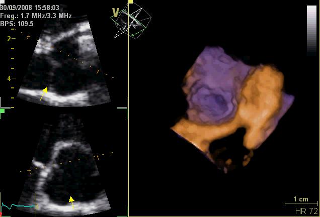
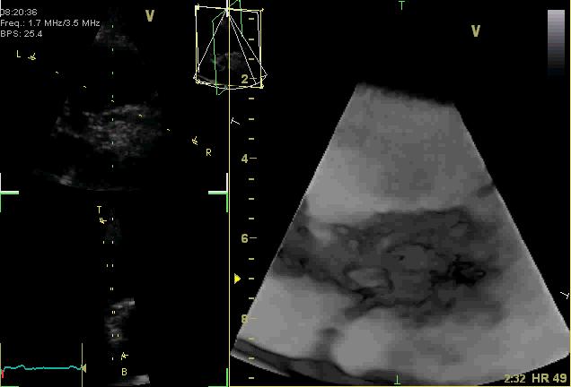
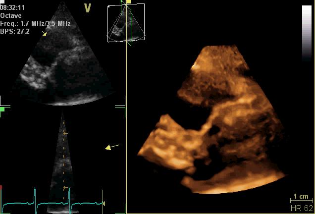
12 3D4D-TTE can be very helpful and even sometimes better and sufficient in comparison to 3D TEE. old examples of 2005 with old machines The additional value of information is obvious. Surface imaging is a complete new modality than sectional scanning. Universitätsmedizin Leipzig (2016) - Hagendorff - 3D- and Multidimensional Echocardiography 12

 - Hagendorff - 3D- and Multidimensional")
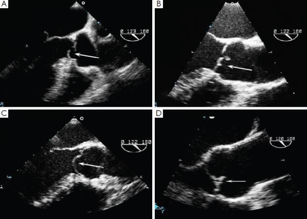
13 Example of 2D-imaging Aortic regurgitation type Ia FAA functional aortic annulus; STJ - sinotubular junction; SCA - subcommissural anuloplasty Universitätsmedizin Leipzig (2016) - Hagendorff - 3D- and Multidimensional Echocardiography 13

 Visualisation")


14 2D-TTE-imaging (triplane acquisition a nd deformation imaging) Visualisation of the aortic aneurysm funcional AR-anaylsis cw-spectrum of AR pw-spectrum of subclavian flow Universitätsmedizin Leipzig (2016) - Hagendorff - 3D- and Multidimensional Echocardiography 14


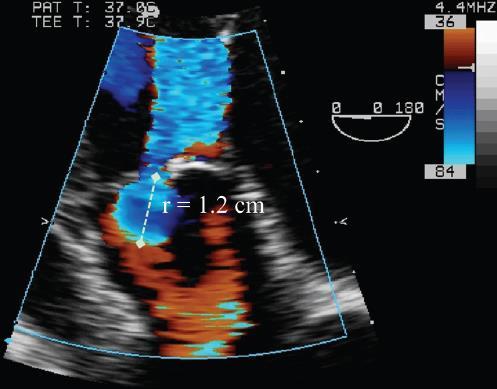
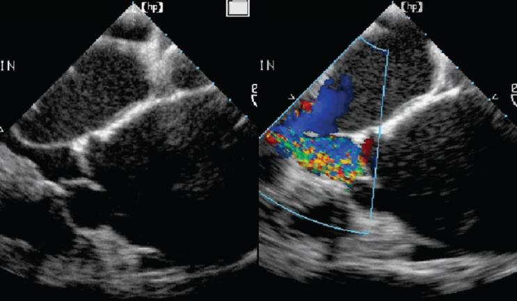
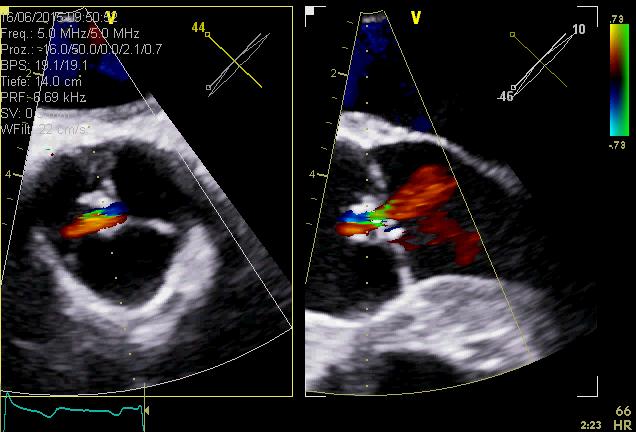
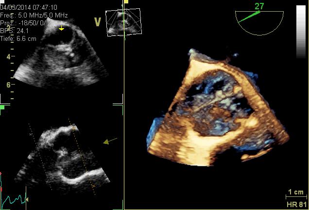
15 The added value of 3D-TTE-imaging imaging of regurgitant flow; surface morphology of aortiv valve and root Universitätsmedizin Leipzig (2016) - Hagendorff - 3D- and Multidimensional Echocardiography 15
 - Hagendorff - 3D- and Multidimensional Echocardiography")
16 pre 3D- and Multidimensional Echocardiography pre post Comparison pre- and post-aortiv valve repair surgery post post post Universitätsmedizin Leipzig (2016) - Hagendorff - 3D- and Multidimensional Echocardiography 16


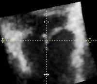
17 The added value of 3D-TTE-imaging Volume measurement by 3D-imaging - There are still problems, which are debatable. LVEDV > 400ml LVESV > 180ml SV total > 220ml Meaasurements using 2Dtechniques in the presence of brilliant image quality. What are the results of the 3D volume measurements? Universitätsmedizin Leipzig (2016) - Hagendorff - 3D- and Multidimensional Echocardiography 17


 - Hagendorff -")
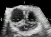
18 The added value of 3D-TTE-imaging Volume measurement by 3D-imaging only in data sets with optimal image quality Excellent image quality in 2D-images. Endocardial contour detection is especially in this example excellent. Universitätsmedizin Leipzig (2016) - Hagendorff - 3D- and Multidimensional Echocardiography 18


19 Important message: 1. No 3D echocardiography was used for LV volume assessment. 2. VARC-criteria for grading of the AR Universitätsmedizin Leipzig (2016) - Hagendorff - 3D- and Multidimensional Echocardiography 19


20 Again no 3D echocardiography was used for LV volume assessment. Universitätsmedizin Leipzig (2016) - Hagendorff - 3D- and Multidimensional Echocardiography 20


 -")

21 Again no 3D echocardiography was used for LVvolume assessment. Why? Universitätsmedizin Leipzig (2016) - Hagendorff - 3D- and Multidimensional Echocardiography 21


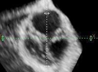
22 The added value of 3D-TTE-imaging Volume measurement by 3D-imaging only in data sets with optimal image quality Comparable image quality in comparison to the 2D-images. Endocardial contour detection is also pssible in triplane and multidimensional documentation. Despite acceptable 3D images the LV volume tools seem to underestimate LV.volume. Universitätsmedizin Leipzig (2016) - Hagendorff - 3D- and Multidimensional Echocardiography 22


 - Hagendorff - 3D- and Multidimensional")
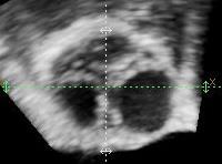
23 The added value of 3D-TTE-imaging Volume measurement by 3D-imaging There are some debatable issues in the software of the 3D volume analysis regarding volume detection of the MV tenting area as well as LVOTvolume measurement Universitätsmedizin Leipzig (2016) - Hagendorff - 3D- and Multidimensional Echocardiography 23


 - Hagendorff - 3D- and")
24 The added value of 3D-TTE-imaging Volume measurement by 3D-imaging LV-volumes determined in correct standardized views in 2D-echocardiography are still higher than LV volumes determined in 3D data sets. Universitätsmedizin Leipzig (2016) - Hagendorff - 3D- and Multidimensional Echocardiography 24


25 If the aortic annulus is visualized, the correct measurements of dimensions are not easy to understand. The intersections of the commissures and the cusps with the sectional plane do not describe exactly the dimension of the virtual annulus at the hinge points of the cusps. Universitätsmedizin Leipzig (2016) - Hagendorff - 3D- and Multidimensional Echocardiography 25


26 Sinutubular junction Anatomical ventriculoarterial junction Crown-like ring Virtual ring formed by the hinge points of the aortic cusps Universitätsmedizin Leipzig (2016) - Hagendorff - 3D- and Multidimensional Echocardiography 26


27 * 1 3 * * * * * * * * * * * 3 is mirror inverted to 1. Universitätsmedizin Leipzig (2016) - Hagendorff - 3D- and Multidimensional Echocardiography 27


 -")

28 Diastole Thus, how to operate correctly? Systole Intersection with the commissure Intersection with the commissure Intersection with the cusp Intersection with the cusp Intersection at the same level of the cups Universitätsmedizin Leipzig (2016) - Hagendorff - 3D- and Multidimensional Echocardiography 28


 -")
29 1. step: adjust the central axis of AV during diastole to label the perpendicular plane through the hinge points 2. step: go to systole to measure the widest expansion of the LVOT 3. step: adjust the annulus plane in the LAX view Universitätsmedizin Leipzig (2016) - Hagendorff - 3D- and Multidimensional Echocardiography 29
 -")

30 Universitätsmedizin Leipzig (2016) - Hagendorff - 3D- and Multidimensional Echocardiography 30
 -")
31 according to de Waroux et al., J Am Coll Cardiol Img 2009;2: Universitätsmedizin Leipzig (2016) - Hagendorff - 3D- and Multidimensional Echocardiography 31


32 Steps for measurement of geometric and effective height Acquire a ZOOM data set of the complete mitral and aortic valve 2. Adjust the central axis of the aortic root in the long axis (systole/diastole) 3. Adjust the central axis of the aortic root in the perpendicular axis 4. Adjust the short axis to the hindge points by translation during diastole 5. Rotate the short axis view to control the sectional short axis plane.. Universitätsmedizin Leipzig (2016) - Hagendorff - 3D- and Multidimensional Echocardiography 32

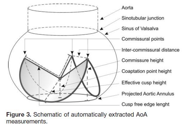
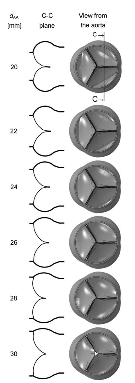
 and is defined as a virtual ring (green line) with 3 anatomical anchor points at the nadir")

33 aus: J Am Coll Cardiol Img. 2013;6(2): doi: /j.jcmg Standardized Imaging for Aortic Annular Sizing: Implications for Transcatheter Valve Selection Normal Anatomy of the Aortic Annulus The aortic annulus accounts for the tightest part of the aortic root (A) and is defined as a virtual ring (green line) with 3 anatomical anchor points at the nadir (green points) of each of the attachments of the 3 aortic leaflets (B). LCC = left coronary cusp; NCC = noncoronary cusp; RCC = right coronary cusp Universitätsmedizin Leipzig (2016) - Hagendorff - 3D- and Multidimensional Echocardiography 33
34 Universitätsmedizin Leipzig (2016) - Hagendorff - 3D- and Multidimensional Echocardiography 34
35 Universitätsmedizin Leipzig (2016) - Hagendorff - 3D- and Multidimensional Echocardiography 35


36 gh = geometric height aus Swanson WM and Clark RE. Dimensions and Geometric Relationships of the Human Aortic Valve as a Function of Pressure. Circ Res 1974; 35: Universitätsmedizin Leipzig (2016) - Hagendorff - 3D- and Multidimensional Echocardiography 36
 2 distance,")

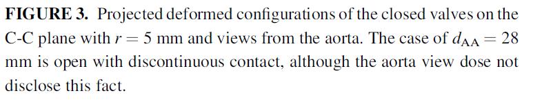
37 The following measurements are possible (performed in special centers). 1 shortest distance from aortic insertion to coaptation line gh 3 - maximum geometric height (coaptation height) 2 distance, assuming a straight course of the cusps and coaptation height of 4mm aus: Schäfers HJ, Schmied W, Marom G, Aicher D, Cusp height in aortic valves. The Journal of Thoracic and Cardiovascular Surgery 146; 2, (2013 Universitätsmedizin Leipzig (2016) - Hagendorff - 3D- and Multidimensional Echocardiography 37
 -")

38 Universitätsmedizin Leipzig (2016) - Hagendorff - 3D- and Multidimensional Echocardiography 38
39 Universitätsmedizin Leipzig (2016) - Hagendorff - 3D- and Multidimensional Echocardiography 39


40 Aortic Regurgitation and Aortic Aneurysm - Epidemiology and Guidelines - Universitätsmedizin Leipzig (2016) - Hagendorff - 3D- and Multidimensional Echocardiography 40

41 Assessment of Cusp Geometry: Effective and Geometric Height Step 1: Check alignements of the commissure between left and non coronary cusp to be perpendicular to the center of the right coronary cusp In asymmetric aortic root geometry the corresponding opposite cusp has to be centered. Universitätsmedizin Leipzig (2016) - Hagendorff - 3D- and Multidimensional Echocardiography 41

 - Hagendorff - 3D- and Multidimensional")
42 Assessment of Cusp Geometry: Effective and Geometric Height Not optimal Step 2: Center in tricuspid valves the central point of all commissures Optimized Universitätsmedizin Leipzig (2016) - Hagendorff - 3D- and Multidimensional Echocardiography 42
43 Cusp Geometry: Scheme of the Sectional Planes for Assessment of Geometric Height NCC LCC In 2D-TTE and TEE only the exact assessment of the right coronary cusp is possible. The left and non coronary cusp has to be analyzed in 3D data sets. RCC Universitätsmedizin Leipzig (2016) - Hagendorff - 3D- and Multidimensional Echocardiography 43


44 Assessment of Cusp Geometry: Effective and Geometric Height Assessment of the geometic height of the right coronary cusp Universitätsmedizin Leipzig (2016) - Hagendorff - 3D- and Multidimensional Echocardiography 44
 - Hagendorff - 3D- and Multidimensional Echocardiography")
45 Assessment of Cusp Geometry: Effective and Geometric Height Assessment of the geometic height of the left coronary cusp Universitätsmedizin Leipzig (2016) - Hagendorff - 3D- and Multidimensional Echocardiography 45
 - Hagendorff - 3D- and Multidimensional Echocardiography")
46 Assessment of Cusp Geometry: Effective and Geometric Height Assessment of the geometic height of the non coronary cusp Universitätsmedizin Leipzig (2016) - Hagendorff - 3D- and Multidimensional Echocardiography 46

 - Hagendorff - 3D- and")
47 Cusp Geometry: Scheme of the Sectional Planes for Assessment of Effektive Height NCC vs LCC In 2D-TTE and TEE only the exact assessment of the right coronary cusp is possible. The left and non coronary cusp has to be analyzed in 3D data sets. NCC vs RCC LCC vs RCC Universitätsmedizin Leipzig (2016) - Hagendorff - 3D- and Multidimensional Echocardiography 47

 - Hagendorff - 3D- and Multidimensional")
48 Assessment of Cusp Geometry: Effective and Geometric Height Assessment of the coaptation length and effective height between the left and non coronary cusp Universitätsmedizin Leipzig (2016) - Hagendorff - 3D- and Multidimensional Echocardiography 48
 - Hagendorff - 3D- and Multidimensional Echocardiography")
49 Assessment of Cusp Geometry: Effective and Geometric Height Assessment of the coaptation length and effective height between the left and right coronary cusp Universitätsmedizin Leipzig (2016) - Hagendorff - 3D- and Multidimensional Echocardiography 49
 - Hagendorff - 3D- and Multidimensional Echocardiography")
50 Assessment of Cusp Geometry: Effective and Geometric Height Assessment of the coaptation length and effective height between the right and non coronary cusp Universitätsmedizin Leipzig (2016) - Hagendorff - 3D- and Multidimensional Echocardiography 50


51 Assessment of Cusp Geometry: Effective and Geometric Height in BAV patients Universitätsmedizin Leipzig (2016) - Hagendorff - 3D- and Multidimensional Echocardiography 51


52 Assessment of Cusp Geometry: Effective and Geometric Height Universitätsmedizin Leipzig (2016) - Hagendorff - 3D- and Multidimensional Echocardiography 52


53 The 3D4D approach: use the best one. Parasternal or TEE: The TEE approach is sometimes not the best. TEE TTE apical Use the approach with the best reflection speckles of the cusp to get their best visualisation. Universitätsmedizin Leipzig (2016) - Hagendorff - 3D- and Multidimensional Echocardiography TTE parasternal aersus apical 53


54 The 3D4D approach: use the best one. Parasternal or TEE: The TEE approach is sometimes not the best. There are more artifacts using the parasternal approach than using the apical approach. Postprocessing is easier using images with better rendering. Universitätsmedizin Leipzig (2016) - Hagendorff - 3D- and Multidimensional Echocardiography 54


55 The orientation of imaging in TTE and TEE It is obvious but sometimes not present. Biplane scanning: If the virtual annulus is perpendicular to the radial scanlines and the hindge points can be visualized in a short axis view. Primary scanning in the short axis view causes a mirror inverted long axis view, primary scanning in the long axis view causes a usual short axis view. Universitätsmedizin Leipzig (2016) - Hagendorff - 3D- and Multidimensional Echocardiography 55


 - Hagendorff - 3D- and")
56 If the primary sectional plane is the long axis view in TTE, the 90 view is again with the blood stream in the LVOT. In TEE - if the primary sectional plane is the long axis view - the short axis view is again with the blood stream in the LVOT, but the view is mirrorinverted. 3D- and Multidimensional Echocardiography Comparisons of views between TTE and TTE TV RA RVOT AV LA PV PV LA l r a RVOT RA TV Universitätsmedizin Leipzig (2016) - Hagendorff - 3D- and Multidimensional Echocardiography 56

 4:671 689 3D- and")


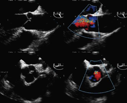
57 according to Rajiah P and SchoenhagenP. The role of computed tomography in pre-procedural planning of cardiovascular surgery and intervention. Insights Imaging (2013) 4: D- and Multidimensional Echocardiography Morphology and calcification of aortic valve: It can be assessed by echocardiography - however, echogeneity is not always the same thing. Stenotic orifice areas can normally well be determined by 2D- and 3D4D techniques. 3D-data set 3D- surface rendering 3D-data set biplane Biplane and 3D spatial resolution is sufficient and at least comparable to CT. Universitätsmedizin Leipzig (2016) - Hagendorff - 3D- and Multidimensional Echocardiography 57


 - Hagendorff - 3D- and Multidimensional")
58 The documentation of special cardiac structures: Anatomy of coronary ostia and their relation to the aoric cusps The distances between annulus and the coronary ostia as well as the lenght of the cusps can be easily measured using sectional planes within a 3D4D data set (furthermore the dynamic aspect of the ostial movement can be visualized and analyzed in 3D- TEE data sets. Universitätsmedizin Leipzig (2016) - Hagendorff - 3D- and Multidimensional Echocardiography 58


59 Visualization of coronary ostia by using FlexiSlice and 2-Click-Crop 3D4Ddata-set: short axis view aortic valve 3D4Ddata-set: long axis view ostium of the LCA 3D4Ddata-set: Flexislice axis of the aortic valve 2-Click- Cropview of the ostium of the RCA Universitätsmedizin Leipzig (2016) - Hagendorff - 3D- and Multidimensional Echocardiography 59


60 The proximal part of the right coronary artery is often well visible by 3D-TTE. Measurements of the cusps and aortic root dimensions can be performed. Universitätsmedizin Leipzig (2016) - Hagendorff - 3D- and Multidimensional Echocardiography 60


61 Aortic regurgitation Type Id: The assessment of left ventricular function by TEE not very new but still unknown: Analysis of deformation imaging in TEE Universitätsmedizin Leipzig (2016) - Hagendorff - 3D- and Multidimensional Echocardiography 61


62 Aortic regurgitation Type Id: the pre-surgical state prior to aortic valve repaier Universitätsmedizin Leipzig (2016) - Hagendorff - 3D- and Multidimensional Echocardiography 62


63 Aortic regurgitation Type Id: the post-surgical state post to aortic valve repaier Universitätsmedizin Leipzig (2016) - Hagendorff - 3D- and Multidimensional Echocardiography 63
64 Left ventricular function after aortic valve repair : What is normal? What is a normal effect in excentric left ventricular hypertrophy due to volume overload? What is the normal sequelae after surgical repair? Can the reverse remodeling be monitored in the follow-up? What is normal in the follow-up? Global strain prior to surgery: -14.2% Global strain prior to surgery: -10.8% Universitätsmedizin Leipzig (2016) - Hagendorff - 3D- and Multidimensional Echocardiography 64


65 Left ventricular function after mitral valve annuloplasty: What is normal? A global strain of -16% one year after surgery? preoperative - 28% - 16% postoperative Universitätsmedizin Leipzig (2016) - Hagendorff - 3D- and Multidimensional Echocardiography 65

66 Example:the monitoring of MitraClip-patients. The acute effect of clipping can be monitored. There is an improvement of left ventricular deformation, if the anterior-posterior diameter of the mitral annulus is reduced by the clipping procedure. Universitätsmedizin Leipzig (2016) - Hagendorff - 3D- and Multidimensional Echocardiography 66


 - Hagendorff - 3D- and")
67 Navigation in the 3D4D-data set enables all views to the aortic valve (auto-alignement, 2-click-cropping and flip crop); Estimation of effective regurgitant orifice by flexislice in a 3D4D color coded data set Universitätsmedizin Leipzig (2016) - Hagendorff - 3D- and Multidimensional Echocardiography 67


68 Additional information and better diagnostic impact: Quantification of an excentric regurgitation in biscuspid aortic valve Case: ERO cm 2 Universitätsmedizin Leipzig (2016) - Hagendorff - 3D- and Multidimensional Echocardiography 68


69 Multidimensional analysis of aortic arch: Objective measurements of aortic dimensions Universitätsmedizin Leipzig (2016) - Hagendorff - 3D- and Multidimensional Echocardiography 69


70 Summary: 1. 3D echocardiography enables a completely new modality of imaging in echocardiography the visualization of surfaces (endocardium and the cusps). 2. Biplane and triplane simultaneous sectional planes enables a better and more acurate standardization of imaging with improvement of measurements of anatomical structures. 3. Postprocessing in 3D data sets offers the possibility of new views (e.g. en-face view of the coronary ostia, etc.) 4. Especially for the decicion making and the planning of the surgical strategy 3D echocardiography can provide important informations. 5. The higher the image quality, the better the information. 6. Thus, training and expertise in 3D echocardiography is a prerequisite for a better diagnosis. Universitätsmedizin Leipzig (2016) - Hagendorff - 3D- and Multidimensional Echocardiography 70


71 Thank You for Your Attention Universitätsmedizin Leipzig (2016) - Hagendorff - 3D- and Multidimensional Echocardiography 71
Aortic Regurgitation and Aortic Aneurysm - Epidemiology and Guidelines -
 Reconstruction of the Aortic Valve and Root - A Practical Approach - Aortic Regurgitation and Aortic Aneurysm Wednesday 14 th September - 9.45 Practice must always be founded on sound theory. Leonardo
3-Dimensional Echocardiography in Aortic Valve Repair 13. September Saarland University Medical Center - Homburg/Saar, Germany
 3-Dimensional Echocardiography in Aortic Valve Repair 13. September 2018 14.00 14.30 Saarland University Medical Center - Homburg/Saar, Germany Prof. Dr. med. Andreas Hagendorff, Universitätsklinikum Leipzig
Part II: Fundamentals of 3D Echocardiography: Acquisition and Application
 Part II: Fundamentals of 3D Echocardiography: Acquisition and Application Dr. Bruce Bollen 3D matrix array TEE probes provide options for both 2D and 3D imaging. Indeed, their utility in obtaining multiple
JOINT MEETING 2 Tricuspid club Chairpersons: G. Athanassopoulos, A. Avgeropoulou, M. Khoury, G. Stavridis
 JOINT MEETING 2 Tricuspid club Chairpersons: G. Athanassopoulos, A. Avgeropoulou, M. Khoury, G. Stavridis Similarities and differences in Tricuspid vs. Mitral Valve Anatomy and Imaging. Echo evaluation
When Does 3D Echo Make A Difference?
 When Does 3D Echo Make A Difference? Wendy Tsang, MD, SM Assistant Professor, University of Toronto Toronto General Hospital, University Health Network 1 Practical Applications of 3D Echocardiography Recommended
Questions of the webinar "Imaging in TAVI procedures" Answered by Andreas Hagendorff, Victoria Delgado and Bernard Cosyns
 Questions of the webinar "Imaging in TAVI procedures" Answered by Andreas Hagendorff, Victoria Delgado and Bernard Cosyns 1. The incidence in AR I think that this question focuses on the incidence in AR
Imaging in TAVI. Jeroen J Bax Dept of Cardiology Leiden Univ Medical Center The Netherlands Davos, feb 2013
 Imaging in TAVI Jeroen J Bax Dept of Cardiology Leiden Univ Medical Center The Netherlands Davos, feb 2013 Research grants: Medtronic, Biotronik, Boston Scientific, St Jude, BMS imaging, GE Healthcare,
ECHOCARDIOGRAPHY DATA REPORT FORM
 Patient ID Patient Study ID AVM - - Date of form completion / / 20 Initials of person completing the form mm dd yyyy Study period Preoperative Postoperative Operative 6-month f/u 1-year f/u 2-year f/u
Failed Aortic Valve Repairs Lessons Learned
 Failed Aortic Valve Repairs Lessons Learned A. Stephane Lambert, MD, FRCPC Munir Boodhwani, MD, MMSc, FRCSC University of Ottawa Heart Institute Ottawa Ontario No Disclosure Why do repairs fail? Basic
Severity of AS Degree of AV calcification (? Bicuspid AV), annulus size, & aortic root
, annulus size, & aortic root") The role of Cardiac Imaging modalities in evaluation & selection of patients for Trans-catheter Aortic Valve Implantation Dr.Saeed AL Ahmari Consultant Cardiologist Prince Sultan Cardaic Center, Riyadh
The role of Cardiac Imaging modalities in evaluation & selection of patients for Trans-catheter Aortic Valve Implantation Dr.Saeed AL Ahmari Consultant Cardiologist Prince Sultan Cardaic Center, Riyadh
Aortic Valve Repair a Modular and Geometric Approach. H.-J. Schäfers Dept. of Thoracic and Cardiovascular Surgery University Hospital of Saarland
 Aortic Valve Repair a Modular and Geometric Approach H.-J. Schäfers Dept. of Thoracic and Cardiovascular Surgery University Hospital of Saarland 12.09.2018 Limitations: Purely echocardiographic, does not
Dr Winnie Sze-Wun Chan. Cardiac Team Deputy Team Head Department of Radiology and Imaging Queen Elizabeth Hospital Hong Kong
 Dr Winnie Sze-Wun Chan Cardiac Team Deputy Team Head Department of Radiology and Imaging Queen Elizabeth Hospital Hong Kong Why? Is CT reliable? How to perform the CT study? How to interpret the CT study?
Image Assistance in TAVI Why CT? Won-Jang Kim, MD, PhD Clinical Assistant Professor of Medicine, Heart Institute, Asan Medical Center, Seoul, Korea
 Image Assistance in TAVI Why CT? Won-Jang Kim, MD, PhD Clinical Assistant Professor of Medicine, Heart Institute, Asan Medical Center, Seoul, Korea Major Uses of CT in TAVI Ileofemoral Patient Arterial
Technical consideration of aquiring and analyzing 3D TEE volume data sets (EchoPac ) Phasic changes of the aortic root throughout the cardiac cycle
 Phasic changes of the aortic root throughout the cardiac cycle") Technical consideration of aquiring and analyzing 3D TEE volume data sets (EchoPac ) Phasic changes of the aortic root throughout the cardiac cycle Specific application in aortic regurgitation and aortic
Technical consideration of aquiring and analyzing 3D TEE volume data sets (EchoPac ) Phasic changes of the aortic root throughout the cardiac cycle Specific application in aortic regurgitation and aortic
What are the best diagnostic tools to quantify aortic regurgitation?
 What are the best diagnostic tools to quantify aortic regurgitation? Agnès Pasquet, MD, PhD Pôle de Recherche Cardiovasculaire Institut de Recherche Expérimentale et Clinique Université catholique de Louvain
Revealing new insights. irotate electronic rotation and xplane adjustable biplane imaging. Ultrasound cardiology. irotate and xplane
 Ultrasound cardiology irotate and xplane Revealing new insights irotate electronic rotation and xplane adjustable biplane imaging Annemien van den Bosch and Jackie McGhie Department of Cardiology, Erasmus
Pre-procedural CT angiography for Transcatheter Aortic Valve Implantation: What a Radiologist Needs to Know?
 Pre-procedural CT angiography for Transcatheter Aortic Valve Implantation: What a Radiologist Needs to Know? E O Dwyer, C O Brien, I Murphy, C Shortt, O Buckley Department of Radiology, AMNCH, Dublin,
Conflict of Interests
 The Left Ventricle: How Should We Quantify Its Size and Function; Is It Time for 3D in Everyone? Roberto M Lang, MD Conflict of Interests Philips Medical Imaging Research Grants Speakers bureau Advisory
Back to Basics: Common Errors In Quantitation In Everyday Practice
 Back to Basics: Common Errors In Quantitation In Everyday Practice Deborah Agler, ACS, RDCS, FASE October 9, 2017 ASE: Echo Florida Rebecca T. Hahn, MD Director of Interventional Echocardiography Professor
Anatomy of aortic valve and root Emmanuel Lansac MD PhD
 Anatomy of aortic valve and root Emmanuel Lansac MD PhD Cardiac Surgery Institut Mutualiste Montsouris, Paris, France The aortic valve : a passive or dynamic structure? Leonardo da Vinci 1508 Quadr Anat
The Role of Imaging in Transcatheter Aortic Valve Implantation
 The Role of Imaging in Transcatheter Aortic Valve Implantation Helmut Baumgartner Westfälische Wilhelms-Universität Münster Division of Adult Congenital and Valvular Heart Disease Department of Cardiovascular
Anatomy of aortic valve and root
 Anatomy of aortic valve and root Emmanuel Lansac, Isabelle Di Centa Cardiac Surgery Institut Mutualiste Montsouris, Paris, France The aortic valve : a passive or dynamic structure? Leonardo da Vinci 1508
Outline. EuroScore II. Society of Thoracic Surgeons Score. EuroScore II
 SURGICAL RISK IN VALVULAR HEART DISEASE: WHAT 2D AND 3D ECHO CAN TELL YOU AND WHAT THEY CAN'T Ernesto E Salcedo, MD Professor of Medicine University of Colorado School of Medicine Director of Echocardiography
Aortic valve repair: When and how to employ this novel approach?
 Aortic valve repair: When and how to employ this novel approach? Konstadinos A Plestis, MD System Chief of Cardiac Thoracic and Vascular Surgery Main Line Health Care System Professor Sidney Kimmel Medical
Joseph E. Bavaria, M.D. Roberts Measy Professor and Vice Chief CardioVascular Surgery Director: Thoracic Aortic Surgery Program University of
 Joseph E. Bavaria, M.D. Roberts Measy Professor and Vice Chief CardioVascular Surgery Director: Thoracic Aortic Surgery Program University of Pennsylvania, USA AVRS Philadelphia Sept 2016 Pictures courtesy
Certificate in Clinician Performed Ultrasound (CCPU) Syllabus. Rapid Cardiac Echo (RCE)
 Syllabus. Rapid Cardiac Echo (RCE)") Certificate in Clinician Performed Ultrasound (CCPU) Syllabus Rapid Cardiac Echo (RCE) Purpose: Rapid Cardiac Echocardiography (RCE) This unit is designed to cover the theoretical and practical curriculum
Certificate in Clinician Performed Ultrasound (CCPU) Syllabus Rapid Cardiac Echo (RCE) Purpose: Rapid Cardiac Echocardiography (RCE) This unit is designed to cover the theoretical and practical curriculum
B-Mode measurements protocols:
 Application Note How to Perform the Most Commonly Used Measurements from the Cardiac Measurements Package associated with Calculations of Cardiac Function using the Vevo Lab Objective The Vevo LAB offline
New Cardiovascular Devices and Interventions: Non-Contrast MRI for TAVR Abhishek Chaturvedi Assistant Professor. Cardiothoracic Radiology
 New Cardiovascular Devices and Interventions: Non-Contrast MRI for TAVR Abhishek Chaturvedi Assistant Professor Cardiothoracic Radiology Disclosure I have no disclosure pertinent to this presentation.
Aortic Valve Repair - Alternative to Replacement
 Aortic Valve Repair - Alternative to Replacement Seite 1 Dept. of Thoracic and Cardiovascular Surgery University Hospital of Saarland Homburg/ Saar Germany Seite 2 Aortic Valve - Historic Repair Attempts
Lessons From The Computer Model and How We Do Root Replacement
 Lessons From The Computer Model and How We Do Root Replacement Ehud Raanani, MD Cardiac Surgery Leviev Cardiothoracic and Vascular Center Sheba Medical Center Sackler School of Medicine, Tel Aviv University
Results of Aortic Valve Preservation and Repair
 Results of Aortic Valve Preservation and Repair Department of Cardiothoracic and Vascular Surgery Cliniques Universitaires St. Luc Brussels, Belgium Gebrine Elkhoury Institutional experience in AV preservation
NEW GUIDELINES. A Guideline Protocol for the Assessment of Aortic Regurgitation From the British Society of Echocardiography Education Committee
 NEW GUIDELINES A Guideline Protocol for the Assessment of Aortic Regurgitation From the British Society of Echocardiography Education Committee Gill Wharton, Prathap Kanagala (Lead Authors) Richard Steeds
Aortic Valve Repair: The Brussels Approach Laurent de Kerchove, MD, PhD Cliniques Universitaires St-Luc, IREC, UCL, Brussels, Belgium
 Reconstruction of the Aortic Valve and Root: A Practical Approach September 14 th -16 th, Homburg/Saar, Germany Aortic Valve Repair: The Brussels Approach Laurent de Kerchove, MD, PhD Cliniques Universitaires
Imaging to select patients for Transcatheter TV
 Imaging to select patients for Transcatheter TV Jeroen J Bax Dept of Cardiology Leiden Univ Medical Center The Netherlands San Diego, february 2018 Research grants: Medtronic, Biotronik, Boston Scientific,
Aortic Regurgitation & Aorta Evaluation
 VALVULAR HEART DISEASE Regurgitation Valvular Lessions 2017 Aortic Regurgitation & Aorta Evaluation Jorge Eduardo Cossío-Aranda MD, FACC Chairman of Outpatient Care Department Instituto Nacional de Cardiología
Conflict of Interests
 Introduction to Interventional Echocardiography Roberto M Lang, MD Tomtec Conflict of Interests Research Grants Philips Medical Imaging Research Grants Speakers bureau Advisory bureau 1 Structural Heart
ICE: Echo Core Lab-CRF
 APPENDIX 1 ICE: Echo Core Lab-CRF Study #: - Pt Initials: 1. Date of study: / / D D M M M Y Y Y Y 2. Type of Study: TTE TEE 3. Quality of Study: Poor Moderate Excellent Ejection Fraction 4. Ejection Fraction
Functional anatomy of the aortic root. ΔΡΟΣΟΣ ΓΕΩΡΓΙΟΣ Διεσθσνηής Καρδιοθωρακοτειροσργικής Κλινικής Γ.Ν. «Γ. Παπανικολάοσ» Θεζζαλονίκη
 Functional anatomy of the aortic root ΔΡΟΣΟΣ ΓΕΩΡΓΙΟΣ Διεσθσνηής Καρδιοθωρακοτειροσργικής Κλινικής Γ.Ν. «Γ. Παπανικολάοσ» Θεζζαλονίκη What is the aortic root? represents the outflow tract from the LV provides
VMS Quick Reference Guide
 VMS Quick Reference Guide Connecting to the VMS Workstation Ensure Ultrasound system is up and running prior to starting VMS. Connect the video cable from the ultrasound machine to the VMS Integrated Station.
Introduction to TEE using Heartworks Echocardiography Simulator
 Introduction to TEE using Heartworks Echocardiography Simulator Steven M. Ewer, MD Assistant Professor Division of Cardiovascular Medicine University of Wisconsin School of Medicine & Public Health Version
Transoesophageal echocardiography and decision making in valve surgery
 Transoesophageal echocardiography and decision making in valve surgery Intraoperative evaluation of the surgical results in aortic valve / root surgery Catherine Szymanski Disclosures None Sino-tubular
MAYON VOLCANO: FAST FACTS
 MAYON VOLCANO: FAST FACTS Type of Volcano: Stratovolcano Elevation: 2.46 km Base Diameter: 20 km Base Circumference: 62.8 km Area: 314.1 km 2 Reference: http://www.phivolcs.dost.gov.ph/html/update_vmepd/volcano/volcanolist/mayon.htm
Echo Assessment Pre-TAVI
 Disclosure Statement of Financial Interest Within the past 12 months, I or my spouse/partner have had a financial Interest /arrangement or affiliation with the organization(s) listed below Echocardiographic
Joseph E. Bavaria, M.D. Roberts Measy Professor and Vice Chief CardioVascular Surgery Director: Thoracic Aortic Surgery Program University of
 Joseph E. Bavaria, M.D. Roberts Measy Professor and Vice Chief CardioVascular Surgery Director: Thoracic Aortic Surgery Program University of Pennsylvania, USA North American Valve Repair, Philadelphia
3D Printing & Echocardiography
 ASE SOTA Feb 19, 2018 3D Printing & Echocardiography Stephen H. Little, MD John S. Dunn Chair in Cardiovascular Research and Education, Associate professor, Weill Cornell Medicine Disclosures Personal
Imaging Assessment of Aortic Stenosis/Aortic Regurgitation
 Imaging Assessment of Aortic Stenosis/Aortic Regurgitation Craig E Fleishman, MD FACC FASE The Heart Center at Arnold Palmer Hospital for Children, Orlando SCAI Fall Fellows Course 2014 Las Vegas Disclosure
ΔΙΑΔΕΡΜΙΚΗ ΑΝΤΙΜΕΤΩΠΙΣΗ ΔΟΜΙΚΩΝ ΠΑΘΗΣΕΩΝ: Ο ΡΟΛΟΣ ΤΗΣ ΑΠΕΙΚΟΝΙΣΗΣ ΣΤΟ ΑΙΜΟΔΥΝΑΜΙΚΟ ΕΡΓΑΣΤΗΡΙΟ ΣΤΗΝ ΤΟΠΟΘΕΤΗΣΗ MITRACLIP
 ΔΙΑΔΕΡΜΙΚΗ ΑΝΤΙΜΕΤΩΠΙΣΗ ΔΟΜΙΚΩΝ ΠΑΘΗΣΕΩΝ: Ο ΡΟΛΟΣ ΤΗΣ ΑΠΕΙΚΟΝΙΣΗΣ ΣΤΟ ΑΙΜΟΔΥΝΑΜΙΚΟ ΕΡΓΑΣΤΗΡΙΟ ΣΤΗΝ ΤΟΠΟΘΕΤΗΣΗ MITRACLIP ΒΛΑΣΗΣ ΝΙΝΙΟΣ MD MRCP ΚΛΙΝΙΚΗ ΑΓΙΟΣ ΛΟΥΚΑΣ ΘΕΣΣΑΛΟΝΙΚΗ CONFLICT OF INTEREST PROCTOR
British Society of Echocardiography
 British Society of Echocardiography Affiliated to the British Cardiac Society A Minimum Dataset for a Standard Adult Transthoracic Echocardiogram From the British Society of Echocardiography Education
Chamber Quantitation Guidelines: What is New?
 Chamber Quantitation Guidelines: What is New? Roberto M Lang, MD J AM Soc Echocardiogr 2005; 18:1440-1463 1 Approximately 10,000 citations iase in itune Cardiac Chamber Quantification: What is New? Database
The stentless bioprosthesis has many salient features that
 Aortic Valve Replacement with the Medtronic Freestyle Xenograft Using the Subcoronary Implantation Technique D. Michael Deeb, MD The stentless bioprosthesis has many salient features that make it an attractive
The Bicuspid AV Surgical Conisiderations
 The Bicuspid AV Surgical Conisiderations Ehud Raanani, MD Cardiothoracic Surgery, Sheba Medical Center Sackler School of Medicine, Tel Aviv University MAY 15, 2014 Homburg BAV Repair Congenital variations
Aortic Stenosis: Spectrum of Disease, Low Flow/Low Gradient and Variants
 Aortic Stenosis: Spectrum of Disease, Low Flow/Low Gradient and Variants Martin G. Keane, MD, FASE Professor of Medicine Lewis Katz School of Medicine at Temple University Basic root structure Parasternal
Optimal Imaging Technique Prior to TAVI -Echocardiography-
 2014 KSC meeting Optimal Imaging Technique Prior to TAVI -Echocardiography- Geu-Ru Hong, M.D. Ph D Associate Professor of Medicine Division of Cardiology, Severance Cardiovascular Hospital Yonsei University
Quantification of Cardiac Chamber Size
 2017 KSE 2017-11-25 Quantification of Cardiac Chamber Size Division of Cardiology Keimyung University Dongsan Medical Center In-Cheol Kim M.D., Ph.D. LV size and function Internal linear dimensions PLX
Organic mitral regurgitation
 The best in heart valve disease Organic mitral regurgitation Ewa Szymczyk Department of Cardiology Medical University of Lodz, Poland I have nothing to declare Organic mitral regurgitation leaflet abnormality
Annular Stabilization Techniques in the Context of Aortic Valve Repair
 Annular Stabilization Techniques in the Context of Aortic Valve Repair Prashanth Vallabhajosyula, MD MS University of Pennsylvania, Philadelphia, Pennsylvania 2 nd North American Aortic Valve Repair Symposium
Reconstruction of the Aortic Valve and Root A Practical approach Failures after aortic valve repair. Diana Aicher. September 16 th -18 th 2015
 Reconstruction of the Aortic Valve and Root A Practical approach Failures after aortic valve repair Diana Aicher September 16 th -18 th 2015 Classification of failures- root repair 51/810 acute/ intraoperative
The Key Questions in Mitral Valve Interventions. Where Are We in 2018?
 The Key Questions in Mitral Valve Interventions Where Are We in 2018? Gilles D. DREYFUS, MD, FRCS, FESC Professor of Cardiothoracic Surgery 30 GIORNATE CARDIOLOGICHE TORINESI - OCT 2018 Are guidelines
Introduction. Aortic Valve. Outflow Tract and Aortic Valve Annulus
 Chapter 1: Surgical anatomy of the aortic and mitral valves Jordan RH Hoffman MD, David A. Fullerton MD, FACC University of Colorado School of Medicine, Department of Surgery, Division of Cardiothoracic
Congenital. Unicuspid Bicuspid Quadricuspid
 David Letterman s Top 10 Aortic Stenosis The victim can be anyone: Echo is the question and the answer!!!! Hilton Head Island Echocardiography Conference 2012 Timothy E. Paterick, MD, JD, MBA Christopher
Case 47 Clinical Presentation
 93 Case 47 C Clinical Presentation 45-year-old man presents with chest pain and new onset of a murmur. Echocardiography shows severe aortic insufficiency. 94 RadCases Cardiac Imaging Imaging Findings C
Valvular Regurgitation: Can We Do Better Than Colour Doppler?
 Valvular Regurgitation: Can We Do Better Than Colour Doppler? A/Prof David Prior St Vincent s Hospital Melbourne Sports Cardiology Valvular Regurgitation Valve regurgitation volume loads the ventricles
Preprocedural evaluation for TAVR
 KEBE 30/05/15 Preprocedural evaluation for TAVR Ioannis Iakovou, MD, PhD Interventional Cardiology Onassis Cardiac Surgery Center Athens, Greece Clinical Pathway: Developing Peri- Procedural Protocols
Index. B B-type natriuretic peptide (BNP), 76
, 76") Index A ACCESS-EU registry, 158 159 Acute kidney injury (AKI), 76, 88 Annular enlargement, RV, 177 178 Annuloplasty chordal cutting, 113 complete ring, 99 etiology-specific ring, 100 evolution, 98 flexible
Index A ACCESS-EU registry, 158 159 Acute kidney injury (AKI), 76, 88 Annular enlargement, RV, 177 178 Annuloplasty chordal cutting, 113 complete ring, 99 etiology-specific ring, 100 evolution, 98 flexible
Normal TTE/TEE Examinations
 Normal TTE/TEE Examinations Geoffrey A. Rose, MD FACC FASE Sanger Heart & Vascular Institute Before you begin imaging... Obtain the patient s Height Weight BP PLAX View PLAX View Is apex @ 9-10 o clock?
Imaging Guide Echocardiography
 Imaging Guide Guide to Small Animal Echocardiography using the Vevo Imaging Systems System Compatibility: This guide contains instructions and suggestions for work on the Vevo2100, VevoLAZR, Vevo 3100
Regurgitant Lesions. Bicol Hospital, Legazpi City, Philippines July Gregg S. Pressman MD, FACC, FASE Einstein Medical Center Philadelphia, USA
 Regurgitant Lesions Bicol Hospital, Legazpi City, Philippines July 2016 Gregg S. Pressman MD, FACC, FASE Einstein Medical Center Philadelphia, USA Aortic Insufficiency Valve anatomy and function LVOT and
Management of TR in Patients Undergoing Mitral Interventions
 Management of TR in Patients Undergoing Mitral Interventions Stephen H. Little, MD John S. Dunn Chair in Cardiovascular Research and Education, Associate professor, Weill Cornell Medicine shlittle@houstonmethodist.org
What I Have Learned from 3D Imaging of Heart Valve Disease
 What I Have Learned from 3D Imaging of Heart Valve Disease Rebecca T. Hahn, MD Director of Interventional Echocardiography Columbia University Core Lab Director for multiple tricuspid device trials for
cardiac imaging planes planning basic cardiac & aortic views for MR
 cardiac imaging planes planning basic cardiac & aortic views for MR Dianna M. E. Bardo, M. D. Assistant Professor of Radiology & Cardiovascular Medicine Director of Cardiac Imaging cardiac imaging planes
Cardiac MRI in ACHD What We. ACHD Patients
 Cardiac MRI in ACHD What We Have Learned to Apply to ACHD Patients Faris Al Mousily, MBChB, FAAC, FACC Consultant, Pediatric Cardiology, KFSH&RC/Jeddah Adjunct Faculty, Division of Pediatric Cardiology
PART II ECHOCARDIOGRAPHY LABORATORY OPERATIONS ADULT TRANSTHORACIC ECHOCARDIOGRAPHY TESTING
 PART II ECHOCARDIOGRAPHY LABORATORY OPERATIONS ADULT TRANSTHORACIC ECHOCARDIOGRAPHY TESTING STANDARD - Primary Instrumentation 1.1 Cardiac Ultrasound Systems SECTION 1 Instrumentation Ultrasound instruments
Late secondary TR after left sided heart disease correction: is it predictibale and preventable
 Late secondary TR after left sided heart disease correction: is it predictibale and preventable Gilles D. Dreyfus Professor of Cardiothoracic surgery Nath J, et al. JACC 2004 PREDICT Incidence of secondary
2D/3D in Evaluation of Atrial Septum
 2D/3D in Evaluation of Atrial Septum Roberto M Lang, MD OSTIUM SECUNDUM ASD: 2D AND 3D TNSESOPHAGEAL ECHO 1 Biplane views 90 0 3D Acquisi on Acquire 3D volume Lang RM et al. JASE 2012;25:3 46. Right atrial
The Bicuspid AV Surgical Considerations
 The Bicuspid AV Surgical Considerations Ehud Raanani, MD Cardiothoracic Surgery, Sheba Medical Center Sackler School of Medicine, Tel Aviv University September 12, 2014 Homburg BAV Repair Congenital variations
New ASE Guidelines: What you must know
 New ASE Guidelines: What you must know Federico M Asch MD, FASE, FACC Chair, ASE Guidelines and Standards Committee Medstar Washington Hospital Center Medstar Health Research Institute Georgetown University
Cardiac ultrasound protocols
 Cardiac ultrasound protocols IDEXX Telemedicine Consultants Two-dimensional and M-mode imaging planes Right parasternal long axis four chamber Obtained from the right side Displays the relative proportions
Martin G. Keane, MD, FASE Temple University School of Medicine
 Martin G. Keane, MD, FASE Temple University School of Medicine Measurement of end-diastolic LV internal diameter (LVIDd) made by properly-oriented M-Mode techniques in the Parasternal Long Axis View (PLAX):
8/31/2016. Mitraclip in Matthew Johnson, MD
 Mitraclip in 2016 Matthew Johnson, MD 1 Abnormal Valve Function Valve Stenosis Obstruction to valve flow during that phase of the cardiac cycle when the valve is normally open. Hemodynamic hallmark - pressure
PROSTHETIC VALVE BOARD REVIEW
 PROSTHETIC VALVE BOARD REVIEW The correct answer D This two chamber view shows a porcine mitral prosthesis with the typical appearance of the struts although the leaflets are not well seen. The valve
Imaging Strategies for Endovascular Cardiovascular Procedures and Percutaneous Aortic Valves. Roy K Greenberg, MD
 Imaging Strategies for Endovascular Cardiovascular Procedures and Percutaneous Aortic Valves Roy K Greenberg, MD Disclosure Research support Cook Inc, Boston Scientific, W.L.Gore, Cordis, Vascutek, Terarecon
Quantification of Aortic Regurgitation
 Quantification of Aortic Regurgitation ASE Review 2018 Boston Susan E Wiegers, MD, FASE, FACC Professor of Medicine And thanks to Dr. Roberto Lang Disclosure None related to this presentation 1 Objectives
Vevo 2100 System Cardio Measurements. Dieter Fuchs, PhD FUJIFILM VisualSonics, Inc.
 Vevo 2100 System Cardio Measurements Dieter Fuchs, PhD FUJIFILM VisualSonics, Inc. dfuchs@visualsonics.com Instructions This document is a guideline on how to assess cardiac function in rodents imaged
Introduction. Cardiac Imaging Modalities MRI. Overview. MRI (Continued) MRI (Continued) Arnaud Bistoquet 12/19/03
 MRI (Continued) Arnaud Bistoquet 12/19/03") Introduction Cardiac Imaging Modalities Arnaud Bistoquet 12/19/03 Coronary heart disease: the vessels that supply oxygen-carrying blood to the heart, become narrowed and unable to carry a normal amount
Introduction Cardiac Imaging Modalities Arnaud Bistoquet 12/19/03 Coronary heart disease: the vessels that supply oxygen-carrying blood to the heart, become narrowed and unable to carry a normal amount
3D Printing & Echocardiography
 Echo Hawaii Jan 18, 2018 3D Printing & Echocardiography Stephen H. Little, MD John S. Dunn Chair in Cardiovascular Research and Education, Associate professor, Weill Cornell Medicine Rapid Prototyping
Disclosure Statement of Financial Interest Saibal Kar, MD, FACC
 MitraClip Therapy Saibal Kar, MD, FACC, FAHA, FSCAI Director of Interventional Cardiac Research Program Director, Interventional Cardiology Heart Institute, Cedars-Sinai Medical Center, Los Angeles, CA
Atrial Septal Defects
 Supplementary ACHD Echo Acquisition Protocol for Atrial Septal Defects The following protocol for echo in adult patients with atrial septal defects (ASDs) is a guide for performing a comprehensive assessment
What echo measurements are key prior to MitraClip?
 APHP CHU Bichat - Claude Bernard What echo measurements are key prior to MitraClip? Eric Brochet,MD Cardiology Department Hopital Bichat Paris France No disclosure Conflict of interest Case 69 y.o man
Cardioband: una chance per l insufficienza mitralica funzionale
 HEARTLINE Genova 10-11 novembre 2017 Cardioband: una chance per l insufficienza mitralica funzionale Sergio Berti Ospedale del Cuore Fondazione C.N.R. Reg Toscana Massa/Pisa Reduction of Septo Lateral
Aortic valve repair is an accepted option for aortic valve
 Complex Aortic Valve Disease in Children Christopher W. Baird, MD,* and Pedro J. del Nido, MD Aortic valve repair is an accepted option for aortic valve pathologic conditions in children and young adults.
PVL Assessment. Is paravalvular regurgitation after TAVR still an important consideration in 2018?
 Joint Meeting 1 Aortic and Mitral Club Chairpersons: S.Adamopoulos, M. Vavuranakis, L. Michalis, P. Nihoyannopoulos PVL Assessment. Is paravalvular regurgitation after TAVR still an important consideration
Reconstruction of the Aortic Valve and Root A Practical approach Why and when to repair the aortic valve. Diana Aicher. September 16 th - 18 th 2015
 Reconstruction of the Aortic Valve and Root A Practical approach Why and when to repair the aortic valve Diana Aicher September 16 th - 18 th 2015 Why repair the aortic valve? Aortic Valve Replacement
Sparing aortic valve techniques
 Surgical Technique Sparing aortic valve techniques Rubén Álvarez-Cabo Cardiac Surgery Department, Heart Area, Central University Hospital of Asturias (HUCA), Oviedo, Spain Correspondence to: Rubén Álvarez-Cabo.
TAVR: Echo Measurements Pre, Post And Intra Procedure
 2017 ASE Florida, Orlando, FL October 10, 2017 8:00 8:25 AM 25 min TAVR: Echo Measurements Pre, Post And Intra Procedure Muhamed Sarić MD, PhD, MPA Director of Noninvasive Cardiology Echo Lab Associate
LV FUNCTION ASSESSMENT: WHAT IS BEYOND EJECTION FRACTION
 LV FUNCTION ASSESSMENT: WHAT IS BEYOND EJECTION FRACTION Jamilah S AlRahimi Assistant Professor, KSU-HS Consultant Noninvasive Cardiology KFCC, MNGHA-WR Introduction LV function assessment in Heart Failure:
How to Assess and Treat Obstructive Lesions
 How to Assess and Treat Obstructive Lesions Erwin Oechslin, MD, FESC, FRCPC, Director, Congenital Cardiac Centre for Adults Peter Munk Cardiac Centre University Health Network/Toronto General Hospital
S. Bruce Greenberg, MD FNASCI and President, NASCI Professor of Radiology and Pediatrics University of Arkansas for Medical Sciences
 S. Bruce Greenberg, MD FNASCI and President, NASCI Professor of Radiology and Pediatrics University of Arkansas for Medical Sciences No financial disclosures Aorta Congenital aortic stenosis/insufficiency
Conflict of Interests
 New Approaches to Systolic Function: 4D Roberto M Lang, MD Conflict of Interests Philips Medical Imaging Research Grants Speakers bureau Advisory bureau Tomtec Research Grants Epsilon Research Grants 1
Federico M Asch MD, FASE MedStar Heart and Vascular Institute Georgetown University Washington, DC
 TAVR: When Things go Wrong Federico M Asch MD, FASE MedStar Heart and Vascular Institute Georgetown University Washington, DC Disclosures Academic Echo Core Lab Abbott / St Jude Medical Edwards Medtronic