US of Renovascular Hypertension. Jonathan R. Dillman, MD, MSc Associate Professor Director, Thoracoabdominal Imaging
|
|
|
- Thomas Bailey
- 6 years ago
- Views:
Transcription

1 US of Renovascular Hypertension Jonathan R. Dillman, MD, MSc Associate Professor Director, Thoracoabdominal Imaging
2 Disclosures Nothing Relevant Unrelated grant funding Siemens US Toshiba US
3 Objectives 1. Renovascular hypertension background, causes & complications 2. Renal Doppler US approaches direct vs. indirect 3. Does it work? (i.e., the literature) 4. Role of US?
4 Pediatric Hypertension Most often primary (essential) >6 years-old Multiple secondary, treatable causes Renovascular (renin-mediated) = 5-10% Suprarenal aortic narrowing Renal artery stenosis Tullus K, et al. Lancet 2008 Gomes RS, et al. Pediatr Nephrol 2011

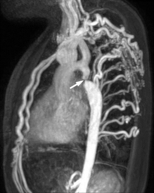
5 Renovascular Hypertension Castelli P, et al. AJR 2013

6 Renovascular Hypertension Clinically suspected when hypertension severe or refractory to multiple drugs Untreated Complications: Hypertensive encephalopathy/stroke Left ventricular hypertrophy/diastolic dysfunction -opathies kidney, retina Detection allows: Endovascular or surgical cure Fewer complications Less intense medical therapy
7 Causes of Aortic & Renal Artery Narrowing Developmental non-inflammatory arteriopathy: Idiopathic/fibromuscular dysplasia Complex medial/peri-medial dysplasia with intimal fibroplasia NF1 Williams syndrome Tuberous sclerosis Congenital aortic coarctation/interruption (thoracic or abdominal) Inflammatory arteritis (including radiation-induced) Catheter-related thromboembolic disease Extrinsic compression

8 Imaging of Renovascular Hypertension Why (or why not) US? Advantages: Lack of ionizing radiation exposure Low cost Widespread availability Disadvantages: Operator dependent requires operator experience/skill Small/accessory/intra-renal arteries challenging to assess Suboptimal diagnostic performance?

9 Renal Doppler US Technique Renal Doppler US exam Standard gray-scale imaging Color Doppler Main renal artery Spectral Doppler Main renal artery Intra-renal spectral Doppler How many samples from each kidney? Upper, mid and lower?

10 Gray-Scale US What to look for Morphologically abnormal kidneys Focal scarring Dysplasia/hypoplasia Retroperitoneal masses Adrenal Pheochromocytoma Neuroblastoma Adrenocortical neoplasm Renal (Wilms, RCC, others)
 Renal Aorta Ratio (RAR) Renal Intrarenal Ratio")
11 Doppler US What to look for Abnormal spectral Doppler waveforms Parvus et tardus Parvus = weak/small Tardus = delayed/late Delayed acceleration Low resistive index Abnormal Doppler velocities Peak systolic velocity (PSV) Renal Aorta Ratio (RAR) Renal Intrarenal Ratio (RIR)

/PSV Abnormal <0.5 Granata A, et al.")
12 Doppler US Abnormal Waveforms Assess distal to stenosis Acceleration time Time from onset of systole to PSV Abnormal >0.07 sec Resistive index Compares peak systolic and end diastolic velocities = (PSV-EDV)/PSV Abnormal <0.5 Granata A, et al. J Ultrasound 2009


13 Abnormal Waveforms How Good is Visual Assessment? Loss of ESP enabled identification of RAS with 95% sensitivity, 97% specificity, a 92% positive predictive value, a 98% negative predictive value, and 96% overall accuracy *ESP = early systolic compliance peak/ reflective-wave complex Stavros A, et al. Radiology 1992


14 It s Not Just Tardus RI Matters
15 Doppler US Abnormal Velocities Interrogate length of main renal artery Peak systolic velocity (PSV) Abnormal > cm/s Main renal artery PSV normalized to aortic PSV Renal-Aorta Ratio (RAR) Abnormal >3-3.5 Main renal artery PSV normalized to intra-renal PSV Renal-Intrarenal Ratio (RIR) Abnormal >5 (50% stenosis in adults) Sensitivity = 88%; specificity = 88% Li JC, et al. J Ultrasound Med 2006

16 Doppler US Direct vs. Indirect Assessment Direct: interrogate main renal artery with color/spectral Doppler Challenging! Intra-renal stenosis, >1 renal artery? Indirect: interrogate intra-renal arteries with spectral Doppler Abnormal wave waveform and/or low RI = proximal narrowing? Aorta, main renal artery, or intra-renal KEY POINT: if ABNORMAL, sample more proximal (main renal arteries & suprarenal aorta)


17 Doppler US 12 year-old boy with incidentally detected HTN in ED


18 Doppler US Teenage girl with newly diagnosed hypertension

19 Don t Forget Causes of renal arterial narrowing in children are generally different from adults Atherosclerotic disease RARE in children Pediatric narrowings OFTEN syndromic Thus: Adults = usually central Children = central, peripheral, or both Multifocal, bilateral?
 Aorta/main renal artery > segmental extrarenal (n=18) > segmental intra-renal (n=2) Excluding syndromes: Vo, et al.")
20 Syndromic Does it Matter? Including syndromes: Stanley, et al (n=97) Aorta/main renal artery > segmental extrarenal (n=18) > segmental intra-renal (n=2) Excluding syndromes: Vo, et al (n=21) 15 intra-renal segmental lesions 6 main & 3 accessory lesions Stanley JC, et al. J Vasc Surg 2006 Vo NJ, et al. Pediatr Radiol 2006
21 So, Does it Work in Kids? Chhadia, et al. (2013): 62 children (124 kidneys) Renal Doppler ultrasound and catheter angiography in ALL PSV >180 cm/sec, accel time >70 msec, parvus et tardus Sensitivity = 65% (detected 11/17 lesions, 4 misses segmental) Specificity = 94% Reasonable specificity, BUT sensitivity not good enough! Chhadia S, et al. Pediatr Radiol 2013
: 99 children with renal artery stenosis 36 unilateral major extraparenchymal stenosis 47 bilateral stenosis 16 intrarenal small")
22 Does it Work in Kids? Trautmann, et al. (2016): 99 children with renal artery stenosis 36 unilateral major extraparenchymal stenosis 47 bilateral stenosis 16 intrarenal small vessel disease US sensitivity = 63% US specificity = 95% Trautmann S, et al. Pediatr Nephrol 2016
23 Does it Work in Kids? Castelli, et al. (2014): 48 patients with confirmatory testing (CTA, MRA, angiography) Overall diagnostic performance: Sensitivity = 90% Specificity = 68% Patients with catheter angiography & renal artery stenosis (n=23): Sensitivity = 75% Conclusion: Not Good Enough! Specificity = 70% Best performance if: Older, aortic or main renal artery narrowing Castelli PK, et al. Pediatr Radiol 2014


24 The Problem (Challenge) RK = 8.8 cm LK = 8.6 cm 10 yo boy with refractory hypertension, normal CTA


25 The Problem (Challenge) Upper, RI = 0.6 Mid, RI = 0.63 Lower, RI = 0.47 Left kidney Doppler US

26 The Problem (Challenge) RI = 0.47 RI = 0.49 Additional left lower pole images

27 The Problem (Challenge) Outside Hospital CTA

28 The Problem (Challenge) Catheter Angiogram


29 The Problem Another Example 11 yo boy with refractory hypertension
 May")

30 Role of Renal Doppler US? Appropriate initial test in children (adds value) May direct further work-up e.g., CTA/MRA of aorta May provide alternative diagnosis Retroperitoneal mass Unsuspected renal parenchymal abnormality
31 Final Take-Aways Generally, CANNOT STOP with US Positive US almost certainly need more aortic or renal artery imaging CTA/MRA for aortic narrowings Catheter angiography for renal artery lesions (bilateral, multifocal, intrarenal disease?) Negative US almost certainly need more imaging if suspicion moderate/high
Duplex Ultrasound of the Renal Arteries. Duplex Ultrasound. In the Beginning
 Duplex Ultrasound of the Renal Arteries DIMENSIONS IN HEART AND VASCULAR CARE 2013 PENN STATE HEART AND VASCULAR INSTITUTE ROBERT G. ATNIP MD PROFESSOR OF SURGERY AND RADIOLOGY Duplex Ultrasound Developed
Ultrasound of the Renal Arteries
 Ultrasound of the Renal Arteries Greg Curry Vascular Ultrasound Workshop Aug 2017 The Examination Technique Pathophysiology Role of US then and now Background Live Scanning Ultrasound Population: 20% Hypertensive
What Do We Know? Disclosure Statement: 3/11/2015. Deep abdominal imaging
 Marsha M. Neumyer, BS, RVT, FSVU, FSDMS, FAIUM International Director Vascular Diagnostic Educational Services Vascular Resource Associates Harrisburg, PA Disclosure Statement: CME Calendar QR Code Marsha
11 TH ANNUAL VASCULAR NONINVASIVE TESTING SYMPOSIUM NOVEMBER 10, 2018
 11 TH ANNUAL VASCULAR NONINVASIVE TESTING SYMPOSIUM NOVEMBER 10, 2018 RENAL ARTERY DISEASE AND RENOVASCULAR HYPERTENSION 1 WHAT IS RENOVASCULAR HYPERTENSION? https://my.clevelandclinic.org/health/diseases/16459-renovascular-hypertension
Transducer Selection. Renal Artery Duplex Exam. Renal Scan. Renal Scan Echogenicity. How to Perform an Optimal Renal Artery Doppler Examination
 How to Perform an Optimal Renal Artery Doppler Examination Director of Ultrasound Education & Quality Assurance Baylor College of Medicine Division of Maternal-Fetal Medicine Maternal Fetal Center Imaging
Diagnosis of Renal Artery Stenosis (RAS)
") May 2001 Diagnosis of Renal Artery Stenosis (RAS) Kurt Fink, Harvard Medical School, Year III Epidemiology Hypertension -Affects 60 million Americans Essential HTN >95% of cases Secondary HTN 1-5% of cases
May 2001 Diagnosis of Renal Artery Stenosis (RAS) Kurt Fink, Harvard Medical School, Year III Epidemiology Hypertension -Affects 60 million Americans Essential HTN >95% of cases Secondary HTN 1-5% of cases
Case 8038 Renal allograft complicated with renal artery stenosis
 Case 8038 Renal allograft complicated with renal artery stenosis Santiago I, Canelas A, Pinto AP Section: Cardiovascular Published: 2009, Nov. 30 Patient: 61 year(s), male Clinical History A 61-year-old
What effects will proximal or distal disease have on an waveform?
 Spectral Doppler Interpretation Director Director of of Ultrasound Ultrasound Education Education & & Quality Quality Assurance Assurance Baylor Baylor College College of of Medicine Medicine Division
DISCLOSURE TEST YOUR WAVEFORM IQ. Partial volume artifact. 86 yo female with right arm swelling, picc line. AVF on left? Dx?
 Deborah Rubens University of Rochester Rochester, NY DISCLOSURE Neither I nor my immediate family have a financial relationship with a commercial organization that may have a direct or indirect interest
Vascular Imaging in the Pediatric Abdomen. Jonathan Swanson, MD
 Vascular Imaging in the Pediatric Abdomen Jonathan Swanson, MD Goals and Objectives To understand the imaging approach, appearance, and clinical manifestations of the common pediatric abdominal vascular
Abdominal Doppler Mastering the next level of vascular anatomy in the belly. Cindy A. Owen, RDMS, RVT
 Abdominal Doppler Mastering the next level of vascular anatomy in the belly Cindy A. Owen, RDMS, RVT Introduction Abdominal Doppler is a tough exam Success is dependent on: Patient body habitus Patient
What effects will proximal or distal disease have on a waveform?
 Spectral Doppler Interpretation Director of Ultrasound Education & Quality Assurance Baylor College of Medicine Division of Maternal-Fetal Medicine Maternal Fetal Center Imaging Manager Texas Children
Renal Artery Stenosis With Severe Hypertension: A Case Report
 CASE REPORT Renal Artery Stenosis With Severe Hypertension: A Case Report Suwaid MA ABSTRACT Background: Renal artery stenosis (RAS) is found in 77% of hypertensive patients and is responsible for 1-2%
Renal Doppler Hospital Based Study and Review of Literature
 ORIGINAL RESEARCH www.ijcmr.com Hospital Based Study and Review of Literature Jahangeer Ahmad Bhat 1, Murassa Shamshad 2, Aresalan Malik 1, Iqbal Bhat 3, Iqbal Dar 3 ABSTRACT Introduction: Study was done
No financial or commercial relationships to disclose
 Deanna New, RVT No financial or commercial relationships to disclose IAC REQUIREMENTS: The main duty of a sonographer is to make the physician or radiologists job easier by capturing images and doing
Visceral Vascular Ultrasound. Joel Thompson, MD, MPH Borg & Ide Imaging
 Visceral Vascular Ultrasound Joel Thompson, MD, MPH Borg & Ide Imaging Objectives: Review major abdominal vascular structures Identify normal peak systolic velocity (PSV) for major abdominal arteries.
AIUM Practice Guideline for the Performance of Renal Artery Duplex Sonography
 AIUM Practice Guideline for the Performance of Renal Artery Duplex Sonography 2008 by the American Institute of Ultrasound in Medicine The American Institute of Ultrasound in Medicine (AIUM) is a multidisciplinary
Goals. Access flow and renal artery stenosis evaluation by Doppler ultrasound. Reimbursement. WHY use of Doppler Ultrasound
 Access flow and renal artery stenosis evaluation by Doppler ultrasound Adina Voiculescu, MD Interventional Nephrology Brigham and Women s Hospital Boston Instructor at Harvard Medical School Understand
Renal artery stenosis
 Renal artery stenosis Dr. Alexander Woywodt Consultant Renal Physician, Royal Preston Hospital Preston, 31.10.2007 Menu anatomy of the renal arteries diseases of the large renal arteries atherosclerotic
Renovascular hypertension in children and adolescents
 Renovascular hypertension in children and adolescents M I E C Z Y S L AW L I T W I N D E P T. O F N E P H R O LO G Y & A R T E R I A L H Y P E R T E N S I O N T H E C H I L D R E N S M E M O R I A L H
Evaluation of Colour Duplex Ultrasound Scanning in Diagnosis of Renal Artery Stenosis, Compared to Angiography: A Prospective Study on 53 Patients
 Eur J Vasc Endovasc Surg 14, 305-309 (1997) Evaluation of Colour Duplex Ultrasound Scanning in Diagnosis of Renal Artery Stenosis, Compared to Angiography: A Prospective Study on 53 Patients M. Mollo,
Renal Artery Ultrasound
 Ask the Expert Renal Artery Ultrasound Looking for windows of opportunity... by Peter Coombs AMS Peter Coombs is Co-ordinator of the Graduate Diploma of Ultrasound at Monash University, Melbourne. Dear
NON-ATHEROSCLEROTIC PATHOLOGY OF THE CAROTID ARTERIES
 NON-ATHEROSCLEROTIC PATHOLOGY OF THE CAROTID ARTERIES Leslie M. Scoutt, MD, FACR Professor of Diagnostic Radiology & Surgery Vice Chair, Dept of Radiology & Biomedical Imaging Chief, Ultrasound Section
Pre-and Post Procedure Non-Invasive Evaluation of the Patient with Carotid Disease
 Pre-and Post Procedure Non-Invasive Evaluation of the Patient with Carotid Disease Michael R. Jaff, D.O., F.A.C.P., F.A.C.C. Assistant Professor of Medicine Harvard Medical School Director, Vascular Medicine
Ultrasound Imaging of The Posterior Circulation
 Ultrasound Imaging of The Posterior Circulation Michigan Sonographers Society 2 Nd Annual Fall Vascular Conference Larry N. Raber RDMS-RVT Clinical Manager General Ultrasound/Neurovascular Laboratory Cleveland
Radiologic Importance of a High- Resistive Vertebral Artery Doppler Waveform on Carotid Duplex Ultrasonography
 CME Article Radiologic Importance of a High- Resistive Vertebral Artery Doppler Waveform on Carotid Duplex Ultrasonography Esther S. H. Kim, MD, MPH, Megan Thompson, Kristine M. Nacion, BA, Carmel Celestin,
ASDIN 7th Annual Scientific Meeting DISCLOSURES TECHNICAL CONSIDERATIONS TECHNICAL CONSIDERATIONS UTILITY OF ULTRASOUND IN EVALUATING ACCESS
 DISCLOSURES UTILITY OF ULTRASOUND IN EVALUATING ACCESS DYSFUNCTION None Vandana Dua Niyyar, MD Assistant Professor of Medicine, Division of Nephrology, Emory University UTILITY OF ULTRASOUND IN ACCESS
Carotid Abnormalities Coils, Kinks and Tortuosity David Lorelli M.D., RVT, FACS Michigan Vascular Association Conference Saturday, October 20, 2012
 Carotid Abnormalities Coils, Kinks and Tortuosity David Lorelli M.D., RVT, FACS Michigan Vascular Association Conference Saturday, October 20, 2012 Page 1 Table of Contents Carotid Anatomy Carotid Duplex
GUNDERSEN/LUTHERAN ULTRASOUND DEPARTMENT POLICY AND PROCEDURE MANUAL
 GUNDERSEN/LUTHERAN ULTRASOUND DEPARTMENT POLICY AND PROCEDURE MANUAL SUBJECT: Carotid Duplex Ultrasound SECTION: Vascular Ultrasound ORIGINATOR: Deborah L. Richert, BSVT, RDMS, RVT DATE: October 15, 2015
Renal Artery Disease. None > 65,000,000. Learning objectives: Renal Artery Disease
 Renal Artery Disease Robert D. McBane, M.D. Division of Cardiology Mayo Clinic Rochester Financial Disclosure Information Renal Artery Disease Robert McBane, MD None To appreciate: Learning objectives:
Fibromuscular Dysplasia. Miranda Forrest Baker College
 Fibromuscular Dysplasia Miranda Forrest Baker College Overview Case Study Patient Information Exam Images Findings FMD Types Signs and Symptoms Treatment Case Study Patient Information Female 57 years
Current Role of Renal Artery Stenting in Patients with Renal Artery Stenosis
 Current Role of Renal Artery Stenting in Patients with Renal Artery Stenosis Young-Guk Ko, M.D. Severance Cardiovascular Hospital, Yonsei University College of Medicine, Seoul, Korea Etiology Fibromuscular
Contrast Enhanced Ultrasound of Parenchymal Masses in Children
 Contrast Enhanced Ultrasound of Parenchymal Masses in Children Sue C Kaste, DO On behalf of Beth McCarville, MD St. Jude Children s Research Hospital Memphis, TN Overview Share St. Jude experience with
This is not FMD! Fibromuscular Dysplasia Diagnosis, Treatment and Surveillance. Disclosures
 Disclosures Advisor: Innovein, inc Fibromuscular Dysplasia Diagnosis, Treatment and Surveillance Marlene Grenon, MD Department of Surgery April 2017 40 year-old woman referred for arm and leg weakness
Imaging Strategy For Claudication
 Who are the Debators? Imaging Strategy For Claudication Duplex Ultrasound Alone is Adequate to Select Patients for Endovascular Intervention - Pro: Dennis Bandyk MD No Disclosures PRO - Vascular Surgeon
Fibromuscular Dysplasia (FMD) of the renal arteries Angiographic features and therapeutic options
 of the renal arteries Angiographic features and therapeutic options") Fibromuscular Dysplasia (FMD) of the renal arteries Angiographic features and therapeutic options Poster No.: C-0630 Congress: ECR 2012 Type: Educational Exhibit Authors: K. I. Ringe, B. Meyer, F. Wacker,
Fibromuscular Dysplasia (FMD) of the renal arteries Angiographic features and therapeutic options Poster No.: C-0630 Congress: ECR 2012 Type: Educational Exhibit Authors: K. I. Ringe, B. Meyer, F. Wacker,
Carotid Artery Doppler
 Carotid Artery Doppler Patient Position supine or semisupine head slightly hyper extended rotated 45 away from the side being examined. Higher frequency linear transducers (7 MHz) Vessels should be imaged
Minimally Invasive Treatment Options for Renal Artery FMD
 Minimally Invasive Treatment Options for Renal Artery FMD FMDSA Meeting 2016 Alan H. Matsumoto, M.D., FSIR, FACR, FAHA Professor and Chair Department of Radiology & Medical Imaging University of Virginia
A CASE OF HYPERTENSION AND ACUTE RENAL FAILURE OBJECTIVES
 A CASE OF HYPERTENSION AND ACUTE RENAL FAILURE Maricel Pilapil-Pureza WLA Nephrology OBJECTIVES After the presentation, the attendee will be able to: 1. Discuss when to suspect for secondary causes of
Recommendations for Follow-up After Vascular Surgery Arterial Procedures SVS Practice Guidelines
 Recommendations for Follow-up After Vascular Surgery Arterial Procedures 2018 SVS Practice Guidelines vsweb.org/svsguidelines About the guidelines Published in the July 2018 issue of Journal of Vascular
Original Article Assessment of diagnostic value of a new Doppler index for renal artery stenosis and comparison with conventional methods
 Int J Clin Exp Med 2017;10(6):9513-9517 www.ijcem.com /ISSN:1940-5901/IJCEM0052288 Original Article Assessment of diagnostic value of a new Doppler index for renal artery stenosis and comparison with conventional
Measure #195 (NQF 0507): Radiology: Stenosis Measurement in Carotid Imaging Reports National Quality Strategy Domain: Effective Clinical Care
: Radiology: Stenosis Measurement in Carotid Imaging Reports National Quality Strategy Domain: Effective Clinical Care") Measure #195 (NQF 0507): Radiology: Stenosis Measurement in Carotid Imaging Reports National Quality Strategy Domain: Effective Clinical Care 2017 OPTIONS FOR INDIVIDUAL MEASURES: CLAIMS ONLY MEASURE TYPE:
Measure #195 (NQF 0507): Radiology: Stenosis Measurement in Carotid Imaging Reports National Quality Strategy Domain: Effective Clinical Care 2017 OPTIONS FOR INDIVIDUAL MEASURES: CLAIMS ONLY MEASURE TYPE:
Vascular Sonography Examination
 Vascular Sonography Examination The purpose of The American Registry of Radiologic Technologists (ARRT ) Vascular Sonography Examination is to assess the knowledge and cognitive skills underlying the intelligent
Special Lecture 11/08/2013. Hypertension Dr. HN Mayrovitz
 Special Lecture 11/08/2013 Hypertension Dr. HN Mayrovitz Arterial Blood Pressure (ABP) Major Factors Summarized Sympathetic Hormones Arteriole MAP ~ Q x TPR + f (V / C) SV x HR Renal SBP Hypertension =
Quality ID #195 (NQF 0507): Radiology: Stenosis Measurement in Carotid Imaging Reports National Quality Strategy Domain: Effective Clinical Care
: Radiology: Stenosis Measurement in Carotid Imaging Reports National Quality Strategy Domain: Effective Clinical Care") Quality ID #195 (NQF 0507): Radiology: Stenosis Measurement in Carotid Imaging Reports National Quality Strategy Domain: Effective Clinical Care 2018 OPTIONS FOR INDIVIDUAL MEASURES: REGISTRY ONLY MEASURE
Quality ID #195 (NQF 0507): Radiology: Stenosis Measurement in Carotid Imaging Reports National Quality Strategy Domain: Effective Clinical Care 2018 OPTIONS FOR INDIVIDUAL MEASURES: REGISTRY ONLY MEASURE
Pediatric Retroperitoneal Masses Radiologic-Pathologic Correlation
 Acta Radiológica Portuguesa, Vol.XVIII, nº 70, pág. 61-70, Abr.-Jun., 2006 Pediatric Retroperitoneal Masses Radiologic-Pathologic Correlation Marilyn J. Siegel Mallinckrodt Institute of Radiology, Washington
Mesenteric/Splanchnic Artery Duplex Imaging
 VASCULAR TECHNOLOGY PROFESSIONAL PERFORMANCE GUIDELINES Mesenteric/Splanchnic Artery Duplex Imaging This Guideline was prepared by members of the Society for Vascular Ultrasound (SVU) as a template to
Carotid US: More than just a chart on the wall
 Carotid US: More than just a chart on the wall Leslie M. Scoutt, MD, FACR Professor of Diagnostic Radiology & Surgery Vice Chair, Dept of Radiology & Biomedical Imaging Chief, Ultrasound Section Medical
Vascular Ultrasound: Current state, current needs, future directions
 Vascular Ultrasound: Current state, current needs, future directions Laurence Needleman, MD Thomas Jefferson University Hospitals Sidney Kimmel Medical College of Thomas Jefferson University Disclosures
CT Versus MR for the Runoff
 CT Versus MR for the Runoff Robert R. Edelman, M.D. Dept. of Radiology NorthShore University HealthSystem Feinberg School of Medicine, Northwestern University Magnetic Resonance Computed Tomography Radio
Prof. Nabil CHAKFE et coll.
 Prof. Nabil CHAKFE et coll. For the Department of Vascular Surgery and Kidney Transplantation University Hospital of Strasbourg, FRANCE Popliteal artery entrapment: misdiagnosed Epidemiology Prevalence:
7/10/2015. Deborah J. Rubens, MD RENAL DOPPLER IT MAKES THE DIAGNOSIS! DISCLOSURES. Renal Doppler It Makes the Diagnosis!
 Renal Doppler It Makes the Diagnosis! None DISCLOSURES Deborah J. Rubens, M.D. Professor of Imaging Sciences, Oncology and Biomedical Engineering University of Rochester Medical Center Associate Director,
How to assess the hemodynamic importance of a renal artery stenosis. Felix Mahfoud, MD Saarland University Hospital Homburg/Saar, Germany
 How to assess the hemodynamic importance of a renal artery stenosis Felix Mahfoud, MD Saarland University Hospital Homburg/Saar, Germany How to assess renal artery stenosis severity 1. Non-invasive assessments
Pediatric Imaging Review
 Pediatric Imaging Review Castelli et al. Renin-Mediated Hypertension in Children Pediatric Imaging Review Patricia K. Castelli 1 Jonathan R. Dillman 1 Ethan. Smith 1 Ranjith Vellody 2 Kyung Cho 2 James
Lower Extremity Artery: Physiologic Testing
 Master Title Ultrasound for Initial Evaluation of Lower Extremity Arterial Occlusive Disease: WHY? Gregory L. Moneta MD Professor and Chief Knight Cardiovascular Institute Division of Vascular Surgery
STRUCTURED EDUCATION REQUIREMENTS IMPLEMENTATION DATE: JULY 1, 2016
 STRUCTURED EDUCATION REQUIREMENTS Vascular Sonography The purpose of structured education is to provide the opportunity for individuals to develop mastery of discipline-specific knowledge that, when coupled
RENAL ARTERY PTA. JH PEREGRIN IKEM, Prague
 RENAL ARTERY PTA JH PEREGRIN IKEM, Prague PTRA/Stenting PTRA technical success rate > 90 % In some patients helps control hypertension In some patients can improve kidney function Serious complications
Rare Cause of Cephalalgia in a Young Woman - a Case Report
 Acta Medica Marisiensis 2015;61(4):382-386 DOI: 10.1515/amma-2015-0045 CASE REPORT Rare Cause of Cephalalgia in a Young Woman - a Case Report Varga Andreea, Szakacs Xantus Timea *, Gliga Mirela, Podoleanu
The Role of US in Chronic Mesenteric Ischemia. Sagar S. Gandhi, MD Vascular Health Alliance Greenville Health System
 The Role of US in Chronic Mesenteric Ischemia Sagar S. Gandhi, MD Vascular Health Alliance Greenville Health System No Disclosures Mesenteric Ischemia Anatomy Presentation Diagnostic tools Treatment Celiac
A Closer Look: Renal Artery Stenosis. Renal artery stenosis (RAS) is defined as a TOPICS FROM CHEP. Shawn s stenosis
 is defined as a TOPICS FROM CHEP. Shawn s stenosis") TOPICS FROM CHEP A Closer Look: Renal Artery Stenosis On behalf of the Canadian Hypertension Education Program (CHEP), Dr. Tobe gives an overview of renal artery stenosis, including the prevalence, screening
TOPICS FROM CHEP A Closer Look: Renal Artery Stenosis On behalf of the Canadian Hypertension Education Program (CHEP), Dr. Tobe gives an overview of renal artery stenosis, including the prevalence, screening
HD Scanning: Velocities and Volume Flow
 HD Scanning: Velocities and Volume Flow Non-Invasive Lab Symposium West Orange, NJ April 27, 2018 Volume Flow Cindy Sturt, MD, FACS, RVT 500,000 Americans on dialysis 20-25% annual mortality 65% 5 year
Image Formation (10) 2 Evaluation and Selection of Representative Images (10)
 2 Evaluation and Selection of Representative Images (10)") STRUCTURED SELF ASSESSMENT CONTENT SPECIFICATIONS SSA LAUNCH DATE: JANUARY 1, 2018 Vascular Sonography The purpose of continuing qualifications requirements (CQR) is to assist registered technologists
STRUCTURED SELF ASSESSMENT CONTENT SPECIFICATIONS SSA LAUNCH DATE: JANUARY 1, 2018 Vascular Sonography The purpose of continuing qualifications requirements (CQR) is to assist registered technologists
Diagnostic Performance of the Modern Imaging Modalities in Renovascular Disease: An Evaluation
 DOI: 10.7860/IJARS/2016/18883:2159 Radiology Section Original Article Diagnostic Performance of the Modern Imaging Modalities in Renovascular Disease: An Evaluation Surg Cdr Brijesh K Soni, Surg Cdr SN
A Case for Mandatory Routine Graft Surveillance of lower extremity bypass grafts. Avishai Meyer UCHSC resident, Surgery May 8, 2006
 A Case for Mandatory Routine Graft Surveillance of lower extremity bypass grafts Avishai Meyer UCHSC resident, Surgery May 8, 2006 Outline: Definition Background of terms and studies U/S surveillance What
168/112mmHg.,ex-smoker 20 42,
 48 40 168/112mmHg,ex-smoker 2042, 48 36.5,168/112mmHg,HR89/min,RR18/min,97%RA 163cm,101kg,BMI38.0 Cushing,, Cre0.68mg/dl,K2.8mEq/L,TSH0.862μIU/ml 1+,WBC1-4/HF,RBC1/HF 10 Hypertension. 2018 ;71:1269-1324
Multislice CTA for Renal Artery Stenting
 Multislice CT for Renal rtery Stenting How CT can be a useful modality for diagnosing and managing renal artery stenosis for stent placement. Y MICHEL WHOLEY, MD, M; JMES WU, ; WILLIM C.L. WU, MD, FCC;
Carotid Doppler: Doppler wave forms obtained from the common, external and internal carotid arteries. As well as the vertebral and subclavian
 Competency Carotid Doppler: Doppler wave forms obtained from the common, external and internal carotid arteries. As well as the vertebral and subclavian arteries. Preferred angle is 60 degrees or less.
Beyond Stenosis Severity: Top 5 Important Duplex Characteristics to Identify in a Patient with Carotid Disease
 Beyond Stenosis Severity: Top 5 Important Duplex Characteristics to Identify in a Patient with Carotid Disease Jan M. Sloves RVT, RCS, FASE Technical Director New York Cardiovascular Associates Disclosures
Intravascular Ultrasound in the Treatment of Complex Aortic Pathologies. Naixin Kang, M.D. Vascular Surgery Fellow April 26 th, 2018
 Intravascular Ultrasound in the Treatment of Complex Aortic Pathologies Naixin Kang, M.D. Vascular Surgery Fellow April 26 th, 2018 DISCLOSURES Nothing To Disclose 2 ENDOVASCULAR AORTIC INTERVENTION Improved
Dr Doris M. W Kinuthia
 Dr Doris M. W Kinuthia Objectives Normal blood pressures in children Measurement of blood pressure in children Aetiology of Hypertension in children Evaluation of children with hypertension Treatment of
Secondary Hypertension: A Real World Approach
 Secondary Hypertension: A Real World Approach Evan Brittain, MD December 7, 2012 Kingston, Jamaica Disclosures None Real World Causes Renovascular Hypertension Endocrine Obstructive Sleep Apnea Pseudosecondary
Postoperative AV Fistula Evaluation. Postoperative examination protocol. Postoperative AVF Protocol. Hemodialysis Access Surveillance
 Hemodialysis Access Surveillance Postoperative AV Fistula Evaluation Failure of maturation Stenosis Perigraft mass/fluid collection Joseph L. Mills, Sr., M.D. Professor of Surgery Chief, Division of Vascular
Evaluation of renal artery stenosis with hemodynamic parameters of Doppler sonography
 Evaluation of renal artery stenosis with hemodynamic parameters of Doppler sonography Jian-chu Li, MD, a Yu-xin Jiang, MD, a Shu-yang Zhang, MD, b Lei Wang, MD, c Yun-shu Ouyang, MD, a and Zhen-hong Qi,
1. Long images of aorta (prox, mid, and dist) with AP measurements. 2. Trans images of aorta (prox, mid, and dist) with R/L measurements.
 with AP measurements. 2. Trans images of aorta (prox, mid, and dist) with R/L measurements.") Aorta 1. Long images of aorta (prox, mid, and dist) with AP measurements. 2. Trans images of aorta (prox, mid, and dist) with R/L measurements. 3. Long images of R/L common iliac arteries with AP measurements.
Aorta 1. Long images of aorta (prox, mid, and dist) with AP measurements. 2. Trans images of aorta (prox, mid, and dist) with R/L measurements. 3. Long images of R/L common iliac arteries with AP measurements.
Approach to patient with hypertension. Dr. Amitesh Aggarwal
 Approach to patient with hypertension Dr. Amitesh Aggarwal Definition A systolic blood pressure ( SBP) >139 mmhg and/or A diastolic (DBP) >89 mmhg. Based on the average of two or more properly measured,
Vascular Surgery Cases: Detours. Brian F. Stull, RDMS, RVT UNC REX Healthcare Vascular Specialists
 Vascular Surgery Cases: Detours Brian F. Stull, RDMS, RVT UNC REX Healthcare Vascular Specialists Brian.Stull@Unchealth.unc.edu Objectives Anatomy of a bypass graft Where does it connect, where does it
RENAL AND MESENTERIC ARTERY STENTS Are There Standard Velocity Criteria for Restenosis?
 RENAL AND MESENTERIC ARTERY STENTS Are There Standard Velocity Criteria for Restenosis? R. Eugene Zierler, M.D. The D. E. Strandness, Jr. Vascular Laboratory University of Washington Medical Center Division
Lower Extremity Arterial Doppler
 Lower Extremity Arterial Doppler 1. Spectral Doppler waveform should be taken in distal aorta and common iliac arteries. 2. R/L common femoral artery (CFA) color Doppler with velocity and B-mode. 3. R/L
MESENTERIC ISCHEMIA. Phillip J Bendick, PhD
 MESENTERIC ISCHEMIA Phillip J Bendick, PhD Arterial Celiac - Hepatic - Splenic Superior Mesenteric Artery Inferior Mesenteric Artery Venous Mesenteric system Porto - hepatic system Inferior Vena Cava Acute
Carotid Imaging IT S ABOUT MORE THAN JUST OBTAINING THE IMAGES
 Carotid Imaging IT S ABOUT MORE THAN JUST OBTAINING THE IMAGES No financial or commercial relationships to disclose Carotid artery disease: Stroke is one of the most serious causes of mortality and morbidity
Radiologic Evaluation of Peripheral Arterial Disease
 January 2003 Radiologic Evaluation of Peripheral Arterial Disease Grace Tye, Harvard Medical School Year III Patient D.M. CC: 44 y/o male with pain in his buttocks Occurs after walking 2 blocks. Pain is
Section II: Patient Interview Grade: 5
 Only written competency completed with this EXACT form will be accepted for grading. No modifications to the LAYOUT of the form will be accepted for a written competency. Failure to comply will result
Codes Requiring Authorization from MedSolutions (MSI): Updated 3/2014
: Updated 3/2014") s Requiring Authorization from MedSolutions (): Updated 3/2014 0042T Cerebral Perfusion Analysis using CT with contrast 0159T CAD, including computer algorithm analysis, BREAST MRI 0195T prepare interspace,
s Requiring Authorization from MedSolutions (): Updated 3/2014 0042T Cerebral Perfusion Analysis using CT with contrast 0159T CAD, including computer algorithm analysis, BREAST MRI 0195T prepare interspace,
8/20/18. The Doppler Effect. Objectives. What is the Doppler Effect. Doppler principles. Spectral Waveform. Image recognition. Vascular Ultrasound
 Vascular Ultrasound: Physics and Haemodynamics Objectives Doppler principles Spectral Waveform Key factors Haemodynamics: Stenosis Waveforms Image recognition Vascular Ultrasound: A flawed paradigm What
1Pulse sequences for non CE MRA
 MRI: Principles and Applications, Friday, 8.30 9.20 am Pulse sequences for non CE MRA S. I. Gonçalves, PhD Radiology Department University Hospital Coimbra Autumn Semester, 2011 1 Magnetic resonance angiography
ADI Procedure Codes. August 2016 Revised April 2017 Page 1 of 7 ADI Procedure Codes
 Code Description 70450 CT Head without contrast 70460 CT Head with contrast 70470 CT Head with & without contrast 70480 CT Orbit, et al without contrast 70481 CT Orbit, et al with contrast 70482 CT Orbit,
Case 9799 Stanford type A aortic dissection: US and CT findings
 Case 9799 Stanford type A aortic dissection: US and CT findings Accogli S, Aringhieri G, Scalise P, Angelini G, Pancrazi F, Bemi P, Bartolozzi C Department of Diagnostic and Interventional Radiology, University
Deb Coghlan AMS (Vascular and General ) Brisbane, Australia
 Brisbane, Australia") Deb Coghlan AMS (Vascular and General ) Brisbane, Australia ANEURYSMAL DIISEASE The infrarenal aorta enlarges with age, and is the commonest site for arterial aneurysms. An aneurysm is a permanent focal
Deb Coghlan AMS (Vascular and General ) Brisbane, Australia ANEURYSMAL DIISEASE The infrarenal aorta enlarges with age, and is the commonest site for arterial aneurysms. An aneurysm is a permanent focal
Lung sequestration and Scimitar syndrome
 Lung sequestration and Scimitar syndrome Imaging approaches M. Mearadji International Foundation for Pediatric Imaging Aid Rotterdam, The Netherlands Pulmonary sequestration Pulmonary sequestration (PS)
Sonography in Renovascular Hypertension
 Review rticle Sonography in Renovascular Hypertension Hsin-Yi Lee, MD, Edward G. Grant, MD Objective. To familiarize practitioners with different sonographic manifestations of renal artery compromise and
Coral Reef Aorta- Treatment Options?
 Chronic mesenteric ischemia (CMI) Coral Reef Aorta- Treatment Options? Bala Ramanan Vascular Fellow, UCSF CMI is a life-threatening problem that can result in death from inanition or bowel infarction Incidence
Carotid Imaging. Dr Andrew Farrall. Consultant Neuroradiologist
 20121123 SSCA http://www.neuroimage.co.uk/network Andrew Farrall Carotid Imaging Dr Andrew Farrall Consultant Neuroradiologist SFC Brain Imaging Research Centre (www.sbirc.ed.ac.uk), SINAPSE Collaboration
Optimising your Doppler settings for an accurate PI. Alison McGuinness Mid Yorks Hospitals
 Optimising your Doppler settings for an accurate PI Alison McGuinness Mid Yorks Hospitals Applications Both maternal uterine and fetal circulations can be studied with doppler sonography Uterine arteries
Imaging for Peripheral Vascular Disease
 Imaging for Peripheral Vascular Disease James G. Jollis, MD Director, Rex Hospital Cardiovascular Imaging Imaging for Peripheral Vascular Disease 54 year old male with exertional calf pain in his right
The European Consensus on Fibromuscular Dysplasia
 The European Consensus on Fibromuscular Dysplasia Alexandre Persu, M.D.-PhD Cardiology Department Cliniques Universitaires Saint-Luc Catholic University of Louvain Brussels, Belgium Eur J Clin Invest.
Carotid Ultrasound: Improving Ultrasound
 Carotid Ultrasound: Improving Ultrasound Edward I. Bluth, M.D., F.A.C.R. Chairman Emeritus, Department of Radiology, Ochsner Clinic Foundation, New Orleans, Louisiana Professor, Ochsner Clinical School,
Indications: following: embolization. artery that has diseases 5. The evaluation. of suspected. such entities. a cold hand. biopsy
 Peripheral Arterial Ultrasound Protocol Using Color and Spectral Doppler Reviewed by: Mark Yuhasz, MD Last Review Date: January 2015 Contact: (866) 761 4200, Option 1 Indications: The indications for peripheral
The role for contrast-enhanced ultrasonography outside of focal liver lesions
 The role for contrast-enhanced ultrasonography outside of focal liver lesions Paul S. Sidhu King s College Hospital, London, UK Introduction Contrast-enhanced ultrasonography (US) of focal liver lesions
ViosWorks: A Paradigm Shift in Cardiac MR Imaging
 Figure 1. ViosWorks image of a patient with shunted pulmonary venous return. Image courtesy of Dr. Shreyas Vasanawala, Stanford University. ViosWorks: A Paradigm Shift in Cardiac MR Imaging The value of
Abdominal Aortic Doppler Waveform in Patients with Aorto-iliac Disease
 Eur J Vasc Endovasc Surg (2010) 39, 714e718 Abdominal Aortic Doppler Waveform in Patients with Aorto-iliac Disease G. Styczynski a, *, C. Szmigielski a, J. Leszczynski b, A. Kuch-Wocial a, M. Szulc a a