Surgical Procedures. Direct suture of small ASDs Patch repair Transcatheter closure with a prosthetic device called occluder
|
|
|
- Marlene Montgomery
- 6 years ago
- Views:
Transcription
1 PEDIATRIC Review
2 Surgical Procedures Atrial Septal Defect repair: Direct suture of small ASDs Patch repair Transcatheter closure with a prosthetic device called occluder Balloon atrial septostomy (Rashkind) Transcatheter procedure using an inflated balloon passed across the interatrial septum to create an ASD
3 Balloon valvotomy(balloon valvuloplasty) Transcatheter procedure using an inflated balloon to relieve obstruction in a stenosed valve. Blalock-Taussig shunt Palliative procedure used in cyanotic defects Direct anastomosis of the right subclavian artery to right pulmonary artery Directs blood flow from systemic to pulmonary circulation
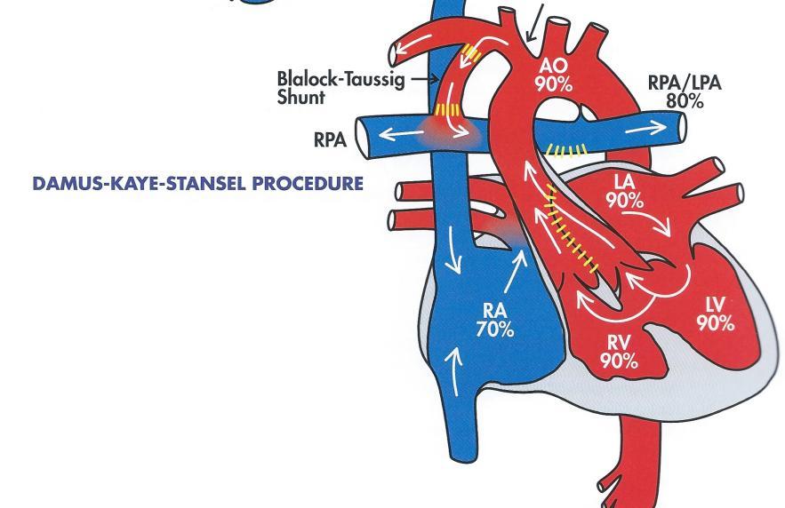
4 Damus-Kaye-Stansel procedure Palliative procedure used to establish systemic arterial flow in the treatment of one functional ventricle associated with sub-aortic obstruction Main PA is divided, proximal end is anastomosed to ascending aorta, bypassing the AV. Distal end of PA is closed. Pulmonary flow is maintained with a B-T shunt from RSCA to RPA
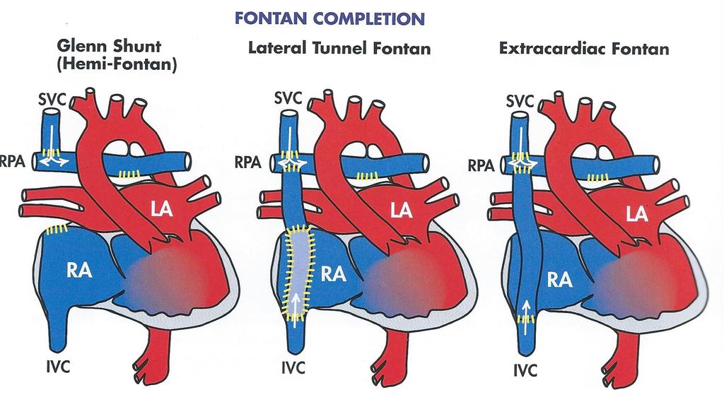
5 Fontan procedure Staged surgical repair for defects characterized by one functional ventricle The goal is to separate pulmonary and systemic circulation Stage 1: if cyanosis is present, B-T shunt or PA banding if overcirculation Stage 2: Glenn: SVC to RPA Stage 3 Fontan completion: IVC to RPA, by placing an intracardiac tunnel within the RA( lateral tunnel Fontan) or creating a conduit for IVC outside the RA(extracardiac Fontan)
6 Glenn procedure or Glenn shunt Single ventricle defects, the goal is to separate pulmonary and systemic circulations Replaces B-T shunts with anastomosis of SVC to RPA Venous return from upper body flows directly into the RPA Also known as Hemi-Fontan procedure
7 Jatene procedure Arterial switch used to connect the PA to RV and AO to the LV when the great arteries are completely transposed. The aorta and pulmonary artery are detached from their roots and repositioned over their counterpart PA is anastomosed to aortic root becoming a neopulmonary artery Ao is anastomosed to PA root becoming a neo-aorta Coronary arteries are re-attached to neo-aorta
8 Konno procedure A procedure for aortic root replacement used in the treatment of left ventricular outflow tract obstruction in association with aortic valve stenosis Av and coronaries are removed, LVOT is opened and enlarged, an aortic or pulmonary homograph replaces the AV. Coronaries are transferred to new aorta
9 Mustard procedure Atrial switch procedure utilizing a surgically created intra-atrial tunnel to redirect flow in the atria The interatrial septum is excised and a patch is used to create a tunnel The tunnel redirects oxygenated pulmonary venous return into the RV where is ejected out of the transposed aorta Deoxygenated cavae flow is redirected into the left ventricle, where is ejected out of the transposed pulmonary artery
10 Norwood procedure Use in the management of hypoplastic left heart syndrome to convert the RV from a pulmonary function to a systemic and completely separate pulmonary and systemic circulation Stage 1 palliation: RV is converted into the main pumping chamber by dividing the main PA and attaching the pulmonary root and valve to the base of the aorta. An ASD is opened. A B-T shunt is created Stage 2 Glenn procedure- SVC to RPA Stage 3 Fontan completion-ivc to RPA
11 PDA repair PDA ligation: the PDA is tied off at both ends with sutures and cut in the middle Transcatheter coil occlusion: the PDA is closed with a spring-type device called a coil. Potts shunt Palliative procedure, establish a systemic to pulmonary connection and increase pulmonary circulation Anastomoses the descending Ao to LPA
12 Pulmonary artery band Palliative procedure used in patients with pulmonary over-circulation. An adjustable band is placed around the main PA and tightened, restricting flow to the branches and lungs
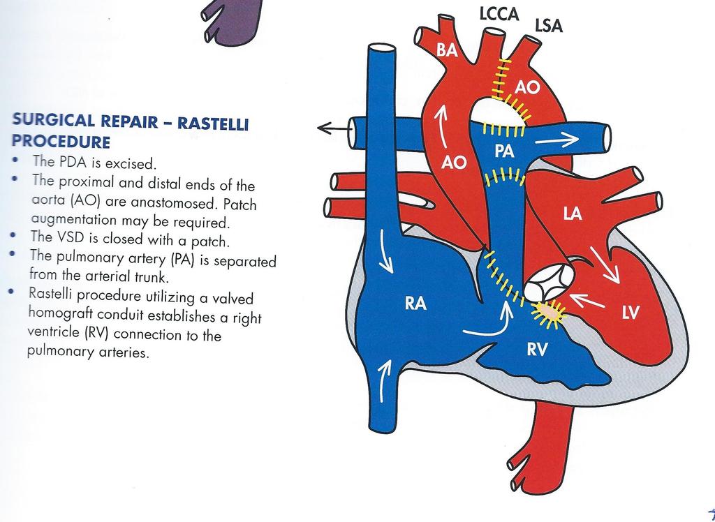
13 Rastelli procedure Used in a variety of cyanotic defects presenting in combination with VSD and severe RVOT obstruction Repair includes: Patch repair of VSD Excision of obstructive tissue in the RV Surgical closure of abnormal or damaged PV Creation of an extracardiac valved conduit from the RV to the bifurcation of the main PA
14 Ross procedure Procedure for the replacement of a stenotic aortic valve Aortic root is removed The patient s own pulmonary valve is excised and used to replace the aortic valve PV is replaced with a valved homograft
15 Waterston shunt Palliative procedure used to establish a systemicto-pulmonary connection and increase pulmonary circulation by directly anastomosing the ascending aorta and RPA
16
17
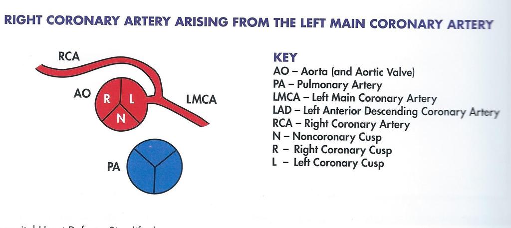
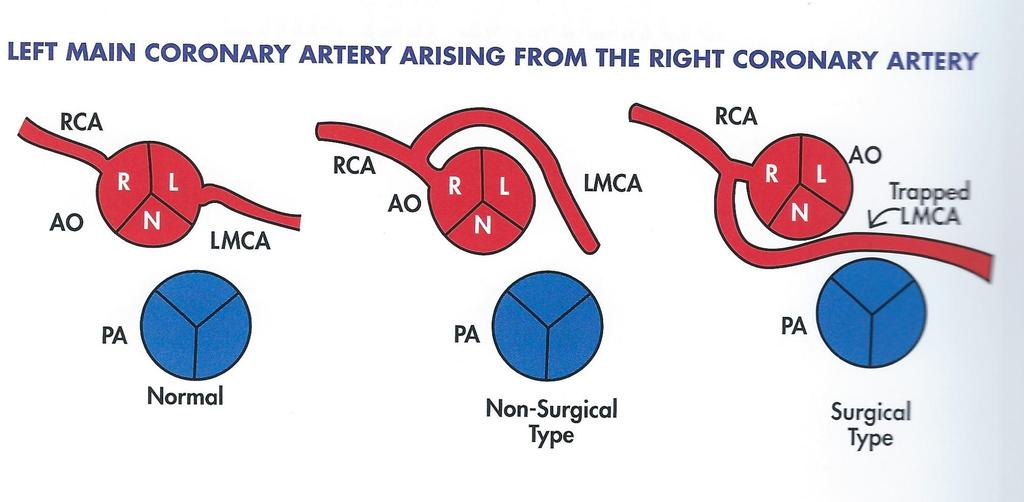
18 Anomalous Coronary Arteries Left Main Coronary Artery Arising from the Pulmonary Artery Most common defect among anomalous coronary arteries and requires surgical intervention Systemic left main coronary SaO2 levels 70% Left Main Coronary Artery Arising from the RCA Less serious defect, there is fully saturated flow in the LMCA If LMCA is trapped between the 2 great arteries it might get trapped and cause symptoms during exercise. RCA Arising from the LMCA Also known as single coronary artery The vessel is fully saturated and no surgical repair is necessary

19
20
21
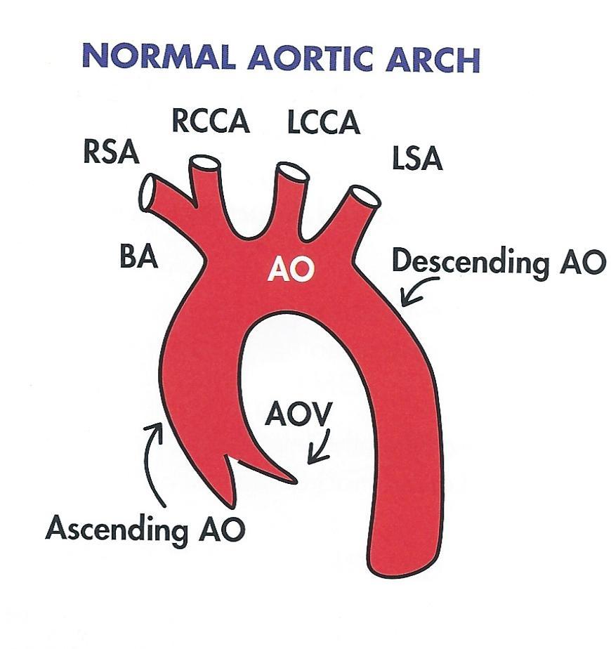
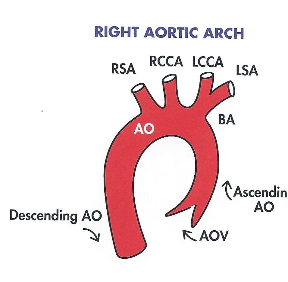
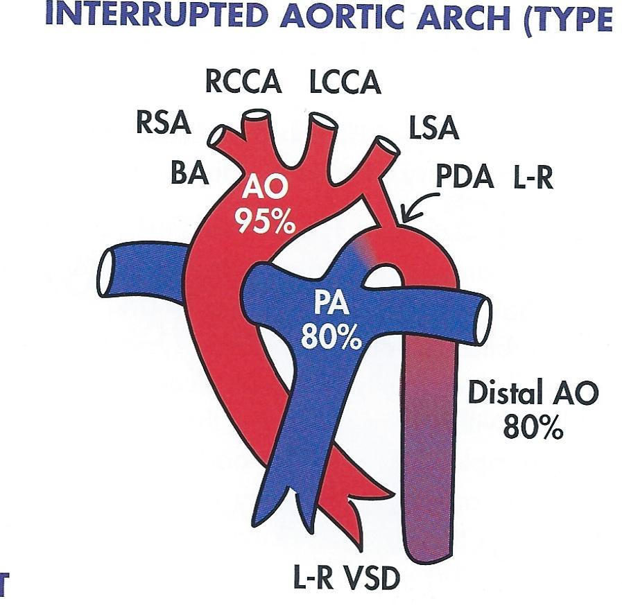
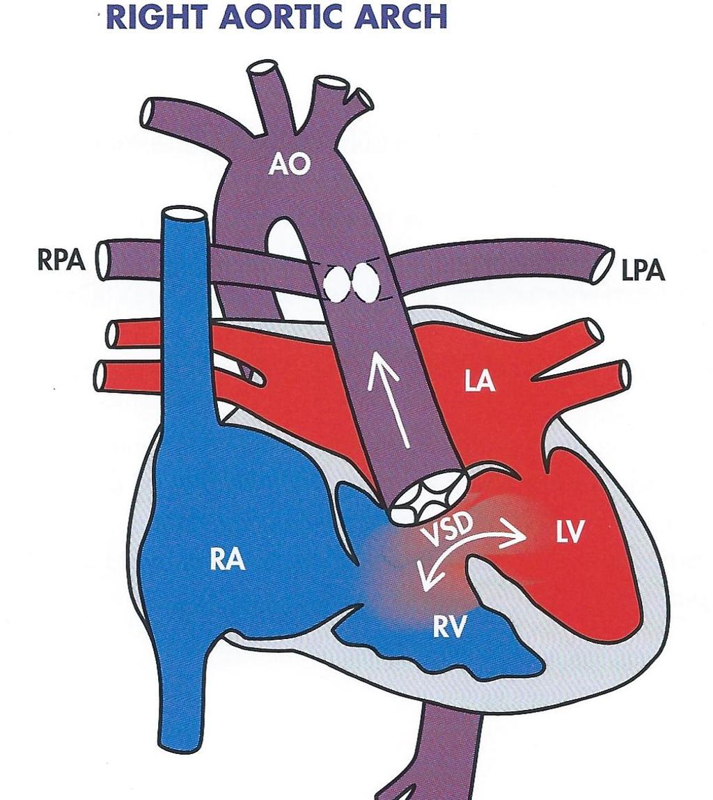
22 Aortic Arch Abnormalities Right aortic Arch Is essentially a mirror image of the normal left aortic arch Is highly associated with TOF There is usually no surgical repair Interrupted Aortic Arch Is an absence of a segment of the descending thoracic aorta Type A-interruption occurs distal to LSA Type B-interruption occurs distal to LCCA and just proximal to LSA Type C-occurs distal to brachiocephalic artery
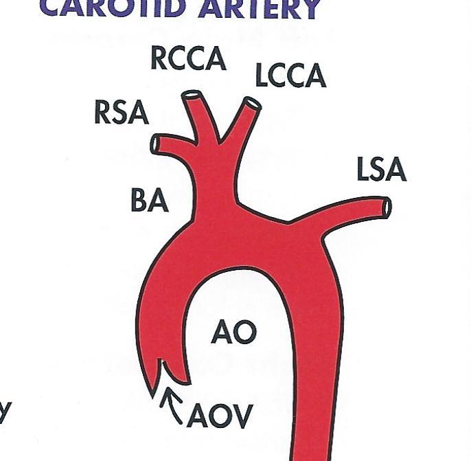
23 Persistent Right Aortic Arch Is a type of vascular ring characterized by the presence of an extra, right sided branch of the aorta that reconnects the aortic trunk distally. Abnormal origin of LCCA This is an abnormality where the LCCA arises from the brachiocephalic artery
24
25

26
27 Persistent Left Superior Vena Cava Is the most common congenital abnormality occurring in association with other congenital defects. There are 2 types: PLSVC drains directly into coronary sinus(90%) PLSVC connects to LA PLSVC draining into RA via CS CS is enlarged RA and RV maybe dilated Coronary Sinus ASD PLSVC connects to LA CS is almost always enlarged Unroofed coronary sinus Associated cardiac abnormalities ASD-VSD TOF Endocardial cushion defect Cor triatriatum
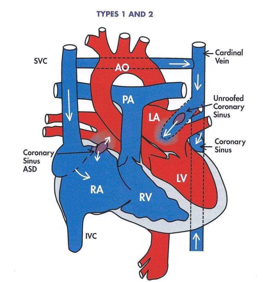
28 Persistent Left SVC
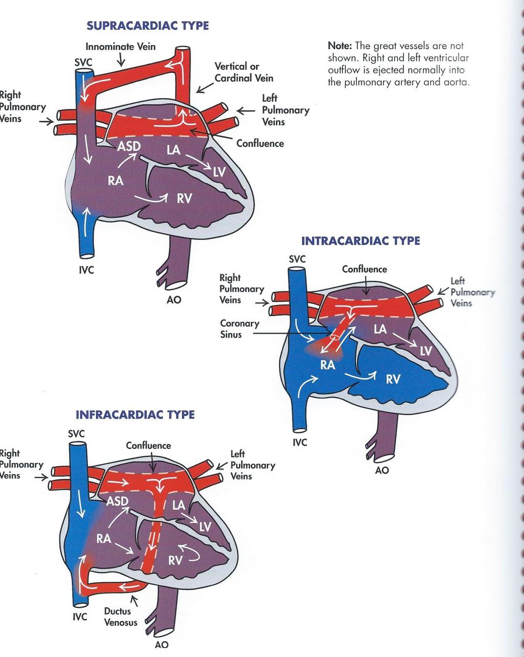
29 Total Anomalous Pulmonary Venous Return Occurs when all 4 pulmonary veins drain oxygenated into the vena cavae Oxygenated and deoxygenated blood myxung takes place in the vena cavae and RA An interatrial communication with R-L shunting is required Types: Supracardiac Intracardiac Infracardiac Echocardiographic documentation of all 4 pulmonary veins is important.
30
31 Tricuspid Atresia Is a cyanotic abnormality characterized by complete failure of the TV to form RA and RV are not connected, PA and AO maybe normally related or transposed An atrial septal defect with R-L shunting will be present RV is hypoplastic RA maybe enlarged and hypertrophic LV is usually enlarged with decreased ventricular function as a result of volume overload Pulmonary circulation maybe dependent on a PDA or VSD Patients with large VSD and no associated pulmonary obstructions will exhibit pulmonary circulation and have minimal cyanosis
32 Associated Cardiac Abnormalities may include: Transposition of the great arteries D-type Transposition of the great arteries L-type Persistent Left Superior Vena Cava Coartation of the aorta Right aortic arch Surgical Repair-Fontan Procedure B-T shunt at 4-6 weeks Glenn procedure at 4-6 months Fontan completion at 2-3 years
33 Tricuspid Atresia with D-Transposition Occurs when the TV fails to form and the AO and PA arise from the wrong ventricles. An atrial septal defect will be present A large VSD with left-to-right shunting will almost always be present Patients with no associated pulmonary obstructions ans a large VSD will usually have abnormally high pulmonary flow and severe pulmonary hypertension Cyanosis will usually be present. Subvalvular aortic stenosis is common Associated cardiac abnormalites: Coartation of the aorta Interrupted aortic arch

34 Tricuspid Atresia with D-transposition
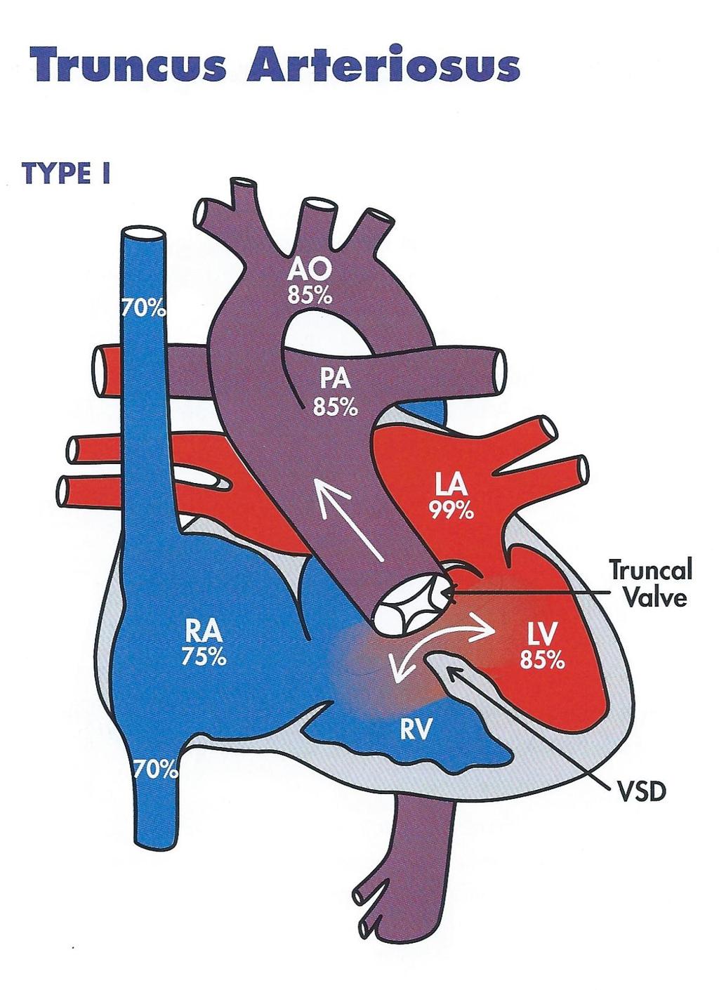
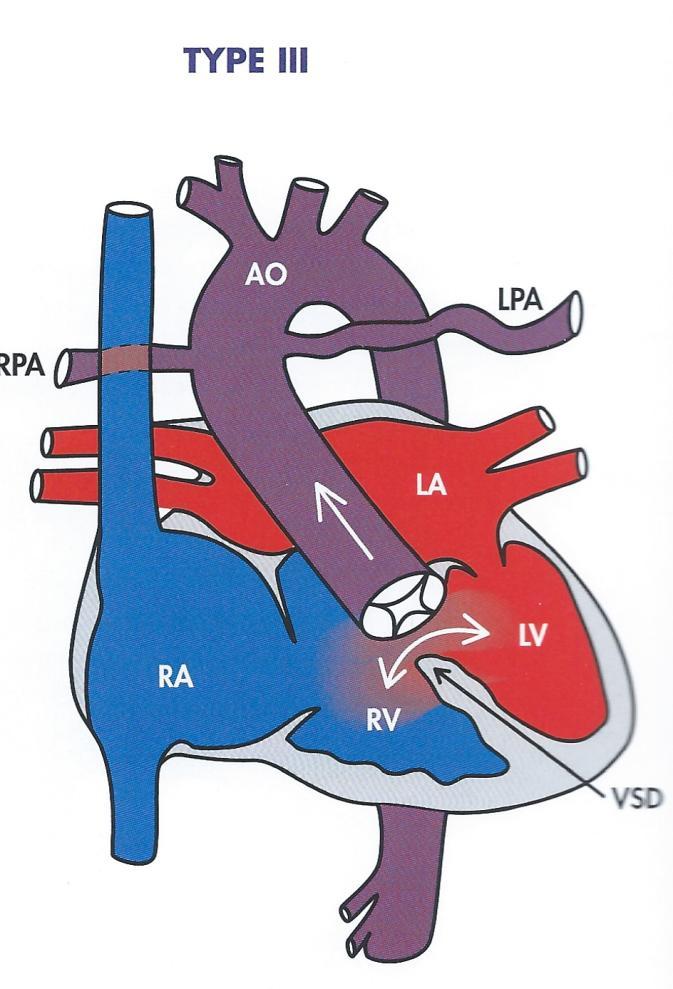
35 Truncus Arteriosus Is a rare abnormality in which the embryologic truncus fails to divide There is a single great artery with a common trunk overriding a large ventricular septal defect. The aortic and pulmonic valves are fused into a large semilunar valve with multiple leaflets called truncal valve Truncus arteriosus is classified into 4 types: Type I: a single pulmonary trunk arises from the common arterial trunk with left and right pulmonary arteries arising from the pulmonary trunk Type II: the left and right pulmonary arteries arise separately from the posterior portion of the common arterial trunl Type III: the left and right pulmonary arteries arise as separate vessels from the lateral portion of the common trunk, widely separated from one another Type IV: this type is considered pulmonary atresia, neither of the pulmonary arteries arises from the common trunk
36
37 Truncus Arteriosus
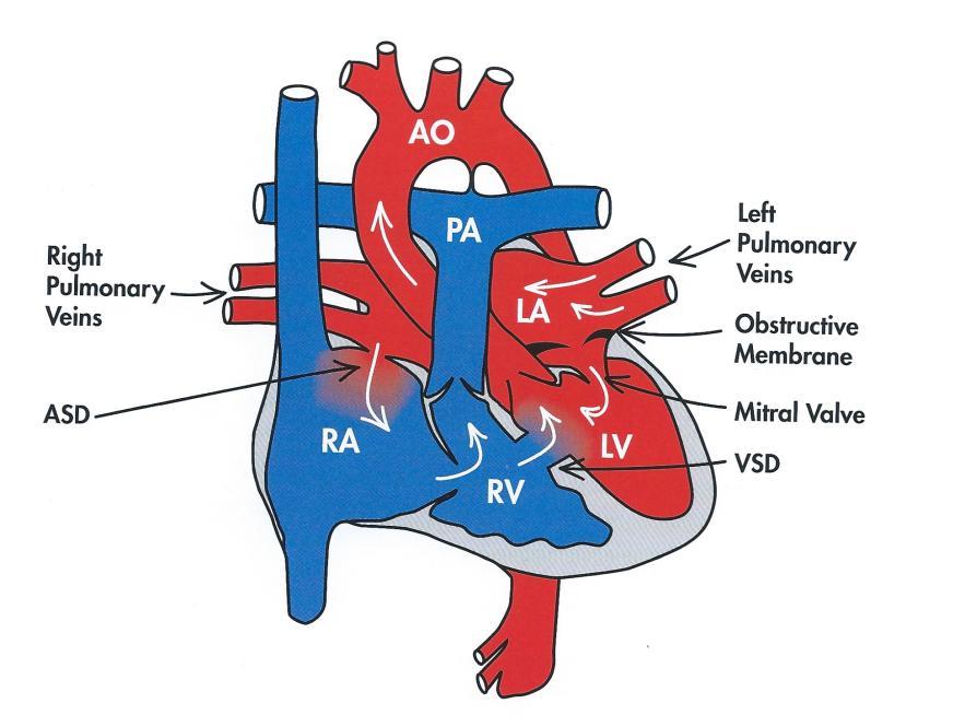
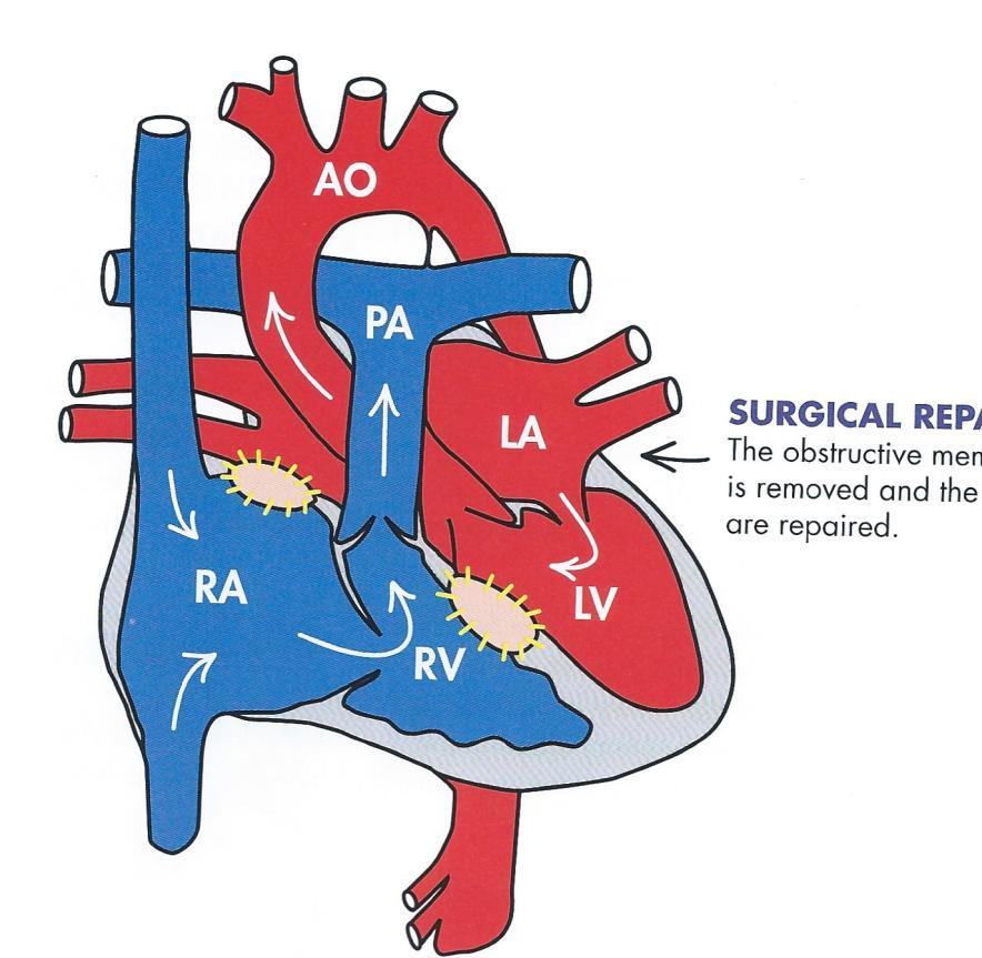
38 Cor Triatriatum Triatrial heart, is a rare abnormality characterized by the presence of a third atrial on the left side resulting from the incomplete absorption of the common pulmonary vein during fetal development There will almost always be an atrial septal defect with left-to-right shunting In most cases a VSD will also be present Echocardiography: Always document pulmonary inflow in multiple views Evaluate the direction of flow and the gradient of any shunts Do not confuse cor triatriatum with mitral stenosis. Mitral stenosis is rarely seen in neonates Associated cardiac abnormalities Coartation of the aorta AV Canal Pulmonary stenosis PAPVR
39 Cor Triatriatum
40 Branch Stenosis and Pulmonary Artery Band Is typically a vasoconstrictive response, is one of the most common pediatric heart conditions, especially in preemies and full term neonates. Usually occurs as a result of PHTN in order to reduce flow in response to elevated pressure in the lung Will usually resolve over time once the underlying cause of PHTN has been corrected Always measure the gradients across the RVOT, PV, MPA, RPA and LPA Any gradient above 16mmHg is considered abnormal

41 Pulmonary Banding
42 Hypoplastic Left Heart Syndrome Is a cyanotic abnormality characterized by severe underdevelopment of the left ventricle aortic valve and mitral valve and ascending aorta. HLHS is the most common cause of death among neonates with congenital heart defects The LV and ascending AO are severely hypoplastic Aortic atresia or aortic stenosis is common A coartation of the aortic isthmus opposite the PDA will usually be present Varying degree of hypoplasia of the left atrium RV is dilated and may be hypertrophic An atrial septal defect will usually be present Intracardiac mixing occurs in the RV. Systemic circulation is dependent on the PDA with right-to-left shunting
43 Surgical Repair is performed in 3 stages: Norwood-stage I (B-T shunt) Norwood-stage II (Glenn procedure) Norwood-stage III (Fontan completion)
44


45 Hypoplastic Left heart syndrome Norwood Stage I
46 Syndromes and Conditions Associated with Congenital Heart Disease (CHD) Apert s syndrome: A genetic disorder resulting from premature closure of the cranial sutures between the bones of the skull. It is characterized by malformations of the skull and facial features and usually involves webbing and/or fusion of the bony structures of the hands and feet. Associated CHD: VSD ASD Cri du chat: A chromosomal disorder characterized by a distinctive cat-like cry and abnormalities of the skull and face. Associated CHD: PDA VSD ASD (secundum) Endocardial cushion defect (AV canal) Tetralogy of Fallot Truncus arteriosus
47 DiGeorge syndrome: A chromosomal disorder resulting in the absence of the thymus and parathyroid glands. Associated CHD: VSD PDA Interrupted aortic arch Tetralogy of Fallot Truncus arteriousus Down syndrome (Trisomy 21): A chromosomal disorder caused by the presence of extra genetic material from chromosome 21. It is characterized by distinctive facial features and mental deficiency ranging mild to severe. Associated CHD: VSD ASD (primum) Endocardial cushion defect (AV canal) Duchenne and Becker muscular dystrophies (MD): Duchenne MD is a type distinguished by progressive muscle degeneration, skeletal distortions (especially of the spine), paralysis, and eventual death. It almost exclusively affects boys. Becker MD is a milder variant of Duchenne. Associated CHD: Cardiomyopathy
48 Eisenmenger s syndrome: A progressive condition that develops in patients with an underlying heart defect with left-to-right shunting and pulmonary hypertension. Eventually, the pressure in the lungs becomes great enough to reverse the direction of the shunt resulting in cyanosis and organ damage. Associated CHD: VSD PDA Single ventricle Endocardial cushion defect (AV canal) Truncus arteriosus Glycogen storage disease: Any one of several genetic metabolic disorders involving defects in the enzymes responsible for the synthesis and degradation of glycogen. Associated CHD: Left ventricular hypertrophy Cardiomyopathy
49 Kawasaki disease: A disease of unknown etiology that may cause inflammation in the coronary arteries leading to coronary aneurysms. It is seen predominantly in children under the age of 5 (80%). Associated CHD: Enlarged coronary arteries, especially the left main coronary artery Marfan s syndrome: A genetic connective tissue disorder characterized by a tall lean body type with disproportionally long arms, legs, fingers, and toes; flat feet; stooped shoulders; and abnormal joint flexibility. Associated CHD: Aortic root dilation Mitral valve prolapse Aortic valve prolapse
50 Maternal risk factors: Both environmental factors and maternal medical conditions may increase the risk of CHD. The most common maternal risks include: Insulin dependent diabetes (VSD, AV canal, truncus arteriosus) Alcohol and drug abuse (VSD, ASD, tetralogy of Fallot) Phenylketonuria, or PKU (coarctation of the aorta, tetralogy of Fallot) Rubella (PDA, pulmonary stenosis, ASD) Lupus (heart block) Antiseizure medications (ASD, tetralogy of Fallot, VSD) Lithium (Ebstein s malformation, ASD, tricuspid atresia) Mitral atresia: The underdevelopment or absence of a normal mitral valve between the left atrium and left ventricle. Associated CHD: Hypoplastic left heart syndrome Single ventricle Noonan s syndrome: A genetic disorder characterized nu short stature, malformations of the breast bone ( pigeon breast ), webbed neck, and distinctive facial features. Associated CHD: Pulmonary stenosis ASD VSD Cardiomyopathy
51 Oral clefts: Cleft lip and cleft palate are the 2 main types of oral clefts. Infants with oral clefts are approximately 16 times more likely to have CHD than infants with normal nasal and palate development. Associated CHD: ASD PDA VSD Tetralogy of Fallot Scimitar syndrome: A variant of partial anomalous pulmonary venous return in which one or both of the right pulmonary veins drains into the inferior vena cava (instead of the left atrium) through a large anomalous pulmonary vein. Associated CHD: Dextrocardia ASD VSD Coarctation of the aorta Tetralogy of Fallot Pulmonary stenosis
52 Resources for this presentation: Congenital heart defects, simplified A Pediatric Registry Review By Ken Heiden RDCS (AE,PE) RVT
Appendix A.1: Tier 1 Surgical Procedure Terms and Definitions
 Appendix A.1: Tier 1 Surgical Procedure Terms and Definitions Tier 1 surgeries AV Canal Atrioventricular Septal Repair, Complete Repair of complete AV canal (AVSD) using one- or two-patch or other technique,
Absent Pulmonary Valve Syndrome
 Absent Pulmonary Valve Syndrome Fact sheet on Absent Pulmonary Valve Syndrome In this condition, which has some similarities to Fallot's Tetralogy, there is a VSD with narrowing at the pulmonary valve.
Congenital Heart Defects
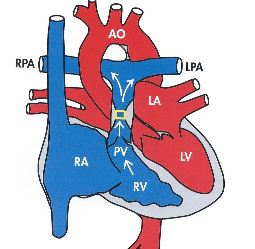
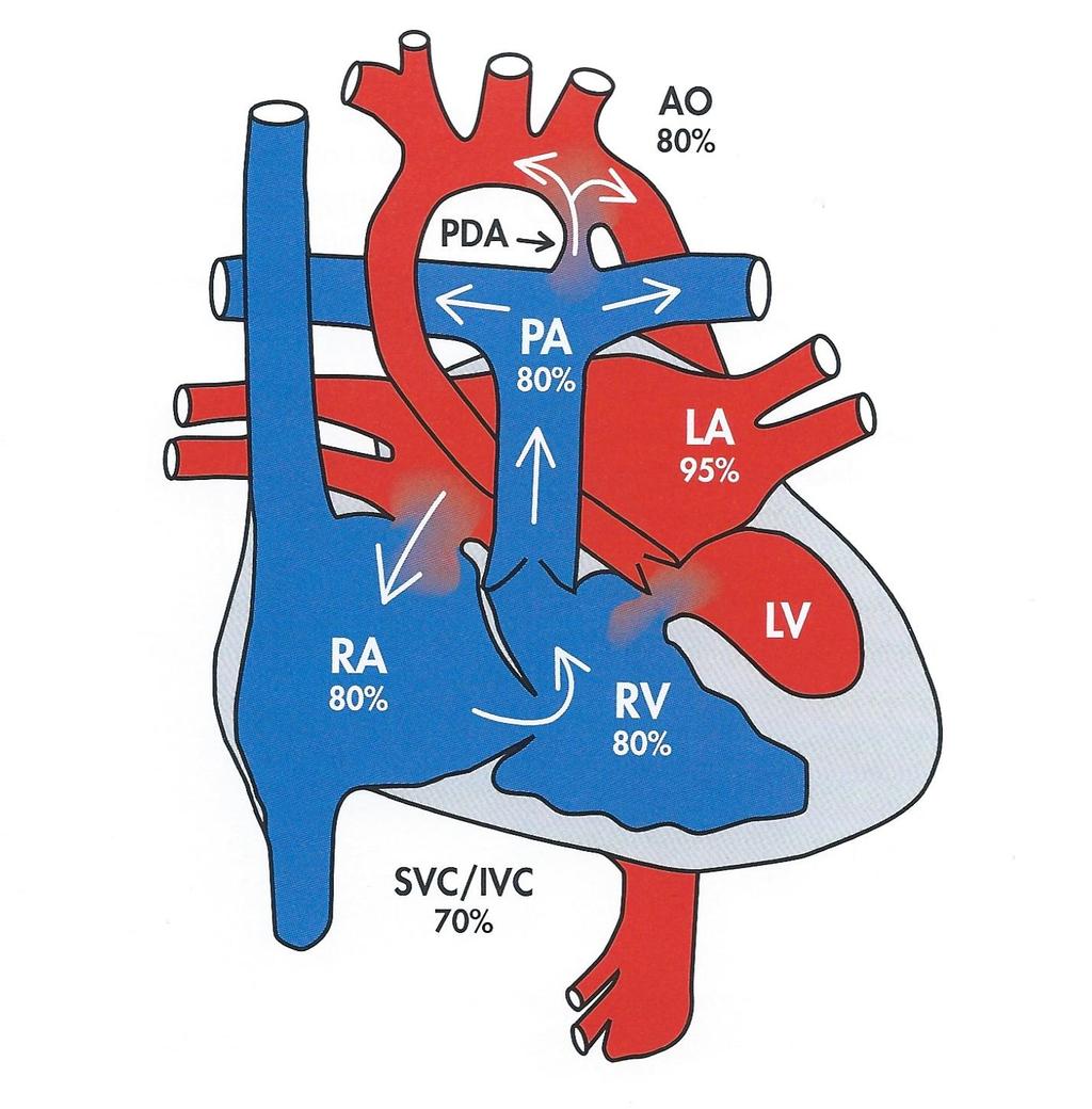
 Normal Heart Congenital Heart Defects 1. Patent Ductus Arteriosus The ductus arteriosus connects the main pulmonary artery to the aorta. In utero, it allows the blood leaving the right ventricle to bypass
Common Defects With Expected Adult Survival:
 Common Defects With Expected Adult Survival: Bicuspid aortic valve :Acyanotic Mitral valve prolapse Coarctation of aorta Pulmonary valve stenosis Atrial septal defect Patent ductus arteriosus (V.S.D.)
Congenital Heart Disease An Approach for Simple and Complex Anomalies
 Congenital Heart Disease An Approach for Simple and Complex Anomalies Michael D. Pettersen, MD Director, Echocardiography Rocky Mountain Hospital for Children Denver, CO None Disclosures 1 ASCeXAM Contains
NASCI 2012 Segmental Analysis
 NASCI 2012 Segmental Analysis Frandics Chan, M.D., Ph.D. Stanford University Medical Center Lucile Packard Department Children s of Radiology Hospital Menagerie of Congenital Cardiac Lesions 1. Absent
Pediatric Echocardiography Examination Content Outline
 Pediatric Echocardiography Examination Content Outline (Outline Summary) # Domain Subdomain Percentage 1 Anatomy and Physiology Normal Anatomy and Physiology 10% 2 Abnormal Pathology and Pathophysiology
Adult Congenital Heart Disease: What All Echocardiographers Should Know Sharon L. Roble, MD, FACC Echo Hawaii 2016
 1 Adult Congenital Heart Disease: What All Echocardiographers Should Know Sharon L. Roble, MD, FACC Echo Hawaii 2016 DISCLOSURES I have no disclosures relevant to today s talk 2 Why should all echocardiographers
Cardiac Catheterization Cases Primary Cardiac Diagnoses Facility 12 month period from to PRIMARY DIAGNOSES (one per patient)
") PRIMARY DIAGNOSES (one per patient) Septal Defects ASD (Atrial Septal Defect) PFO (Patent Foramen Ovale) ASD, Secundum ASD, Sinus venosus ASD, Coronary sinus ASD, Common atrium (single atrium) VSD (Ventricular
PRIMARY DIAGNOSES (one per patient) Septal Defects ASD (Atrial Septal Defect) PFO (Patent Foramen Ovale) ASD, Secundum ASD, Sinus venosus ASD, Coronary sinus ASD, Common atrium (single atrium) VSD (Ventricular
Data Collected: June 17, Reported: June 30, Survey Dates 05/24/ /07/2010
 Job Task Analysis for ARDMS Pediatric Echocardiography Data Collected: June 17, 2010 Reported: Analysis Summary For: Pediatric Echocardiography Exam Survey Dates 05/24/2010-06/07/2010 Invited Respondents
Adel Hasanin Ahmed 1 ASD
 Adel Hasanin Ahmed 1 ASD Atrial septal defect (ASD) is the commonest form of congenital heart disease seen in adults. The commonest form of defect is the secundum ASD, accounting for two thirds of cases,
CONGENITAL HEART DISEASE (CHD)
") CONGENITAL HEART DISEASE (CHD) DEFINITION It is the result of a structural or functional abnormality of the cardiovascular system at birth GENERAL FEATURES OF CHD Structural defects due to specific disturbance
CONGENITAL HEART DISEASE (CHD) DEFINITION It is the result of a structural or functional abnormality of the cardiovascular system at birth GENERAL FEATURES OF CHD Structural defects due to specific disturbance
CMR for Congenital Heart Disease
 CMR for Congenital Heart Disease * Second-line tool after TTE * Strengths of CMR : tissue characterisation, comprehensive access and coverage, relatively accurate measurements of biventricular function/
Children with Single Ventricle Physiology: The Possibilities
 Children with Single Ventricle Physiology: The Possibilities William I. Douglas, M.D. Pediatric Cardiovascular Surgery Children s Memorial Hermann Hospital The University of Texas Health Science Center
Anomalous Systemic Venous Connection Systemic venous anomaly
 World Database for Pediatric and Congenital Heart Surgery Appendix B: Diagnosis (International Paediatric and Congenital Cardiac Codes (IPCCC) and definitions) Anomalous Systemic Venous Connection Systemic
Heart and Lungs. LUNG Coronal section demonstrates relationship of pulmonary parenchyma to heart and chest wall.
 Heart and Lungs Normal Sonographic Anatomy THORAX Axial and coronal sections demonstrate integrity of thorax, fetal breathing movements, and overall size and shape. LUNG Coronal section demonstrates relationship
ECHOCARDIOGRAPHIC APPROACH TO CONGENITAL HEART DISEASE: THE UNOPERATED ADULT
 ECHOCARDIOGRAPHIC APPROACH TO CONGENITAL HEART DISEASE: THE UNOPERATED ADULT Karen Stout, MD, FACC Divisions of Cardiology University of Washington Medical Center Seattle Children s Hospital NO DISCLOSURES
By Dickens ATURWANAHO & ORIBA DAN LANGOYA MAKchs, MBchB CONGENTAL HEART DISEASE
 By Dickens ATURWANAHO & ORIBA DAN LANGOYA MAKchs, MBchB CONGENTAL HEART DISEASE Introduction CHDs are abnormalities of the heart or great vessels that are present at birth. Common type of heart disease
Congenital heart disease: When to act and what to do?
 Leading Article Congenital heart disease: When to act and what to do? Duminda Samarasinghe 1 Sri Lanka Journal of Child Health, 2010; 39: 39-43 (Key words: Congenital heart disease) Congenital heart disease
Coarctation of the aorta
 T H E P E D I A T R I C C A R D I A C S U R G E R Y I N Q U E S T R E P O R T Coarctation of the aorta In the normal heart, blood flows to the body through the aorta, which connects to the left ventricle
Echocardiographic assessment in Adult Patients with Congenital Heart Diseases
 Echocardiographic assessment in Adult Patients with Congenital Heart Diseases Athanasios Koutsakis Cardiologist, Cl. Research Fellow George Giannakoulas Ass. Professor in Cardiology 1st Cardiology Department,
Heart and Soul Evaluation of the Fetal Heart
 Heart and Soul Evaluation of the Fetal Heart Ivana M. Vettraino, M.D., M.B.A. Clinical Associate Professor, Michigan State University College of Human Medicine Objectives Review the embryology of the formation
SURGICAL TREATMENT AND OUTCOME OF CONGENITAL HEART DISEASE
 SURGICAL TREATMENT AND OUTCOME OF CONGENITAL HEART DISEASE Mr. W. Brawn Birmingham Children s Hospital. Aims of surgery The aim of surgery in congenital heart disease is to correct or palliate the heart
3/14/2011 MANAGEMENT OF NEWBORNS CARDIAC INTENSIVE CARE CONFERENCE FOR HEALTH PROFESSIONALS IRVINE, CA. MARCH 7, 2011 WITH HEART DEFECTS
 CONFERENCE FOR HEALTH PROFESSIONALS IRVINE, CA. MARCH 7, 2011 MANAGEMENT OF NEWBORNS WITH HEART DEFECTS A NTHONY C. CHANG, MD, MBA, MPH M E D I C AL D I RE C T OR, HEART I N S T I T U T E C H I LDRE N
Notes: 1)Membranous part contribute in the formation of small portion in the septal cusp.
Membranous part contribute in the formation of small portion in the septal cusp.") Embryology 9 : Slide 16 : There is a sulcus between primitive ventricular and bulbis cordis that will disappear gradually and lead to the formation of one chamber which is called bulboventricular chamber.
Embryology 9 : Slide 16 : There is a sulcus between primitive ventricular and bulbis cordis that will disappear gradually and lead to the formation of one chamber which is called bulboventricular chamber.
Making Sense of Cardiac Views and Imaging Characteristics for 13 Congenital Heart Defects (CHDs)
") Making Sense of Cardiac Views and Imaging Characteristics for 13 Congenital Heart Defects (CHDs) Manny Gaziano, MD, FACOG obimages.net obimages.net@gmail.com Acknowledgements: Krista Wald, RDMS, sonographer,
Making Sense of Cardiac Views and Imaging Characteristics for 13 Congenital Heart Defects (CHDs) Manny Gaziano, MD, FACOG obimages.net obimages.net@gmail.com Acknowledgements: Krista Wald, RDMS, sonographer,
Slide 1. Slide 2. Slide 3 CONGENITAL HEART DISEASE. Papworth Hospital NHS Trust INTRODUCTION. Jakub Kadlec/Catherine Sudarshan INTRODUCTION
 Slide 1 CONGENITAL HEART DISEASE Jakub Kadlec/Catherine Sudarshan NHS Trust Slide 2 INTRODUCTION Most common congenital illness in the newborn Affects about 4 9 / 1000 full-term live births in the UK 1.5
The Chest X-ray for Cardiologists
 Mayo Clinic & British Cardiovascular Society at the Royal College of Physicians, London : 21-23-October 2013 Cases-Controversies-Updates 2013 The Chest X-ray for Cardiologists Michael Rubens Royal Brompton
5.8 Congenital Heart Disease
 5.8 Congenital Heart Disease Congenital heart diseases (CHD) refer to structural or functional heart diseases, which are present at birth. Some of these lesions may be discovered later. prevalence of Chd
Anatomy & Physiology
 1 Anatomy & Physiology Heart is divided into four chambers, two atrias & two ventricles. Atrioventricular valves (tricuspid & mitral) separate the atria from ventricles. they open & close to control flow
Congenital Heart Disease: Physiology and Common Defects
 Congenital Heart Disease: Physiology and Common Defects Jamie S. Sutherell, M.D, M.Ed. Associate Professor, Pediatrics Division of Cardiology Director, Medical Student Education in Pediatrics Director,
Congenital Heart Disease: Cyanotic Lesions. Amitesh Aggarwal
 Congenital Heart Disease: Cyanotic Lesions Amitesh Aggarwal 12 y/o male admitted because of dyspnea and cyanosis Patient has been cyanotic since few months after birth Has episodes of tachypnea and worsening
Foetal Cardiology: How to predict perinatal problems. Prof. I.Witters Prof.M.Gewillig UZ Leuven
 Foetal Cardiology: How to predict perinatal problems Prof. I.Witters Prof.M.Gewillig UZ Leuven Cardiopathies Incidence : 8-12 / 1000 births ( 1% ) Most frequent - Ventricle Septum Defect 20% - Atrium Septum
Congenital Heart Disease II: The Repaired Adult
 Congenital Heart Disease II: The Repaired Adult Doreen DeFaria Yeh, MD FACC Assistant Professor, Harvard Medical School MGH Adult Congenital Heart Disease Program Echocardiography Section, no disclosures
Cardiovascular Pathophysiology: Right to Left Shunts aka Cyanotic Lesions
 Cardiovascular Pathophysiology: Right to Left Shunts aka Cyanotic Lesions Ismee A. Williams, MD, MS iib6@columbia.edu Pediatric Cardiology Learning Objectives To discuss the hemodynamic significance of
Cardiovascular Pathophysiology: Right to Left Shunts aka Cyanotic Lesions Ismee A. Williams, MD, MS Pediatric Cardiology
 Cardiovascular Pathophysiology: Right to Left Shunts aka Cyanotic Lesions Ismee A. Williams, MD, MS iib6@columbia.edu Pediatric Cardiology Learning Objectives To discuss the hemodynamic significance of
Tetralogy of Fallot (TOF) repair, Ventriculotomy Coarctation repair, Other
 repair, Ventriculotomy Coarctation repair, Other") Tier 1 Surgery Form Date of Surgery DD/MM/YYYY Primary Cardiac Procedure Select the patient's primary surgical procedure. If the patient has multiple operating room visits, these should be reported on
Tier 1 Surgery Form Date of Surgery DD/MM/YYYY Primary Cardiac Procedure Select the patient's primary surgical procedure. If the patient has multiple operating room visits, these should be reported on
Uptofate Study Summary
 CONGENITAL HEART DISEASE Uptofate Study Summary Acyanotic Atrial septal defect Ventricular septal defect Patent foramen ovale Patent ductus arteriosus Aortic coartation Pulmonary stenosis Cyanotic Tetralogy
Cardiac Emergencies in Infants. Michael Luceri, DO
 Cardiac Emergencies in Infants Michael Luceri, DO October 7, 2017 I have no financial obligations or conflicts of interest to disclose. Objectives Understand the scope of congenital heart disease Recognize
MEDICAL MANAGEMENT WITH CAVEATS 1. In one study of 50 CHARGE patients with CHD, 75% required surgery. 2. Children with CHARGE may be resistant to chlo
 CARDIOLOGY IN CHARGE SYNDROME: FOR THE PHYSICIAN Angela E. Lin, M.D. Teratology Program/Active Malformation Surveillance, Brigham and Women's Hospital, Old PBBH-B501, 75 Francis St., Boston, MA 02115 alin@partners.org
Index. cardiology.theclinics.com. Note: Page numbers of article titles are in boldface type.
 Index Note: Page numbers of article titles are in boldface type. A ACHD. See Adult congenital heart disease (ACHD) Adult congenital heart disease (ACHD), 503 512 across life span prevalence of, 504 506
World Database for Pediatric and Congenital Heart Surgery Appendix A: Surgical Procedure Terms and Definitions
 World Database for Pediatric and Congenital Heart Surgery Appendix A: Surgical Procedure Terms and Definitions All surgeries are Tier 2 surgeries unless otherwise noted. Anomalous Systemic Venous Connection
The complications of cardiac surgery:
 The complications of cardiac surgery: a walk on the Dark Side? Prof Rik De Decker Red Cross Children s Hospital CME Nov/Dec 2011 http://www.cmej.org.za Why should you care? You are about to leave your
Surgical Interventions for Congenital Heart Disease
 Chapter 46 Surgical Interventions for Congenital Heart Disease Alden M. Parsons, G. William Henry, and Michael R. Mill Our understanding of the complexities of congenital heart disease, a deviation from
ADULT CONGENITAL HEART DISEASE. Stuart Lilley
 ADULT CONGENITAL HEART DISEASE Stuart Lilley More adults than children have congenital heart disease Huge variety of congenital lesions from minor to major Heart failure, re-operation and arrhythmia are
Appendix A.2: Tier 2 Surgical Procedure Terms and Definitions
 Appendix A.2: Tier 2 Surgical Procedure Terms and Definitions Tier 2 surgeries Anomalous Systemic Venous Connection Anomalous Systemic Venous Connection Repair Repair includes a range of surgical approaches,
Hypoplastic Left Heart Syndrome: Echocardiographic Assessment
 Hypoplastic Left Heart Syndrome: Echocardiographic Assessment Craig E Fleishman, MD, FACC, FASE Director, Non-invasive Cardiac Imaging The Hear Center at Arnold Palmer Hospital for Children, Orlando SCAI
The Double Switch Using Bidirectional Glenn and Hemi-Mustard. Frank Hanley
 The Double Switch Using Bidirectional Glenn and Hemi-Mustard Frank Hanley No relationships to disclose CCTGA Interesting Points for Discussion What to do when. associated defects must be addressed surgically:
Pediatric Board Review Congenital Heart Disease. Steven H. Todman, M.D. Pediatric Cardiologist Louisiana State University
 Pediatric Board Review Congenital Heart Disease Steven H. Todman, M.D. Pediatric Cardiologist Louisiana State University Our Mission To discuss various types of congenital heart disease that are commonly
Congenital Heart Disease
 Congenital Heart Disease Mohammed Alghamdi, MD, FRCPC, FAAP, FACC Associate Professor and Consultant Pediatric Cardiology, Cardiac Science King Fahad Cardiac Centre King Saud University INTRODUCTION CHD
List of Videos. Video 1.1
 Video 1.1 Video 1.2 Video 1.3 Video 1.4 Video 1.5 Video 1.6 Video 1.7 Video 1.8 The parasternal long-axis view of the left ventricle shows the left ventricular inflow and outflow tract. The left atrium
Adult Echocardiography Examination Content Outline
 Adult Echocardiography Examination Content Outline (Outline Summary) # Domain Subdomain Percentage 1 2 3 4 5 Anatomy and Physiology Pathology Clinical Care and Safety Measurement Techniques, Maneuvers,
Notes by Sandra Dankwa 2009 HF- Heart Failure DS- Down Syndrome IE- Infective Endocarditis ET- Exercise Tolerance. Small VSD Symptoms -asymptomatic
 Congenital Heart Disease: Notes. Condition Pathology PC Ix Rx Ventricular septal defect (VSD) L R shuntsdefect anywhere in the ventricle, usually perimembranous (next to the tricuspid valve) 30% 1)small
Chapter 2 Cardiac Interpretation of Pediatric Chest X-Ray
 Chapter 2 Cardiac Interpretation of Pediatric Chest X-Ray Ra-id Abdulla and Douglas M. Luxenberg Key Facts The cardiac silhouette occupies 50 55% of the chest width on an anterior posterior chest X-ray
9/8/2009 < 1 1,2 3,4 5,6 7,8 9,10 11,12 13,14 15,16 17,18 > 18. Tetralogy of Fallot. Complex Congenital Heart Disease.
 Current Indications for Pediatric CTA S Bruce Greenberg Professor of Radiology Arkansas Children s Hospital University of Arkansas for Medical Sciences greenbergsbruce@uams.edu 45 40 35 30 25 20 15 10
CYANOTIC CONGENITAL HEART DISEASES. PRESENTER: DR. Myra M. Koech Pediatric cardiologist MTRH/MU
 CYANOTIC CONGENITAL HEART DISEASES PRESENTER: DR. Myra M. Koech Pediatric cardiologist MTRH/MU DEFINITION Congenital heart diseases are defined as structural and functional problems of the heart that are
DEVELOPMENT OF THE CIRCULATORY SYSTEM L E C T U R E 5
 DEVELOPMENT OF THE CIRCULATORY SYSTEM L E C T U R E 5 REVIEW OF CARDIAC ANATOMY Heart 4 chambers Base and apex Valves Pericardial sac 3 layers: epi, myo, endo cardium Major blood vessels Aorta and its
Assessing Cardiac Anatomy With Digital Subtraction Angiography
 485 JACC Vol. 5, No. I Assessing Cardiac Anatomy With Digital Subtraction Angiography DOUGLAS S., MD, FACC Cleveland, Ohio The use of intravenous digital subtraction angiography in the assessment of patients
Patent ductus arteriosus PDA
 Patent ductus arteriosus PDA Is connecting between the aortic end just distal to the origin of the LT sub clavian artery& the pulmonary artery at its bifurcation. Female/male ratio is 2:1 and it is more
Case 47 Clinical Presentation
 93 Case 47 C Clinical Presentation 45-year-old man presents with chest pain and new onset of a murmur. Echocardiography shows severe aortic insufficiency. 94 RadCases Cardiac Imaging Imaging Findings C
Adult congenital heart disease Complex plumbing made simple
 Adult congenital heart disease Complex plumbing made simple James Oliver Leeds Bayer Disclosures Q 1. With respect to atrial septal defects: 1. Severe right heart volume loading is generally a contraindication
ULTRASOUND OF THE FETAL HEART
 ULTRASOUND OF THE FETAL HEART Cameron A. Manbeian, MD Disclosure Statement Today s faculty: Cameron Manbeian, MD does not have any relevant financial relationships with commercial interests or affiliations
Pathological physiology of cardiovascular system Congenital heart diseases
 Pathological physiology of cardiovascular system Congenital heart diseases Rácz Oliver, Sedláková Eva Institute of Pathological Physiology, Medical School, P.J. Šafárik University Oliver Rácz, Eva Sedláková
Born Blue. Anesthesia and CHD. Kristine Faust, CRNA, MS, MBA, DNAP
 Born Blue Anesthesia and CHD Kristine Faust, CRNA, MS, MBA, DNAP Disclosures Disclosures None to Report Objectives Review all congenital defects in which the patient is blue Describe physiology of the
Atrial Septal Defects
 Supplementary ACHD Echo Acquisition Protocol for Atrial Septal Defects The following protocol for echo in adult patients with atrial septal defects (ASDs) is a guide for performing a comprehensive assessment
"Lecture Index. 1) Heart Progenitors. 2) Cardiac Tube Formation. 3) Valvulogenesis and Chamber Formation. 4) Epicardium Development.
 Heart Progenitors. 2) Cardiac Tube Formation. 3) Valvulogenesis and Chamber Formation. 4) Epicardium Development.") "Lecture Index 1) Heart Progenitors. 2) Cardiac Tube Formation. 3) Valvulogenesis and Chamber Formation. 4) Epicardium Development. 5) Septation and Maturation. 6) Changes in Blood Flow during Development.
"Lecture Index 1) Heart Progenitors. 2) Cardiac Tube Formation. 3) Valvulogenesis and Chamber Formation. 4) Epicardium Development. 5) Septation and Maturation. 6) Changes in Blood Flow during Development.
September 28-30, 2018
 September 28-30, 2018 Course Director Optimizing Detection of Congenital Heart Disease: Important Anatomic Cardiac Regions The Top 5 Critical Anatomic Regions in Fetal Cardiac Imaging Alfred Abuhamad,
ISUOG Basic Training. Obtaining & Interpreting Heart Views Correctly Alfred Abuhamad, USA. Basic training. Editable text here
 ISUOG Basic Training Obtaining & Interpreting Heart Views Correctly Alfred Abuhamad, USA Learning Objectives 6, 7 & 8 At the end of the lecture you will be able to: describe how to assess cardiac situs
Surgical Management Of TAPVR. Daniel A. Velez, M.D. Congenital Cardiac Surgeon Phoenix Children s Hospital
 Surgical Management Of TAPVR Daniel A. Velez, M.D. Congenital Cardiac Surgeon Phoenix Children s Hospital No Disclosures Goals Review the embryology and anatomy Review Surgical Strategies for repair Discuss
World Database for Pediatric and Congenital Heart Surgery Appendix A: Surgical Procedure Terms and Definitions
 World Database for Pediatric and Congenital Heart Surgery Appendix A: Surgical Procedure Terms and Definitions All surgeries are Tier 2 surgeries unless otherwise noted. Anomalous Systemic Venous Connection
Suggested Readings. Khonsiari S. Cardiac surgery: safeguards and pittfalls in operative techniques. Philadelphia: Lippincott Williams and
 Suggested Readings Adams FH, Emmanouilides GC, Riemenshneider TA. Heart disease in infants, children, and adolescent. Baltimore: Williams and Wilkins; 1987. Elliot LP. Cardiac imaging in infants, children,
Surgical options for tetralogy of Fallot
 Surgical options for tetralogy of Fallot Serban Stoica FRCS(CTh) MD ACHD study day, 19 September 2017 Anatomy Physiology Children Adults Complications Follow up Anatomy Etienne Fallot (1850-1911) VSD Overriding
UPDATE FETAL ECHO REVIEW
 UPDATE 1 FETAL ECHO REVIEW Study Alert for RDCS Candidates D A V I E S P U B L I S H I N G I N C. Fetal Echo Review Study Alert U P D A T E D A U G U S T 1, 2 0 1 2 Nikki Stahl, RT(R)(M)(CT), RDMS, RVT
Paediatric Cardiology. Acyanotic CHD. Prof F F Takawira
 Paediatric Cardiology Acyanotic CHD Prof F F Takawira Aetiology Chromosomal Down syndrome, T13, T18 Genetic syndromes (gene defects) Velo-Cardio-facial (22 del) Genetic syndromes (undefined aetiology)
4a.i. 4a.ii. Form 12: Pre Transplant Status Report. Height and Weight. Status.
 PHTS - Form : Pre Transplant Report Page of 5 Patient Details Hidden Show Show/Hide Annotations Stickies: Toggle All Toggle Open Toggle Resolved Form : Pre Transplant Report Print this Form t Started Was
Systematic approach to Fetal Echocardiography. Objectives. Introduction 11/2/2015
 Systematic approach to Fetal Echocardiography. Pediatric Echocardiography Conference, JCMCH November 7, 2015 Rajani Anand Objectives Fetal cardiology pre-test Introduction Embryology and Physiology of
Cardiovascular MRI of Adult Congenital Heart Disease
 Cardiovascular MRI of Adult Congenital Heart Disease Anil K. Attili, MD Cardiovascular Magnetic Resonance imaging of Adult Congenital Heart Disease Anil Attili, M.D. Assistant Professor of Radiology /Cardiology
TGA Surgical techniques: tips & tricks (Arterial switch operation)
") TGA Surgical techniques: tips & tricks (Arterial switch operation) Seoul National University Children s Hospital Woong-Han Kim Surgical History 1951 Blalock and Hanlon, atrial septectomy 1954 Mustard et
TGA Surgical techniques: tips & tricks (Arterial switch operation) Seoul National University Children s Hospital Woong-Han Kim Surgical History 1951 Blalock and Hanlon, atrial septectomy 1954 Mustard et
Screening for Critical Congenital Heart Disease
 Screening for Critical Congenital Heart Disease Caroline K. Lee, MD Pediatric Cardiology Disclosures I have no relevant financial relationships or conflicts of interest 1 Most Common Birth Defect Most
Adult Congenital Heart Disease: The New Reality. Disclosures
 Adult Congenital Heart Disease: The New Reality Kathryn Rouine-Rapp, MD Professor of Anesthesia Disclosures I have nothing to disclose 1 Outline Historic perspective Our reality Common lesions Guidelines
When is Risky to Apply Oxygen for Congenital Heart Disease 부천세종병원 소아청소년과최은영
 When is Risky to Apply Oxygen for Congenital Heart Disease 부천세종병원 소아청소년과최은영 The Korean Society of Cardiology COI Disclosure Eun-Young Choi The author have no financial conflicts of interest to disclose
Introduction. Pediatric Cardiology. General Appearance. Tools of Assessment. Auscultation. Vital Signs
 Introduction Pediatric Cardiology An introduction to the pediatric patient with heart disease: M-III Lecture Douglas R. Allen, M.D. Assistant Professor and Director of Community Pediatric Cardiology at
Heart Development and Congenital Heart Disease
 Heart Development and Congenital Heart Disease Sally Dunwoodie s.dunwoodie@victorchang.edu.au Developmental and Stem Cell Biology Division Victor Chang Cardiac Research Institute for the heart of Australia...
Outline. Congenital Heart Disease. Special Considerations for Special Populations: Congenital Heart Disease
 Special Considerations for Special Populations: Congenital Heart Disease Valerie Bosco, FNP, EdD Alison Knauth Meadows, MD, PhD University of California San Francisco Adult Congenital Heart Program Outline
Supplemental Table 1. ICD-9 Codes for Diagnoses and Procedures
 Supplemental Table 1. ICD-9 Codes for Diagnoses and Procedures ICD-9 Code Description Heart Failure 402.01 Malignant hypertensive heart disease with heart failure 402.11 Benign hypertensive heart disease
Reconstruction of right ventricular outflow with a valved homograft conduit
 Thorax (1974), 29, 617. Reconstruction of right ventricular outflow with a valved homograft conduit D. J. WHEATLEY, S. PRUSTY, and D. N. ROSS Department of Surgery, National Heart Hospital, London WI Wheadey,
The Rastelli procedure has been traditionally used for repair
 En-bloc Rotation of the Truncus Arteriosus A Technique for Complete Anatomic Repair of Transposition of the Great Arteries/Ventricular Septal Defect/Left Ventricular Outflow Tract Obstruction or Double
Giovanni Di Salvo MD, PhD, FESC Second University of Naples Monaldi Hospital
 Giovanni Di Salvo MD, PhD, FESC Second University of Naples Monaldi Hospital VSD is one of the most common congenital cardiac abnormalities in the newborn. It can occur as an isolated finding or in combination
What is the Definition of Small Systemic Ventricle. Hong Ryang Kil, MD Department of Pediatrics, College of Medicine, Chungnam National University
 What is the Definition of Small Systemic Ventricle Hong Ryang Kil, MD Department of Pediatrics, College of Medicine, Chungnam National University Contents Introduction Aortic valve stenosis Aortic coarctation
Low-dose prospective ECG-triggering dual-source CT angiography in infants and children with complex congenital heart disease: first experience
 Low-dose prospective ECG-triggering dual-source CT angiography in infants and children with complex congenital heart disease: first experience Ximing Wang, M.D., Zhaoping Cheng, M.D., Dawei Wu, M.D., Lebin
ORIGINAL RESEARCH PAPER
 ORIGINAL RESEARCH PAPER ROLE OF CT PULMONARY ANGIOGRAPHY IN CONGENITAL HEART DISEASES IN PAEDIATRIC POPULATION Radiology KEY WORDS: Congenital heart disease, CT pulmonary angiography, pediatric heart disease,
Preoperative Echocardiographic Assessment of Uni-ventricular Repair
 Preoperative Echocardiographic Assessment of Uni-ventricular Repair Salem Deraz, MD Pediatric Cardiologist, Aswan Heart Centre Magdi Yacoub Heart Foundation Uni-ventricular repair A single or series of
Adults with Congenital Heart Disease
 Adults with Congenital Heart Disease Edward K. Rhee, MD, FACC Director, Pediatric-Adult Congenital Arrhythmia Service SJHMC Disclosures & Disclaimer I have no lucrative financial relationships with industry
Cardiovascular Imaging Interactive Case Discussions
 Cardiovascular Imaging Interactive Case Discussions Satinder Singh MD, FCCP Professor of Radiology & Medicine (Division of CV Diseases) Chief Cardiopulmonary Radiology Director Cardiac CT UAB SCBT.MR September
Cardiac CT in Infants with Congenital heart disease Sunrise Session. LaDonna Malone, MD May 17, 2018
 Cardiac CT in Infants with Congenital heart disease Sunrise Session LaDonna Malone, MD May 17, 2018 None Disclosures Objectives Describe cardiac CT techniques used in infants with congenital heart disease.
The role of intraoperative TOE in congenital cardiac surgery
 The role of intraoperative TOE in congenital cardiac surgery Justiaan Swanevelder Dept of Anaesthesia Groote Schuur and Red Cross War Memorial Children s Hospitals University of Cape Town, South Africa
Most common fetal cardiac anomalies
 Most common fetal cardiac anomalies Common congenital heart defects CHD % of cardiac defects Chromosomal Infants Fetuses anomaly (%) 22q11 deletion (%) VSD 30 5~10 20~40 10 PS 9 5 (PA w/ VSD) HLHS 7~9
Cardiac Radiology In-Training Test Questions for Diagnostic Radiology Residents
 Cardiac Radiology In-Training Test Questions for Diagnostic Radiology Residents March, 2013 Sponsored by: Commission on Education Committee on Residency Training in Diagnostic Radiology 2013 by American