How to Assess and Treat Obstructive Lesions
|
|
|
- Buddy Sullivan
- 6 years ago
- Views:
Transcription
1 How to Assess and Treat Obstructive Lesions Erwin Oechslin, MD, FESC, FRCPC, Director, Congenital Cardiac Centre for Adults Peter Munk Cardiac Centre University Health Network/Toronto General Hospital Toronto, ON, Canada
2 Outline Background Diagnostic Work-Up General Principles Doppler echocardiography CMR / CT Exercise testing Specific Lesions LVOT obstruction Right ventricular outflow tract obstruction
3 BACKGROUND RIGHT and LEFT ventricular outflow tract obstructions are common Simple vs complex Level of obstruction Subvalvular Valvular Supravalvular Combined lesions
4 Diagnostic Work-Up (1) GENERAL PRINCIPLES Comprehensive CLINICAL assessment Medical history Family history! Learning difficulties! Physical examination Murmurs / ejection click? BD difference upper and lower extremities / brachiofemoral pulse delay Syndromic appearance Turner / Noonan syndrome, microdeletion 22q11.2 syndrome amongst others
5 Diagnostic Work-Up (2) GENERAL PRINCIPLES Non-invasive imaging Chest X-ray (not routinely; when clinically indicated) Doppler echocardiography Cardiovascular MR (CMR) Computed tomography (CT) Exercise testing Diagnostic heart catheterization To verify the gradients across the LVOT / RVOT
6 Doppler Echocardiography (1) FIRST LINE NON-INVASIVE INVESTIGATION Anatomy / morphology (intracardiac!) Type of obstruction Level of obstruction (isolated/multiple levels) Functional assessment Diameters / volumes (left ventricle) Ventricular hypertrophy (concentric vs eccentric) Ventricular function (systolic and diastolic function) GRADIENTS across RVOT / LVOT Fixed vs dynamic obstruction RV systolic pressure
7 Doppler Echocardiography (2) LIMITATIONS and PRECAUTIONS Investigator dependence Expertise in CHD is required! Poor window / echo quality Doppler gradients can be misleading RVOT obstruction; serial stenoses at multiple levels Tubular stenosis (aortic coarctation/conduits) Pressure recovery Modified Bernoulli equation does not apply to tubular stenosis / stenoses in series RVSP is more accurate to estimate severity of RVOT obstruction
8 Specific Indications for CMR CMR is Superior to Echocardiography RVOT obstruction RV to PA conduits Extracardiac anatomy Pulmonary arteries (peripheral PA stenosis, aneurysm) Aorta (supravalvular AS, aortic coarctation, aneurysm, dissection)
9 Exercise Testing Important role in the timing of interventions and reinterventions in asymptomatic patients Asymptomatic Patient Exercise Testing* Normal *Symptoms Inadequate BP response Regular Follow-up Intervention
10 Outline Background Diagnostic Work-Up General Principles Doppler echocardiography CMR / CT Exercise testing Specific Lesions LVOT obstruction Right ventricular outflow tract obstruction
11 Left Ventricular Outflow Tract Obstruction Level of obstruction: Subvalvular Valvular (75%) Supravalvular
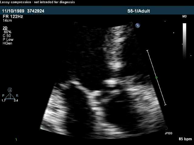
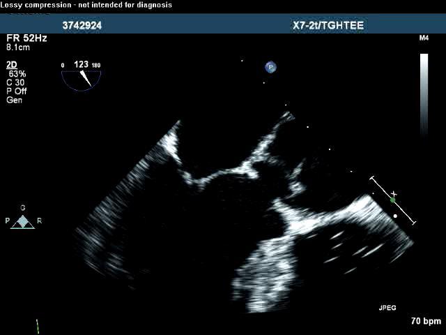
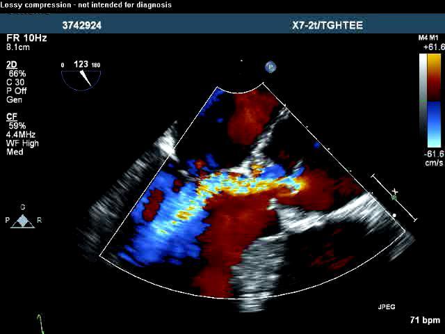
12 Subaortic Stenosis (1) BACKGROUND Presentation Isolated Associated defects: VSD, AVSD, conotruncal anomalies Shone complex! Prevalence: 6-7% in GUCH population Morphologic appearance Fibrous ridge Fibromuscular narrowing Differential: HCM
13 General Principles Subaortic Stenosis (2) DIAGNOSTIC WORK-UP Doppler Echocardiography Morphology / associated congenital anomalies(!) Functional assessment: Overestimation / underestimation of the gradient (tubular stenosis / localization of VSD) Aortic regurgitation TEE / 3-D echocardiography: to characterize complex anatomy of the LVOT
14
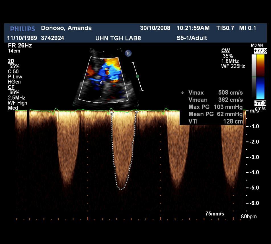
15 Subaortic Stenosis (3) INDICATIONS FOR INTERVENTION Peak gradient >50 / mean gradient >30 mmhg Symptoms?? Symptoms and peak gradient >50mmHg or mean gradient >30 mmhg or If combined with progressive AR

16 Subaortic Stenosis (3) INDICATIONS FOR INTERVENTION Peak gradient <50 / mean grad. <30mmHg and progressive AR and LV-ESD > 50 mm and LV-EF <55% Class I C????????

17 Subaortic Stenosis (4) INDICATIONS FOR INTERVENTION?? Mean grad. of 30mmHg Class IIb C Mean grad.<30mmhg, if LV hypertrophy Pregnancy considered Strenuous exercise Class IIb C Trivial SAS to prevent AR Class III C????????
18 Supravalvular Aortic Stenosis (1) BACKGROUND Rare (<7% of all fixed forms of LVOTO) Morphologic appearance: Localized fibrous diaphragm (distal to ostia of the coronary arteries) External hourglass deformity with luminal narrowing Diffuse stenosis of the ascending aorta Association with Williams-Beuren syndrome Diffuse hypoplasia of the entire aorta Involvement of coronary arteries Stenosis of major branches of the aorta/pas
19 Supravalvular Aortic Stenosis (2) DIAGNOSTIC WORK-UP General Principles Doppler echocardiography CMR Anatomic description Overestimation of the gradient! Precise anatomic description Identification of additional lesions (branches of the aorta/pas/renal arteries Exercise testing

20 Supravalvular AS (4) INDICATIONS FOR INTERVENTION????

21 Supravalvular AS (5) INDICATIONS FOR INTERVENTION??????
22 Aortic Coarctation (1) BACKGROUND Generalized AORTOPATHY! Cystic media necrosis with increased stiffness of the aorta and carotid arteries Aortic dilatation Endothelial dysfunction Morphologic appearance: Discrete narrowing Diffuse hypoplasia / geometry of the aortic arch
23 Aortic Coarctation (2) BACKGROUND Presentation: Isolated Associated CHD: Bicuspid aortic valve (up to 85%) Shone complex! Turner syndrome Williams-Beuren syndrome Congenital Rubella syndrome Non-congenital pathologies: Takayasu-arteritis, trauma
24 Aortic Coarctation (3) DIAGNOSTIC WORK-UP Specific considerations Hemodynamically relevant aortic coarctation: BP difference > 20 mmhg between right arm and lower extremities Brachio-femoral pulse delay Palpable collaterals Chest X-Ray: Rib-notching Kinking / double contouring of the descending aorta (figure -3 sign) Dilatation of ascending aorta
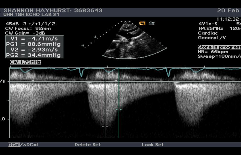
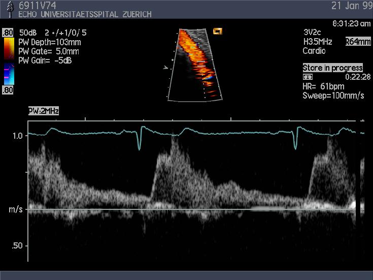
25 Aortic Coarctation (4) DIAGNOSTIC WORK-UP Doppler Echocardiography Gradients are not useful! Tubular stenosis Aortic collaterals in native aortic coarctation Increased systolic flow velocity due to the lack of aortic compliance after repair even in the absence of stenosis Diastolic run-off : most reliable sign for significant aortic coarctation
26 ??????
27 Aortic Coarctation (6) DIAGNOSTIC WORK-UP CMR / CT Preferred diagnostic modality to evaluate the entire aorta (geometry of the arch!) Detection Site, extent, and degree of stenosis (collaterals!) Complications after repair (aneurysm, re-stenosis)

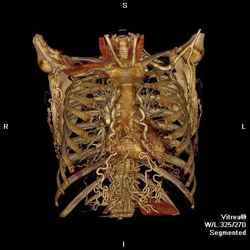
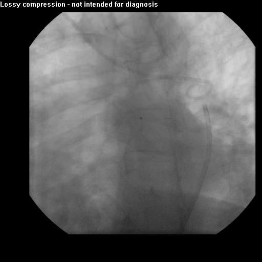
28 Native aortic coarctation
29 Aortic Coarctation (6) DIAGNOSTIC WORK-UP Cardiac Catheterization Reference method to evaluate severity of aortic coarctation Hemodynamically relevant stenosis Peak-to-peak gradient >20 mmhg in the absence of well-developed collaterals
30 Aortic Coarctation (7) INDICATIONS FOR INTERVENTION Class a Level b All patients with a non-invasive pressure difference > 20 mmhg between upper and lower limbs, regardless of symptoms but with upper limb hypertension (> 140/90 mmhg in adults), pathologic blood pressure response during exercice, or significant LVH should have intervention I C Peak to peak grad > 20mmHg Peak to peak grad < 20 mmhg if significant collaterals Peak to peak grad > 20mmHg and hypertension PG < 20 mmhg and collateral flow!
31 Aortic Coarctation (9) SURGERY vs BALLOON DILATATION/STENT Aortic coarctation Native coarctation Recurrent/residual aortic coarctation Anatomy suitable? No Yes Surgery Balloon/stent
32 Aortic Coarctation (9) CO-MORBIDITIES Aortic stenosis / regurgitation (bicuspid aortic valve!) Aneurysm of the ascending aorta Diameter > 50 mm or 27.5 mm/m 2 Aneurysm at previous aortic site Aneurysm of the circle of Willis
33 Aortic Coarctation (10) FOLLOW-UP At least every 2 years at a GUCH centre Long term complications Arterial hypertension (geometry of the aortic arch normal, gothic, crenel may play a role) Role of exercise induced hypertension? Recurring or residual aortic coarctation Aneurysm of the ascending aorta or at the intervention site (patch repair!) risk for rupture/death Aortic valve anomaly / Shone complex Aneurysm of the circle of Willis
34 Outline Background Diagnostic Work-Up General Principles Doppler echocardiography CMR / CT Exercise testing Specific Lesions LVOT obstruction Right ventricular outflow tract obstruction
35 RVOT Obstruction (1) BACKGROUND Level of stenosis Subinfundibular: associated with VSD! Infundibular: secondary to valvular PS or associated with VSD (!), TOF Dynamic obstruction Valvular: 7-12% of all CHDs Intrinsic pulmonary wall abnormality Supravalvular: isolated (seldom) or associated with syndromes Noonan, Williams-Beuren, Keutel, congenital Rubella syndrome
36 RVOT Obstruction (2) NATURAL HISTORY Mild valvular PS does not progress Progressive stenosis Infundibular stenosis Moderate valvular stenosis (calcification of the cusps) Reactive infundibular hypertrophy with dynamic obstruction of the RVOT Supravalvular PS
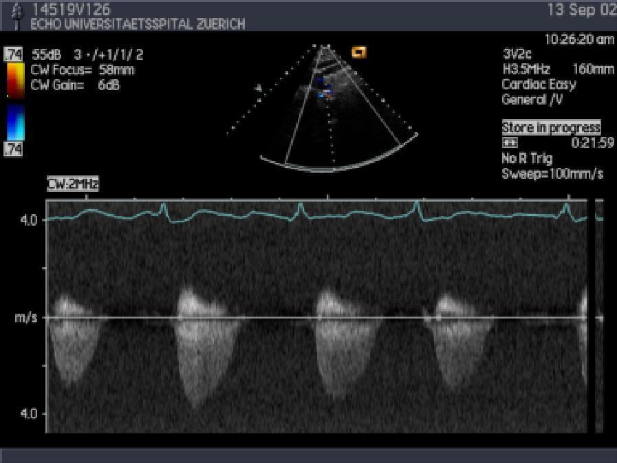
37 Chest X-ray RVOT Obstruction (3) DIAGNOSIC WORK-UP Calcification of the cups Dilatation of pulmonary trunk / right heart Doppler echocardiography - Pitfalls Overestimation of the gradient (!) Stenoses in series / tubular stenosis Contamination with VSD jet Underestimation of the gradient (!) Doppler probe is not aligned to blood flow (e.g. double chambered RV)

38 Subvalvular and Valvular PS
39 RVOT Obstruction (4) HOW TO ASSESS SEVERITY? Severity based on peak gradient across RVOT mild (peak gradient < 36 mm Hg, peak velocity < 3m/s) moderate (36 to 64 mm Hg; 3-4 m/s) severe (>64 mm Hg, peak velocity >4m/s) TR velocity with estimation of RV systolic pressure should always be used in addition when assessing severity
40 RVOT Obstruction (5) DIAGNOSTIC WORK-UP CMR / CT Infundibular and supravalvular/peripheral PS (Assessment of conduits) Dilatation of PAs Differential blood flow to the right/left lung (Nuclear techniques) Differential blood flow to the right/left lung Catheterization Confirmation of the level and severity of stenosis
41 RVOT Obstruction (6) INDICATIONS FOR INTERVENTION Balloon dilatation Pulmonary valvular stenosis (nondysplastic pulmonic valve) Peripheral PS (balloon dilatation plus stent) Surgery Infundibular stenosis Severely dysplastic pulmonic valve Hypoplastic pulmonic annulus Associated lesions requiring surgery (severe PR, TR)

42 RVOT Obstruction (7) INDICATIONS FOR INTERVENTION VALVULAR PS: Symptoms: PG / MG >50/>30 mmhg Less than moderate PR No Symptoms: PG / MG >60/>40 mmhg and less than moderate PR
43 VALVULAR PS (8) BALLOON VALVOTOMY vs SURGERY ASYMPTOMATIC Valvular PS Suitable for Balloon Valvuloplasty? YES NO PVR is the only option Balloon Valvuloplasty when PG > 64 mmhg Class I C Surgery when RVSP > 80 mmhg TR velocity > 4.3 m/sec
44 Summary Comprehensive assessment Clinical assessment Non-invasive +/- invasive imaging Exercise testing Critical interpretation of Doppler gradients! RVSP is more accurate to estimate severity of RVOTO Guidelines are a useful handbook. It is the cook who makes the difference
RVOTO adult and post-op
 Right ventricular outflow tract obstruction in the adult: native and post-op Helmut Baumgartner Westfälische Wilhelms-Universität Münster Adult Congenital and Valvular Heart Disease Center University of
Cardiac MRI in ACHD What We. ACHD Patients
 Cardiac MRI in ACHD What We Have Learned to Apply to ACHD Patients Faris Al Mousily, MBChB, FAAC, FACC Consultant, Pediatric Cardiology, KFSH&RC/Jeddah Adjunct Faculty, Division of Pediatric Cardiology
Debate in Management of native COA; Balloon Versus Surgery
 Debate in Management of native COA; Balloon Versus Surgery Dr. Amira Esmat, El Tantawy, MD Professor of Pediatrics Consultant Pediatric Cardiac Interventionist Faculty of Medicine Cairo University 23/2/2017
HISTORY. Question: What category of heart disease is suggested by the fact that a murmur was heard at birth?
 HISTORY 23-year-old man. CHIEF COMPLAINT: Decreasing exercise tolerance of several years duration. PRESENT ILLNESS: The patient is the product of an uncomplicated term pregnancy. A heart murmur was discovered
Echocardiographic assessment in Adult Patients with Congenital Heart Diseases
 Echocardiographic assessment in Adult Patients with Congenital Heart Diseases Athanasios Koutsakis Cardiologist, Cl. Research Fellow George Giannakoulas Ass. Professor in Cardiology 1st Cardiology Department,
PATENT DUCTUS ARTERIOSUS (PDA)
") PATENT DUCTUS ARTERIOSUS (PDA) It is a channel that connect the pulmonary artery with the descending aorta (isthumus part). It results from the persistence of patency of the fetal ductus arteriosus after
PATENT DUCTUS ARTERIOSUS (PDA) It is a channel that connect the pulmonary artery with the descending aorta (isthumus part). It results from the persistence of patency of the fetal ductus arteriosus after
ECHOCARDIOGRAPHIC APPROACH TO CONGENITAL HEART DISEASE: THE UNOPERATED ADULT
 ECHOCARDIOGRAPHIC APPROACH TO CONGENITAL HEART DISEASE: THE UNOPERATED ADULT Karen Stout, MD, FACC Divisions of Cardiology University of Washington Medical Center Seattle Children s Hospital NO DISCLOSURES
Left Ventricular Outflow Tract Obstruction
 Left Ventricular Outflow Tract Obstruction Department of Paediatrics Left Ventricular Outflow Tract Obstruction Subvalvular aortic stenosis Aortic Stenosis Supravalvular aortic stenosis Aortic Coarctation
Aortic Stenosis: LVOT Obstruction
 Aortic Stenosis: LVOT Obstruction Raymond Stainback, MD 7 th annual Houston Echo Review 2016: Boot Camp for the Echo Board Murmur: Additional heart or vascular sound due to normal or abnormal turbulent
Difficult echocardiography in an adult patient with repaired congenital heart disease
 Difficult echocardiography in an adult patient with repaired congenital heart disease Markus Schwerzmann, MD Center for Congenital Heart Disease University of Bern Clinical history 19 year old male, 168
Comprehensive Hemodynamics By Doppler Echocardiography. The Echocardiographic Swan-Ganz Catheter.
 Comprehensive Hemodynamics By Doppler Echocardiography. The Echocardiographic Swan-Ganz Catheter. Itzhak Kronzon, MD, FASE, FACC, FESC, FAHA, FACP, FCCP North Shore HS, LIJ/Lenox Hill Hospital, New York
Echo Doppler Assessment of Right and Left Ventricular Hemodynamics.
 Echo Doppler Assessment of Right and Left Ventricular Hemodynamics. Itzhak Kronzon, MD, FASE, FACC, FESC, FAHA, FACP, FCCP Northwell, Lenox Hill Hospital, New York Professor of Cardiology Hofstra University
TGA atrial vs arterial switch what do we need to look for and how to react
 TGA atrial vs arterial switch what do we need to look for and how to react Folkert Meijboom, MD, PhD, FES Dept ardiology University Medical entre Utrecht The Netherlands TGA + atrial switch: Follow-up
Comprehensive Echo Assessment of Aortic Stenosis
 Comprehensive Echo Assessment of Aortic Stenosis Smonporn Boonyaratavej, MD, MSc King Chulalongkorn Memorial Hospital Bangkok, Thailand Management of Valvular AS Medical and interventional approaches to
Adult Congenital Heart Disease: What All Echocardiographers Should Know Sharon L. Roble, MD, FACC Echo Hawaii 2016
 1 Adult Congenital Heart Disease: What All Echocardiographers Should Know Sharon L. Roble, MD, FACC Echo Hawaii 2016 DISCLOSURES I have no disclosures relevant to today s talk 2 Why should all echocardiographers
Aortic Coarctation Imaging and Management in Adults. Michael D. Hope, MD
 Aortic Coarctation Imaging and Management in Adults Michael D. Hope, MD 1 Background 2 Imaging - Morphology 3 Imaging - Hemodynamics 4 Associations and Complications Campbell M. British Heart Journal 1970
Aortic Stenosis: Spectrum of Disease, Low Flow/Low Gradient and Variants
 Aortic Stenosis: Spectrum of Disease, Low Flow/Low Gradient and Variants Martin G. Keane, MD, FASE Professor of Medicine Lewis Katz School of Medicine at Temple University Basic root structure Parasternal
CMR for Congenital Heart Disease
 CMR for Congenital Heart Disease * Second-line tool after TTE * Strengths of CMR : tissue characterisation, comprehensive access and coverage, relatively accurate measurements of biventricular function/
LEFT VENTRICULAR OUTFLOW OBSTRUCTION WITH A VSD: OPTIONS FOR SURGICAL MANAGEMENT
 LEFT VENTRICULAR OUTFLOW OBSTRUCTION WITH A VSD: OPTIONS FOR SURGICAL MANAGEMENT 10-13 March 2017 Ritz Carlton, Riyadh, Saudi Arabia Zohair AlHalees, MD Consultant, Cardiac Surgery Heart Centre LEFT VENTRICULAR
P = 4V 2. IVC Dimensions 10/20/2014. Comprehensive Hemodynamic Evaluation by Doppler Echocardiography. The Simplified Bernoulli Equation
 Comprehensive Hemodynamic Evaluation by Doppler Echocardiography Itzhak Kronzon, MD North Shore LIJ/ Lenox Hill Hospital New York, NY Disclosure: Philips Healthcare St. Jude Medical The Simplified Bernoulli
Cardiovascular MRI of Adult Congenital Heart Disease
 Cardiovascular MRI of Adult Congenital Heart Disease Anil K. Attili, MD Cardiovascular Magnetic Resonance imaging of Adult Congenital Heart Disease Anil Attili, M.D. Assistant Professor of Radiology /Cardiology
Congenital Heart Disease
 Congenital Heart Disease Mohammed Alghamdi, MD, FRCPC, FAAP, FACC Associate Professor and Consultant Pediatric Cardiology, Cardiac Science King Fahad Cardiac Centre King Saud University INTRODUCTION CHD
CONGENITAL HEART DISEASE (CHD)
") CONGENITAL HEART DISEASE (CHD) DEFINITION It is the result of a structural or functional abnormality of the cardiovascular system at birth GENERAL FEATURES OF CHD Structural defects due to specific disturbance
CONGENITAL HEART DISEASE (CHD) DEFINITION It is the result of a structural or functional abnormality of the cardiovascular system at birth GENERAL FEATURES OF CHD Structural defects due to specific disturbance
Case 47 Clinical Presentation
 93 Case 47 C Clinical Presentation 45-year-old man presents with chest pain and new onset of a murmur. Echocardiography shows severe aortic insufficiency. 94 RadCases Cardiac Imaging Imaging Findings C
Adult Echocardiography Examination Content Outline
 Adult Echocardiography Examination Content Outline (Outline Summary) # Domain Subdomain Percentage 1 2 3 4 5 Anatomy and Physiology Pathology Clinical Care and Safety Measurement Techniques, Maneuvers,
Coarctation of the Aorta
 Interventional Management of Coarctation of the Aorta Lee Benson MD Professor Pediatrics (Cardiology) Director, Cardiac Diagnostic & Interventional Unit The Hospital for Sick Children Toronto, Canada Outline
Aortic Regurgitation & Aorta Evaluation
 VALVULAR HEART DISEASE Regurgitation Valvular Lessions 2017 Aortic Regurgitation & Aorta Evaluation Jorge Eduardo Cossío-Aranda MD, FACC Chairman of Outpatient Care Department Instituto Nacional de Cardiología
Giovanni Di Salvo MD, PhD, FESC Second University of Naples Monaldi Hospital
 Giovanni Di Salvo MD, PhD, FESC Second University of Naples Monaldi Hospital VSD is one of the most common congenital cardiac abnormalities in the newborn. It can occur as an isolated finding or in combination
ECHOCARDIOGRAPHY DATA REPORT FORM
 Patient ID Patient Study ID AVM - - Date of form completion / / 20 Initials of person completing the form mm dd yyyy Study period Preoperative Postoperative Operative 6-month f/u 1-year f/u 2-year f/u
HEMODYNAMIC ASSESSMENT
 HEMODYNAMIC ASSESSMENT INTRODUCTION Conventionally hemodynamics were obtained by cardiac catheterization. It is possible to determine the same by echocardiography. Methods M-mode & 2D echo alone can provide
Uptofate Study Summary
 CONGENITAL HEART DISEASE Uptofate Study Summary Acyanotic Atrial septal defect Ventricular septal defect Patent foramen ovale Patent ductus arteriosus Aortic coartation Pulmonary stenosis Cyanotic Tetralogy
Stage of Valvular AS. Outline 10/14/16. Low-flow and Other Challenges to the Assessment of Aortic Stenosis. Severe AS
 Low-flow and Other Challenges to the Assessment of Aortic Stenosis Nithima Ratanasit, MD, FACC, FASE Siriraj Hospital, Mahidol University Bangkok, Thailand Outline Types of low-flow aortic stenosis Assessment
PROSTHETIC VALVE BOARD REVIEW
 PROSTHETIC VALVE BOARD REVIEW The correct answer D This two chamber view shows a porcine mitral prosthesis with the typical appearance of the struts although the leaflets are not well seen. The valve
ROLE OF CONTRAST ENHANCED MR ANGIOGRAPHY IN AORTIC COARCTATION
 ROLE OF CONTRAST ENHANCED MR ANGIOGRAPHY IN AORTIC COARCTATION By Adel El Badrawy, Ahmed Abdel Razek, Nermin Soliman, Hala El Marsafawy *, Sameh Amer** From Radiodiagnosis, Pediatric Cardiology* & Cardiothoracic
Congenital Heart Disease An Approach for Simple and Complex Anomalies
 Congenital Heart Disease An Approach for Simple and Complex Anomalies Michael D. Pettersen, MD Director, Echocardiography Rocky Mountain Hospital for Children Denver, CO None Disclosures 1 ASCeXAM Contains
DECLARATION OF CONFLICT OF INTEREST. No disclosures
 DECLARATION OF CONFLICT OF INTEREST No disclosures Congenital Aortic Valve Disease and Aortopathy: Recent Advances Sub- and Supravalvular Aortic Stenosis Westfälische Wilhelms-Universität Münster Helmut
Echocardiography in Adult Congenital Heart Disease
 Echocardiography in Adult Congenital Heart Disease Michael Vogel Kinderherz-Praxis München CHD missed in childhood Subsequent lesions after repaired CHD Follow-up of cyanotic heart disease CHD missed in
CONGENITAL HEART DEFECTS IN ADULTS
 CONGENITAL HEART DEFECTS IN ADULTS THE ROLE OF CATHETER INTERVENTIONS Mario Carminati CONGENITAL HEART DEFECTS IN ADULTS CHD in natural history CHD with post-surgical sequelae PULMONARY VALVE STENOSIS
Imaging Assessment of the Pulmonary Valve in Stenosis/Atresia and Regurgitation
 Imaging Assessment of the Pulmonary Valve in Stenosis/Atresia and Regurgitation Craig E Fleishman, MD FACC FASE The Heart Center at Arnold Palmer Hospital for Children SCAI Fall Fellows Course 2014 Las
2/4/2011. Nathan Kerner, M.D.
 Nathan Kerner, M.D. Definition Elevated pressures - cut off usually >40 mmhg pulmonary artery systolic pressure (PASP) Usually associated with elevated pulmonary vascular resistance (PVR) measured in dynessec/cm
Aortic Valve Practice Guidelines: What Has Changed and What You Need to Know
 Aortic Valve Practice Guidelines: What Has Changed and What You Need to Know James F. Burke, MD Program Director Cardiovascular Disease Fellowship Lankenau Medical Center Disclosure Dr. Burke has no conflicts
The background of the Cardiac Sonographer Network News masthead is a diagnostic image:
 Number 5 Welcome Number 5 Welcome to the newsletter created just for you: sonographers who perform pediatric echocardiograms in primarily adult echo labs. Each issue features tips on echocardiography of
Hemodynamic Assessment. Assessment of Systolic Function Doppler Hemodynamics
 Hemodynamic Assessment Matt M. Umland, RDCS, FASE Aurora Medical Group Milwaukee, WI Assessment of Systolic Function Doppler Hemodynamics Stroke Volume Cardiac Output Cardiac Index Tei Index/Index of myocardial
Adult Congenital Heart Disease: A Growing Problem. Dr. Gary Webb Cincinnati Children s Hospital Heart Institute
 Adult Congenital Heart Disease: A Growing Problem Dr. Gary Webb Cincinnati Children s Hospital Heart Institute ACHD Resources in Ohio ACHAHEART.ORG Situations When We Might Help When you don t know a
Congenital. Unicuspid Bicuspid Quadricuspid
 David Letterman s Top 10 Aortic Stenosis The victim can be anyone: Echo is the question and the answer!!!! Hilton Head Island Echocardiography Conference 2012 Timothy E. Paterick, MD, JD, MBA Christopher
cardiac imaging planes planning basic cardiac & aortic views for MR
 cardiac imaging planes planning basic cardiac & aortic views for MR Dianna M. E. Bardo, M. D. Assistant Professor of Radiology & Cardiovascular Medicine Director of Cardiac Imaging cardiac imaging planes
Imaging Assessment of Aortic Stenosis/Aortic Regurgitation
 Imaging Assessment of Aortic Stenosis/Aortic Regurgitation Craig E Fleishman, MD FACC FASE The Heart Center at Arnold Palmer Hospital for Children, Orlando SCAI Fall Fellows Course 2014 Las Vegas Disclosure
Pediatric Echocardiography Examination Content Outline
 Pediatric Echocardiography Examination Content Outline (Outline Summary) # Domain Subdomain Percentage 1 Anatomy and Physiology Normal Anatomy and Physiology 10% 2 Abnormal Pathology and Pathophysiology
The production of murmurs is due to 3 main factors:
 Heart murmurs The production of murmurs is due to 3 main factors: high blood flow rate through normal or abnormal orifices forward flow through a narrowed or irregular orifice into a dilated vessel or
By Dickens ATURWANAHO & ORIBA DAN LANGOYA MAKchs, MBchB CONGENTAL HEART DISEASE
 By Dickens ATURWANAHO & ORIBA DAN LANGOYA MAKchs, MBchB CONGENTAL HEART DISEASE Introduction CHDs are abnormalities of the heart or great vessels that are present at birth. Common type of heart disease
Data Collected: June 17, Reported: June 30, Survey Dates 05/24/ /07/2010
 Job Task Analysis for ARDMS Pediatric Echocardiography Data Collected: June 17, 2010 Reported: Analysis Summary For: Pediatric Echocardiography Exam Survey Dates 05/24/2010-06/07/2010 Invited Respondents
5.8 Congenital Heart Disease
 5.8 Congenital Heart Disease Congenital heart diseases (CHD) refer to structural or functional heart diseases, which are present at birth. Some of these lesions may be discovered later. prevalence of Chd
Case submission for CSI Asia-Pacific Case 2
 Case submission for CSI Asia-Pacific 2018- Case 2 Title Page Case category: Coarctation and ducts, valves Title: Simultaneous balloon aortic valvuloplasty with transcatheter closure of large hypertensive
ASCeXAM / ReASCE. Practice Board Exam Questions. Tuesday Morning
 ASCeXAM / ReASCE Practice Board Exam Questions Tuesday Morning Congenital Heart Disease in Adults Congenital Heart Disease Cases Diastolic Function Pericardial Disease Congenital Heart Disease Cases Michael
MRI (AND CT) FOR REPAIRED TETRALOGY OF FALLOT
 FOR REPAIRED TETRALOGY OF FALLOT") MRI (AND CT) FOR REPAIRED TETRALOGY OF FALLOT Linda B Haramati MD, MS Departments of Radiology and Medicine Bronx, New York OUTLINE Pathogenesis Variants Initial surgical treatments Basic MR protocols
MRI (AND CT) FOR REPAIRED TETRALOGY OF FALLOT Linda B Haramati MD, MS Departments of Radiology and Medicine Bronx, New York OUTLINE Pathogenesis Variants Initial surgical treatments Basic MR protocols
Aortic Stenosis and Perioperative Risk With Non-cardiac Surgery
 Aortic Stenosis and Perioperative Risk With Non-cardiac Surgery Aortic stenosis (AS) is characterized as a high-risk index for cardiac complications during non-cardiac surgery. A critical analysis of old
가천의대길병원소아심장과최덕영 PA C IVS THE EVALUATION AND PRINCIPLES OF TREATMENT STRATEGY
 가천의대길병원소아심장과최덕영 PA C IVS THE EVALUATION AND PRINCIPLES OF TREATMENT STRATEGY PA c IVS (not only pulmonary valve disease) Edwards JE. Pathologic Alteration of the right heart. In: Konstam MA, Isner M, eds.
Right Ventricle Steven J. Lester MD, FACC, FRCP(C), FASE Mayo Clinic, Arizona
, FASE Mayo Clinic, Arizona") Right Ventricle Steven J. Lester MD, FACC, FRCP(C), FASE Mayo Clinic, Arizona 1. In which scenario will applying the simplified Bernoulli equation to the peak tricuspid regurgitation velocity and adding
Right Ventricle Steven J. Lester MD, FACC, FRCP(C), FASE Mayo Clinic, Arizona 1. In which scenario will applying the simplified Bernoulli equation to the peak tricuspid regurgitation velocity and adding
Notes by Sandra Dankwa 2009 HF- Heart Failure DS- Down Syndrome IE- Infective Endocarditis ET- Exercise Tolerance. Small VSD Symptoms -asymptomatic
 Congenital Heart Disease: Notes. Condition Pathology PC Ix Rx Ventricular septal defect (VSD) L R shuntsdefect anywhere in the ventricle, usually perimembranous (next to the tricuspid valve) 30% 1)small
MITRAL STENOSIS. Joanne Cusack
 MITRAL STENOSIS Joanne Cusack BSE Breakdown Recognition of rheumatic mitral stenosis Qualitative description of valve and sub-valve calcification and fibrosis Measurement of orifice area by planimetry
TSDA Boot Camp September 13-16, Introduction to Aortic Valve Surgery. George L. Hicks, Jr., MD
 TSDA Boot Camp September 13-16, 2018 Introduction to Aortic Valve Surgery George L. Hicks, Jr., MD Aortic Valve Pathology and Treatment Valvular Aortic Stenosis in Adults Average Course (Post mortem data)
Imaging of Repaired Tetralogy of Fallot in Adults
 SURGICAL MORPHOLOGY and IMAGING of CONGENITAL HEART DISEASE WORKSHOP 22 nd SEPTEMBER, 2016 Imaging of Repaired Tetralogy of Fallot in Adults Tan Ju-Le MBBS, MRCP, FAMS, FACC, FESC Director, Senior Consultant
Tricuspid and Pulmonic Valve Disease
 Chapter 31 Tricuspid and Pulmonic Valve Disease David A. Tate Acquired disease of the right-sided cardiac valves is much less common than disease of the leftsided counterparts, possibly because of the
The production of murmurs is due to 3 main factors:
 Heart murmurs The production of murmurs is due to 3 main factors: high blood flow rate through normal or abnormal orifices forward flow through a narrowed or irregular orifice into a dilated vessel or
Cases in Adult Congenital Heart Disease
 Cases in Adult Congenital Heart Disease Sabrina Phillips, MD FACC FASE Associate Professor of Medicine The University of Oklahoma Health Sciences Center No Disclosures I Have Palpitations 18 Year old Man
RIGHT VENTRICULAR SIZE AND FUNCTION
 RIGHT VENTRICULAR SIZE AND FUNCTION Edwin S. Tucay, MD, FPCC, FPCC, FPSE Philippine Society of Echocardiography Quezon City, Philippines Echo Mission, BRTTH, Legaspi City, July 1-2, 2016 NO DISCLOSURE
Aortic arch anomalies Coarctation of the Aorta Interrupted Aortic Arch Echocardiography
 Aortic arch anomalies Coarctation of the Aorta Interrupted Aortic Arch Echocardiography V.Tomek, J. Marek, J. Škovránek, J. Gilík No disclosures Kardiocentrum, University Hospital Motol, Prague, Czech
Echocardiographic Evaluation of the Cardiomyopathies. Stephanie Coulter, MD, FACC, FASE April, 2016
 Echocardiographic Evaluation of the Cardiomyopathies Stephanie Coulter, MD, FACC, FASE April, 2016 Cardiomyopathies (CMP) primary disease intrinsic to cardiac muscle Dilated CMP Hypertrophic CMP Infiltrative
Fetal Tetralogy of Fallot
 36 Fetal Tetralogy of Fallot E.D. Bespalova, R.M. Gasanova, O.A.Pitirimova National Scientific and Practical Center of Cardiovascular Surgery, Moscow Elena D. Bespalova, MD Professor, Director Rena M,
Cardiac Catheterization Cases Primary Cardiac Diagnoses Facility 12 month period from to PRIMARY DIAGNOSES (one per patient)
") PRIMARY DIAGNOSES (one per patient) Septal Defects ASD (Atrial Septal Defect) PFO (Patent Foramen Ovale) ASD, Secundum ASD, Sinus venosus ASD, Coronary sinus ASD, Common atrium (single atrium) VSD (Ventricular
PRIMARY DIAGNOSES (one per patient) Septal Defects ASD (Atrial Septal Defect) PFO (Patent Foramen Ovale) ASD, Secundum ASD, Sinus venosus ASD, Coronary sinus ASD, Common atrium (single atrium) VSD (Ventricular
G. AORTIC STENOSIS (AS)
") G. AORTIC STENOSIS (AS) DEFINITION THE FACTS Aortic stenosis (AS) is a narrowing/thickening/obstruction of the aortic valve (AOV) that impedes systolic flow traveling from the left ventricle, through the
G. AORTIC STENOSIS (AS) DEFINITION THE FACTS Aortic stenosis (AS) is a narrowing/thickening/obstruction of the aortic valve (AOV) that impedes systolic flow traveling from the left ventricle, through the
DOPPLER HEMODYNAMICS (1) QUANTIFICATION OF PRESSURE GRADIENTS and INTRACARDIAC PRESSURES
 QUANTIFICATION OF PRESSURE GRADIENTS and INTRACARDIAC PRESSURES") THORAXCENTRE DOPPLER HEMODYNAMICS (1) QUANTIFICATION OF PRESSURE GRADIENTS and INTRACARDIAC PRESSURES J. Roelandt DOPPLER HEMODYNAMICS Intracardiac pressures and pressure gradients Volumetric measurement
THORAXCENTRE DOPPLER HEMODYNAMICS (1) QUANTIFICATION OF PRESSURE GRADIENTS and INTRACARDIAC PRESSURES J. Roelandt DOPPLER HEMODYNAMICS Intracardiac pressures and pressure gradients Volumetric measurement
The Doppler Examination. Katie Twomley, MD Wake Forest Baptist Health - Lexington
 The Doppler Examination Katie Twomley, MD Wake Forest Baptist Health - Lexington OUTLINE Principles/Physics Use in valvular assessment Aortic stenosis (continuity equation) Aortic regurgitation (pressure
2019 Qualified Clinical Data Registry (QCDR) Performance Measures
 Performance Measures") 2019 Qualified Clinical Data Registry (QCDR) Performance Measures Description: This document contains the 18 performance measures approved by CMS for inclusion in the 2019 Qualified Clinical Data Registry
2019 Qualified Clinical Data Registry (QCDR) Performance Measures Description: This document contains the 18 performance measures approved by CMS for inclusion in the 2019 Qualified Clinical Data Registry
Most common fetal cardiac anomalies
 Most common fetal cardiac anomalies Common congenital heart defects CHD % of cardiac defects Chromosomal Infants Fetuses anomaly (%) 22q11 deletion (%) VSD 30 5~10 20~40 10 PS 9 5 (PA w/ VSD) HLHS 7~9
Pregnancy, Heart Disease and Imaging. Hemodynamics. Decreased systemic vascular resistance. Physiology anemia
 Pregnancy, Heart Disease and Imaging Sangeeta Shah, MD, FASE, FACC Associate Professor, Ochsner Clinical School of Medicine Advanced CV Imaging and Adult Congenital Heart Disease New Orleans, LA Hemodynamics
3/14/2011 MANAGEMENT OF NEWBORNS CARDIAC INTENSIVE CARE CONFERENCE FOR HEALTH PROFESSIONALS IRVINE, CA. MARCH 7, 2011 WITH HEART DEFECTS
 CONFERENCE FOR HEALTH PROFESSIONALS IRVINE, CA. MARCH 7, 2011 MANAGEMENT OF NEWBORNS WITH HEART DEFECTS A NTHONY C. CHANG, MD, MBA, MPH M E D I C AL D I RE C T OR, HEART I N S T I T U T E C H I LDRE N
ADULT CONGENITAL HEART DISEASE. Stuart Lilley
 ADULT CONGENITAL HEART DISEASE Stuart Lilley More adults than children have congenital heart disease Huge variety of congenital lesions from minor to major Heart failure, re-operation and arrhythmia are
Congenital Heart Defects
 Normal Heart Congenital Heart Defects 1. Patent Ductus Arteriosus The ductus arteriosus connects the main pulmonary artery to the aorta. In utero, it allows the blood leaving the right ventricle to bypass
Transposition of the great arteries
 EuroEcho 2010 - Teaching course on CHD Transposition of the great arteries - Follow-up after the arterial switch Gertjan Tj. Sieswerda, MD PhD Nothing to disclose Interuniversitary Institute for Congenital
2) VSD & PDA - Dr. Aso
 VSD & PDA - Dr. Aso") 2) VSD & PDA - Dr. Aso Ventricular Septal Defect (VSD) Most common cardiac malformation 25-30 % Types of VSD: According to position perimembranous, inlet, muscular. According to size small, medium, large.
2) VSD & PDA - Dr. Aso Ventricular Septal Defect (VSD) Most common cardiac malformation 25-30 % Types of VSD: According to position perimembranous, inlet, muscular. According to size small, medium, large.
Congenital Heart Disease Cases
 Congenital Heart Disease Cases Sabrina Phillips, MD FACC FASE Mayo Clinic Congenital Heart Disease Center 2013 MFMER slide-1 No Disclosures 2013 MFMER slide-2 1 CASE 1 2013 MFMER slide-3 63 year old Woman
Candice Silversides, MD Toronto Congenital Cardiac Centre for Adults University of Toronto Toronto, Canada
 PVR Following Repair of TOF Now? When? Candice Silversides, MD Toronto Congenital Cardiac Centre for Adults University of Toronto Toronto, Canada Late Complications after TOF repair Repair will be necessary
Pregnancy and Heart Disease Sharon L. Roble, MD Echo Hawaii 2016
 1 Pregnancy and Heart Disease Sharon L. Roble, MD Echo Hawaii 2016 DISCLOSURES I have no disclosures relevant to today s talk 2 Cardiovascular Effects of Pregnancy Anatomic Ventricular muscle mass increases
Valvular Heart Disease
 Valvular Heart Disease B K Singh, MD, FACC Disclosures: None 1 CARDIAC CYCLE S2 S2=A2P2 S1=M1T1 S4 S1 S3 2 JVP Carotid S1 Slitting of S2 S3 S4 Ejection click Opening snap Dynamic Auscultation What is the
New Cardiovascular Devices and Interventions: Non-Contrast MRI for TAVR Abhishek Chaturvedi Assistant Professor. Cardiothoracic Radiology
 New Cardiovascular Devices and Interventions: Non-Contrast MRI for TAVR Abhishek Chaturvedi Assistant Professor Cardiothoracic Radiology Disclosure I have no disclosure pertinent to this presentation.
2018 Guideline for the Management of Adults with Congenital Heart Disease
 2018 Guideline for the Management of Adults with Congenital Heart Disease A Selection of Tables and Figures ACC.org/GMSACHD 2018 Guideline for the Management of Adults with Congenital Heart Disease A report
25 different brand names >44 different models Sizes mm
 Types of Prosthetic Valves BIOLOGIC STENTED Porcine xenograft Pericardial xenograft STENTLESS Porcine xenograft Pericardial xenograft Homograft (allograft) Autograft PERCUTANEOUS MECHANICAL Bileaflet Single
What are the best diagnostic tools to quantify aortic regurgitation?
 What are the best diagnostic tools to quantify aortic regurgitation? Agnès Pasquet, MD, PhD Pôle de Recherche Cardiovasculaire Institut de Recherche Expérimentale et Clinique Université catholique de Louvain
Echo Assessment Pre-TAVI
 Disclosure Statement of Financial Interest Within the past 12 months, I or my spouse/partner have had a financial Interest /arrangement or affiliation with the organization(s) listed below Echocardiographic
What is the Definition of Small Systemic Ventricle. Hong Ryang Kil, MD Department of Pediatrics, College of Medicine, Chungnam National University
 What is the Definition of Small Systemic Ventricle Hong Ryang Kil, MD Department of Pediatrics, College of Medicine, Chungnam National University Contents Introduction Aortic valve stenosis Aortic coarctation
Back to Basics: Common Errors In Quantitation In Everyday Practice
 Back to Basics: Common Errors In Quantitation In Everyday Practice Deborah Agler, ACS, RDCS, FASE October 9, 2017 ASE: Echo Florida Rebecca T. Hahn, MD Director of Interventional Echocardiography Professor
Echocardiography in Congenital Heart Disease
 Chapter 44 Echocardiography in Congenital Heart Disease John L. Cotton and G. William Henry Multiple-plane cardiac imaging by echocardiography can noninvasively define the anatomy of the heart and the
PREGNANCY AND CONGENITAL HEART DISEASE
 PREGNANCY AND CONGENITAL HEART DISEASE SIDDHARTH JADHAV M.D. Assistant Professor of Radiology E.B. Singleton Department of Pediatric Radiology Texas Children's Hospital COMMERCIAL DISCLOSURE - None Objectives
HISTORY. Question: What type of heart disease is suggested by this history? CHIEF COMPLAINT: Decreasing exercise tolerance.
 HISTORY 15-year-old male. CHIEF COMPLAINT: Decreasing exercise tolerance. PRESENT ILLNESS: A heart murmur was noted in childhood, but subsequent medical care was sporadic. Easy fatigability and slight
The Role of Imaging in Transcatheter Aortic Valve Implantation
 The Role of Imaging in Transcatheter Aortic Valve Implantation Helmut Baumgartner Westfälische Wilhelms-Universität Münster Division of Adult Congenital and Valvular Heart Disease Department of Cardiovascular
Unusual Causes of Aortic Regurgitation. Case 1
 Unusual Causes of Aortic Regurgitation Judy Hung, MD Cardiology Division Massachusetts General Hospital Boston, MA No Disclosures Case 1 54 year old female with h/o cerebral aneurysm and vascular malformation
AORTIC COARCTATION. Synonyms: - Coarctation of the aorta
 AORTIC COARCTATION Synonyms: - Coarctation of the aorta Definition: Aortic coarctation is a congenital narrowing of the aorta, usually located after the left subclavian artery, near the ductus or the ligamentum
Section 1: Initial Evaluation for Valvular Heart Disease Table 1: Initial Evaluation of an Asymptomatic Patient
 Section 1: Initial Evaluation for Valvular Heart Disease Table 1: Initial Evaluation of an Asymptomatic Patient Indication 1. Unexplained murmur or abnormal heart sounds 2. Reasonable suspicion of valvular
MAKING SENSE OF MODERATE GRADIENTS IN PATIENTS WITH SYMPTOMATIC AORTIC STENOSIS
 MAKING SENSE OF MODERATE GRADIENTS IN PATIENTS WITH SYMPTOMATIC AORTIC STENOSIS David A. Orsinelli, MD, FACC, FASE Professor, Internal Medicine Director, Structural Heart Imaging The Ohio State University