CT for Myocardial Characterization of Cardiomyopathy. Byoung Wook Choi, Yonsei University Severance Hospital, Seoul, Korea
|
|
|
- Peregrine Bennett
- 6 years ago
- Views:
Transcription
1 CT for Myocardial Characterization of Cardiomyopathy Byoung Wook Choi, Yonsei University Severance Hospital, Seoul, Korea
2 Cardiomyopathy Elliott P et al. Eur Heart J 2008;29: The European Society of Cardiology All rights reserved. For Permissions, please journals.permissions@oxfordjournals.org
3 Etiology, treatment, prognosis, & imaging Etiology Unknown Genetic & Familial Secondary Treatment Supportive care Transplantation Prognosis Progressive heart failure Sudden cardiac death Imaging role: Structural and functional characterization Identifying etiology which is treatable or controllable. Predicting prognosis Imaging modality Echocardiography MRI CT
4 Imaging methods & protocol Structural change Dilatation of ventricle Hypertrophy of myocardium Regional abnormality Enlargement of atria or pulmonary veins Functional change Systolic dysfunction Diastolic dysfunction Tissue characterization Enhancement Perfusion T1/T2/T2* value ECV Calcification MRI Morphology and function Perfusion Late gadolinium enhancement T1 mapping & ECV fraction Coronary angiography (optional) CT Coronary angiography Delayed enhancement CT & MRI


5 Delayed enhancement Kim RJ Cardiovascular MRI and MRA Necrosis Infiltration Fibrosis
 sarcoid Transmural Infarction (most common) Myocarditis, severe Sarcoid, chronic Areas of abnormal contrast enhancement can also be used as")
6 Etiology by enhancement pattern Mesocardial HCMP DCMP Pulmonary hypertension Patchy Sarcoid Amyloid Myocarditis Subendoardial Infarction Amyloid Hypereosinophilic syndrome Histiocytoid cardiomyopathy Cardiac transplant Subepicardial Myocarditis (most common) sarcoid Transmural Infarction (most common) Myocarditis, severe Sarcoid, chronic Areas of abnormal contrast enhancement can also be used as guide for endomyocardial biopsy to increase diagnostic yield RadioGraphics 2009;29:89-103
7 Prognostic implication of LGE A certain amount of irreversible myocardial damage may cause adverse ventricular remodeling. à ventricular dilatation and dysfunction Myocardial areas affected by irreversible damage will not respond to heart failure drug therapy Irreversible myocardial damage may trigger arrhythmic events causing sudden cardiac death. The extent of total scar (cutoff >5% LV mass) might be the most important parameter in patients with ICM and NICM The number of separate scarred areas independent predictor of outcomes with hazard ratio of 1.7
8 JAMA. 2013;309(9): doi: /jama Date of download: 6/9/2013 Copyright 2012 American Medical Association. All rights reserved.


9 Diffuse fibrosis? TI 280ms TI 280ms TI 220ms
10 T1mapping & ECV Absolute value (time) Quantification of tissue value T1 mapping pre-contrast enhancement (native) post-contrast enhancement Extracellular volume fraction (ECV) R1 myo =1/T1 myo post -1/T1 myo pre R1 blood =1/T1 blood post 1/T1 blood pre ECV= R1 myo / R1 blood (100 HCT)
11 Standardization and consideration of protocol Native T1 mapping Heart rate 1.5T vs. 3.0T 3.0T: T ± 39 ms T1 value: 1.5T < 3T Post-contrast T1 mapping Contrast agent, dose Infusion vs. bolus: similar Waiting time: min Cardiac phase: systolic < diastolic JCMR 2012;14:17 Region: nonseptal septum JCMR 2011;13:75
12 ECV predicts outcome Circulation 2012
13 ECV in myocardium without LGE Eur Heart J 2012;33:1268

14 Why not CT? Circulation 1979;60:284 Circulation 2006;113:
15 Advantage of CT delayed enhancement Delayed imaging in conjunction with coronary artery angiography Isotropic 3D volume data No need an adjustment like TI in MRI


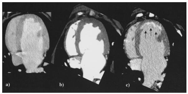
16 Application to cardiomyopathy Myocarditis HCMP J Am Coll Cardiol 2012;60 Eur Radiol 2013;23: (ASCI squre)
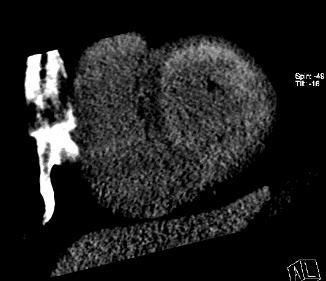
17 AMYLOIDOSIS SARCOIDOSIS HYPERTROPHIC Yonsei University: Unpublished data
18 Yonsei University: Unpublished data
19 Clinical application of delayed CT Myocardial Nonischemic infarction cardiomyopathy Emergency Differentiation room from ischemic cardiomyopathy Coronary angiography + myocardial Pattern infarction diagnosis (culprit for artery) specific etiology Chronic Volume fraction myocardial of hyper-enhanced infarction myocardium Infarct sizing for risk stratification Transmural extent

 LGE MRI Baseline EF= 53.5% 16 weeks EF= 36.")
20 Dual-energy delayed CT for quantifying DIFFUSE fibrosis Rabbit model with myocardial fibrosis induced by Doxorubicin Doxorubicin induced cardiomyopathy (1.0mg/kg twice a week for 6 weeks) Imaging Dual-energy CT (2 nd generation dual-source) MRI T1-mapping MRI (MOLLI, 3T-unit) LGE MRI Baseline EF= 53.5% 16 weeks EF= 36.7% JACC CVI: in revision


21 Extracellular volume fraction vs. Collagen volume fraction JACC CVI: in revision
22 ECV measurement with dual energy CT: Advantages Might be more accurate - measurement error Pre-contrast CT is not required. No misregistration between two sets of images (pre- & post- in MRI) Isotropic whole heart volume data
 Septal: 31.5% Inferior: 42.")
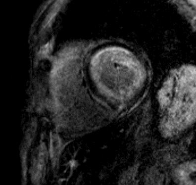
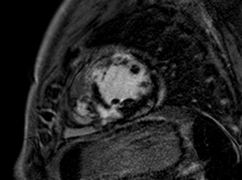
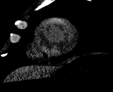
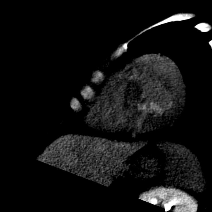
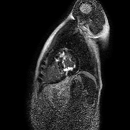
23 Measurement F/75 HCMP, Apical LV MR ECV (Hct 33.8) Septal 33.8% Inferior: 42.6% CT ECV (Hct 32.5) Septal: 31.5% Inferior: 42.8% Radiology: in revision
24 Per-patient analysis PER-SEGMENT ANALYSIS 23 patients with cardiomyopathy MRI ECV= ± 8.61% CT ECV = ± 8.30% P = ICC = MRI ECV= ± 10.50% CT ECV = ± 10.62% P = ICC = 0.940
25 ECV measurement with dual energy CT: Clinical application In addition to differential diagnosis of ischemic and nonischemic cardiomyopathies in combination with CTA and CT MDE Detecting diffuse fibrosis Quantifying diffuse fibrosis
26 summary Assessment of myocardial delayed enhancement is feasible and diagnostic with dual-energy cardiac CT in CMP and shows a good correlation with MRI in volume measurement. Myocardial ECV with dual-energy cardiac CT in cardiomyopathy is feasible to detect and quantify diffuse myocardial fibrosis. ECV calculated with dual-energy cardiac CT showed excellent agreement with that with cardiac MRI, suggesting the potential of myocardial tissue characterization with cardiac CT.


27
Role of CMR in heart failure and cardiomyopathy
 Role of CMR in heart failure and cardiomyopathy Hajime Sakuma Department of Radiology, Mie University Late gadolinium enhancement (LGE) LGE MRI can demonstrate site of necrosis, fibrosis or deposition
Cardiac MRI: Cardiomyopathy
 Cardiac MRI: Cardiomyopathy Laura E. Heyneman, MD I do not have any relevant financial relationships with any commercial interests Cardiac MRI: Cardiomyopathy Laura E. Heyneman, MD Duke University Medical
What s New in Cardiac MRI
 What s New in Cardiac MRI Katie M. Hawthorne, MD Director, Cardiac MRI Main Line Health Philadelphia Cardiovascular Summit November 18, 2017 Cardiac MRI: Disclosure 2 Disclosures No financial disclosures
Case based learning: CMR in Heart Failure
 Case based learning: CMR in Heart Failure Milind Y Desai, MD FACC FAHA FESC Associate Professor of Medicine Heart and Vascular Institute, Cleveland Clinic Cleveland, OH Disclosures: none Use of Gadolinium
Imaging in Heart Failure: A Multimodality Approach. Thomas Ryan, MD
 Imaging in Heart Failure: A Multimodality Approach Thomas Ryan, MD Heart Failure HFrEF HFpEF EF50% Lifetime risk 20% Prevalence 6M Americans Societal costs - $30B 50% 5-year survival 1 Systolic
Multiparametric T1, T2 and T2* MR Imaging
 Multiparametric T1, T2 and T2* MR Imaging Kate Hanneman MD Assistant Professor of Radiology Joint Department of Medical Imaging, University of Toronto Disclosure Neither I nor my immediate family members
The Value of Stress MRI in Evaluation of Myocardial Ischemia
 The Value of Stress MRI in Evaluation of Myocardial Ischemia Dr. Saeed Al Sayari, MBBS, EBCR, MBA Department of Radiology and Nuclear Medicine Mafraq Hospital, Abu Dhabi United Arab Emirates Introduction
Cardiology for the Practitioner Advanced Cardiac Imaging: Worth the pretty pictures?
 Keenan Research Centre Li Ka Shing Knowledge Institute Cardiology for the Practitioner Advanced Cardiac Imaging: Worth the pretty pictures? Howard Leong-Poi, MD, FRCPC Associate Professor of Medicine St.
DELAYED ENHANCEMENT IMAGING IN CHILDREN
 NASCI 38 TH ANNUAL MEENG, SEATLE October 3-5, 21 1. DELAYED ENHANCEMENT IN CHILDREN Shi-Joon Yoo, MD Lars Grosse-Wortmann, MD University of Toronto Canada -1. 1. 1. Magnitude image Magnitude images -1.
A Light in the Dark: Cardiac MRI and Risk Mitigation. J. Ronald Mikolich MD Professor of Internal Medicine Northeast Ohio Medical University (NEOMED)
") A Light in the Dark: Cardiac MRI and Risk Mitigation J. Ronald Mikolich MD Professor of Internal Medicine Northeast Ohio Medical University (NEOMED) Dr. Mikolich has NO financial disclosures relative to
A Light in the Dark: Cardiac MRI and Risk Mitigation J. Ronald Mikolich MD Professor of Internal Medicine Northeast Ohio Medical University (NEOMED) Dr. Mikolich has NO financial disclosures relative to
1. LV function and remodeling. 2. Contribution of myocardial ischemia due to CAD, and
 1 The clinical syndrome of heart failure in adults is commonly associated with the etiologies of ischemic and non-ischemic dilated cardiomyopathy, hypertrophic cardiomyopathy, hypertensive heart disease,
Managing Hypertrophic Cardiomyopathy with Imaging. Gisela C. Mueller University of Michigan Department of Radiology
 Managing Hypertrophic Cardiomyopathy with Imaging Gisela C. Mueller University of Michigan Department of Radiology Disclosures Gadolinium contrast material for cardiac MRI Acronyms Afib CAD Atrial fibrillation
Cardiac Sarcoidosis. Millee Singh DO Non Invasive Cardiology First Coast Heart and Vascluar
 Cardiac Sarcoidosis Millee Singh DO Non Invasive Cardiology First Coast Heart and Vascluar Introduction Multisystem granulomatous disease of unknown etiology characterized by noncaseating granulomas in
Myocardial Fibrosis in Heart Failure
 Myocardial Fibrosis in Heart Failure Dr Leah Iles, MBChB, FRACP The Alfred Hospital and Baker IDI Heart and Diabetes Research Institute, Vic, Australia DECLARATION OF CONFLICT OF INTEREST Nothing to declare
HFPEF Echo with Strain vs. MRI T1 Mapping
 HFPEF Echo with Strain vs. MRI T1 Mapping Erik Schelbert, MD MS Director, Cardiovascular Magnetic Resonance Assistant Professor of Medicine Heart & Vascular Institute University of Pittsburgh Disclosures
Newly Diagnosed Heart Failure patient: When to Order an MRI and Why
 Newly Diagnosed Heart Failure patient: When to Order an MRI and Why Jennifer Dickerson MD Assistant Professor of Clinical Internal Medicine Director, The Ohio State University Echocardiography Laboratory
Update in Nuclear Imaging of Amyloidosis and Sarcoidosis
 Update in Nuclear Imaging of Amyloidosis and Sarcoidosis Balaji Tamarappoo MD, PhD, Cedars-Sinai Heart Institute and Biomedical Imaging Research Institute Cedars-Sinai Medical Center Los Angeles, CA, USA.
XVth Balkan Congress of Radiology Danubius Hotel Helia, October 2017, Budapest, Hungary
 XVth Balkan Congress of Radiology Danubius Hotel Helia, 12-14 October 2017, Budapest, Hungary Ružica Maksimović MRI in Myocarditis Faculty of Medicine, University of Belgrade, Centre for Radiology and
ADVANCED CARDIOVASCULAR IMAGING. Medical Knowledge. Goals and Objectives PF EF MF LF Aspirational
 Medical Knowledge Goals and Objectives PF EF MF LF Aspirational Know the basic principles of magnetic resonance imaging (MRI) including the role of the magnetic fields and gradient coil systems, generation
Cardiac MRI: Clinical Application to Disease
 Cardiac MRI: Clinical Application to Disease Jessi Smith, MD Cardiothoracic imaging, Indiana University Slides courtesy of Stacy Rissing, MD Outline Imaging planes Disease findings Pulse sequences used
Cardiac Imaging. Kimberly Delcour, DO, FACC. Mahi Ashwath, MD, FACC, FASE. Director, Cardiac CT. Director, Cardiac MRI
 Cardiac Imaging Kimberly Delcour, DO, FACC Director, Cardiac CT Mahi Ashwath, MD, FACC, FASE Director, Cardiac MRI Cardiac Imaging Discuss the clinical applications of and indications for: Cardiac CT Nuclear
Why Cardiac MRI? Presented by:
 Why Cardiac MRI? Presented by: Lisa G. Carkner, MD, FACC 1 Disclosures I have no financial disclosures Objectives Review basic principles of Cardiac MRI. What patient characteristics do I need to consider
Pathophysiology of Coronary Microvascular Dysfunction
 Pathophysiology of Coronary Microvascular Dysfunction Cheol Woong Yu, MD, PhD Cardiology Department Division of Internal Medicine Korea University Anam Hospital. Etiologies of Chest Pain without obstructive
Outils d évaluation myocardique par relaxométrie (Myomaps)
") Cardiovascular sciences Outils d évaluation myocardique par relaxométrie (Myomaps) Alain Nchimi Geva T. Magnetic resonance imaging: historical perspective. J Cardiovasc Magn Reson. 2006;8(4):573-80. CMR
Cardiovascular sciences Outils d évaluation myocardique par relaxométrie (Myomaps) Alain Nchimi Geva T. Magnetic resonance imaging: historical perspective. J Cardiovasc Magn Reson. 2006;8(4):573-80. CMR
THE NEW PLACE OF CARDIAC MRI IN AERONAUTICAL FITNESS
 88 th ASMA ANNUAL SCIENTIFIC MEETING DENVER - CO April 30- May 4, 2017 THE NEW PLACE OF CARDIAC MRI IN AERONAUTICAL FITNESS S. BISCONTE (1), J. MONIN (2), N. HUIBAN (3), G. GUIU (2), S. NGUYEN (1), O.
Cardiac MRI: Clinical Application to Disease
 Cardiac MRI: Clinical Application to Disease Stacy Rissing, MD! Cardiothoracic imaging, Indiana University! Outline Imaging planes Disease findings Pulse sequences used for each indication Pathophysiology
Acute Myocarditis Mimicking ST-segment Elevation Myocardial Infarction: Relation Between ECG Changes And Myocardial Damage As Assessed By CMR
 Acute Myocarditis Mimicking ST-segment Elevation Myocardial Infarction: Relation Between ECG Changes And Myocardial Damage As Assessed By CMR G. Nucifora 1, A. Di Chiara 2, D. Miani 1, G. Piccoli 3, M.
The use of Cardiac CT and MRI in Clinical Practice
 The use of Cardiac CT and MRI in Clinical Practice Matthew W. Martinez, MD Assistant Professor of Medicine LVPG - Lehigh Valley Heart Specialists Lehigh Valley Health Network Oct. 3, 2009 DISCLOSURE Relevant
Cardiomyopathy. Cardiomyopathies HOCM. Hypertrophic Obstructive Cardiomyopathy. Systolic Anterior Movement (SAM) of Mitral Valve (Venturi Effect) Cine
 of Mitral Valve (Venturi Effect) Cine") Jens Bremerich Radiology University Hospital Basel Hypertrophic Obstructive Cine VENC Cine (5m/s) Modified Bernoulli Equation: P (in mmhg) = 4 x (Vmax)2 Vmax= 4.2 m/s, P = 70mm Hg Hydrodynamica 738 HOCM
Jens Bremerich Radiology University Hospital Basel Hypertrophic Obstructive Cine VENC Cine (5m/s) Modified Bernoulli Equation: P (in mmhg) = 4 x (Vmax)2 Vmax= 4.2 m/s, P = 70mm Hg Hydrodynamica 738 HOCM
Klinische Anwendung und neue Entwicklungen: Welches Verfahren für welche Indika:on? Kardiales MR. 17. Zürcher Herzkurs
 Klinische Anwendung und neue Entwicklungen: Welches Verfahren für welche Indika:on? Kardiales MR 17. Zürcher Herzkurs 28.09.2017 Dr. med. Gabriella De Pasquale HerzGefässZentrum Zürich Klinik im Park Most
CARDIOMYOPATHY IN CT. Hans- Christoph Becker Professor of Radiology
 CARDIOMYOPATHY IN CT Hans- Christoph Becker Professor of Radiology 1 Cardiomyopathy Heart muscle disease Deterioration of the heart function, heart failure Dyspnea, peripheral edema Risk of arrhythmia,
9/23/2011. Cardiac MRI Evaluation of Cardiomyopathy and Myocarditis. Primary Hypertrophic Cardiomyopathy. Cardiomyopathy.
 Cardiomyopathy Cardiac MRI Evaluation of Cardiomyopathy and Myocarditis Laureen Sena Children s Hospital Boston, MA NASCI 2011 Baltimore, Maryland Primary Hypertrophic ARVD Dilated Restrictive Unclassified
27-year-old professionnal rugby player: asymptomatic
 27-year-old professionnal rugby player: asymptomatic Benefits and limits of cardiac MRI in the young athlete with a suspected heart disease. Philippe PAULE Service de Cardiologie, HIA Clermont Tonnerre,
Review of Cardiac Imaging Modalities in the Renal Patient. George Youssef
 Review of Cardiac Imaging Modalities in the Renal Patient George Youssef ECHO Left ventricular hypertrophy (LVH) assessment Diastolic dysfunction Stress ECHO Cardiac CT angiography Echocardiography - positives
Current Indications for Cardiac MRI: What You See is What You Get?
 Current Indications for Cardiac MRI: What You See is What You Get? Javier Ganame, MD, PhD, FASE No disclosures Cardiology Update, Niagara, Sept 24th, 2016 The Ideal Diagnostic Technique Easy to apply Accurate
Usefulness of Delayed Enhancement by Magnetic Resonance Imaging in Hypertrophic Cardiomyopathy as a Marker of Disease and Its Severity
 Usefulness of Delayed Enhancement by Magnetic Resonance Imaging in Hypertrophic Cardiomyopathy as a Marker of Disease and Its Severity G.D.Aquaro, MD Fondazione G.Monasterio Regione Toscana/CNR Pisa, Italy
Cardiac magnetic resonance imaging in rheumatoid arthritis: promising or misleading? Sophie Mavrogeni MD FESC
 Cardiac magnetic resonance imaging in rheumatoid arthritis: promising or misleading? Sophie Mavrogeni MD FESC Onassis Cardiac Surgery Center Athens Greece Nothing to disclose Financial disclosure Cardiac
Cardiovascular Imaging Stress Echo
 Cardiovascular Imaging Stress Echo Theodora A Zaglavara, MD, PhD Cardiac Imaging Department INTERBALKAN MEDICAL CENTER Thessaloniki GREECE Evolution of Stress Echo: From Innovation to a Widely Established
Sung A Chang Department of Internal Medicine, Division of Cardiology, Sungkyunkwan University School of Medicine, Samsung Medical Center
 CMR Perfusion and Viability A STICH Out of Time? Sung A Chang Department of Internal Medicine, Division of Cardiology, Sungkyunkwan University School of Medicine, Samsung Medical Center Can Imaging Improve
Pearls & Pitfalls in nuclear cardiology
 Pearls & Pitfalls in nuclear cardiology Maythinee Chantadisai, MD., NM physician Division of Nuclear Medicine, Department of radiology, KCMH Principle of myocardial perfusion imaging (MPI) Radiotracer
Rational use of imaging for viability evaluation
 EUROECHO and other imaging modalities 2011 Rational use of imaging for viability evaluation Luc A. Pierard, MD, PhD, FESC, FACC Professor of Medicine Head, Department of Cardiology, CHU Liège, Belgium
A Light in the Dark: Cardiac MRI and Risk Mitigation. J. Ronald Mikolich MD Professor of Internal Medicine Northeast Ohio Medical University (NEOMED)
") A Light in the Dark: Cardiac MRI and Risk Mitigation J. Ronald Mikolich MD Professor of Internal Medicine Northeast Ohio Medical University (NEOMED) Dr. Mikolich has NO financial disclosures relative to
A Light in the Dark: Cardiac MRI and Risk Mitigation J. Ronald Mikolich MD Professor of Internal Medicine Northeast Ohio Medical University (NEOMED) Dr. Mikolich has NO financial disclosures relative to
Advanced MR Imaging in Myocarditis
 Naeem Merchant MD FRCP Professor of Medicine Department of Radiology Department of Cardiac Sciences Cumming School of Medicine University of Calgary Advanced MR Imaging in Myocarditis The Lake Louise Criteria
Etiology, Classification & Management. Sheba Medical Center Cardiology Department Matthew Wright St. George s University of London
 Etiology, Classification & Management Sheba Medical Center Cardiology Department Matthew Wright St. George s University of London Introduction World Health Organization (1995): Diseases of myocardium (heart
Imaging in Ischemic Heart Disease: Role of Cardiac MRI
 Imaging in Ischemic Heart Disease: Role of Cardiac MRI Chiara Bucciarelli Ducci MD, PhD, FESC, FRCP Consultant Senior Lecturer Cardiologist Bristol Heart Institute, University of Bristol, UK Chair elect,
Echocardiographic Evaluation of the Cardiomyopathies. Stephanie Coulter, MD, FACC, FASE April, 2016
 Echocardiographic Evaluation of the Cardiomyopathies Stephanie Coulter, MD, FACC, FASE April, 2016 Cardiomyopathies (CMP) primary disease intrinsic to cardiac muscle Dilated CMP Hypertrophic CMP Infiltrative
Restrictive Cardiomyopathy
 ESC Congress 2011, Paris Imaging Unusual Causes of Cardiomyopathy Restrictive Cardiomyopathy Kazuaki Tanabe, MD, PhD Professor of Medicine Chair, Division of Cardiology Izumo, Japan I Have No Disclosures
while the absence of LGE-CMR may reduce the need for ICD implantation in patients with NIDM who are at low risk for future VF/VT or SCD.
 Midwall replacement fibrosis with late gadolinium enhancement LGE-CMR imaging as an independent predictor of mortality and utility of Cardiovascular Magnetic Resonance Image One-third of all patients with
Acute chest pain: game changer or waste of resources? CMR one stop shop for function, structure and perfusion
 Multidisciplinary Cardiovascular Research Centre Leeds Institute of Genetics, Health and Therapeutics Acute chest pain: game changer or waste of resources? CMR one stop shop for function, structure and
Patterns of Left Ventricular Remodeling in Chronic Heart Failure: The Role of Inadequate Ventricular Hypertrophy
 Abstract ESC 82445 Patterns of Left Ventricular Remodeling in Chronic Heart Failure: The Role of Inadequate Ventricular Hypertrophy FL. Dini 1, P. Capozza 1, P. Fontanive 2, MG. Delle Donne 1, V. Santonato
Cardiac MRI in ACHD What We. ACHD Patients
 Cardiac MRI in ACHD What We Have Learned to Apply to ACHD Patients Faris Al Mousily, MBChB, FAAC, FACC Consultant, Pediatric Cardiology, KFSH&RC/Jeddah Adjunct Faculty, Division of Pediatric Cardiology
Imaging in dilated cardiomyopathy : factors associated with a poor outcome
 Imaging in dilated cardiomyopathy : factors associated with a poor outcome Johan De Sutter, MD, PhD, FESC AZ Maria Middelares Gent and University Gent - Belgium Dilated cardiomyopathy Cardiomyopathy with
MR Assessment of Myocardial Viability
 MR Assessment of Myocardial Viability Definition of Viability Clinical Metabolism: Presence of glucose uptake Perfusion / Perfusion reserve Morphology: Wall thickness, wall thickening Contractility: Recovery
Left atrial function. Aliakbar Arvandi MD
 In the clinic Left atrial function Abstract The left atrium (LA) is a left posterior cardiac chamber which is located adjacent to the esophagus. It is separated from the right atrium by the inter-atrial
Cardiomyopathy. Jeff Grubbe MD FACP, Chief Medical Director, Allstate Life & Retirement
 Cardiomyopathy Jeff Grubbe MD FACP, Chief Medical Director, Allstate Life & Retirement Nebraska Home Office Life Underwriters Association March 20, 2018 1 Cardiomyopathy A myocardial disorder in which
Multimodality Imaging of Anomalous Left Coronary Artery from the Pulmonary
 1 IMAGES IN CARDIOVASCULAR ULTRASOUND 2 3 4 Multimodality Imaging of Anomalous Left Coronary Artery from the Pulmonary Artery 5 6 7 Byung Gyu Kim, MD 1, Sung Woo Cho, MD 1, Dae Hyun Hwang, MD 2 and Jong
The role of Magnetic Resonance Imaging in the diagnosis of viability & Coronary Artery Disease
 The role of Magnetic Resonance Imaging in the diagnosis of viability & Coronary Artery Disease G.P. Spanos, MSc, Phd Head of CardioVascular Imaging Tomographia Diagnostic Center Cardiovascular magnetic
Advanced Imaging MRI and CTA
 Advanced Imaging MRI and CTA Who and why may benefit. Matthew W. Martinez, M.D. FACC Lehigh Valley Health Network Director, Cardiovascular Imaging Learning Objectives Review basics of CMR and CTA Review
HYPERTROPHY: Behind the curtain. V. Yotova St. Radboud Medical University Center, Nijmegen
 HYPERTROPHY: Behind the curtain V. Yotova St. Radboud Medical University Center, Nijmegen Disclosure of interest: none Relative wall thickness (cm) M 0.22 0.42 0.43 0.47 0.48 0.52 0.53 F 0.24 0.42 0.43
How NOT to miss Hypertrophic Cardiomyopathy? Adaya Weissler-Snir, MD University Health Network, University of Toronto
 How NOT to miss Hypertrophic Cardiomyopathy? Adaya Weissler-Snir, MD University Health Network, University of Toronto Introduction Hypertrophic cardiomyopathy is the most common genetic cardiomyopathy,
Φαινόμενο No-Reflow. Απεικόνιση με CMR, κλινική συσχέτιση και προγνωστική σημασία
 Φαινόμενο No-Reflow. Απεικόνιση με CMR, κλινική συσχέτιση και προγνωστική σημασία Θεόδωρος. Καραμήτσος MD PhD Honorary Consultant in Cardiology University of Oxford Centre for Clinical Magnetic Resonance
3/27/2014. Introduction.
 Introduction. Myocardial perfusion & contractility becomes abnormal immediately after the onset of ischaemia, even before the development of the symptoms & ST segment changes. 1 Myocardial Wall Motion
4/11/2017. Cardiomyopathy. John Steuter, MD Bryan Heart. Disclosures. No Conflicts. Cardiomyopathy. WHO Classification
 Cardiomyopathy John Steuter, MD Bryan Heart Disclosures No Conflicts Cardiomyopathy WHO Classification Anatomy & physiology of the LV 1. Dilated Enlarged Systolic dysfunction 2. Hypertrophic Thickened
Perfusion, Viability, Edema and Hemorrhage: How it Can (and Should) Change Clinical Practice. Rohan Dharmakumar, Ph.D.
 Change Clinical Practice. Rohan Dharmakumar, Ph.D.") Perfusion, Viability, Edema and Hemorrhage: How it Can (and Should) Change Clinical Practice Rohan Dharmakumar, Ph.D. Director, Translational Cardiac Imaging Research Associate Director, Biomedical Imaging
Perfusion, Viability, Edema and Hemorrhage: How it Can (and Should) Change Clinical Practice Rohan Dharmakumar, Ph.D. Director, Translational Cardiac Imaging Research Associate Director, Biomedical Imaging
Multiparametric Mapping for Assessment of Cancer Therapy Related Cardiotoxicity
 Multiparametric Mapping for Assessment of Cancer Therapy Related Cardiotoxicity Cory V. Noel, M.D. Medical Director of CMR Pediatric Cardiology Outline What is meant by the term cardiotoxicity? Scope of
Imaging and heart failure
 Imaging and heart failure Jeroen J Bax Dept of Cardiology Leiden Univ Medical Center The Netherlands Davos, feb 2013 Research grants: Medtronic, Biotronik, Boston, St Jude, BMS imaging, GE Healthcare,
Imaging of the Heart Todd Tessendorf MD FACC
 Imaging of the Heart Todd Tessendorf MD FACC Outline Imaging Modalities for Structural Heart Disease ECHO, MRI Imaging Modalities for Ischemic Heart Disease SPECT, PET, CCTA Show lots of pretty pictures
Myocardial Infarction
 Myocardial Infarction MI = heart attack Defined as necrosis of heart muscle resulting from ischemia. A very significant cause of death worldwide. of these deaths, 33% -50% die before they can reach the
Constrictive/Restrictive Cardiomyopathies: Diagnosis and Management Update; Radiation Induced Heart Disease. Alexander (Sandy) Dick, MD
 Dick, MD") Constrictive/Restrictive Cardiomyopathies: Diagnosis and Management Update; Radiation Induced Heart Disease Alexander (Sandy) Dick, MD Outline Pericardial Constriction Diagnosis: Imaging, Hemodynamics
Constrictive/Restrictive Cardiomyopathies: Diagnosis and Management Update; Radiation Induced Heart Disease Alexander (Sandy) Dick, MD Outline Pericardial Constriction Diagnosis: Imaging, Hemodynamics
Current Guidelines for Diagnosis of AMI Chest pain ST change on EKG Cardiac Enzymes
 Noninvasive Cardiac Imaging in Myocardial Infarction Sangchol Lee Sungkyunkwan University Samsung Medical Center Current Guidelines for Diagnosis of AMI Chest pain ST change on EKG Cardiac Enzymes Do We
Case based learning: CMR in Heart Failure
 Case based learning: CMR in Heart Failure Milind Y Desai, MD FACC FAHA FESC Associate Professor of Medicine Heart and Vascular Institute, Cleveland Clinic Cleveland, OH Disclosures: none Use of Gadolinium
What s new in Hypertrophic Cardiomyopathy?
 What s new in Hypertrophic Cardiomyopathy? Dr Andris Ellims HCM Clinic @ The Alfred Hypertrophic Cardiomyopathy = otherwise unexplained LV hypertrophy* 1 in 500 prevalence most common inherited cardiovascular
Functional Mitral Regurgitation
 Club 35 - The best in heart valve disease - Functional Mitral Regurgitation Steven Droogmans, MD, PhD UZ Brussel, Jette, Belgium 08-12-2011 Euroecho & other Imaging Modalities 2011 No conflicts of interest
The Egyptian Journal of Hospital Medicine (July 2018) Vol. 72 (9), Page
 Vol. 72 (9), Page") The Egyptian Journal of Hospital Medicine (July 2018) Vol. 72 (9), Page 5270-5277 An Egyptian study for standardization of myocardial T1 mapping values on a 3 Tesla MRI machine Ahmed Samir Ibrahim, Emad
The Egyptian Journal of Hospital Medicine (July 2018) Vol. 72 (9), Page 5270-5277 An Egyptian study for standardization of myocardial T1 mapping values on a 3 Tesla MRI machine Ahmed Samir Ibrahim, Emad
Detection and Assessment of MI: Use of Imaging Methods. Robert O. Bonow, M.D.
 Detection and Assessment of MI: Use of Imaging Methods Robert O. Bonow, M.D. Detection and Assessment of MI: Use of Imaging Methods Robert O. Bonow, M.D. No Relationships to Disclose Expert Consensus Document
Cardiovascular manifestations of HIV
 Cardiovascular manifestations of HIV Prabhakar Rajiah, MBBS, MD, FRCR Associate Professor of Radiology Associate Director, Cardiac CT and MRI University of Texas Southwestern Medical Center, Dallas, USA
Cardiac Stress MRI: Detection of Ischemia. Disclosures: Dobutamine Stress MR. April 28, 2018
 Cardiac MRI: Detection of Ischemia Cardiac MRI in Today s Clinical Practice Foundations of Cardiovascular Magnetic Resonance Daniel C. Lee, MD, MSc Assistant Professor of Medicine and Radiology Co-Director,
Viability Testing Using Dynamic Echocardiography
 Viability Testing Using Dynamic Echocardiography Theodora A Zaglavara, MD, PhD Director of Echocardiography EUROMEDICA KYANOUS STAVROS HOSPITAL Thessaloniki GREECE Goals of Cardiac Imaging in Coronary
Cardiac Risk Factors and Noninvasive Cardiac Diagnosis-ECG, ECHO, et al. Martin C. Burke, DO, FACOI ACOI IM Board Review Course 2018
 Cardiac Risk Factors and Noninvasive Cardiac Diagnosis-ECG, ECHO, et al. Martin C. Burke, DO, FACOI ACOI IM Board Review Course 2018 No Disclosures The American Heart Association Evidence-Based Scoring
Ventricular Tachycardia Ablation. Saverio Iacopino, MD, FACC, FESC
 Ventricular Tachycardia Ablation Saverio Iacopino, MD, FACC, FESC ü Ventricular arrhythmias, both symptomatic and asymptomatic, are common, but syncope and SCD are infrequent initial manifestations of
IMAGING IN CARDIAC AMYLOIDOSIS ; TRENDS IN DIAGNOSIS AND GUIDING THERAPY
 IMAGING IN CARDIAC AMYLOIDOSIS ; TRENDS IN DIAGNOSIS AND GUIDING THERAPY Mohamed Abo Mandour, MD. Al-Azhar University Cardiac amyloidosis is an under appreciated cause of HF The bottom line pathologic
Importance of CRT team for optimization of the results: a European point of view
 Importance of CRT team for optimization of the results: a European point of view Matteo Bertini, MD, PhD Arcispedale S. Anna Azienda Ospedaliero-Universitaria Cona-Ferrara No conflict of interest to declare
Mapping the Future of Cardiac MR Imaging: Case-based Review of T1 and T2 Mapping Techniques 1
 TECHNICAL ADVANCEMENTS IN CARDIAC MR IMAGING 1594 Note: This copy is for your personal non-commercial use only. To order presentation-ready copies for distribution to your colleagues or clients, contact
Screening of Children and Adolescents at Risk of Sudden Cardiac Arrest: What Is the Utility of Non-Invasive Imaging?
 Screening of Children and Adolescents at Risk of Sudden Cardiac Arrest: What Is the Utility of Non-Invasive Imaging? Beth F. Printz, M.D., Ph.D. Medical Director, Non-Invasive Imaging Rady Children s Hospital,
DOWNLOAD PDF MYOCARDIAL CONTRAST TWO DIMENSIONAL ECHOCARDIOGRAPHY (DEVELOPMENTS IN CARDIOVASCULAR MEDICINE)
") Chapter 1 : Imaging Cardiovascular Medicine Stanford Medicine contrast two-dimensional echocardiography (MC-2DE), a new and exciting diagnostic methodology for assessment of myocardial perfusion, which
Chapter 1 : Imaging Cardiovascular Medicine Stanford Medicine contrast two-dimensional echocardiography (MC-2DE), a new and exciting diagnostic methodology for assessment of myocardial perfusion, which
Conflict Disclosures. Vermont Cardiac Network. Outline. Series Learning Objectives 4/27/2016. Scott E. Friedman April 28, 2016
 Conflict Disclosures Vermont Cardiac Network The Speaker has reported no significant financial relationship with any companies whose product may be germane to the content of their presentations or who
Cardiomyopathy. ACOI IM Board Review 2018 Martin C. Burke DO, FACOI
 Cardiomyopathy ACOI IM Board Review 2018 Martin C. Burke DO, FACOI No Disclosures Cardiomyopathies Definition: diseases of heart muscle 1980 WHO: unknown causes Not clinically relevant 1995 WHO: diseases
Novel echocardiographic modalities: 3D echo, speckle tracking and strain rate imaging. Potential roles in sports cardiology. Stefano Caselli, MD, PhD
 Novel echocardiographic modalities: 3D echo, speckle tracking and strain rate imaging. Potential roles in sports cardiology. Stefano Caselli, MD, PhD Ospedale San Pietro Fatebenefratelli Rome, Italy Differential
Hypertrophic Cardiomyopathy: beyond gradient and wall thickness
 Hypertrophic Cardiomyopathy: beyond gradient and wall thickness Michael H. Picard, M.D. Massachusetts General Hospital Harvard Medical School no disclosures special thanks to A. Baggish 1 Hypertrophic
QCVC Committees Scientific Activities Central Hall General Information FAC. SPECT tomography has the advantage of quantifying biventricular volumes.
 QCVC Committees Scientific Activities Central Hall General Information FAC Thematic Units Arrhythmias and Electrophysiology Basic Research Bioengineering and Medical Informatics Cardiac Surgical Intensive
Coronary interventions
 Controversial issues in the management of ischemic heart failure Coronary interventions Maciej Lesiak Department of Cardiology, University Hospital in Poznan none DECLARATION OF CONFLICT OF INTEREST CHF
Role of cardiovascular magnetic resonance in the guidelines of the European Society of Cardiology
 von Knobelsdorff-Brenkenhoff and Schulz-Menger Journal of Cardiovascular Magnetic Resonance (2016) 18:6 DOI 10.1186/s12968-016-0225-6 RESEARCH Open Access Role of cardiovascular magnetic resonance in the
Patterns of late myocardial enhancement in cardiac MRI
 Patterns of late myocardial enhancement in cardiac MRI Poster No.: C-0683 Congress: ECR 2010 Type: Educational Exhibit Topic: Cardiac Authors: A. F. L. Carneiro, A. S. R. Preto, R. C. Ramos, A. J. B. S.
Peri-operative Troponin Measurements - Pathophysiology and Prognosis
 Peri-operative Troponin Measurements - Pathophysiology and Prognosis Allan S. Jaffe, MD.* Consultant - Cardiology & Laboratory Medicine Professor of Medicine Chair, CCLS Division, Department of Laboratory
MRI Sequences: What to use for what
 MRI Sequences: What to use for what MRI basics T 1 and T 2 relaxation Common Imaging Protocols Mechanical function (cine) Tissue characterization LGE Edema imaging (T 2 weighted) T1 Special protocols MRA
Isolated congenital coronary anomalies: Evaluation by multislice-ct or MRI
 Isolated congenital coronary anomalies: Evaluation by multislice-ct or MRI B.K. Velthuis, Dept. of Radiology UMC Utrecht, the Netherlands ESC 2010 Coronary artery anomalies CAA Uncommon 0.3-5% normal population
Cardial MRI; Approaching the Level of Gold Standard for Viability Assessment
 Cardial MRI; Approaching the Level of Gold Standard for Viability Assessment 용환석 고려대학교구로병원영상의학과 Viability Hibernating myocardium a state of myocardial hypocontractility during chronic hypoperfusion, in
Case based interactive discussion Encourage debate Cover common conditions seen in MRI Give you the good and the bad of what we do ESC Guidelines and
 Dr Dan Sado Honorary Senior Lecturer in Cardiology, Kings College London Consultant in Cardiology and CMR Lead, Kings College Hospital South London Cardiology SpR Imaging Training Lead Case based interactive
Clinical history. 73 yo man with chest pain Systemic hypertension and WG Stress EKG N Stress echocardiogram: Cardiac catheterization: no CAD
 CASE 8 Clinical history 73 yo man with chest pain Systemic hypertension and WG Stress EKG N Stress echocardiogram: Concentric hypertrophy Hypokinesis of LV-Inf Cardiac catheterization: no CAD Technique
Rotation: Echocardiography: Transthoracic Echocardiography (TTE)
") Rotation: Echocardiography: Transthoracic Echocardiography (TTE) Rotation Format and Responsibilities: Fellows rotate in the echocardiography laboratory in each clinical year. Rotations during the first
Rotation: Echocardiography: Transthoracic Echocardiography (TTE) Rotation Format and Responsibilities: Fellows rotate in the echocardiography laboratory in each clinical year. Rotations during the first