12/02/2012. Chapter 19: Blood Vessels. Blood Vessel Structure and Function
|
|
|
- Ethel Thornton
- 6 years ago
- Views:
Transcription
1 Chapter 19: Blood Vessels 12/02/2012 Blood Vessel Structure and Function 3 major types: arteries capillaries veins as heart contracts it forces blood into the large arteries leaving the ventricles then blood moves into arteries that get smaller and smaller, finally reaching smallest branches the arterioles, which feed into the capillary beds of body organs and tissues blood drains from capillaries into venules, which are the smallest veins. then on to the larger veins that merge to form the large veins that empty into the heart arteries arterioles capillaries venules veins BV stretch for about 100,000 km through the internal body Arteries carry blood away from the heart Branch and fork as they form smaller and smaller divisions Veins carry blood towards the heart Join/ merge together into larger vessels approaching the heart In systemic circuit Arteries carry oxygenated blood Veins carry deoxygenated blood
2 In pulmonary circuit Arteries carry deoxygenated blood to the lungs The veins carry oxygenated blood from the lungs to the heart Umbilical vessels of a fetus also differ in the roles of veins and arteries Only capillaries have intimate contact with tissue cells and directly serve cellular needs Exchanges between blood and tissue occur primarily through the gossamer thin capillary walls Structure of Blood Vessel Walls 3 distinct layers/ tunics (covering)that surround a central blood containing space> lumen Tunica intima Innermost tunic Tunic is in intimate contact with the blood in the lumen Contain simple squamous epithelium that lines the lumen of all vessels Endothelium is continuous with the endocardial lining of the heart and its flat cells fit closely together> forming a slick surface that minimizes friction as blood moves through the lumen In vessels larger then 1 mm a sub endothelial layer> consisting of basement membrane and loose CT > supports the endothelium Arteries have an internal elastic membrane > VEINS DON T Tunica media Mostly circularly arranged smooth muscle Sheets of elastin Contains the most elastin Activity of the smooth muscle is regulated by sympathetic vasomotor nerve fibers of the autonomic nervous system & chemicals Depending on body s need at any given moment regulation causes: Vasoconstriction Lumen diameter decreases as smooth muscle contracts

3 Vasodilation Lumen diameter increase as SM relaxes Activity is critical in regulating circulatory dynamics because small changes in vessel diameter greatly influence blood flow and pressure Bulkiest layer in the arteries Bear the chief responsibility for maintaining blood pressure and circulation Tunica Adventitia/ Externa: Outer most layer Composed largely of loosely woven collagen fibers that protect & reinforce the vessel & anchor it to surrounding structures Is infiltrated with nerve fibers, lymphatic vessels and in larger veins> a network of elastic fibers In larger arteries> it contains a system of tiny BV, the vasa vasorum vessels of vessels nourish the more external tissues of the blood vessel wall the innermost (luminal) portion of vessels obtain their nutrients directly from blood in lumen. The 3 vessel types vary in length, diameter, wall thickness and tissue make up
4 Arterial System 3 groups: elastic muscular arterioles Elastic Arteries / Conducting Arteries thick walled arteries near the heart aorta and its major branches longest in diameter 2.5 cm 1 cm the most elastic > highest portion of elastin their large lumens make them low resistance pathways that conduct blood from the heart to medium sized arteries arteries are sometimes called conducting arteries pressure reservoirs expanding and recoiling as the heart ejects blood blood flows fairly continuously rather than starting and stopping with pulsating rhythm of heart beat atherosclerosis BV become hard Blood flows more intermittently Smooth out pressure fluctuations Recoil helps maintain pressure & flow of blood Muscular Arteries/ Distributing Arteries Deliver blood to specific organs.3mm 1cm diameter Have the thickest tunic media of all vessels Contains more smooth muscle and less elastic tissue More active in vasoconstriction and less capable of stretching There is an elastic membrane on each face of tunica media Arterioles
5 Smallest of arteries have a lumen diameter of 10um.3mm larger arteries have all 3 tunics but tunica media is chiefly smooth muscle with a few scattered elastic fibers smaller arteries> lead into capillary beds a little bit more then a single layer of smooth muscle cells spiraling around endothelial lining minute to minute blood flow into capillary beds is determined by arteriolar diameter varies in response to changing: neural hormonal local chemical influences when arterioles constrict > the tissues served are largely bypassed when arterioles dilate> blood flow into the local capillaries increases Capillaries Microscopic capillaries are the smallest BV Exceedingly thin walls consist of just a thin tunica intima Length = 1mm Lumen diameter = 8 10 um Just large enough for RBCs to slip through single file Function= exchange Pericytes At strategic locations along the outer surface of some capillaries Smooth muscle like cells that stabilize the capillary wall & help control capillary permeability
6 Most tissues have a rich capillary supply Exceptions: Tendon & ligaments= poorly vascularized Cartilage & epithelial lack capillaries Receive nutrients from BV in nearby CT Avascular cornea & lens of eye receive nutrients from aqueous humor capillaries= back alleys & driveways that provide direct access to nearly every cell in the body ideal suited for their role of exchange between blood and interstitial fluid. gases nutrients hormones Types of Capillaries 3 types: continuous fenestrated sinusoidal Continuous Capillaries abundant in skin and muscles continuous endothelial cells are joined by tight junctions
7 providing an uninterrupted lining these junctions are usually incomplete & leave gap of unjoined membrane called intercellular clefts large enough to allow limited passage of fluids and small solutes Fenestrated Capillaries similar to continuous capillaries except: endothelial cells are riddled with oval pores/ fenestrations delicate membrane covers the fenestrations much more permeable to fluids and small solutes than continuous capillaries found wherever active capillary absorption or filtrate formation occurs in small intestine receive nutrients from digested food endocrine organs allow homes rapid entry into blood fenestrated capillaries with perpetually open pores occur in kidneys where rapid filtration of blood plasma is essential areas of active absorption or filtration Sinusoidal Capillaries highly modified, leaky capillaries found in liver bone marrow spleen

8 adrenal medulla have large irregular shaped lumens are usually fenestrated endothelial lining has fewer tight junctions & larger intercellular clefts than ordinary capillaries structural adaptations allow large molecules, proteins and even blood cells to pass between the blood and surrounding tissues most permeable Capillary Beds do not function independently they form capillary beds interweaving network microcirculation flow of blood from an arteriole to a venule through a capillary bed consists of 2 types of BV vascular shunt metarteriole + throughfare channels short vessel that directly connects the arteriole and venules at opposite ends of the bed true capillaries actual exchange vessels per bed
9 usually branch off the metarteriole end (proximal end of the shunt) and return to the thoroughfare channel (distal end) occasionally they spring from the terminal arteriole and empty directly into the venule the terminal arteriole feeding the bed leads into a metarteriole a vessel that is structurally intermediate between an arteriole and a capillary metarteriole is continuous with the thoroughfare channel intermediate between a capillary and a venule thoroughfare channel joins the postcapillary venule that drains in the beds. Precapillary sphincters Cuff of smooth muscle Surrounds root of each true capillary at metarteriole Acts as a valve to regulate blood flow into the capillary Blood flowing through a terminal arteriole may go either through the true capillaries or the shunt When relaxed/ open Blood flows through the true capillaries and takes part in exchanges with tissue cells When contracted/ closed Blood flows through shunts and bypasses the tissue cells Local chemical condition and arteriole vasomotor nerve fibers regulate the amount of blood entering a capillary bed A bed may be completely flooded with blood or bypassed depending on the condition Vigorous exercise Blood is rerouted from your digestive organs to the capillary beds of your skeletal muscle where it is more immediately needed Helps explain why vigorous exercise right after a meal can cause indigestion/ abdominal cramps Venous System Carry blood from the capillary beds toward the heart along route, the diameter of successive venous vessels increases
10 walls gradually thicken as progress from venules to larger and larger veins Venules capillaries unit to form venules range from um diameter smallest venules> the post capillary venules > consist entirely of endothelium around which pericytes congregate extremely porous fluid and WBCs move easily from the bloodstream through their walls just endothelium & a few fibroblasts larger venules have one or two layers of SM cells ( a scanty tunica media) and a thin tunica externa/ adventiitia Veins venules join to form veins usually have 3 distinct tunics but walls are always thinner & lumen are larger then corresponding arteries little smooth muscle or elastin in the tunica media poorly developed and tends to be thin even in largest veins tunica externa is the heaviest wall layer consisting of thick bundles of collagen fibers & elastic networks can accommodate a fairly large supply at any time because of their large lumens and thin walls. Are capacitance vessels and blood reservoirs Can hold up to 65% of the body s blood supply at any time. Normally only partly filled. Walls can be much thinner then those of arterial walls without danger of bursting because the blood pressure in veins is low.
11 Low pressure condition demands several structural adaptations to ensure veins return blood o the hear at the same rate it was pumped into the circulation One adaptation: Large diameter lumens Offer relatively little resistance to flow Venous Valves Another adaptation is valves that prevent blood from flowing backward Venous valves Formed from folds of the tunica intima Resemble the SL valves in both structure and function Most abundant in veins of limbs where gravity opposes the upward flow of blood Absent in veins of the thoracic and abdominal body cavities Effectiveness is demonstrated by ability for the vein to remain flat/ collapsed despite the pull of gravity Homeostatic Imbalance Varicose veins Veins that are tortuous and dilated because of incompetent/ leaky valves Adults usually suffer in lower limbs Factors : Hereditary Age Conditions that hinder venous return Prolonged standing in one position Pregnancy
12 Obesity Elevated venous pressure Venous Sinuses Such as: Coronary sinuses of heart Dural venous sinuses of the brain Receive cerebrospinal fluid and blood draining from the brain Reinforced by the though dura matter that covers brains surface Highly specialized, flattened veins with extremely thin walls Composed of only endothelium Supported by tissues that surround them Rather then tunics Vascular Anastomoses BV form special interconnections called vascular anastomoses Most organs receive blood from more than one arterial branch and the arteries supplying the same territory often merge forming> arterial anastomoses Provide alternate pathways called collateral channels for blood to reach a given body region If one branch is cut or blocked by a clot this channel can provide sufficient blood to area Occur around joints where active movement may hinder blood flow through one channel Also common in abdominal organ, heart and brain Arteries that supply the retina, kidneys and spleen either do no anastomose or have a poorly developed collateral circulation If blood flow is interrupted> cells supplied by such vessel die Arteriovenous anastomoses
13 Ex: The metarteriole thoroughfare channel shunts of capillary beds that connect arterioles and venules Veins interconnect much more freely then arteries Venous anastomoses are common Are abundant so on occluded vein rarely blocks blood flow or leads to tissue death Part 2 Physiology of Circulation Introduction to Blood Flow, Blood Pressure and Resistance Blood Flow (BF) The volume of blood flowing through a vessel, an organ or the entire circulation in a given period ml/min considering entire vascular system blood flow is equivalent to CO under resting conditions its relatively constant at any given moment blood flow through individual body organs may vary widely according to their immediate needs can be regulated independently for various tissues & organs Blood Pressure (BP) force per unit area exerted on a vessel wall by the contained blood expressed by mm Hg refers to systemic blood pressure in the largest arteries near the heart average aortic bp= 90mm Hg
14 the differences in bp within the vascular system pressure gradient provides driving force that keeps blood moving from high pressure through the body 90> 90 = no flow! 90>30= flow! pressure to low F = P1 - P2 R Resistance opposition to flow/ inversely related is a measure of the amount of friction blood encounters as it passes through the vessels measure of total frictional forces that impede flow generally use the term peripheral resistance most friction is encountered in the peripheral/ systemic circulation well away from the heart major determinant of BF because a change in blood vessels radius increases resistance to the 4th power. Resistance varies inversely with (radius)4!! 3 important sources of resistance: blood viscosity vessel diameter vessel length
15 Blood Viscosity viscosity internal resistance to flow that exists in all fluids related to the thickness/ stickiness of fluid the great the viscosity the less easily molecules slide past one another & the more difficult it is to get and keep the fluid moving BV is fairly constant Conditions such as polycythemia (excessive RBCs) can increase blood viscosity Resistance If RBC is low Anemia s Blood is less viscous Peripheral resistance declines Due to formed elements, plasma proteins Total Blood Vessel Length Relationship between total BV length + resistance is straight forward: Longer vessel= greater resistance BV Diameter Changes frequently & significantly alters peripheral resistance Blood viscosity and vessel length are normally unchanging How? Principles of fluid flow
16 Fluids close to the wall of a tube/ channel is slowed by friction as its passes along the wall Fluids in the center flows more freely and faster Laminar flow / streamlining In a tube of any given size the relative speed and position of fluid in the different regions of the tubes cross section remain constant The smaller the tube= the greater the friction Relatively more fluid contacts the walls Resistance varies inversely with the 4th power of the vessel radius If the radius of a vessel doubles, the resistance drops to 1/16 of its original value Large arteries close to the heart which do not change dramatically in diameter contribute little to peripheral resistance Small diameter arterioles are major determinants of peripheral resistance Because they can enlarge or constrict in response to neural and chemical controls When blood encounters an abrupt change in vessel diameter or rough or protruding areas of the tube wall (fatty plaque)> the smooth laminar blood is replaced by turbulent flow Irregular fluid motion where blood from the difference lamina mixes Turbulence dramatically increases resistance Relationship Between Flow, Pressure, and Resistance Blood flow (F) is directly proportional to the difference in BP (ΔP) between two points in the circulation that is blood pressure or hydrostatic pressure gradient When ΔP Blood flow speeds up ΔP Blood flow declines BF is inversely proportional to the peripheral resistance (R) in the systemic circuit If R BF F= ΔP/ R
17 R is far more important than ΔP in influencing local BF because R can easily be changed by altering blood vessel diameter Arterioles dilate> resistance= BF to that area Even though systemic pressure is unchanged or may actually be failing Systemic Blood Pressure Any fluid driven by a pump through a circuit of closed channels operates under pressure The nearer the fluid is to the pump > the greater the pressure exerted on the fluid. BF in BV is the same Blood flows through the BVs along a pressure gradient Always moving from higher > lower pressure areas The pumping action of the heart generates blood flow Pressure results when flow is opposed by resistance Systemic blood pressure= highest in aorta Declines through pathway to finally reach 0 mmhg in the right atrium The steepest drop in BP occurs in the arterioles Offers the greatest resistance to BF As long as pressure gradients exist, no matter how small, blood continues to flow until it completes the circuit back to the heart Arterial Pressure 2 factors: 1. How much the elastic arteries close to the heart can stretch compliance/ dispensability 2. The volume of blood forced into them at any time If amounts of blood entering and leaving the elastic arteries in a given period were equal, arterial pressure would be constant
18 Instead BP is pulsatile in the elastic arteries near the heart It increases and decreases with rise and fall Systolic Pressure As L ventricle contracts/ systole & expels blood into the aorta > it imparts kinetic pressure energy to the blood Stretches the elastic aorta as aortic pressure reaches its peak Averages 120 mm Hg in healthy adults Blood moves forward into arterial bed bc pressure in aorta is higher than the pressure in the more distal vessels Diastolic Pressure During diastole> the aortic valve closes> preventing blood from flowing back into the heart Walls recoil Maintaining sufficient pressure to keep the blood from flowing forward into the smaller vessels During this time aortic pressure drops to its lowest levels Elastic arteries = pressure reservoirs that operate as auxiliary pumps to keep blood circulating throughout the period of diastole/ relaxation The volume and energy of blood stored in the elastic arteries during systole are given back during diastole Pulse pressure Difference between the systolic and diastolic pressures systolic bp diastolic bp felt as a pulse during systole, as ventricular contraction forces blood into the elastic arteries and expands them indicates a vigor contraction of ventricle provides info on elasticity of aorta & major arteries stroke volume & faster blood ejection from heart (result of increased contractility) raises pulse pressure temporarily
19 atherosclerosis chronically increases pulse pressure because they elastic arteries become less stretchy Mean Arteriole Pressure (MAP) pressure that propels the blood to the tissues diastole lasts longer then systole so MAP is not simply the value halfway between systolic and diastolic pressures its roughly equal to the diastolic pressure pulse 1/3 of the pulse pressure. MAP= diastolic pressure + pulse pressure/3 Systolic bp = 120 mmhg Diastolic bp= 80 mmhg MAP= 80 mmhg + (120 80= 40mm Hg)/ 3= 93 mm Hg MAP & Pulse pressure both decline with increasing distance from the heart MAP loses ground between the blood and vessels walls Pulse pressure is gradually phased out in the less elastic muscular arteries Elastic rebound of vessels ceases to occur At end of arterial tree Blood flow is steady and the pulse pressure has dissipated Capillary Blood Pressure by time blood reaches capillaries blood pressure has dropped to approximately 35 mm Hg and by the end of the capillary beds is only around 17 mm Hg low capillary pressures are desirable because: 1. Capillaries are fragile and high pressures would rupture them 2. Capillaries are extremely permeable and thus even the low capillaries pressure can force solute containing fluids (filtrate) out of the blood stream into the interstitial space
20 Fluids are important for continuously refreshing the interstitial fluid Lost of exchange at low bp Venous Blood Pressure Steady and changes very little during the cardiac cycle The pressure gradient in veins, from venules to the termini of the vena cavae, is only about 15 mmhg Difference in pressure between an artery and a vein becomes very clear when the vessels are cut The blood flows evenly from the wound, but a lacerated artery spurts blood The very low pressure in the venous system reflects the cumulative effects of peripheral resistance Dissipates most of the energy of blood pressure as heat during each circuit Despite the structural modifications of veins > large lumens and valves > venous pressure is normally too low to promote adequate venous return. 3 functional adaptations are critically important to venous return: the muscular pump consists of skeletal muscle activity as the muscles surrounding the deep veins contract and relax, they milk blood toward the heart once blood passes each successive valves > it can t flow back standing professions such as hairdressers and dentists often have swollen ankles> blood pools in their feet and legs skeletal muscle inactivity reduces venous return the respiratory pump moves blood upward toward the heart as pressure changes in the ventral body cavity during breathing as we inhale > abdominal pressure increases, squeezing local veins and forcing blood towards the heart at the same time, the pressure in the chest decreases allowing thoracic veins to expand and speeding blood entry into the right atrium Sympathetic venoconstriction Reduces the volume of blood in the veins
21 The capacitance vessels As layer of smooth muscle around the veins constricts under sympathetic control, venous volume is reduced and blood is pushed toward the heart All 3 functional adaptations increase venous return > which increases stroke volume (by Frank sterling law), therefore increases cardiac output. Maintaining Blood Pressure Homeostatic mechanisms that regulate cardiovascular dynamics are those that maintain BP Cardio output Blood flow of entire circulation Peripheral resistance Blood volume Homeostatic mechanisms in place to maintain adequate flow to tissues under a variety of circumstances Keeps us from keeling over when we get up too quickly etc. CO and peripheral resistance relate to blood pressure: F= ΔP/R CO= ΔP/R ΔP= CO X R BP varies directly with CO and R Also with blood volume because CO depends on blood volume Heart cant pump out what doesn t enter the chambers In theory, a change in any of these variables would cause a corresponding change in blood pressure What really happens: Changes in one variable that threaten blood pressure homeostasis are quickly compensated for by changes in the other variables CO is equal to stroke volume ml/beat times HR beats/min Normal CO is L/min
 largely controls SV During stress the cardio acceleratory center takes over activating sympathetic NS increasing: HR By acting on")
22 Main factors determining cardiac output: Venous return SV Neural and hormonal controls HR Medulla is in charge of heart rate most of the time Parasympathetic vagus nerves maintains the resting heart rate During resting conditions venous return (end diastolic volume) largely controls SV During stress the cardio acceleratory center takes over activating sympathetic NS increasing: HR By acting on SA node SV By enhancing cardiac muscle contractility, which decreases end systolic volume Enhanced CO increases MAP Factors that regulate Blood Pressure: Short term Regulation By the nervous system and blood borne hormones alters blood pressure by changing peripheral resistance and CO Long term resistance
23 Alters blood volume via the kidneys Short Term Regulation: NEURAL CONTROLS Alter both CO & peripheral resistance Neural controls of peripheral resistance are directed at 2 main goals: Maintaining adequate MAP By altering blood vessel diameter on a moment to moment basis Very small changes in blood vessels diameter cause substantial changes in peripheral resistance Systemic blood pressure Under conditions of low volume All vessels except those supplying the heart and brain are constricted to allow as much blood as possible to flow to those two vital organs Altering Blood distribution To respond to specific demands of various organs During exercise blood is shunted temporarily from the digestive organs to the skeletal muscles Most neural controls operate via reflex arcs involving baroreceptors & afferent fibers Reflexes are integrated in the cardiovascular center of the medulla Their output travels via autonomic fibers to the heart and vascular smooth muscles Inputs from chemoreceptors & higher brain centers also influence the neural control mechanism Role of the Cardiovascular Center Clusters of neurons in the medulla oblongata act together to integrate BP control by altering CO & blood vessel diameter Cardiovascular center Consists of: Cardiac centers Cardioacceleratory and cardioinhibitory centers Vasomotor center Controls diameter of BVs
24 Transmits impulses at a fairly steady rate along sympathetic efferent called vasomotor fibers Exit from T1 through L2 Innervate the smooth muscle of BV, mainly arterioles Arterioles are almost always in a state of moderate constriction called vasomotor tone Constant input, especially to arterioles (NE) Degree of vasomotor varies from organ to organ Arterioles of the skin & digestive viscera receive vasomotor impulses more frequently & tend to be more strongly constricted than skeletal muscles Any increase in sympathetic activity produces generalized vasoconstriction & raises BP Decreased sympathetic activity allows the vascular muscle to relax somewhat + lowers BP to basal levels Modified by inputs from: 1. Baroreceptors Pressure sensitive mechanoreceptors that respond to changes in arterial pressure and stretch) 2. Chemoreceptors Receptors that respond to changes in blood levels of CO2, O2, H+ 3. Higher brain centers Baroreceptors Reflexes When arterial BP rises it activates Baroreceptors Stretch receptors located in the carotid sinuses dilations in the internal carotid arteries which provide the major blood supply to the brain aortic arch walls of nearly every large artery of the neck and thorax
25 when stretched baroreceptors send a rapid stream of impulses to the cardiovascular center inhibiting the vasomotor and Cardioacceleratory centers & stimulating cardioinhibitory center result is a decrease in BP increasing MAP stretches receptors response via vasomotor center is: 3 mechanisms bring this about: Arteriolar Vasodilation Decreased output from the vasomotor center allows arterioles to dilate As peripheral resistance falls> so does MAP Venodilation Decreased output from the vasomotor center also allows veins to dilate Shifts blood to the venous reservoirs Decreases venous return & CO Decreased CO Impulses to the cardiac centers inhibit sympathetic activity and stimulate parasympathetic activity Reducing HR and contractile force As CO falls> so does MAP In opposite situation: A decline in MAP initiates reflex vasoconstriction and increases CO Bringing BP back up Peripheral resistance and cardiac output are regulated in tandem to minimize changes in blood pressure Goal

26 Rapidly responding baroreceptors protect the circulation against short term/ acute changes in BP Ex: when you stand up after reclining blood pressure falls Baroreceptors take part in carotid sinus reflex Protect the blood supply to your brain Whereas those activated in the aortic reflex Help maintain adequate blood pressure in your systemic circuit as a whole They are relatively ineffective in protecting us against sustained pressure changes Development of chronic hypertension Baroreceptors adapt to monitor pressure changes at a higher set point Chemoreceptor Reflexes when CO2 levels rise or ph levels falls or O2 content of the blood drops sharply chemoreceptors in the aortic arch and large arteries of the neck transmit impulses to the Cardioacceleratory center which then increases CO & to the vasomotor center which causes reflex vasoconstriction rise in blood pressure that follows speeds the return of blood to the heart and lungs
27 most prominent chemoreceptors are the carotid and aortic bodies located close by the baroreceptors in the carotid sinuses and aortic arch chemoreceptors play a larger role in regulating respiratory rate than BP Influence of Higher Brain Centers reflexes that regulate BP are integrated in the medulla oblongata of the brain stem fight or flight response mediated by hypothalamus also mediates redistribution of blood flow and other cardiovascular responses that occur during exercises and changes in body temp, stress Short Term Regulation: HORMONAL CONTROLS help regulate BP in short term regulation by changes in peripheral resistance in long term regulation by changes in blood volume Paracrines (local chemicals) Primarily serve to match blood flow to the metabolic need of a particular tissue Massive release of paracrines can affect BP Short term effects of hormones: Adrenal Medulla Hormones During periods of stress Adrenal gland release epinephrine and NE to the blood Fight or flight Enhance sympathetic NS response by CO and promoting generalized vasoconstriction Nicotine mimics effects of catecholamines Amino acid hormones?? Angiotensin II When BP or Blood volume are low Kidneys release renin Acts as an enzyme, ultimately generating angiotensin II Stimulates intense vasoconstriction> promoting rapid rise in systemic BP Also stimulates release of aldosterone & ADH
28 Act in long term regulation of BP by enhancing blood volume Atrial Natriuretic Peptide (ANP) Atria of the heart produces ANP Leads to reduction in blood volume and BP Antagonizes aldosterone and prods the kidneys to excrete more Na & water from the body Reducing blood volume Also causes generalized vasoconstriction Antidiuretic Hormone (ADH) Produced by hypothalamus / posterior pituitary Stimulates kidneys to conserve water Not usually important in short term BP regulation > therefore long term effect When BP falls to dangerously low levels (during hemorrhage) much more ADH is released and helps restore arterial pressure by causing intense vasoconstriction Endothelium derived factors: Endothelin is a potent vasoconstrictor Platelet derived growth factor vasoconstrictor Nitric oxide (NO) Induces brief vasodilation Inflammatory chemicals: Histamine, prostacyclin Release during inflammatory response Potent vasodilators Alcohol
29 Inhibits ADH release Depresses vasomotor center Promotes vasodilation Especially skin Long Term Regulation: RENAL MECHANISMS Unlike short term controls of BP that alter peripheral resistance and CO Long term controls alter Blood Volume Renal mechanisms mediate long term regulation as well as responding to short term changes in blood Baroreceptors quickly adapt to prolonged chronic episodes of high or low pressure this is where the kidneys step in to restore and maintain BP homeostasis by regulating blood volume blood volume varies with age, body size and sex but renal mechanisms keep it close to 5L anything that changes blood volume will change BP blood volume= major determinant of CO excessive salt intake> promotes water retention an increase in blood volume is followed by a increase in BP anything that increases blood volume raises MAP because of the greater fluid load in the vascular tree also stimulate the kidneys to eliminate water this reduces blood volume & BP a decrease in blood volume = a fall/ decrease in BP blood loss & dehydration that occurs during vigorous exercise are causes of reduced Blood volume a sudden drop in pressure signals internal bleeding and blood volume to low to support normal circulation triggers renal mechanisms that increase blood volume and BP blood volume can be stabilized or maintained within normal limits only when blood volume is stable
30 kidneys act both directly & indirectly to regulate arterial pressure and provide the major long term mechanisms of BP control Direct Renal Mechanism alters blood volume independently of hormones when either blood volume or BP is increase increases the rate at which the fluids filter from the blood stream to the kidney tubules = insufficient time to reclaim water, not enough time to absorb more of it leaves the body as urine BP and blood volume decrease Therefore water is conserved and returned to the blood stream and blood pressure rises. As BV go so does arterial blood pressure Arterial Pressure Filtration by Kidneys
31 Urine Formation Blood Volume Mean Arterial Pressure Indirect Renal Mechanism Regulate BP indirectly via the renin angiotensin aldosterone mechanism When arterial BP Certain cells in kidneys release then enzyme renin into the blood Renin enzymatically cleaves angiotensin a plasma protein made by the liver converting it to angiotensin I angiotensin converting enzyme (ACE) converts angiotensin I angiotensin II ACE activity is associated with the capillary endothelium in various body tissues > particularly the lungs Angiotensin II acts in 4 ways to stabilize arterial BP & extracellular fluid volume: Stimulate adrenal cortex to secrete aldosterone Enhances renal reabsorption of Na As Na moves into blood stream water follows conserves blood volume.

32 Also it directly stimulates Na reabsorption by kidneys Prods the posterior pituitary to release ADH promotes more water reabsorption by the kidneys Triggers the sensation of thirst by activating the hypothalamic thirst center Encourages water consumption, ultimately resorting blood volume and so BP Is a potent vasoconstrictor Increasing BP by increasing peripheral resistance. MAP Clinical Monitoring of Circulatory Efficiency Measuring pulse and blood pressure Vital signs Pulse BP Body temp

33 Resp rate Measured by: Taking a Pulse Alternating expansion and recoil of arteries during each cardiac cycle allow us to feel a pressure wave > pulse Is transmitted through the arterial tree Can feel it in any artery that lies close to the surface Push surface artery against firm tissue Usually radial pulse is used to measure pulse Many more spots to take pulse Pressure points Pulse points Compressed to stop blood flow into distal tissues during hemorrhage Activity, postural changes and emotions effect pulse rates Normal healthy man: sitting down pulse = 70 Lying down = 66 Suddenly stands= 80 Vigorous exercise or emotional upsets= Sympathetic NS effects on heart
34 Measuring Blood Pressure most often you measure systemic arterial blood pressure indirectly in the brachial artery of the arm by the auscultatory method steps: 1. wrap the blood pressure cuff/ sphygmomanometer snuggly around the person s arm superior to the elbow 2. inflate cuff until the cuff pressure exceeds systolic pressure blood flow into the arm stops and a brachial pulse cannot be felt or hear artery fully collapsed> no flow 3. reduce cuff pressure gradually and listen (auscultate) with a stethoscope for sounds of brachial artery systolic pressure the 1st soft tapping sounds are heard> first point at which a small amount of blood is spurting through the constricted artery as pressure cuff is reduced further, sounds called the sounds of Korotkoff become louder and more distinct when artery is no longer constricted and blood flows freely the sounds can no longer be heard diastolic pressure pressure at which the sounds disappear
 or DP (above 90)")
35 Homeostatic Imbalances in Blood Pressure normal adult at rest: SP= DP= Elevations in BP occur as normal adaptations during changes in Posture Physical exertion Emotional upset Fever Age Sex Weight Race Hypertension Chronically elevated BP, characterized by a sustained increase in either SP (above 140) or DP (above 90) Prehypertension
36 If BP valves are elevated but not yet in hypertensive range High risk individuals Common and dangerous disease Primary Hypertension Hereditary Diet Obesity Age Diabetes Stress Smoking Can be cured Secondary Hypertension Obstructed renal arteries Kidney disease Endocrine disorders Hypotension Low blood pressure Below 90/60 mm Hg No cause for concern Look in text book for more information pg. 711 Blood Flow Through Body Tissues: Tissue Perfusion Blood flow through body tissue> tissue perfusion is involve in: 1. Delivering O2 and nutrients to tissue cells & removing wastes from them 2. Exchanging gases in the lungs 3. Absorbing nutrients from the digestive tract 4. Forming urine in the kidneys The rate of blood flow to each tissue and organ is almost exactly the right amount for proper function At rest : brain receives 13% of total BF heart 4% kidneys 20% abdominal organs 24%
37 skeletal muscles 20% during exercise nearly all of the increased CO flushes into the skeletal muscles and bf to the kidneys and digestive organ declines Velocity of BF inversely related to cross sectional area speed/velocity of BF changes as blood travels through the systemic circulation fastest at the aorta & other larger arteries like a river total cross sectional area is least slowest in capillaries large total cross sections like a lake picks up speed again in veins river again as arteriole system branchesfi total cross sectional area of vascular bed increases= velocity of blood flow proportionately even though individual branches have smaller lumen > their combined gross sectional areas, therefore the volume of blood they can hold, are much greater then that of aortas aorta 2.5cm squared combined cross section 4500 cm squared this difference results in fast blood flow in aorta slow blood flow in capillaries beneficial because it allows adequate time for exchanges between blood and tissue cells as capillaries combine to form 1st venules and then veins, total cross sectional area declines and velocity increases cross sectional area of vena cava is 8cm squared velocity of bf varies from 10 to 30 cm/s in those vessels depending on activity of skeletal muscle pump Autoregulation: Local Regulation of Blood Flow Autoregulation Automatic adjustment of bf at any instant Local conditions regulate this process independently of any control by nerves or hormones MAP is the same everywhere in the body & homeostatic mechanisms adjust CO as needed to maintain that constant pressure
38 Changes in bf through individual organs are controlled intrinsically by modifying the diameter of local arterioles feeding the capillaries Local conditions in the arterioles feeding the capillaries beds of an organ have little effect on pressure in the muscular artery feeding that organ or in the large elastic arteries As long as the circulatory feedback mechanisms maintain a relatively constant MAP Local demand regulates the amount of blood delivered to various areas Summary: Organs regulate their own bf by varying the resistance of their arterioles Classed as Metabolic Chemical Myogenic Physical Metabolic Controls (Local) When BF is too low to meet a tissue s metabolic needs> oxygen levels and metabolic products accumulate Metabolic products act as paracrines Changes serve as Autoregulation stimuli that lead to autonomic increases in tissue bf Metabolic factors that regulate bf: Low O2 in H+ from CO2 & lactic acid K+ Adenosine Prostaglandins Inflammatory chemicals Many act directly to relax vascular SM Some may act by causing vascular endothelial cells to release nitric oxide Nitric Oxide (NO) Powerful vasodilator
39 Acts via a cyclic GMP second messenger system Quickly destroyed Potent vasodilator effects are brief Major player in controlling local vasodilation Often over riding sympathetic vasoconstriction when tissues need more blood flow Endothelium also releases potent vasoconstrictors, including endothelins, are among the most potent vasoconstrictors Net result of metabolically controlled Autoregulation is immediate vasodilation of the arterioles serving the capillary beds of the needy tissues and dilation of their pre capillary sphincters Bf to the area rises temporarily Allowing blood to surge through the true capillaries and become available to the tissue cells Inflammatory chemicals (histamine, kinins and prostaglandins) released in injury, infection or allergice reactions also cause local vasodilation Helps defense mechanisms clear microorganisms and toxins from the area Promotes healing Myogenic Controls Fluctuations in systemic blood pressure would cause problems for individual organs if it were not for the myogenic responses of vascular SM Myo= muscle Gen= origin Inadequate blood perfusion through an organ is quickly followed by decline in organs metabolic rate and possible organ death if prolonged Excessively high arterial pressure and tissue perfusion can be dangerous > combination can rupture more fragile BVs Vascular SM prevents these problems by responding directly to passive stretch (caused by increased intravascular pressure) with increased tone Tone resists the stretch > causes vasoconstriction Reduced stretch vasodilation increased blood flow into tissue Keep tissue perfusion fairly constant despite most variations in systemic pressure

40 Both metabolic and myogenic factors determine the final auto regulatory response of a tissue Reactive hyperemia Increased bf into a tissue that occurs after the blood supply to the area has been temporarily blocked Headstand Results from both myogenic response and metabolic wastes that accumulated in that area when blocked 1. Active Hyperemia 1. Increased accumulation of waste products due to increased O2 1. Result of increased activity of tissues 2. Stimulation of vasodilation 2. Reactive Hyperemia 1. Due to blockage of bf to an area of tissue for a period of time (i.e sitting in one position for long period of time) 1. Still generates waste products and uses oxygen 1. Not able to get rid of waste products 2. As soon as we change position bf will increase in that area to get rid of waste product
41 Blood Flow in Special Areas Each organ has special requirements and functions that are revealed in its pattern of Autoregulation Autoregulation in the brain, heart and kidneys is extraordinarily efficient Maintains adequate perfusion even when MAP fluctuates Skeletal Muscles Bf varies with fiber type and muscle activity Capillary density and bf are greater in red/slow oxidative fibers than in white/ fast glycolytic fibers Resting skeletal muscles receive about 1L of blood/ minute Only about 25% of their capillaries are open Myogenic and general neural mechanisms predominate Active muscles> bf increases (hyperemia) in direct proportion to their greater metabolic activity Called active hyperemia Occurs mostly in response to the decreased oxygen concentration and accumulated metabolic factors that result from the revved up metabolism of working muscles Systemic adjustments mediated by the vasomotor center must also occur to ensure that blood delivery to the muscle is both faster and more abundant During exercise Sympathetic NS system activity increases, releases: NE > causes vasoconstriction of the vessels of blood reservoirs Digestive viscera and skin Therefore more blood goes to the muscles working SNS and local metabolic controls have opposing effects on arteriolar diameter During: Local controls override sympathetic vasoconstriction Blood flow to skeletal muscles can increase tenfold Virtually all capillaries in the active muscles open to accommodate the increased flow Factor determining how long muscles can contract vigorously is the ability of the CVS to deliver adequate O2 & nutrients & remove waste products The Brain Blood flow is maintained and constant Brain is unable to store essential nutrients Metabolic Controls:
42 Cerebral bf is regulated by one of the body s most precise autoregulatory system & is tailored to local neural need Ex: Make a fist with your right hand > the neurons in the left cerebral cortex controlling that movement receive more blood than the adjoining neurons Very sensitive to declining ph & increased CO2 levels cause vasodilation Resulting in acidic conditions in brain tissue Low levels of O2 are a much less potent stimulus for Autoregulation Very high levels of CO2 abolish autoregulatory mechanisms & depress brain activity Myogenic mechanism Protects it from possibly damaging changes in bp When MAP Cerebral vessels dilate to ensure adequate brain perfusion When MAP Cerebral vessels constrict to protect the small more fragile vessels father along the pathway from excessive pressure During special cases: Brain ischemia > rising intracranial pressure (as with brain tumors) Brain regulates its own bf by triggering a rise in systemic bp However, when systemic bp changes are extreme: Brain becomes vulnerable Fainting occurs when MAP falls below 60mm Hg The Skin Bf through skin a. Supplies nutrients to cells Autoregulation b. Helps regulate body temp
43 neural intervention primary function of the cutaneous circulation c. Provides blood reservoir Neural intervention Body temp Bf can change from 50 ml/min to 2500ml/min Capability reflects neural adjustments of bf through arterioles and arteriovenous anastomoses (richly supplied with sympathetic nerve endings) Controlled by reflexes initiated by temperature receptors or signals from higher CNS centers Exposure to heat/ body temp = hypothalamic thermostat signals for vasomotor stimulation of skin vessels Result: warm blood flushes into the capillary beds and heat radiates from the skin surface When cold/ body temp = superficial skin vessels strongly constrict Blood almost entirely bypasses the capillaries associated with the arteriovenous anastomoses > diverting warm blood inward to organs The Lungs Unusual Relatively short pathway Pulmonary arteries and arterioles are structurally like veins and venules Thin walls & large lumens Resistance to bf is low > less pressure is needed to propel blood through those vessels Therefore pressure is much lower then systemic 24/10 vs. 120/80 in pulmonary circulation: autoregulatory mechanism is opposite: low pulmonary O2 levels cause local vasoconstriction
44 high levels of O2 promote vasodilation persistent with gas exchange role in this circulation when air sacs are flooded with O2 rich air the pulmonary capillaries become flushed with blood and are ready to receive the O2 load if air sacs are collapsed or blocked with mucus> the O2 content in those areas is low> blood largely bypasses those nonfunctional areas The Heart pumping action of ventricles influences movement of blood through smaller vessels of the coronary circulation contraction= compression of coronary vessels bf through myocardium stops relaxation= high aortic pressure forces blood through the coronary circulation myoglobin in cardiac cells store sufficient O2 to satisfy the cell s O2 needs during systole abnormal/ rapid heartbeat reduces the ability of myocardium to receive adequate O2 and nutrients during diastole resting conditions: bf= 250 ml/min controlled by myogenic mechanisms cardiac cells use as much as 65% of the O2 carried to them in blood bf remains fairly constant despite wide variations in coronary perfusion pressure strenuous exercise: coronary vessels dilate in response to local accumulation of vasodilators (adenosine) bf may 3 4 X any event that decreases the O2 content of the blood releases vasodilators that adjust the O2 supply to the O2 demand enhanced bf is important bc it s the only way to provide more O2 to a vigorously working heart Blood Flow Through Capillaries and Capillary Dynamics Capillaries
45 major point of communication between interstitial fluid & blood bf through capillary networks is slow and intermittent due to vasomotion the on/off opening and closing of precapillary sphincters in response to local autoregulatory controls Capillary Exchange of Respiratory Gases and Nutrients Diffusion is how O2, CO2, most nutrients and metabolic wastes pass between blood and interstitial fluid Net movement always occurs along a concentrated gradient Moving from an area of its higher concentration to lower concentration O2 and nutrients pass from the blood> high concentrations through interstitial fluid to tissue CO2 and metabolic wastes leave the cells> high concentration diffuse into the capillary blood 4 different routes across capillaries for different types of molecules Diffuse through membrane/ lipid bilayer of endothelial cell plasma membranes. Lipid soluble respiratory gases Movement through intercellular clefts small water soluble solutes> amino acids & sugars Fenestrations/ pore water soluble substances Transported in vesicles or caveolae large molecules > proteins Capillaries differ in their leakiness

46 Fluid Movements: Bulk Flow Nutrient gas exchanges = across the capillary walls by diffusion Bulk fluid flows are also going on Fluid is forced out of the capillaries through the clefts at the arterial end of the bed Most of it returns to the bloodstream at venous end Important in determining the relative fluid volumes in the bloodstream and the interstitial space Aprox. 20L of fluid filter out of the capillaries each day before being returned to the blood The direction and amount of flow across capillary walls reflects the balance between two dynamic and opposing forces: Hydrostatic & osmotic pressure Hydrostatic Pressure (HP) The force exerted by a fluid pressing against a wall In capillaries HP is the same as capillary blood pressure Pressure exerted by blood on capillary walls
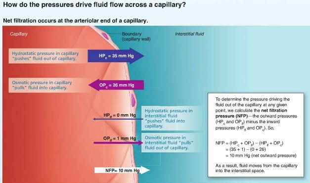
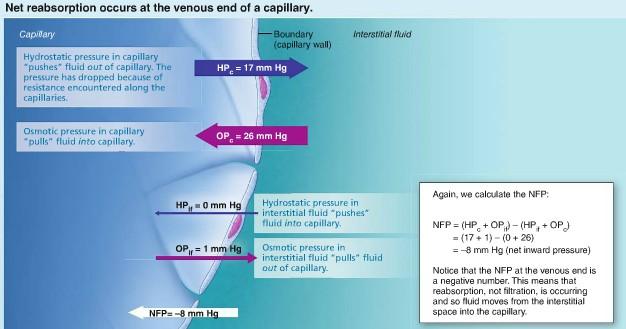
47 Capillary Hydrostatic Pressure (HPC) Forces fluids through capillary walls = filtration Leaving behind cells and most proteins BP drops as bfs along a capillary bed HPC is higher at arterial end (35mm Hg) then at venous end (17 mm Hg) BP which forces fluid out of the capillaries is opposed by the interstitial fluid hydrostatic pressure (HPif) acting outside the capillaries and pushing fluid in Usually very little fluid in the interstitial space Lymphatic vessels constantly withdraw it May vary from slightly to +, traditionally it is assumed to be 0 Colloid Osmotic Pressures (OP) The force opposing HP Created by large nondiffusible molecules Plasma proteins Unable to cross the capillary wall These molecules draw water toward themselves Encouraging osmosis bc the water concentration in their vicinity is lower than it is on opposite side of capillary walls * HP pushes & OP pulls/sucks capillary colloid osmotic pressure (OPc)/ oncotic pressure developed by the abundant amount of plasma proteins, primarily albumin molecules OPc is 26mm Hg Interstitial colloid osmotic pressure (OPif) Is lower.1 5 mm Hg unlike HP> OP doesn t vary significantly from one end of the capillary to the other Hydrostatic Osmotic Pressure Interactions net filtration pressure ( NFP) considers all the forces acting at the capillary bed while net filtration is occurring at the arteriole end a value for NFP at the venous end of the capillary indicates that fluid is moving into the capillaries (called reabsorption) result: net fluid flow is out of the circulation at the arterial end of capillary beds and into the circulation at the venous end

48 more fluid enters the tissue spaces than return to the blood result: a net loss of fluid from the circulation of about 1.5 ml/min lymphatic vessels pick up this fluid and any leaky proteins and return it to the vascular system > account for the relatively low levels of both fluid and proteins in the interstitial space
49

50 Circulatory Shock Any condition that blood vessels are inadequately filled and blood cannot circulate normally Cells die> organ dies Hypovolemic Shock Most common form Low blood volume Results from large scale blood or fluid loss Acute hemorrhage Sever vomiting or diarrhea Extensive burns If blood volume drops rapidly = HR increase in attempt to correct problem Weak thread pulse is first sign Intense vasoconstriction also occurs Shifts blood from the various blood reservoirs into the major circulatory channels & enhances venous return BP is stable at 1st then eventually drops if blood loss continues Replace fluid volume as quickly as possible Vascular Shock Blood volume is normal
Heart. Large lymphatic vessels Lymph node. Lymphatic. system Arteriovenous anastomosis. (exchange vessels)
") Venous system Large veins (capacitance vessels) Small veins (capacitance vessels) Postcapillary venule Thoroughfare channel Heart Large lymphatic vessels Lymph node Lymphatic system Arteriovenous anastomosis
Venous system Large veins (capacitance vessels) Small veins (capacitance vessels) Postcapillary venule Thoroughfare channel Heart Large lymphatic vessels Lymph node Lymphatic system Arteriovenous anastomosis
Cardiovascular System. Blood Vessel anatomy Physiology & regulation
 Cardiovascular System Blood Vessel anatomy Physiology & regulation Path of blood flow Aorta Arteries Arterioles Capillaries Venules Veins Vena cava Vessel anatomy: 3 layers Tunica externa (adventitia):
The Cardiovascular System. The Structure of Blood Vessels. The Structure of Blood Vessels. The Blood Vessels. Blood Vessel Review
 The Cardiovascular System The Blood Vessels The Structure of Blood Vessels Blood Vessel Review Arteries carry blood away from the heart Pulmonary trunk to lungs Aorta to everything else Microcirculation
Physiology of Circulation
 Physiology of Circulation Dr. Ali Ebneshahidi Blood vessels Arteries: Blood vessels that carry blood away from the heart to the lungs and tissues. Arterioles are small arteries that deliver blood to the
Copyright 2010 Pearson Education, Inc. Blood Vessel Structure
 Blood Vessel Structure Structure of Blood Vessel Walls Arteries and veins Tunica intima, tunica media, and tunica externa Lumen Central blood-containing space Capillaries Endothelium with sparse basal
Chapter 19: Blood Vessels. 63 slides
 Chapter 19: Blood Vessels 63 slides 1 Blood Vessels The Blood Vessels are essentially a series of tubes. Three types of blood vessel tubes: Arteries (carry blood away from heart) Arterioles are the smallest
Peripheral Circulation and Regulation
 Peripheral Circulation and Regulation Functions of Peripheral Circulation 1. Contain the blood 2. Exchange nutrients, waste products, and gases with tissues 3. Transport 4. Regulate blood pressure, along
Cardiovascular System Blood Vessels
 Cardiovascular System Blood Vessels Structure of Blood Vessels The three layers (tunics) Tunica intima composed of simple squamous epithelium Tunica media sheets of smooth muscle Contraction vasoconstriction
Cardiovascular system: Blood vessels, blood flow. Latha Rajendra Kumar, MD
 Cardiovascular system: Blood vessels, blood flow Latha Rajendra Kumar, MD Outline 1- Physical laws governing blood flow and blood pressure 2- Overview of vasculature 3- Arteries 4. Capillaries and venules
3/10/2009 VESSELS PHYSIOLOGY D.HAMMOUDI.MD. Palpated Pulse. Figure 19.11
 VESSELS PHYSIOLOGY D.HAMMOUDI.MD Palpated Pulse Figure 19.11 1 shows the common sites where the pulse is felt. 1. Temporal artery at the temple above and to the outer side of the eye 2. External maxillary
Physiology Unit 3 CARDIOVASCULAR PHYSIOLOGY: THE VASCULAR SYSTEM
 Physiology Unit 3 CARDIOVASCULAR PHYSIOLOGY: THE VASCULAR SYSTEM In Physiology Today Hemodynamics F = ΔP/R Blood flow (F) High to low pressure Rate = L/min Pressure (P) Hydrostatic pressure Pressure exerted
Chapter 21: Cardiovascular System: Peripheral Circulation and Regulation
 Chapter 21: Cardiovascular System: Peripheral Circulation and Regulation I. General Features of Blood Vessel Structure A. General Pattern of Circulation 1. Ventricles pump blood into 2. These arteries
Chapter 21 Peripheral circulation and Regulation
 Chapter 21 Peripheral circulation and Regulation I. Blood vessel structure A. Blood flows from large arteries to small capillaries 1. Large arteries contain large amounts of elastic tissue and little smooth
Chapter 21 (1) An Introduction to Blood Vessels and Circulation
 An Introduction to Blood Vessels and Circulation") Chapter 21 (1) An Introduction to Blood Vessels and Circulation Lecture Objectives Compare and contrast the structure of an artery, arteriole, vein, venule, and capillary Discuss the structure and function
Chapter 21 (1) An Introduction to Blood Vessels and Circulation Lecture Objectives Compare and contrast the structure of an artery, arteriole, vein, venule, and capillary Discuss the structure and function
Blood Vessels. Types of Blood Vessels Arteries carry blood away from the heart Capillaries smallest blood vessels. Veins carry blood toward the heart
 C H A P T E R Blood Vessels 20 Types of Blood Vessels Arteries carry blood away from the heart Capillaries smallest blood vessels The site of exchange of molecules between blood and tissue fluid Veins
Types of Blood Vessels
 Chapter 21 Peripheral Circulation and Regulation 21-1 Types of Blood Vessels Capillaries: site of exchange with tissue Arteries in dif. Types & sizes Elastic Muscular Arterioles Veins: thinner walls than
UNIT 4: BLOOD VESSELS
 UNIT 4: BLOOD VESSELS Dr. Moattar Raza Rizvi NRS237, Physiology Generalized Structure of Blood Vessels 1 Tunica interna (tunica intima) Endothelial layer that lines the lumen of all vessels In vessels
Lab Period: Name: Physiology Chapter 14 Blood Flow and Blood Pressure, Plus Fun Review Study Guide
 Lab Period: Name: Physiology Chapter 14 Blood Flow and Blood Pressure, Plus Fun Review Study Guide Main Idea: The function of the circulatory system is to maintain adequate blood flow to all tissues. Clinical
Cardiovascular System B L O O D V E S S E L S 2
 Cardiovascular System B L O O D V E S S E L S 2 Blood Pressure Main factors influencing blood pressure: Cardiac output (CO) Peripheral resistance (PR) Blood volume Peripheral resistance is a major factor
Blood Vessels. Over view. We have about 60,000 miles of blood vessels!
 Blood Vessels Over view 3 types of blood vessels arteries - carry blood away from heart "branch", "diverge", and "fork" veins - carry blood toward heart "join", "merge", and "converge" capillaries - site
Any of these questions could be asked as open question or lab question, thus study them well
 Any of these questions could be asked as open question or lab question, thus study them well describe the factors which regulate cardiac output describe the sympathetic and parasympathetic control of heart
Vascular System Part One
 Vascular System Part One Objectives Trace the route taken by blood as it leaves, and then returns to the heart. Describe the structure of the walls of arteries and veins. Discuss the structure and function
The Cardiovascular System: Vessels and Routes. Pulmonary Circulation H E A R T. Systemic Circulation
 The Cardiovascular System: Vessels and Routes 1. Overview of Blood Circulation A. Pulmonary Circulation Lung Arterioles Pulmonary Artery Capillaries Pulmonary Circulation Venules Pulmonary Veins H E A
Chapter 14 Blood Vessels, Blood Flow and Pressure Exam Study Questions
 Chapter 14 Blood Vessels, Blood Flow and Pressure Exam Study Questions 14.1 Physical Law Governing Blood Flow and Blood Pressure 1. How do you calculate flow rate? 2. What is the driving force of blood
2. capillaries - allow exchange of materials between blood and tissue fluid
 Chapter 19 - Vascular System A. categories and general functions: 1. arteries - carry blood away from heart 2. capillaries - allow exchange of materials between blood and tissue fluid 3. veins - return
Structure and organization of blood vessels
 The cardiovascular system Structure of the heart The cardiac cycle Structure and organization of blood vessels What is the cardiovascular system? The heart is a double pump heart arteries arterioles veins
Cardivascular System Module 5: Structure and Function of Blood Vessels *
 OpenStax-CNX module: m49689 1 Cardivascular System Module 5: Structure and Function of Blood Vessels * Donna Browne Based on Structure and Function of Blood Vessels by OpenStax This work is produced by
Collin County Community College
 Collin County Community College BIOL. 2402 Anatomy & Physiology WEEK 6 Blood Vessels 1 Anatomy of Blood Vessels Walls of blood vessels contain 3 distinct layers : Tunica intima innermost layer includes
Physiology of Circulation. Dr. Hiwa Shafiq 16/12/2018
 Physiology of Circulation Dr. Hiwa Shafiq 16/12/2018 Overview of the circulation The function of the circulation is to: 1. transport nutrients to the body tissues 2. transport waste products away 3. conduct
2402 : Anatomy/Physiology
 Dr. Chris Doumen Lecture 1 2402 : Anatomy/Physiology Hemo Dynamics and Blood Vessels I nt r oduc t i on TextBook Readings Pages 721 through 734. Make use of the figures in your textbook ; a picture is
BIOL 219 Spring Chapters 14&15 Cardiovascular System
 1 BIOL 219 Spring 2013 Chapters 14&15 Cardiovascular System Outline: Components of the CV system Heart anatomy Layers of the heart wall Pericardium Heart chambers, valves, blood vessels, septum Atrioventricular
The Circulatory System (p )
") The Circulatory System (p. 268-281) How Does Gravity Affect Blood Circulation? As with all land animals, the giraffe and the corn snake are constantly subject to the force of gravity The circulatory system
The Circulatory System (p. 268-281) How Does Gravity Affect Blood Circulation? As with all land animals, the giraffe and the corn snake are constantly subject to the force of gravity The circulatory system
Blood Pressure Fox Chapter 14 part 2
 Vert Phys PCB3743 Blood Pressure Fox Chapter 14 part 2 T. Houpt, Ph.D. 1 Cardiac Output and Blood Pressure How to Measure Blood Pressure Contribution of vascular resistance to blood pressure Cardiovascular
BIPN100 F15 Human Physiol I (Kristan) Lecture 14 Cardiovascular control mechanisms p. 1
 Lecture 14 Cardiovascular control mechanisms p. 1") BIPN100 F15 Human Physiol I (Kristan) Lecture 14 Cardiovascular control mechanisms p. 1 Terms you should understand: hemorrhage, intrinsic and extrinsic mechanisms, anoxia, myocardial contractility, residual
BIPN100 F15 Human Physiol I (Kristan) Lecture 14 Cardiovascular control mechanisms p. 1 Terms you should understand: hemorrhage, intrinsic and extrinsic mechanisms, anoxia, myocardial contractility, residual
I. Cardiac Output Chapter 14
 10/24/11 I. Cardiac Output Chapter 14 Cardiac Output, Blood Flow, and Blood Pressure Lecture PowerPoint Copyright The McGraw-Hill Companies, Inc. Permission required for reproduction or display. Cardiac
The Cardiovascular system: physiology of circulation
 Chapter 21 The Cardiovascular system: physiology of circulation blood vessel structure and function physiology of circulation: blood flow, blood pressure, and resistance blood flow the amount of blood
Physiology lecture 15 Hemodynamic
 Physiology lecture 15 Hemodynamic Dispensability (D) : proportional change in volume per unit change in pressure D = V/ P*V It is proportional (divided by the original volume). Compliance (C) : total change
1. Distinguish among the types of blood vessels on the basis of their structure and function.
 Blood Vessels and Circulation Objectives This chapter describes the structure and functions of the blood vessels Additional subjects contained in Chapter 13 include cardiovascular physiology, regulation,
Blood pressure. Formation of the blood pressure: Blood pressure. Formation of the blood pressure 5/1/12
 Blood pressure Blood pressure Dr Badri Paudel www.badripaudel.com Ø Blood pressure means the force exerted by the blood against the vessel wall Ø ( or the force exerted by the blood against any unit area
10. Thick deposits of lipids on the walls of blood vessels, called, can lead to serious circulatory issues. A. aneurysm B. atherosclerosis C.
 Heart Student: 1. carry blood away from the heart. A. Arteries B. Veins C. Capillaries 2. What is the leading cause of heart attack and stroke in North America? A. alcohol B. smoking C. arteriosclerosis
Arteries AWAY. Branch. Typically oxygenated.
 Arteries AWAY Branch Typically oxygenated. Capillaries Smallest. Most abundant. 10 billion. Huge surface area. Exchange Veins TOWARDS Converge. Typically deoxygenated. 3 Layers of the Vascular Wall Tunica
Cardiovascular System: Vessels and Circulation (Chapter 21)
") Cardiovascular System: Vessels and Circulation (Chapter 21) Lecture Materials for Amy Warenda Czura, Ph.D. Suffolk County Community College Eastern Campus Primary Sources for figures and content: Marieb,
Cardiovascular System: Vessels and Circulation (Chapter 21) Lecture Materials for Amy Warenda Czura, Ph.D. Suffolk County Community College Eastern Campus Primary Sources for figures and content: Marieb,
The Cardiovascular and Lymphatic Systems Cardiovascular System Blood Vessels Blood Vessels Arteries Arteries Arteries
 CH 12 The Cardiovascular and s The Cardiovascular and s OUTLINE: Cardiovascular System Blood Vessels Blood Pressure Cardiovascular System The cardiovascular system is composed of Blood vessels This system
Blood Vessels. Chapter 20
 Blood Vessels Chapter 20 Summary of the Characteristics of Arteries and Veins Characteristic Artery Vein Wall thickness thick thin Shape in cross section round flattened Thickest tunic media externa Collagen
BIOLOGY 2060 LECTURE NOTES ANATOMY & PHYSIOLOGY II (A. IMHOLTZ) VESSELS P1 OF 7
 VESSELS P1 OF 7") BIOLOGY 2060 LECTURE NOTES ANATOMY & PHYSIOLOGY II (A. IMHOLTZ) VESSELS P1 OF 7 1. Blood vessels a. Tubes through which the heart pumps blood. b. 3 major types of blood vessels: arteries, capillaries,
BIOLOGY 2060 LECTURE NOTES ANATOMY & PHYSIOLOGY II (A. IMHOLTZ) VESSELS P1 OF 7 1. Blood vessels a. Tubes through which the heart pumps blood. b. 3 major types of blood vessels: arteries, capillaries,
Function: Transportation of. Oxygen Nutrients Waste Hormones gases
 Function: Transportation of Oxygen Nutrients Waste Hormones gases Pericardium: double sac of serous membrane filled with fluid (pericardial fluid to be exact) that surrounds the heart. Parietal pericardium:
Cardiovascular System B L O O D V E S S E L S 3
 Cardiovascular System B L O O D V E S S E L S 3 Fluid Shifts Between Capillaries and Tissue Permeable capillaries allow plasma and solutes to pass into interstitial space interstitial or extracellular
d) Cardiovascular System Higher Human Biology
 Cardiovascular System Higher Human Biology") d) Cardiovascular System Higher Human Biology What can your remember about the heart and blood vessels? What is the Cardiovascular System? The cardiovascular system, also known as the circulatory system,
d) Cardiovascular System Higher Human Biology What can your remember about the heart and blood vessels? What is the Cardiovascular System? The cardiovascular system, also known as the circulatory system,
Physiology Chapter 14 Key Blood Flow and Blood Pressure, Plus Fun Review Study Guide
 Physiology Chapter 14 Key Blood Flow and Blood Pressure, Plus Fun Review Study Guide 1 Main Idea: The function of the circulatory system is to maintain adequate blood flow to all tissues. Clinical Application
Histology of the Cardiac System. Dr. Nabil Khoury Anatomy Department
 Histology of the Cardiac System Dr. Nabil Khoury Anatomy Department Objectives 1. Identify the 3 layers of the heart endocardium, myocardium, epicardium 2. Differentiate cardiacmuscle 3. Define intercalated
The cardiovascular system
 The cardiovascular system Components of the Cardiovascular system Heart Vessels: Arteries Capillaries Veins Functions of CVS: Transportation system where blood is the transporting vehicle Carries oxygen,
Six main classes of blood vessels (on handout) Wall structure of arteries and veins (on handout) Comparison: Arteries vs. Veins (on handout)
 Wall structure of arteries and veins (on handout) Comparison: Arteries vs. Veins (on handout)") Cardiovascular System: Vessels and Circulation (Chapter 21) Lecture Materials for Amy Warenda Czura, Ph.D. Suffolk County Community College Eastern Campus Six main classes of blood vessels Primary Sources
Cardiovascular System: Vessels and Circulation (Chapter 21) Lecture Materials for Amy Warenda Czura, Ph.D. Suffolk County Community College Eastern Campus Six main classes of blood vessels Primary Sources
Blood Pressure Regulation. Faisal I. Mohammed, MD,PhD
 Blood Pressure Regulation Faisal I. Mohammed, MD,PhD 1 Objectives Outline the short term and long term regulators of BP Know how baroreceptors and chemoreceptors work Know function of the atrial reflex.
The Cardiovascular and Lymphatic Systems
 BIOLOGY OF HUMANS Concepts, Applications, and Issues Fifth Edition Judith Goodenough Betty McGuire 12 The Cardiovascular and Lymphatic Systems Lecture Presentation Anne Gasc Hawaii Pacific University and
The Cardiovascular System
 The Cardiovascular System The Cardiovascular System A closed system of the heart and blood vessels The heart pumps blood Blood vessels allow blood to circulate to all parts of the body The function of
Therefore MAP=CO x TPR = HR x SV x TPR
 Regulation of MAP Flow = pressure gradient resistance CO = MAP TPR Therefore MAP=CO x TPR = HR x SV x TPR TPR is the total peripheral resistance: this is the combined resistance of all blood vessels (remember
Veins. VENOUS RETURN = PRELOAD = End Diastolic Volume= Blood returning to heart per cardiac cycle (EDV) or per minute (Venous Return)
 or per minute (Venous Return)") Veins Venous system transports blood back to heart (VENOUS RETURN) Capillaries drain into venules Venules converge to form small veins that exit organs Smaller veins merge to form larger vessels Veins
Veins Venous system transports blood back to heart (VENOUS RETURN) Capillaries drain into venules Venules converge to form small veins that exit organs Smaller veins merge to form larger vessels Veins
Major Function of the Cardiovascular System. Transportation. Structures of the Cardiovascular System. Heart - muscular pump
 Structures of the Cardiovascular System Heart - muscular pump Blood vessels - network of tubes Blood - liquid transport vehicle brachiocephalic trunk superior vena cava right pulmonary arteries right pulmonary
CHAPTER 21 LECTURE OUTLINE
 CHAPTER 21 LECTURE OUTLINE I. INTRODUCTION A. One main focus of this chapter considers hemodynamics, the means by which blood flow is altered and distributed and by which blood pressure is regulated. B.
Chapter 21! Chapter 21 Blood Vessels and Circulation! Blood Vessels and Circulation!
 Chapter 21! Blood Vessels and Circulation! SECTION 21-1! Blood vessels differ in size, structure, and functional properties! 2 Major Vessel Types! Arteries - carry blood away from the heart Higher pressure
Chapter 21! Blood Vessels and Circulation! SECTION 21-1! Blood vessels differ in size, structure, and functional properties!
 Chapter 21! Blood Vessels and Circulation! SECTION 21-1! Blood vessels differ in size, structure, and functional properties! 2 1! Major Vessel Types! Arteries - carry blood away from the heart Higher pressure
Blood Pressure. a change in any of these could cause a corresponding change in blood pressure
 Blood Pressure measured as mmhg Main factors affecting blood pressure: 1. cardiac output 2. peripheral resistance 3. blood volume a change in any of these could cause a corresponding change in blood pressure
Extra notes for lab- 1 histology. Slide 1 : cross section in the elastic artery ( aortic arch, ascending aorta, descending aorta )
") Extra notes for lab- 1 histology Slide 1 : cross section in the elastic artery ( aortic arch, ascending aorta, descending aorta ) - twin of ascending aorta is the pulmonary trunk. Ascending aorta represents
Extra notes for lab- 1 histology Slide 1 : cross section in the elastic artery ( aortic arch, ascending aorta, descending aorta ) - twin of ascending aorta is the pulmonary trunk. Ascending aorta represents
Cardiovascular Anatomy Dr. Gary Mumaugh
 Cardiovascular Anatomy Dr. Gary Mumaugh Location of Heart Approximately the size of your fist Location o Superior surface of diaphragm o Left of the midline in mediastinum o Anterior to the vertebral column,
Structure. Arteries. 21_01d 4/18/12. The Cardiovascular System: Blood Vessels and Hemodynamics. Dr Badri Paudel GMC
 Goal of the Cardiovascular System: deliver blood to all parts of the body The Cardiovascular System: Blood Vessels and Hemodynamics Dr Badri Paudel GMC Does so by using different types of tubing, attached
Cardiovascular System
 Cardiovascular System Purpose Transport oxygen and nutrients Take waste products away from tissues & organs Things we learned Blood pressure: the force of blood pushing against the walls of blood vessels
Cardiovascular Physiology
 Cardiovascular Physiology Lecture 1 objectives Explain the basic anatomy of the heart and its arrangement into 4 chambers. Appreciate that blood flows in series through the systemic and pulmonary circulations.
Blood Flow and Blood Pressure Regulation *
 OpenStax-CNX module: m44806 1 Blood Flow and Blood Pressure Regulation * OpenStax This work is produced by OpenStax-CNX and licensed under the Creative Commons Attribution License 4.0 By the end of this
IB TOPIC 6.2 THE BLOOD SYSTEM
 IB TOPIC 6.2 THE BLOOD SYSTEM TERMS TO KNOW circulation ventricle artery vein THE BLOOD SYSTEM 6.2.U1 - Arteries convey blood at high pressure from the ventricles to the tissues of the body Circulation
The Circulatory System. Lesson Overview. Lesson Overview The Circulatory System
 33.1 THINK ABOUT IT More than one-third of the 1.2 million Americans who suffer a heart attack each year die. This grim evidence shows that the heart and the circulatory system it powers are vital to life.
Blood flows away from the heart in arteries, to the capillaries and back to the heart in the veins
 Cardiovascular System Summary Notes The cardiovascular system includes: The heart, a muscular pump The blood, a fluid connective tissue The blood vessels, arteries, veins and capillaries Blood flows away
The Circulatory System
 The Circulatory System Key Questions What are the functions of the circulatory system? How does the heart pump blood through the body? What are three types of blood vessels? Vocabulary myocardium atrium
Cardiovascular system
 Cardiovascular system L-4 Blood pressure & special circulation Dr Than Kyaw 27 February 2012 Blood Pressure (BP) Pressure generation and flow Blood is under pressure within its closed system. Pressure
Blood Pressure Regulation. Slides 9-12 Mean Arterial Pressure (MAP) = 1/3 systolic pressure + 2/3 diastolic pressure
 = 1/3 systolic pressure + 2/3 diastolic pressure") Sheet physiology(18) Sunday 24-November Blood Pressure Regulation Slides 9-12 Mean Arterial Pressure (MAP) = 1/3 systolic pressure + 2/3 diastolic pressure MAP= Diastolic Pressure+1/3 Pulse Pressure CO=MAP/TPR
Sheet physiology(18) Sunday 24-November Blood Pressure Regulation Slides 9-12 Mean Arterial Pressure (MAP) = 1/3 systolic pressure + 2/3 diastolic pressure MAP= Diastolic Pressure+1/3 Pulse Pressure CO=MAP/TPR
Chapter 9. Body Fluid Compartments. Body Fluid Compartments. Blood Volume. Blood Volume. Viscosity. Circulatory Adaptations to Exercise Part 4
 Body Fluid Compartments Chapter 9 Circulatory Adaptations to Exercise Part 4 Total body fluids (40 L) Intracellular fluid (ICF) 25 L Fluid of each cell (75 trillion) Constituents inside cell vary Extracellular
Chapter 21. Blood Vessels and Circulation
 Chapter 21 Openstax: Chapter 20 Blood Vessels and Circulation Chapter 21 Learning Outcomes After completing Chapter 21, you will be able to: 1. Distinguish among the types of blood vessels based on their
Citation Jarvis S (2018) Vascular system 1: anatomy and physiology. Nursing Times [online]; 114: 4,
![Citation Jarvis S (2018) Vascular system 1: anatomy and physiology. Nursing Times [online]; 114: 4,](/thumbs/96/127077409.jpg "Citation Jarvis S (2018) Vascular system 1: anatomy and physiology. Nursing Times [online]; 114: 4,") Vascular system Keywords Arteries/Veins// Blood flow/fluid movement This article has been double-blind peer reviewed In this article... Anatomy of the vascular system and structure of blood vessels The
Vascular system Keywords Arteries/Veins// Blood flow/fluid movement This article has been double-blind peer reviewed In this article... Anatomy of the vascular system and structure of blood vessels The
Circulation. Sinoatrial (SA) Node. Atrioventricular (AV) Node. Cardiac Conduction System. Cardiac Conduction System. Linked to the nervous system
 Node. Atrioventricular (AV) Node. Cardiac Conduction System. Cardiac Conduction System. Linked to the nervous system") Circulation Cardiac Conduction System AHS A H S Your body resembles a large roadmap. There are routes or arteries that take you downtown to the heart of the city and veins that take you to the outskirts
Circulation Cardiac Conduction System AHS A H S Your body resembles a large roadmap. There are routes or arteries that take you downtown to the heart of the city and veins that take you to the outskirts
Ch. 12 The Circulatory System. The heart. The heart is a double pump. A quick note on arteries vs. veins. = the muscular pump of the CV system
 Ch. 12 The Circulatory System The heart A.k.a. the cardiovascular system Blood was discussed in Ch. 11 Focus of Ch. 12: heart and blood vessels = the muscular pump of the CV system ~ 100,000 heartbeats/day!
1. Label the Diagram using the following terms: artery, arterioles, vein, venules, capillaries, valve, inner wall, middle wall, outer wall
 Bio 20 Ms. Nyboer Arteries, Veins, Capillaries, and the Heart Structure and Function Workbook Use your textbook (Ch. 10) and notes to fill in this workbook Part A: Arteries, Veins, Capillaries 1. Label
The Cardiovascular System
 PowerPoint Lecture Slide Presentation by Patty Bostwick-Taylor, Florence-Darlington Technical College The Cardiovascular System 11PART B The Heart: Cardiac Output Cardiac output (CO) Amount of blood pumped
Circulatory System Review
 Circulatory System Review 1. Know the diagrams of the heart, internal and external. a) What is the pericardium? What is myocardium? What is the septum? b) Explain the 4 valves of the heart. What is their
Chapter 21. Blood Vessels and Circulation
 Chapter 21 Blood Vessels and Circulation INTRODUCTION A. One main focus of this chapter considers hemodynamics, the means by which blood flow is altered and distributed and by which blood pressure is regulated.
1. Which of the following blood vessels has a thin elastic layer? A. Aorta. B. Pulmonary artery. C. Posterior vena cava. D. Mesenteric capillary.
 CIRCULATORY SYSTEM 1. Which of the following blood vessels has a thin elastic layer? A. Aorta. B. Pulmonary artery. C. Posterior vena cava. D. Mesenteric capillary. 2. Capillary beds are equipped with
Cardiovascular System. Biology 105 Lecture 15 Chapter 12
 Cardiovascular System Biology 105 Lecture 15 Chapter 12 Outline I. Functions of cardiovascular system II. Components of the cardiovascular system: I. Blood vessels II. Heart III. Regulation of the heartbeat
Approximately the size of your fist Location Superior surface of diaphragm Left of the midline in mediastinum Anterior to the vertebral column,
 Dr. Gary Mumaugh Approximately the size of your fist Location Superior surface of diaphragm Left of the midline in mediastinum Anterior to the vertebral column, posterior to the sternum Posteriorly the
37 1 The Circulatory System
 H T H E E A R T 37 1 The Circulatory System The circulatory system and respiratory system work together to supply cells with the nutrients and oxygen they need to stay alive. a) The respiratory system:
Figure ) The specific chamber of the heart that is indicated by letter A is called the. Diff: 1 Page Ref: 364
 The specific chamber of the heart that is indicated by letter A is called the. Diff: 1 Page Ref: 364") Essentials of Anatomy and Physiology, 9e (Marieb) Chapter 11 The Cardiovascular System Short Answer Figure 11.1 Using Figure 11.1, identify the following: 1) The Purkinje fibers are indicated by label.
Essentials of Anatomy and Physiology, 9e (Marieb) Chapter 11 The Cardiovascular System Short Answer Figure 11.1 Using Figure 11.1, identify the following: 1) The Purkinje fibers are indicated by label.
Cardiovascular system
 BIO 301 Human Physiology Cardiovascular system The Cardiovascular System: consists of the heart plus all the blood vessels transports blood to all parts of the body in two 'circulations': pulmonary (lungs)
Unit 1: Human Systems. The Circulatory System
 Unit 1: Human Systems The Circulatory System nourish all cells with oxygen, glucose, amino acids and other nutrients and carry away carbon dioxide, urea and other wastes Purposes Transport chemical messengers
Control of blood tissue blood flow. Faisal I. Mohammed, MD,PhD
 Control of blood tissue blood flow Faisal I. Mohammed, MD,PhD 1 Objectives List factors that affect tissue blood flow. Describe the vasodilator and oxygen demand theories. Point out the mechanisms of autoregulation.
Anatomy and Physiology, Spring 2015 Exam II: Form A April 9, Name Student Number
 Anatomy and Physiology, Spring 2015 Exam II: Form A April 9, 2015 Name Student Number For Questions 1 2 refer to the following table. 1 Ventricular pressure is greater than aortic 6 AV valve is open 2
Cardiac Output 1 Fox Chapter 14 part 1
 Vert Phys PCB3743 Cardiac Output 1 Fox Chapter 14 part 1 T. Houpt, Ph.D. Regulation of Heart & Blood Pressure Keep Blood Pressure constant if too low, not enough blood (oxygen, glucose) reaches tissues
During exercise the heart rate is 190 bpm and the stroke volume is 115 ml/beat. What is the cardiac output?
 The Cardiovascular System Part III: Heart Outline of class lecture After studying part I of this chapter you should be able to: 1. Be able to calculate cardiac output (CO) be able to define heart rate
Pearson's Comprehensive Medical Assisting Administrative and Clinical Competencies
 Pearson's Comprehensive Medical Assisting Administrative and Clinical Competencies THIRD EDITION CHAPTER 27 The Cardiovascular System Lesson 1: Overview of the Cardiovascular System Lesson Objectives Upon
Chapter 9, Part 2. Cardiocirculatory Adjustments to Exercise
 Chapter 9, Part 2 Cardiocirculatory Adjustments to Exercise Electrical Activity of the Heart Contraction of the heart depends on electrical stimulation of the myocardium Impulse is initiated in the right
PHYSIOLOGY WEEK 9. vascular physiology - ED Primary Exam Teaching
 PHYSIOLOGY WEEK 9 vascular physiology - ED Primary Exam Teaching CONCEPTS OF BLOOD FLOW Vascular distensibility - when pressure in the arterioles is increased, this dilates the arterioles and therefore
This is a TRANSPORT system that allows every cell: i) uptake of nutrients ( ex. oxygen, glucose) ii) excretes wastes (ex C02, ammonia)
 uptake of nutrients ( ex. oxygen, glucose) ii) excretes wastes (ex C02, ammonia)") Biology 20 Unit D This is a TRANSPORT system that allows every cell: i) uptake of nutrients ( ex. oxygen, glucose) ii) excretes wastes (ex C02, ammonia) requires action of: heart: muscular force to move
Biology 20 Unit D This is a TRANSPORT system that allows every cell: i) uptake of nutrients ( ex. oxygen, glucose) ii) excretes wastes (ex C02, ammonia) requires action of: heart: muscular force to move
Circulation And Blood. Circulation And Blood. Circulation And Blood. Circulation And Blood. Blood 10/22/2012
 Cells in our body build their own membranes and organelles Make their own ATP Assemble their own enzymes and other proteins And may manufacture substances used elsewhere in the body To do these things,