Cardiac MRI: Clinical Application to Disease
|
|
|
- Lambert Jackson
- 6 years ago
- Views:
Transcription

1 Cardiac MRI: Clinical Application to Disease Jessi Smith, MD Cardiothoracic imaging, Indiana University Slides courtesy of Stacy Rissing, MD
2 Outline Imaging planes Disease findings Pulse sequences used for each indication Pathophysiology being evaluated

3 Imaging planes

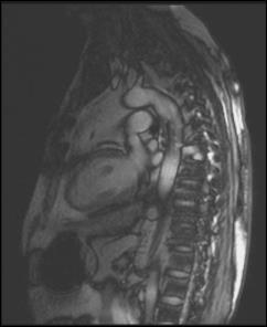
4 3 Plane localizer

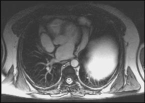
5 Axial Haste

6 2 Chamber Planned from axial images- usually HASTE

7 2 Chamber Planned from axial images- usually HASTE

8 Pseudo short axis Planned from 2 chamber cine


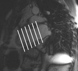
9 Pseudo short axis BASE APEX


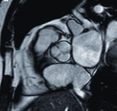
10 4 chamber Planned from Pseudo SA and 2 Chamber
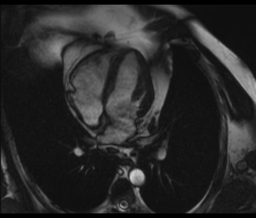
11 4 chamber

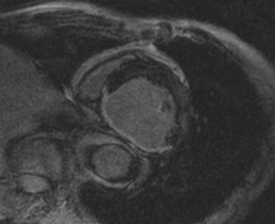
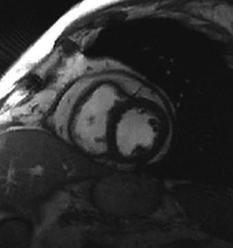
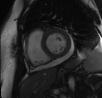
12 True short axis

13 LVOT Left ventricular outflow tract Planned off of pseudo SA
14 LVOT1 and LVOT2


15 Trans-aortic valve view Planned off LVOT1 and LVOT2
16 Trans-aortic valve view
17 Trans-aortic valve view

18 What are we looking for?
19 CMR for Myocardial Disease Coronary artery disease Is there viable myocardium? Function and viability study Infiltrative myocardial disease Sarcoidosis and Amyloidosis Myocarditis Most idiopathic, some cases with viral etiology Cardiomyopathy Dilated CMO, Hypertrophic CMO, and ARVC
20 CMR for Myocardial Disease Coronary artery disease Is there viable myocardium? Function and viability study Infiltrative myocardial disease Sarcoidosis and Amyloidosis Myocarditis Most idiopathic, some cases with viral etiology Cardiomyopathy Dilated CMO, Hypertrophic CMO, and ARVC

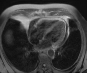
21 Coronary Artery Disease The Function and Viability Study Usual clinical question: Would this patient benefit from CABG? Goal is to distinguish myocardium that has the ability to contract versus myocardium that is replaced by fibrosis or scar. Fibrosis and scar will not regain function after CABG David Letterman
22 Coronary Artery Disease The Function and Viability Study The Basic Protocol 3 plane localizer Axial haste Cine bright blood (SSFP) 2C, SA, 4C Delayed enhancement SA, 2C, 4C Key sequence, looking for scar Pattern is subendocardial


23 Function and viability Delayed enhancement = scar Scar!
24 CMR for Myocardial Disease Coronary artery disease Is there viable myocardium? Function and viability study Infiltrative myocardial disease Sarcoidosis and Amyloidosis Myocarditis Most idiopathic, some cases with viral etiology Cardiomyopathy Dilated CMO, Hypertrophic CMO, and ARVC
25 CMR for Myocardial Disease Coronary artery disease Is there viable myocardium? Function and viability study Infiltrative myocardial disease Sarcoidosis and Amyloidosis Myocarditis Most idiopathic, some cases with viral etiology Cardiomyopathy Dilated CMO, Hypertrophic CMO, and ARVC

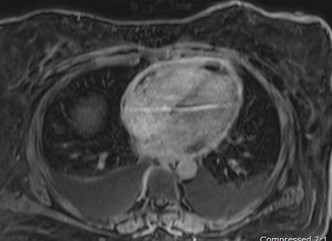
26 Infiltrative myocardial disease Cardiac Sarcoidosis Presence of non-caseating granulomas in the pericardium, myocardium, or endocardium leading to clinical sequelae Myocardial granulomas have been associated with cardiac arrhythmia and even sudden death Reggie White
27 Sarcoidosis MR Protocol Function and viability study + T2WI 3-plane loc, axial haste Cine bright blood (SSFP) 2C, SA, and 4C Focal areas of myocardial thickening, wall motion abnormality, aneurysm formation T2-weighted images SA +/- 4C or 2C Myocardial edema and active inflammation Delayed enhancement in SA, 2C, and 4C Inflammation and/or granulomatous involvement Non-ischemic pattern (not confined to coronary distrib) Patchy or mid-myocardial in distribution


28 Cardiac Sarcoidosis Delayed enhancement, short axis images Courtesy Ricardo Cury


29 Cardiac Sarcoidosis Sparrow P J et al. Radiographics 2009;29: by Radiological Society of North America
30 CMR for Myocardial Disease Coronary artery disease Is there viable myocardium? Function and viability study Infiltrative myocardial disease Sarcoidosis and Amyloidosis Myocarditis Most idiopathic, some cases with viral etiology Cardiomyopathy Dilated CMO, Hypertrophic CMO, and ARVC
31 Cardiac Amyloidosis Amyloid proteins are abnormally deposited within heart, especially myocardium Nobody famous Imaging findings: LV wall thickening, atrial enlargement, pericardial effusion, patchy or diffuse LV delayed enhancement
32 Cardiac Amyloidosis MR protocol Essentially function and viability study 3-plane loc, axial haste Cine SSFP 2C, SA, 4C views Cardiac function often reduced Myocardial wall thickness and mass Delayed enhancement SA, 2C, 4C views Eval for infiltrative myocardial process

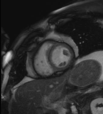
33 Infiltrative disease Figure 3a. Cardiac amyloidosis in a 75-year-old man with multiple myeloma. Amyloidosis Really thick myocardium Cummings K W et al. Radiographics 2009;29: by Radiological Society of North America


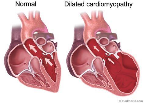
34 Cardiac Amyloidosis delayed enhancement More typical, patchy Classic, diffuse subendocardial

35 CMR for Myocardial Disease Coronary artery disease Is there viable myocardium? Function and viability study Infiltrative myocardial disease Sarcoidosis and Amyloidosis Myocarditis Most idiopathic, some cases with viral etiology Cardiomyopathy Dilated CMO, Hypertrophic CMO, and ARVC

36 Myocarditis Inflammation of the myocardium with necrosis of the adjacent myocytes; usually infectious Classic diagnostic picture: Suddenly decreased systolic function Big dilated heart Otherwise healthy person Shortly after viral illness Rachel Hunter
37 Myocarditis 3-plane loc, axial haste Cine bright blood (SSFP) 2C, SA, 4C Evaluate cardiac function usually reduced T2-weighted images SA and 4C Myocardial edema and inflammation Relative to skeletal muscle Want plenty of skeletal muscle in the FOV get that arm! Pre and post contrast T1-weighted images SA and 4C Ratio myocardial enhance to skeletal muscle enhance RE-RUN if possible!! Hyperemia and inflammation Delayed enhancement images SA, 4C, 2C Inflammation and scar


38 Myocarditis Sneak in that arm! Post contrast Pre contrast We do a similar measurement for T2-weighted images

39 Myocarditis Patchy mid myocardial delayed enhancement
40 CMR for Myocardial Disease Coronary artery disease Is there viable myocardium? Function and viability study Infiltrative myocardial disease Sarcoidosis and Amyloidosis Myocarditis Most idiopathic, some cases with viral etiology Cardiomyopathy (CMO) Dilated CMO, Hypertrophic CMO, and ARVC
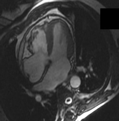
41 Cardiomyopathy Dilated Cardiomyopathy Disease of the myocardium LV becomes enlarged (dilated) and cannot pump blood as effectively to the body (heart failure) Can have no symptoms end stage heart failure Usually idiopathic need to r/o other causes Viral infxn, genetic, CAD, metabolic, lots more
42
43 Dilated Cardiomyopathy MR protocol Basic function and viability 3-plane loc, axial haste Cine SSFP 2C, SA, 4C Evaluate function and chamber sizes Delayed enhancement SA, 2c, 4C Evaluate for infarct
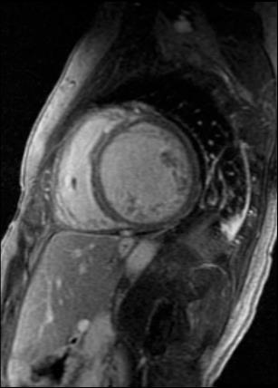
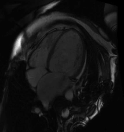


44 Dilated Cardiomyopathy LV too big LV too big Poor LV function

45 Hypertrophic Cardiomyopathy Disease of the myocardium Myocardial thickening, hypertrophy Leading cause sudden cardiac death in young athletes
46 Hypertrophic Cardiomyopathy MR Protocol 3-plane loc, axial haste SSFP 2C, SA, 4C LV wall thickness, chamber size, function LVOT1, LVOT2 LV outflow tract obstruction (important) +/- cine AV and FQ aortic valve Aortic valve appearance and function Delayed enhancement SA, 2C, 4C Myocardial scarring, fibrosis - prognosis


47 60-year-old woman with symmetric hypertrophic cardiomyopathy who underwent cardiac MRI before surgical intervention for mitral valve regurgitation and left ventricle (LV) outflow tract obliteration. Hypertrophic Cardiomyopathy Muscle too thick!! 2008 by American Roentgen Ray Society Belloni E et al. AJR 2008;191:


48 Hypertrophic Cardiomyopathy Patchy delayed enhancement = scar Can be source of arrhythmia

49 Arrhythmogenic Right Ventricular Cardiomyopathy ARVC Fibrofatty infiltration of the right ventricle Arrhythmia and possible sudden cardiac death
50 ARVC MR Protocol Much shorter protocol now! 3 plane loc, axial haste Cine SSFP 2C, SA, 4C RV size and wall thickness, function Coronal single shot TRUFI set up sequence HTR cine RV-axial and RV-sagittal High attention to RV aneurysm, dyskinesia

51 ARVC


52 Valve disease Looking for stenosis or regurgitation Aortic Mitral Tricuspid Pulmonic Barbara Walters
53 Aortic valve Stenosis or regurgitation Morphology- cusps? Helpful sequences to obtain: Cine SSFP LVOT and trans-aortic valve Look for the jet of aortic regurgitation Phase contrast (Flow quant)

54 Aortic valve- regurgitation

55 Aortic valve- regurgitation


56 Aortic valve- stenosis often seen with bicuspid aortic valve
57 Mitral valve Stenosis or regurgitation Cine bright blood (SSFP/SPGR) SA, 2C, 4C, and LVOT Phase contrast not as helpful for MV

58 Mitral valve- regurgitation
59 Pericardial disease Pericarditis Pericardial constriction Pericardial effusion Simple, blood, or malignant


60 Pericarditis Pericardial inflammation Usually due to infection Double IR images 4C and SA views Evaluate for pericardial thickening Early and delayed enhancement Evaluate for pericardial inflammation Bob Dylan


61 Pericarditis Way too thick! Way to bright! Double IR Delayed Enhancement
62 Pericarditis
63 Pericardial Constriction Thickened, fibrotic pericardium forms a non-compliant shell around the heart, which prevents heart from expanding when blood enters Most common in US = radiation therapy and surgery Most common worldwide = infection (TB) Key sequences: Double IR Pericardial thickening High temporal resolution SA cine Septal bounce Delayed enhancement - Pericardial inflammation TAG LINES!! Tethering of pericardial layers

64 Pericardium- normal
65 Pericardial constriction
66 Pericardial constriction
67 Pericardial fluid
68 Take home points Practice your cardiac imaging planes Help build MR protocol for specific diseases Cine SSFP is your workhorse Delayed enhancement for inflammation, infiltration, or infarction Phase contrast for flow quantification (valves) T1, T2, and contrast images for tissue characterization
69 Thank You
70 How many people do cardiac MRI at place Radiologist or cardiologist present for the study Do they give you a tailored protocol or run same protocol for all hearts?
Cardiac MRI: Clinical Application to Disease
 Cardiac MRI: Clinical Application to Disease Stacy Rissing, MD! Cardiothoracic imaging, Indiana University! Outline Imaging planes Disease findings Pulse sequences used for each indication Pathophysiology
Cardiac MRI: Cardiomyopathy
 Cardiac MRI: Cardiomyopathy Laura E. Heyneman, MD I do not have any relevant financial relationships with any commercial interests Cardiac MRI: Cardiomyopathy Laura E. Heyneman, MD Duke University Medical
cardiac imaging planes planning basic cardiac & aortic views for MR
 cardiac imaging planes planning basic cardiac & aortic views for MR Dianna M. E. Bardo, M. D. Assistant Professor of Radiology & Cardiovascular Medicine Director of Cardiac Imaging cardiac imaging planes
Cardiovascular manifestations of HIV
 Cardiovascular manifestations of HIV Prabhakar Rajiah, MBBS, MD, FRCR Associate Professor of Radiology Associate Director, Cardiac CT and MRI University of Texas Southwestern Medical Center, Dallas, USA
Constrictive Pericarditis Pitfalls in MR Diagnosis Cylen Javidan-Nejad Associate Professor Mallinckrodt Institute of Radiology Washington University
 Constrictive Pericarditis Pitfalls in MR Diagnosis Cylen Javidan-Nejad Associate Professor Mallinckrodt Institute of Radiology Washington University in St. Louis Goal o To review the imaging criteria of
Echocardiographic Evaluation of the Cardiomyopathies. Stephanie Coulter, MD, FACC, FASE April, 2016
 Echocardiographic Evaluation of the Cardiomyopathies Stephanie Coulter, MD, FACC, FASE April, 2016 Cardiomyopathies (CMP) primary disease intrinsic to cardiac muscle Dilated CMP Hypertrophic CMP Infiltrative
Role of CMR in heart failure and cardiomyopathy
 Role of CMR in heart failure and cardiomyopathy Hajime Sakuma Department of Radiology, Mie University Late gadolinium enhancement (LGE) LGE MRI can demonstrate site of necrosis, fibrosis or deposition
Imaging in Heart Failure: A Multimodality Approach. Thomas Ryan, MD
 Imaging in Heart Failure: A Multimodality Approach Thomas Ryan, MD Heart Failure HFrEF HFpEF EF50% Lifetime risk 20% Prevalence 6M Americans Societal costs - $30B 50% 5-year survival 1 Systolic
Managing Hypertrophic Cardiomyopathy with Imaging. Gisela C. Mueller University of Michigan Department of Radiology
 Managing Hypertrophic Cardiomyopathy with Imaging Gisela C. Mueller University of Michigan Department of Radiology Disclosures Gadolinium contrast material for cardiac MRI Acronyms Afib CAD Atrial fibrillation
Can SCMR CMR protocol recommendations
 Can SCMR CMR protocol recommendations V1.3 - April 2009 CanSCMR CMR Protocol and SOP Recommendation 2009 (15 minutes) 2 Planning of LV fct. real time multiple axes Realtime 3 cine long axis 6 long axes
Etiology, Classification & Management. Sheba Medical Center Cardiology Department Matthew Wright St. George s University of London
 Etiology, Classification & Management Sheba Medical Center Cardiology Department Matthew Wright St. George s University of London Introduction World Health Organization (1995): Diseases of myocardium (heart
ADVANCED CARDIOVASCULAR IMAGING. Medical Knowledge. Goals and Objectives PF EF MF LF Aspirational
 Medical Knowledge Goals and Objectives PF EF MF LF Aspirational Know the basic principles of magnetic resonance imaging (MRI) including the role of the magnetic fields and gradient coil systems, generation
Cardiac Imaging. Kimberly Delcour, DO, FACC. Mahi Ashwath, MD, FACC, FASE. Director, Cardiac CT. Director, Cardiac MRI
 Cardiac Imaging Kimberly Delcour, DO, FACC Director, Cardiac CT Mahi Ashwath, MD, FACC, FASE Director, Cardiac MRI Cardiac Imaging Discuss the clinical applications of and indications for: Cardiac CT Nuclear
9/23/2011. Cardiac MRI Evaluation of Cardiomyopathy and Myocarditis. Primary Hypertrophic Cardiomyopathy. Cardiomyopathy.
 Cardiomyopathy Cardiac MRI Evaluation of Cardiomyopathy and Myocarditis Laureen Sena Children s Hospital Boston, MA NASCI 2011 Baltimore, Maryland Primary Hypertrophic ARVD Dilated Restrictive Unclassified
Consequences of Cardiomyopathy. thickened and stiff, the left ventricle is most often affected. This results in a lack of pumping
 Roseanne Baird March 30, 2010 BI 104 Dr. Hammoudi Consequences of Cardiomyopathy Cardiomyopathy is a disease in which the myocardium of the heart becomes enlarged, thickened and stiff, the left ventricle
Why Cardiac MRI? Presented by:
 Why Cardiac MRI? Presented by: Lisa G. Carkner, MD, FACC 1 Disclosures I have no financial disclosures Objectives Review basic principles of Cardiac MRI. What patient characteristics do I need to consider
Case based learning: CMR in Heart Failure
 Case based learning: CMR in Heart Failure Milind Y Desai, MD FACC FAHA FESC Associate Professor of Medicine Heart and Vascular Institute, Cleveland Clinic Cleveland, OH Disclosures: none Use of Gadolinium
Cardiovascular Nursing Practice: A Comprehensive Resource Manual and Study Guide for Clinical Nurses 2 nd Edition
 Cardiovascular Nursing Practice: A Comprehensive Resource Manual and Study Guide for Clinical Nurses 2 nd Edition Table of Contents Volume 1 Chapter 1: Cardiovascular Anatomy and Physiology Basic Cardiac
MRI Sequences: What to use for what
 MRI Sequences: What to use for what MRI basics T 1 and T 2 relaxation Common Imaging Protocols Mechanical function (cine) Tissue characterization LGE Edema imaging (T 2 weighted) T1 Special protocols MRA
What s New in Cardiac MRI
 What s New in Cardiac MRI Katie M. Hawthorne, MD Director, Cardiac MRI Main Line Health Philadelphia Cardiovascular Summit November 18, 2017 Cardiac MRI: Disclosure 2 Disclosures No financial disclosures
Restrictive Cardiomyopathy
 ESC Congress 2011, Paris Imaging Unusual Causes of Cardiomyopathy Restrictive Cardiomyopathy Kazuaki Tanabe, MD, PhD Professor of Medicine Chair, Division of Cardiology Izumo, Japan I Have No Disclosures
Adel Hasanin Ahmed 1
 Adel Hasanin Ahmed 1 PERICARDIAL DISEASE The pericardial effusion ends anteriorly to the descending aorta and is best visualised in the PLAX. PSAX is actually very useful sometimes for looking at posterior
DISCLOSURE. Echocardiography in Systemic Diseases: Questions. Relevant Financial Relationship(s) None. Off Label Usage None 5/7/2018
 None. Off Label Usage None 5/7/2018") Echocardiography in Systemic Diseases: Questions Sunil Mankad, MD, FACC, FCCP, FASE Associate Professor of Medicine Mayo Clinic College of Medicine Director, Transesophageal Echocardiography Associate
Echocardiography in Systemic Diseases: Questions Sunil Mankad, MD, FACC, FCCP, FASE Associate Professor of Medicine Mayo Clinic College of Medicine Director, Transesophageal Echocardiography Associate
Case # 1. Page: 8. DUKE: Adams
 Case # 1 Page: 8 1. The cardiac output in this patient is reduced because of: O a) tamponade physiology O b) restrictive physiology O c) coronary artery disease O d) left bundle branch block Page: 8 1.
Proceedings of the 34th World Small Animal Veterinary Congress WSAVA 2009
 www.ivis.org Proceedings of the 34th World Small Animal Veterinary Congress WSAVA 2009 São Paulo, Brazil - 2009 Next WSAVA Congress : Reprinted in IVIS with the permission of the Congress Organizers MANAGEMENT
Patterns of late myocardial enhancement in cardiac MRI
 Patterns of late myocardial enhancement in cardiac MRI Poster No.: C-0683 Congress: ECR 2010 Type: Educational Exhibit Topic: Cardiac Authors: A. F. L. Carneiro, A. S. R. Preto, R. C. Ramos, A. J. B. S.
Cardiomyopathy. ACOI IM Board Review 2018 Martin C. Burke DO, FACOI
 Cardiomyopathy ACOI IM Board Review 2018 Martin C. Burke DO, FACOI No Disclosures Cardiomyopathies Definition: diseases of heart muscle 1980 WHO: unknown causes Not clinically relevant 1995 WHO: diseases
CARDIAC MRI. Cardiovascular Disease. Cardiovascular Disease. Cardiovascular Disease. Overview
 CARDIAC MRI Dr Yang Faridah A. Aziz Department of Biomedical Imaging University of Malaya Medical Centre Cardiovascular Disease Diseases of the circulatory system, also called cardiovascular disease (CVD),
Cardiac Sarcoidosis. Millee Singh DO Non Invasive Cardiology First Coast Heart and Vascluar
 Cardiac Sarcoidosis Millee Singh DO Non Invasive Cardiology First Coast Heart and Vascluar Introduction Multisystem granulomatous disease of unknown etiology characterized by noncaseating granulomas in
LV FUNCTION ASSESSMENT: WHAT IS BEYOND EJECTION FRACTION
 LV FUNCTION ASSESSMENT: WHAT IS BEYOND EJECTION FRACTION Jamilah S AlRahimi Assistant Professor, KSU-HS Consultant Noninvasive Cardiology KFCC, MNGHA-WR Introduction LV function assessment in Heart Failure:
Cardiac MRI: Appropriateness. Scott Mattson, DO, FACC Lutheran Medical Group Fort Wayne, IN
 Cardiac MRI: Appropriateness Scott Mattson, DO, FACC Lutheran Medical Group Fort Wayne, IN Approaches to Appropriateness The indication The patient The scanner and technologists The interpreting physician
CT for Myocardial Characterization of Cardiomyopathy. Byoung Wook Choi, Yonsei University Severance Hospital, Seoul, Korea
 CT for Myocardial Characterization of Cardiomyopathy Byoung Wook Choi, Yonsei University Severance Hospital, Seoul, Korea Cardiomyopathy Elliott P et al. Eur Heart J 2008;29:270-276 The European Society
Myocardial Infarction
 Myocardial Infarction MI = heart attack Defined as necrosis of heart muscle resulting from ischemia. A very significant cause of death worldwide. of these deaths, 33% -50% die before they can reach the
Objectives 8/17/2011. Challenges in Cardiac Imaging. Challenges in Cardiac Imaging. Basic Cardiac MRI Sequences
 8/17/2011 Traditional Protocol Model for Tomographic Imaging Cardiac MRI Sequences and Protocols Frandics Chan, M.D., Ph.D. Stanford University Medical Center Interpretation Lucile Packard Children s Hospital
Review of Cardiac Imaging Modalities in the Renal Patient. George Youssef
 Review of Cardiac Imaging Modalities in the Renal Patient George Youssef ECHO Left ventricular hypertrophy (LVH) assessment Diastolic dysfunction Stress ECHO Cardiac CT angiography Echocardiography - positives
Cardiomyopathy. Cardiomyopathies HOCM. Hypertrophic Obstructive Cardiomyopathy. Systolic Anterior Movement (SAM) of Mitral Valve (Venturi Effect) Cine
 of Mitral Valve (Venturi Effect) Cine") Jens Bremerich Radiology University Hospital Basel Hypertrophic Obstructive Cine VENC Cine (5m/s) Modified Bernoulli Equation: P (in mmhg) = 4 x (Vmax)2 Vmax= 4.2 m/s, P = 70mm Hg Hydrodynamica 738 HOCM
Jens Bremerich Radiology University Hospital Basel Hypertrophic Obstructive Cine VENC Cine (5m/s) Modified Bernoulli Equation: P (in mmhg) = 4 x (Vmax)2 Vmax= 4.2 m/s, P = 70mm Hg Hydrodynamica 738 HOCM
Conflict Disclosures. Vermont Cardiac Network. Outline. Series Learning Objectives 4/27/2016. Scott E. Friedman April 28, 2016
 Conflict Disclosures Vermont Cardiac Network The Speaker has reported no significant financial relationship with any companies whose product may be germane to the content of their presentations or who
Atlas of Practical Cardiac Applications of MRI
 Atlas of Practical Cardiac Applications of MRI Atlas of Practical Cardiac Applications of MRI Guillcm Pons-LIado, MD. Director, Cardiac Imaging Unit, Cardiology Department, Hospital de la Santa Creu i
Case based learning: CMR in Heart Failure
 Case based learning: CMR in Heart Failure Milind Y Desai, MD FACC FAHA FESC Associate Professor of Medicine Heart and Vascular Institute, Cleveland Clinic Cleveland, OH Disclosures: none Use of Gadolinium
Update on use of cardiac MRI in ARVC/D. Stefan L. Zimmerman, MD Johns Hopkins University Department of Radiology
 Update on use of cardiac MRI in ARVC/D Stefan L. Zimmerman, MD Johns Hopkins University Department of Radiology Outline Background Diagnosis Characteristic imaging findings Genetics of ARVC Genotype phenotype
Ablative Therapy for Ventricular Tachycardia
 Ablative Therapy for Ventricular Tachycardia Nitish Badhwar, MD, FACC, FHRS 2 nd Annual UC Davis Heart and Vascular Center Cardiovascular Nurse / Technologist Symposium May 5, 2012 Disclosures Research
Semiology of the Heart in the 21 st century
 Semiology of the Heart in the 21 st century Workshop Rodrigo Salgado Dept of Radiology Antwerp University Hospital - Belgium Question The cardiothoracic index a. Is something I always mention, because
27-year-old professionnal rugby player: asymptomatic
 27-year-old professionnal rugby player: asymptomatic Benefits and limits of cardiac MRI in the young athlete with a suspected heart disease. Philippe PAULE Service de Cardiologie, HIA Clermont Tonnerre,
Presenter: Steven Brust, HCS-D, HCS-H Product Manager, Home Health Coding Center
 Presenter: Steven Brust, HCS-D, HCS-H Product Manager, Home Health Coding Center Pinpoint & properly assign the appropriate heart failure codes Left- vs. Right-sided Left ventricular failure (LVF) may
Ve V rmont rmon Card Car iac d Netw Ne ork tw Scott E. Friedman April 28, 2016
 Vermont Cardiac Network Scott E. Friedman April 28, 2016 Conflict Disclosures Th S k h d i ifi fi i l l i hi ih The Speaker has reported no significant financial relationship with any companies whose product
Noncoronary Cardiac MDCT
 Noncoronary Cardiac MDCT David A. Bluemke, M.D., Ph.D. Professor, of Radiology and Medicine Johns Hopkins University School of Medicine Baltimore, Maryland Toshiba Disclosures Grant support Noncoronary
The use of Cardiac CT and MRI in Clinical Practice
 The use of Cardiac CT and MRI in Clinical Practice Matthew W. Martinez, MD Assistant Professor of Medicine LVPG - Lehigh Valley Heart Specialists Lehigh Valley Health Network Oct. 3, 2009 DISCLOSURE Relevant
Advances in Ablation Therapy for Ventricular Tachycardia
 Advances in Ablation Therapy for Ventricular Tachycardia Nitish Badhwar, MD, FACC, FHRS Director, Cardiac Electrophysiology Training Program University of California, San Francisco For those of you who
PROSTHETIC VALVE BOARD REVIEW
 PROSTHETIC VALVE BOARD REVIEW The correct answer D This two chamber view shows a porcine mitral prosthesis with the typical appearance of the struts although the leaflets are not well seen. The valve
Pericardial Disease: Case Examples. Echo Fiesta 2017
 Pericardial Disease: Case Examples Echo Fiesta 2017 2014 2014 MFMER MFMER 3346252-1 slide-1 Objectives Have a systematic approach to evaluation of constriction 2014 MFMER 3346252-2 CASE 1 2013 MFMER 3248567-3
Screening of Children and Adolescents at Risk of Sudden Cardiac Arrest: What Is the Utility of Non-Invasive Imaging?
 Screening of Children and Adolescents at Risk of Sudden Cardiac Arrest: What Is the Utility of Non-Invasive Imaging? Beth F. Printz, M.D., Ph.D. Medical Director, Non-Invasive Imaging Rady Children s Hospital,
Echocardiography as a diagnostic and management tool in medical emergencies
 Echocardiography as a diagnostic and management tool in medical emergencies Frank van der Heusen MD Department of Anesthesia and perioperative Care UCSF Medical Center Objective of this presentation Indications
Imaging and heart failure
 Imaging and heart failure Jeroen J Bax Dept of Cardiology Leiden Univ Medical Center The Netherlands Davos, feb 2013 Research grants: Medtronic, Biotronik, Boston, St Jude, BMS imaging, GE Healthcare,
1) Severe, crushing substernal chest pain 2) radiate to the neck, jaw, epigastrium, or left arm. 3- rapid and weak pulse 4- nausea (posterior MI).
 Severe, crushing substernal chest pain 2) radiate to the neck, jaw, epigastrium, or left arm. 3- rapid and weak pulse 4- nausea (posterior MI).") 1) Severe, crushing substernal chest pain 2) radiate to the neck, jaw, epigastrium, or left arm. 3- rapid and weak pulse 4- nausea (posterior MI). 5- cardiogenic shock (massive MIs >40% of the left ventricle)
1) Severe, crushing substernal chest pain 2) radiate to the neck, jaw, epigastrium, or left arm. 3- rapid and weak pulse 4- nausea (posterior MI). 5- cardiogenic shock (massive MIs >40% of the left ventricle)
Constrictive/Restrictive Cardiomyopathies: Diagnosis and Management Update; Radiation Induced Heart Disease. Alexander (Sandy) Dick, MD
 Dick, MD") Constrictive/Restrictive Cardiomyopathies: Diagnosis and Management Update; Radiation Induced Heart Disease Alexander (Sandy) Dick, MD Outline Pericardial Constriction Diagnosis: Imaging, Hemodynamics
Constrictive/Restrictive Cardiomyopathies: Diagnosis and Management Update; Radiation Induced Heart Disease Alexander (Sandy) Dick, MD Outline Pericardial Constriction Diagnosis: Imaging, Hemodynamics
Utility of late gadolinium enhancement in pediatric cardiac MRI
 Pediatr Radiol (2016) 46:1096 1113 DOI 10.1007/s00247-015-3526-2 REVIEW Utility of late gadolinium enhancement in pediatric cardiac MRI Maryam Etesami 1 & Robert C. Gilkeson 1 & Prabhakar Rajiah 1 Received:
European CMR Certification: LIST OF PROCEDURES FORM
 European CMR Certification: LIST OF PROCEDURES FORM Application for: Level 2 Level 3 Candidate is requested to submit a list of 150 (Level 2) or 300 (Level 3) studies reported by her/him as detailed in
Current Indications for Cardiac MRI: What You See is What You Get?
 Current Indications for Cardiac MRI: What You See is What You Get? Javier Ganame, MD, PhD, FASE No disclosures Cardiology Update, Niagara, Sept 24th, 2016 The Ideal Diagnostic Technique Easy to apply Accurate
The Value of Stress MRI in Evaluation of Myocardial Ischemia
 The Value of Stress MRI in Evaluation of Myocardial Ischemia Dr. Saeed Al Sayari, MBBS, EBCR, MBA Department of Radiology and Nuclear Medicine Mafraq Hospital, Abu Dhabi United Arab Emirates Introduction
How NOT to miss Hypertrophic Cardiomyopathy? Adaya Weissler-Snir, MD University Health Network, University of Toronto
 How NOT to miss Hypertrophic Cardiomyopathy? Adaya Weissler-Snir, MD University Health Network, University of Toronto Introduction Hypertrophic cardiomyopathy is the most common genetic cardiomyopathy,
4/11/2017. Cardiomyopathy. John Steuter, MD Bryan Heart. Disclosures. No Conflicts. Cardiomyopathy. WHO Classification
 Cardiomyopathy John Steuter, MD Bryan Heart Disclosures No Conflicts Cardiomyopathy WHO Classification Anatomy & physiology of the LV 1. Dilated Enlarged Systolic dysfunction 2. Hypertrophic Thickened
The Causes of Heart Failure
 The Causes of Heart Failure Andy Birchall HFSN Right heart failure LVSD - HFREF Valve regurgitation or stenosis Dropsy CCF congestive cardiac failure Cor pulmonale Pulmonary hypertension HFPEF LVF Definitions
Imaging of Coronary Artery Disease: II
 Acta Radiológica Portuguesa, Vol.XIX, nº 74, pág. 45-51, Abr.-Jun., 2007 Imaging of Coronary Artery Disease: II Jean Jeudy University of Maryland School of Medicine Department of Diagnostic Radiology Armed
Imaging of the Heart Todd Tessendorf MD FACC
 Imaging of the Heart Todd Tessendorf MD FACC Outline Imaging Modalities for Structural Heart Disease ECHO, MRI Imaging Modalities for Ischemic Heart Disease SPECT, PET, CCTA Show lots of pretty pictures
Adult Echocardiography Examination Content Outline
 Adult Echocardiography Examination Content Outline (Outline Summary) # Domain Subdomain Percentage 1 2 3 4 5 Anatomy and Physiology Pathology Clinical Care and Safety Measurement Techniques, Maneuvers,
Index of subjects. effect on ventricular tachycardia 30 treatment with 101, 116 boosterpump 80 Brockenbrough phenomenon 55, 125
 145 Index of subjects A accessory pathways 3 amiodarone 4, 5, 6, 23, 30, 97, 102 angina pectoris 4, 24, 1l0, 137, 139, 140 angulation, of cavity 73, 74 aorta aortic flow velocity 2 aortic insufficiency
Cardiomyopathy. Jeff Grubbe MD FACP, Chief Medical Director, Allstate Life & Retirement
 Cardiomyopathy Jeff Grubbe MD FACP, Chief Medical Director, Allstate Life & Retirement Nebraska Home Office Life Underwriters Association March 20, 2018 1 Cardiomyopathy A myocardial disorder in which
EAE Teaching Course. Magnetic Resonance Imaging. Competitive or Complementary? Sofia, Bulgaria, 5-7 April F.E. Rademakers
 EAE Teaching Course Magnetic Resonance Imaging Competitive or Complementary? Sofia, Bulgaria, 5-7 April 2012 F.E. Rademakers Complementary? Of Course N Engl J Med 2012;366:54-63 Clinical relevance Treatment
Disclosures. GETTING TO THE HEART OF THE MATTER WITH MULTIMODALITY CARDIAC IMAGING Organ Review Meeting 25 September. Overview
 GETTING TO THE HEART OF THE MATTER WITH MULTIMODALITY CARDIAC IMAGING Organ Review Meeting 25 September Disclosures None relevant to this presentation Mini Pakkal Assistant Professor of Radiology University
Cardiac Radiology In-Training Test Questions for Diagnostic Radiology Residents
 Cardiac Radiology In-Training Test Questions for Diagnostic Radiology Residents March, 2013 Sponsored by: Commission on Education Committee on Residency Training in Diagnostic Radiology 2013 by American
Echocardiographic Cardiovascular Risk Stratification: Beyond Ejection Fraction
 Echocardiographic Cardiovascular Risk Stratification: Beyond Ejection Fraction October 4, 2014 James S. Lee, M.D., F.A.C.C. Associates in Cardiology, P.A. Silver Spring, M.D. Disclosures Financial none
SESSION I CELIA M. OAKLEY
 Postgraduate Medical Journal (May 1975) 51, 271-276. SESSION I Chairman: DR WALLACE BRIGDEN The relation between function and causation in cardiomyopathy CELIA M. OAKLEY M.D., F.R.C.P. Department of Cardiology,
Blood Functions. Blood and the Cardiovascular System. Blood. Plasma. Erythrocytes (RBCs) Erythrocytes (RBCs) 4/7/2017
 Erythrocytes (RBCs) 4/7/2017") Blood Functions Blood and the Cardiovascular System Distribution Delivery of oxygen and nutrients to all body cells; Transport of wastes to lungs and excretory organs; Transport of hormones Regulation
Blood Functions Blood and the Cardiovascular System Distribution Delivery of oxygen and nutrients to all body cells; Transport of wastes to lungs and excretory organs; Transport of hormones Regulation
Cardiac Imaging Tests
 Cardiac Imaging Tests http://www.medpagetoday.com/upload/2010/11/15/23347.jpg Standard imaging tests include echocardiography, chest x-ray, CT, MRI, and various radionuclide techniques. Standard CT and
The Cardiovascular System Part I: Heart Outline of class lecture After studying part I of this chapter you should be able to:
 The Cardiovascular System Part I: Heart Outline of class lecture After studying part I of this chapter you should be able to: 1. Describe the functions of the heart 2. Describe the location of the heart,
Introduction. Cardiac Imaging Modalities MRI. Overview. MRI (Continued) MRI (Continued) Arnaud Bistoquet 12/19/03
 MRI (Continued) Arnaud Bistoquet 12/19/03") Introduction Cardiac Imaging Modalities Arnaud Bistoquet 12/19/03 Coronary heart disease: the vessels that supply oxygen-carrying blood to the heart, become narrowed and unable to carry a normal amount
Introduction Cardiac Imaging Modalities Arnaud Bistoquet 12/19/03 Coronary heart disease: the vessels that supply oxygen-carrying blood to the heart, become narrowed and unable to carry a normal amount
MSRS 6473 Vascular Noninvasive Imaging Procedures
 MSRS 6473 Vascular Noninvasive Imaging Procedures Rex T. Christensen MHA RT (R) (MR) (CT) (ARRT) CIIP Basic Physics Equipment Cardiac Positioning Perfusion Pathology MRI 1 Animal Magnetism MRI Basic Physics
, David Stultz, MD. COCATS 2 and MRI. David Stultz, MD Cardiology Fellow PGY-6 July 11, 2005
 COCATS 2 and MRI David Stultz, MD Cardiology Fellow PGY-6 July 11, 2005 Goals of Conference Quickly review COCATS-2 Review Applications of MRI Understand how to achieve COCATS level 1 MRI without ever
Case based interactive discussion Encourage debate Cover common conditions seen in MRI Give you the good and the bad of what we do ESC Guidelines and
 Dr Dan Sado Honorary Senior Lecturer in Cardiology, Kings College London Consultant in Cardiology and CMR Lead, Kings College Hospital South London Cardiology SpR Imaging Training Lead Case based interactive
Clinical history. 73 yo man with chest pain Systemic hypertension and WG Stress EKG N Stress echocardiogram: Cardiac catheterization: no CAD
 CASE 8 Clinical history 73 yo man with chest pain Systemic hypertension and WG Stress EKG N Stress echocardiogram: Concentric hypertrophy Hypokinesis of LV-Inf Cardiac catheterization: no CAD Technique
MI Acute occlusion of the proximal left anterior descending (LAD) artery is the cause of 40% to 50% of all MIs. *
 artery is the cause of 40% to 50% of all MIs. *") MI *33% -50% die before hospital lethal arrhythmia Sudden Cardiac Death. * Arrhythmias are caused by electrical abnormalities of the ischemic myocardium and conduction system. *Acute occlusion of the proximal
MI *33% -50% die before hospital lethal arrhythmia Sudden Cardiac Death. * Arrhythmias are caused by electrical abnormalities of the ischemic myocardium and conduction system. *Acute occlusion of the proximal
Right-Sided Congestive Heart Failure Basics
 Right-Sided Congestive Heart Failure Basics OVERVIEW Failure of the right side of the heart to pump blood at a sufficient rate to meet the needs of the body or to prevent blood from pooling within the
Utility of Echocardiography
 Hypertrophic Cardiomyopathy and Beyond- Echo Hawaii 2018 Lawrence Rudski MD FRCPC FACC FASE Professor of Medicine Director, Division of Cardiology and Azrieli Heart Center Jewish General Hospital, McGill
MRI of Nonischemic Cardiomyopathy
 Cardiopulmonary Imaging Clinical Perspective Bluemke MRI of Nonischemic Cardiomyopathy Cardiopulmonary Imaging Clinical Perspective David A. Bluemke 1 Bluemke DA Keywords: arrhythmogenic right ventricular
Imaging of cardio-pulmonary treatment related damage. Radiotheraphy and Lung
 Imaging of cardio-pulmonary treatment related damage Dr. Andrea Borghesi Dr. Emanuele Gavazzi Department of Radiology 2 University of Brescia Radiotheraphy and Lung The goal of radiation therapy (RT) is
Cardiac MRI in ACHD What We. ACHD Patients
 Cardiac MRI in ACHD What We Have Learned to Apply to ACHD Patients Faris Al Mousily, MBChB, FAAC, FACC Consultant, Pediatric Cardiology, KFSH&RC/Jeddah Adjunct Faculty, Division of Pediatric Cardiology
What are the best diagnostic tools to quantify aortic regurgitation?
 What are the best diagnostic tools to quantify aortic regurgitation? Agnès Pasquet, MD, PhD Pôle de Recherche Cardiovasculaire Institut de Recherche Expérimentale et Clinique Université catholique de Louvain
CMS Limitations Guide - Radiology Services
 CMS Limitations Guide - Radiology Services Starting October 1, 2015, CMS will update their existing medical necessity limitations on tests and procedures to correspond to ICD-10 codes. This limitations
Saluki heart pathology study
 Heart conditions by MaryDee Sist, DVM Originally published in Baraka Book, Autumn-Winter 2001 For the last decade I have been involved in Saluki heart research. Ouroriginalgoalwastoexaminethe incidence
Cardiac Radiography. Jared D. Christensen, M.D.
 Cardiac Radiography Jared D. Christensen, M.D. Cardiac radiography Jared D. Christensen, M.D. Overview Basic Concepts Technique Normal anatomy Cases Technique 3 Standard Views Posterior-Anterior (PA) Anterior-Posterior
A Light in the Dark: Cardiac MRI and Risk Mitigation. J. Ronald Mikolich MD Professor of Internal Medicine Northeast Ohio Medical University (NEOMED)
") A Light in the Dark: Cardiac MRI and Risk Mitigation J. Ronald Mikolich MD Professor of Internal Medicine Northeast Ohio Medical University (NEOMED) Dr. Mikolich has NO financial disclosures relative to
A Light in the Dark: Cardiac MRI and Risk Mitigation J. Ronald Mikolich MD Professor of Internal Medicine Northeast Ohio Medical University (NEOMED) Dr. Mikolich has NO financial disclosures relative to
M-Mode Echocardiography Is it still Alive? Itzhak Kronzon, MD,FASE. Sampling Rate M-Mode: 1800 / sec 2D: 30 / sec
 M-Mode Echocardiography Is it still Alive? Itzhak Kronzon, MD,FASE Honoraria: Philips Classical M-mode Echocardiography M-Mode offers better time and image resolution. Sampling Rate M-Mode: 1800 / sec
New Cardiovascular Devices and Interventions: Non-Contrast MRI for TAVR Abhishek Chaturvedi Assistant Professor. Cardiothoracic Radiology
 New Cardiovascular Devices and Interventions: Non-Contrast MRI for TAVR Abhishek Chaturvedi Assistant Professor Cardiothoracic Radiology Disclosure I have no disclosure pertinent to this presentation.
Mitral Valve Disease, When to Intervene
 Mitral Valve Disease, When to Intervene Swedish Heart and Vascular Institute Ming Zhang MD PhD Interventional Cardiology Structure Heart Disease Conflict of Interest None Current ACC/AHA guideline Stages
Role of cardiovascular magnetic resonance in the guidelines of the European Society of Cardiology
 von Knobelsdorff-Brenkenhoff and Schulz-Menger Journal of Cardiovascular Magnetic Resonance (2016) 18:6 DOI 10.1186/s12968-016-0225-6 RESEARCH Open Access Role of cardiovascular magnetic resonance in the
Eponymous cardiovascular abnormalities- Imaging review and historical perspectives
 Eponymous cardiovascular abnormalities- Imaging review and historical perspectives Poster No.: C-2567 Congress: ECR 2015 Type: Educational Exhibit Authors: Y. Ahmed, P. Rajiah; Cleveland, Ohio/US Keywords:
Cardiovascular MRI of Adult Congenital Heart Disease
 Cardiovascular MRI of Adult Congenital Heart Disease Anil K. Attili, MD Cardiovascular Magnetic Resonance imaging of Adult Congenital Heart Disease Anil Attili, M.D. Assistant Professor of Radiology /Cardiology
ECG Workshop. Nezar Amir
 ECG Workshop Nezar Amir Myocardial Ischemia ECG Infarct ECG in STEMI is dynamic & evolving Common causes of ST shift Infarct Localisation Left main artery occlusion: o diffuse ST-depression with ST elevation
Index. K Knobology, TTE artifact, image resolution, ultrasound, 14
 A Acute aortic regurgitation (AR), 124 128 Acute aortic syndrome (AAS) classic aortic dissection diagnosis, 251 263 evolutive patterns, 253 255 pathology, 250 251 classifications, 247 248 incomplete aortic