Chapter 19: Blood Vessels. 63 slides
|
|
|
- Corey Wheeler
- 6 years ago
- Views:
Transcription

1 Chapter 19: Blood Vessels 63 slides 1
2 Blood Vessels The Blood Vessels are essentially a series of tubes. Three types of blood vessel tubes: Arteries (carry blood away from heart) Arterioles are the smallest arteries before capillaries. Veins (carry blood to the heart) Venules are the smallest veins after capillaries. Capillaries 2
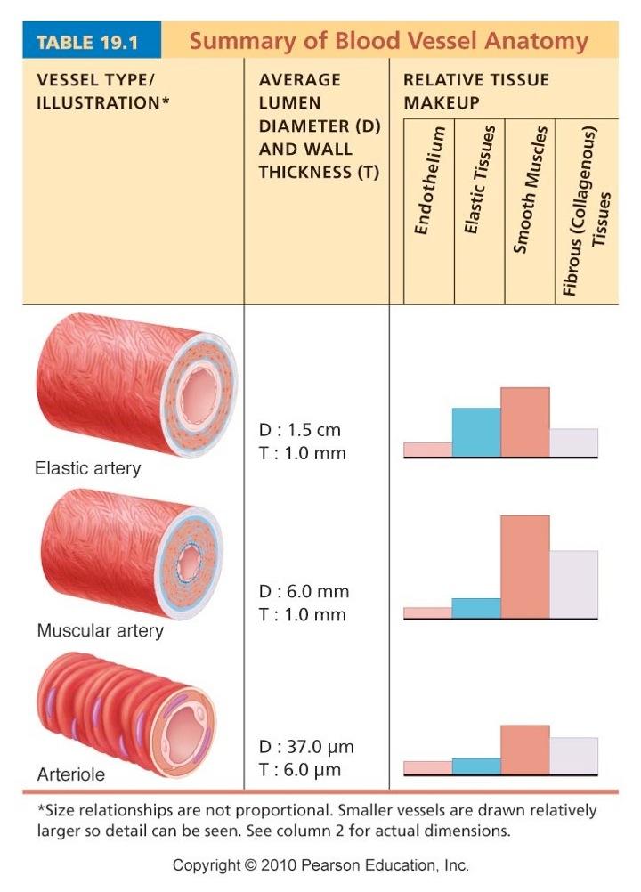
3 Structure of Blood Vessel Walls vessel walls made of layers or tunics tunica intima innermost tunic aka tunica interna. Next to the lumen. Contains the following layers: Endothelium: the simple squamous epithelium Subendothelial Layer: a basement membrane & loose C.T.» only in vessels larger than 1 mm in diameter. tunica media circularly arranged smooth muscle cells sheets of elastin surrounded by internal and external lamina. tunica externa connective tissue, nerves, lymphatics, elastin fibers contains vasa vasorum vessels of the vessels. 3
")
4 Artery & Vein (70x) 4

5 Artery, Vein & Capillary 5
6 6
7 7

8 Relationship of Blood Vessels & Lymphatics 8

9 Relative Proportion of Blood Volume Throughout the Cardiovascular System 9
10 Arterial System Elastic (conducting) Arteries nearest the heart (Aorta parts, major branches). largest diameter at 1 to 2.5 cm. are the most elastic with elastin in all 3 tunics. act as a pressure reservoir, expanding & recoiling. allows blood to flow continuous vs spurting with hardening, this pressure-smoothing effect is lost. smooth muscle here is relatively inactive in vasoconstriction. walls can weaken and balloon over lifetime. 10
11 Arterial System Muscular (distributing) Arteries distal to the elastic arteries account for most of the named arteries in the body. internal diameter range from 0.3 mm to 1 cm. have the thickest tunica media (i.e. most sm. mus.). big role in vasoconstriction. has an elastic lamina on both sides of tunica media 11
12 Arterial System Arterioles smallest of the arteries lumen diameter range of 10 um to 300 um. larger arterioles have all 3 tunics tunica media is mostly smooth muscle with a few scattered elastic fibers. smaller arterioles may not have all 3 tunics are little more than endothelium with a single layer of smooth muscle spiraling around it. controls blood flow into capillaries. 12
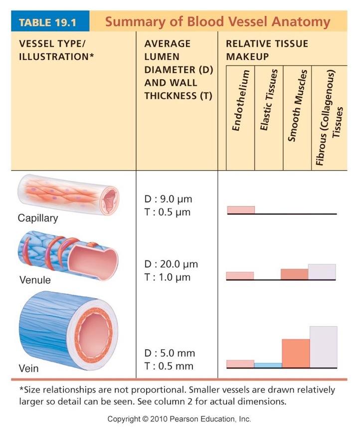
13 Capillaries just consists of the tunica intima. surrounded in some spots by pericytes. a smooth muscle-like cell that stabilizes wall. average length is just 1 mm. diameter is 8 to 10 um. remember the average RBC was only 7.5 um. 3 basic types of capillaries: continuous fenestrated sinusoidal 13
14 Capillaries Continuous Capillaries: abundant in skin and muscles most common type endothelial cells provide an uninterrupted lining. adjacent cells attached by tight junctions. usually incomplete and have gaps of unjoined membrane called intercellular clefts. only small amount of fluid and small solutes can pass. NO intercellular clefts in Brain... blood-brain barrier. 14

15 Continuous Capillary 15
16 Capillaries Fenestrated Capillaries similar to the continuous except: the endothelial cells are riddled with oval pores or fenestrations. fenestrations covered by a delicate membrane called the diaphragm (basal lamina material). makes them more permeable to fluids, solutes. GI tract to absorb larger molecules endocrine organs to receive the larger hormones kidneys where high filtration rate is occurring. 16

17 Fenestrated Capillary 17
18 Capillaries Sinusoidal Capillaries highly modified, leaky capillaries found only in: liver bone marrow lymphoid tissue some endocrine organs have large irregular lumens & fenestrations. allow very large molecules and even cells to leave. in liver the endothelium is discontinuous and have large macrophages called kupffer cells which remove and destroy bacteria Blood flow in this capillary type is slowest. 18

19 Sinusoidal Capillary 19
20 Capillaries tend to form into interweaving networks called capillary beds. has a vascular shunt through it so circulation can be cut-off as needed to rest of capillaries. see figure 19.4 pre-capillary sphincters control entry into capillaries so blood gets shunted past an area. Note: this explains why if you eat just before swimming, the blood that was busy processing digesting is shunted to the now exercising muscles and abdominal cramps and indigestion set in. 20

21 The Capillary Bed 21
22 Venous System Venules the smallest veins entered after capillaries. WBCs adhere to the walls of the postcapillary venule in areas that are inflamed. walls are very porous and WBCs can easily pass. Veins have 3 distinct tunics walls much thinner than artery. lumens look collapsed compared to artery. tunica externa is most developed wall. 22
23 Venous System Venous system is able to hold up to 65% of all blood in body. they are still partially filled at this volume veins called capacitance vessels or blood reservoirs. low pressure here requires help in returning blood: large diameter lowers blood flow resistance. Venous Valves to prevent back flow in limbs no valves in veins of head, chest cavity, abdominal pelvic cavity. contracting skeletal muscle in limbs pushes blood back. 23
24 Venous System Varicose Veins dilated, tortuous veins, mostly found in lower extremities due to incompetent valves. precipitated by: genetics hinderance of venous return conditions prolonged standing in one position obesity... external iliac veins kinked off by abdomen. pregnancy... same. straining increase pressure causes hemorrhoids. 24
25 Venous System Venous Sinuses coronary sinus on heart dural sinuses in brain highly specialized, flattened veins with extremely thin walls of just endothelium. supported by the tissue around them. 25
26 Vascular Anastomoses arterial anastomoses merging arteries into one. venous anastomoses merging veins into one. collateral channels alternate pathways for blood to one area. can be tissue and even life saving if one is cut or blocked by disease. arteriovenous anastomoses pathway to bypass capillary bed. 26
27 Physiology of Circulation Blood Flow volume of blood flowing in an area over time. if referring to the whole body is same as CO. relatively constant under resting conditions. blood flow through individual body organs may vary widely and is related to their immediate needs. Blood Pressure blood s force per unit area exerted on vessel wall expressed in mmhg. unless specified as pulmonary blood pressure, BP refers to the systemic BP in the large arteries near the heart. the pressure gradient (from high pressures upstream to the low pressures down stream) keeps blood moving. 27
28 Physiology of Circulation Resistance opposition to flow... the friction blood encounters. most friction is encountered in systemic circulation. referred to as peripheral resistance. 3 main sources of resistance: blood viscosity the thicker the fluid (blood), the greater the resistance. constant in health people. can vary up or down in disease. vessel length the longer the vessel, the greater the resistance. constant in healthy people. usually increases with obesity. vessel diameter the smaller the diameter, the greater the resistance. can vary with tissue need in healthy and abnormal people. 28
29 Physiology of Circulation more on blood vessel diameter: fluid flowing next to a wall is slowed by friction. fluid flowing in the center flows faster. therefore, in smaller tubes, a greater percentage of the blood volume is next to the vessel wall and will move slower due to the friction it encounters. this phenomenon is called laminar flow or streamlining. resistance varies inversely with the fourth power of the vessel radius. example: if we double the radius of a vessel, the resistance will drop to 1/16 of its original value. R =1/r 4 = 1/ (2x2x2x2) = 1/16. this means the small diameter arterioles have the most impact. turbulent flow (encountered with rough or protruding areas) is an irregular fluid motion... dramatically increases resistance. 29
30 Physiology of Circulation Relationship between Flow, Pressure & Resistance: Blood flow is directly proportional to the difference in blood pressure between two points in circulation, that is, the blood pressure gradient. Formula: F = P/R when there is a bigger drop in blood pressure between point A and point B (the P), flow (F) will increase. when resistance (R) increases the flow (F) will decrease. of the ways to affect flow... resistance will affect flow the most. 30
31 Systemic Blood Pressure the pumping action of the heart creates flow. pressure results when flow meets resistance. systemic blood pressure is highest in the aorta and declines all the way to zero in the right atrium! the steepest pressure drop occurs in the arterioles. 31

32 Blood Pressure in Various Blood Vessels 32
33 Arterial Blood Pressure Arterial BP reflects two factors: how much elastic arteries can be stretched this is their compliance or distensibility. the volume of blood forced into them. systolic pressure due to the forces of left ventricular contraction. 120 mmhg in healthy adult. diastolic pressure due to the recoil forces of the large arteries 70 to 80 mmhg in healthy adult. 33
34 Arterial Blood Pressure pulse pressure is the difference between the systolic and diastolic pressures. it is felt as a throbbing pulsation in an artery during systole as artery walls are stretched. is increased the more stretch resistant a blood vessel becomes: arteriosclerosis in arteries decreases their stretchiness. 34
35 Arterial Blood Pressure mean arterial pressure (MAP) is the pressure that propels blood to the tissues. because diastole usually lasts longer than systole, the MAP is not simply the average. formula: MAP = diastolic pressure + pulse pressure 3 example: person with a BP of 120/80 mmhg: MAP = 80 mmhg + = 93 mmhg NOTE: as you get to the capillaries, the blood flow is steady and pulse pressure is gone. 35
36 Capillary Blood Pressure BP has dropped to about 33 mmhg at the beginning of the capillaries and is only about 15 mmhg as it enters the venules. too high and you push filtrate fluid into the tissues and cause edema. 36
37 Venous Blood Pressure flow is steady and non-pulsatile. goes from about 15 mmhg to 0 mmhg as it enters the right atrium. Assistance to help the venous return: large lumens of veins to hold more blood. valves to prevent back flow of blood. respiratory pump inhalation will increase venous return. muscular pump contracting muscles increase venous return. smooth muscle in veins constricts to increase venous return. 37

38 The Muscular Pump 38
39 Maintaining Blood Pressure Cardiac Output, Blood Pressure and Peripheral Resistance have interrelating variables. changes in one variable that affects blood pressure will be corrected not by one variable, but many variables. major factors enhancing cardiac output in figure 19.7 remember that the heart is primarily controlled by the cardioinhibitory center in the medulla. stroke volume is controlled mainly by venous return (EDV). during stress, the cardioacceleratory center in the medulla, activates the sympathetic n.s. increases heart rate by acting on SA node increases stroke volume by enhancing cardiac contractility (ESV) the enhanced cardiac output results in an increased MAP, which represents an increased blood flow to the tissues. 39

40 Factors Increasing Cardiac Output 40
41 Maintaining Blood Pressure Short-term neural control mechanisms: operate via a reflex arc baroreceptor to vasomotor center of medulla then to vascular smooth muscle. (see figure 19.8) two main goals of short-term neural control: maintain adequate MAP by altering vessel diameter. if BP is low, blood flow to less necessary tissues is diverted. altering blood distribution for demands and situations. if exercising blood goes to muscles more than digestive organs. Warning: vigorous rubbing of the carotid bifurcation where the baroreceptors are can quickly drop a person s heart rate (and therefore CO). Clinically used if person is in supraventricular tachycardia and no drugs ready. 41
42 Baroreceptors Maintain Blood Pressure Baroreceptor Locations: the bifurcation of the common carotids in the aortic arch in the walls of the large arteries of the thorax and neck. activate when stretched (or rubbed vigorously) sends signal to stimulate cardioinhibitory center in medulla. end result is vasodilation of arteries & veins thus dropping BP. also sends signal to inhibit cardioaccelatory center in medulla. end result is a drop in heart rate and heart contractile forces. Main function of Baroreceptors is to protect you from sudden drops in BP with changes of position: laying to standing (BP would drop quickly if not for them). the carotid sinus reflex preserves blood flow to the brain. 42

43 Baroreceptor & BP Homeostasis 43

44 Baroreceptor & BP Homeostasis 44

45 Hormonal Control of BP 45
46 Maintaining Blood Pressure Long-term renal regulation of BP: done by altering blood volume by controlling how much you urinate. direct renal mechanism alters blood volume independently of hormones. increase blood volume and BP will increase the rate of fluid filtration through the kidney and this leads to more fluid ending up in the urine. indirect renal mechanism the renin-angiotensin mechanism. activated by dropping BP, ends with vasoconstriction. Aldosterone secretion to increase Na reabsorption 46

47 Renal Control of Blood Pressure 47

48 Factors Causing an Increased MAP 48
49 Pulses & Blood Pressure Pulse sites can also be used as pressure points to stop blood flow distal to them in case of a severe bleeding injury. Blood Pressure Measurement done by sphygmomanometer and auscultation. pressure is increased above systolic the slowly released listening for the sound of blood to begin flowing again in artery. This is the systolic BP. flow at this point has gone from laminar flow to turbulent flow... this makes Korotkoff sounds! the pressure at which turbulent flow goes back to laminar the sounds vanish (usually) is diastolic BP. 49

50 Easily Palpated Pulse Sites 50
51 Blood Pressure Abnormalities Hypertension systolic BP over 140 mmhg. diastolic BP over 90 mmhg. transient HTN is normal if due to pain, fever, exercise or stress!!! persistent HTN is what needs medical treatment. Hypotension values vary, need a symptom like dizziness. orthostatic HoTN due to slow to respond SNS as we age. i.e. you get dizzy getting up to quick. you will stop urine formation if it gets too low! 51

52 Tissue Perfusion: Rest vs Exercise 52

53 Blood Flow Velocity vs Total Cross-Sectional Vessel Area 53

54 Control of Arteriole Smooth Muscle Diameter 54
55 Blood Flow in Special Location Skeletal Muscle: when muscles become active blood flow increases. is in direct proportion to their metabolic activity is called active or exercise hyperemia. Brain: blood flow is ~750 ml/min and is relatively constant. most sensitive to Carbon Dioxide levels increasing. less sensitive to Oxygen levels dropping. Fainting occurs when MAP is < 60 mmhg. Cerebral edema occurs if MAP is > 160 mmhg. 55
56 Blood Flow in Special Locations Skin: capillaries are in the papillary layer of dermis. epidermis has no blood vessels (diffusion). shunting blood to skin can release heat more heat loss if skin wet (sweating). Lungs: the arteries here look like veins microscopically systolic pressure is 24 mmhg. diastolic pressure is 8 mmhg. low oxygen in lung will constrict blood vessels. consistent with gas exchange done here. areas with disease have blood shunted past them! 56

57 4 Capillary Transport Mechanisms 57

58 4 Capillary Transport Mechanisms 58
59 Capillary Flow Dynamics Bulk Flow: about 20 Liter of fluid is filtered of the capillaries daily before returning to the blood stream. direction of this flow and the volume is affected by: Capillary Hydrostatic Pressure (HPc): this force filters blood through the capillary walls leaving behind the cells and most proteins. Arterial End HPc is 35 mmhg Venous End HPc is 17 mmhg the Interstitial Fluid Hydrostatic Pressure (HPif) pushes fluid back into the capillary also, the capillary colloid osmotic pressure (OPc) or oncotic pressure at about 26 mmhg pulls fluid into it. 59

60 Fluid Flows at Capillaries Remember that unlike HP, the OP will not vary much as the proteins predominately stay inside the capillary and help contribute to the ability to pull water back in. 60

61 Fluid Flows at Capillaries 61

62 Events & Signs of Compensated Hypovolemic Shock 62

63 Pulmonary Circulation 63
Heart. Large lymphatic vessels Lymph node. Lymphatic. system Arteriovenous anastomosis. (exchange vessels)
") Venous system Large veins (capacitance vessels) Small veins (capacitance vessels) Postcapillary venule Thoroughfare channel Heart Large lymphatic vessels Lymph node Lymphatic system Arteriovenous anastomosis
Venous system Large veins (capacitance vessels) Small veins (capacitance vessels) Postcapillary venule Thoroughfare channel Heart Large lymphatic vessels Lymph node Lymphatic system Arteriovenous anastomosis
Blood Vessels. Over view. We have about 60,000 miles of blood vessels!
 Blood Vessels Over view 3 types of blood vessels arteries - carry blood away from heart "branch", "diverge", and "fork" veins - carry blood toward heart "join", "merge", and "converge" capillaries - site
Cardiovascular System Blood Vessels
 Cardiovascular System Blood Vessels Structure of Blood Vessels The three layers (tunics) Tunica intima composed of simple squamous epithelium Tunica media sheets of smooth muscle Contraction vasoconstriction
UNIT 4: BLOOD VESSELS
 UNIT 4: BLOOD VESSELS Dr. Moattar Raza Rizvi NRS237, Physiology Generalized Structure of Blood Vessels 1 Tunica interna (tunica intima) Endothelial layer that lines the lumen of all vessels In vessels
Physiology of Circulation
 Physiology of Circulation Dr. Ali Ebneshahidi Blood vessels Arteries: Blood vessels that carry blood away from the heart to the lungs and tissues. Arterioles are small arteries that deliver blood to the
Chapter 21 (1) An Introduction to Blood Vessels and Circulation
 An Introduction to Blood Vessels and Circulation") Chapter 21 (1) An Introduction to Blood Vessels and Circulation Lecture Objectives Compare and contrast the structure of an artery, arteriole, vein, venule, and capillary Discuss the structure and function
Chapter 21 (1) An Introduction to Blood Vessels and Circulation Lecture Objectives Compare and contrast the structure of an artery, arteriole, vein, venule, and capillary Discuss the structure and function
Blood Vessels. Types of Blood Vessels Arteries carry blood away from the heart Capillaries smallest blood vessels. Veins carry blood toward the heart
 C H A P T E R Blood Vessels 20 Types of Blood Vessels Arteries carry blood away from the heart Capillaries smallest blood vessels The site of exchange of molecules between blood and tissue fluid Veins
Collin County Community College
 Collin County Community College BIOL. 2402 Anatomy & Physiology WEEK 6 Blood Vessels 1 Anatomy of Blood Vessels Walls of blood vessels contain 3 distinct layers : Tunica intima innermost layer includes
Copyright 2010 Pearson Education, Inc. Blood Vessel Structure
 Blood Vessel Structure Structure of Blood Vessel Walls Arteries and veins Tunica intima, tunica media, and tunica externa Lumen Central blood-containing space Capillaries Endothelium with sparse basal
2. capillaries - allow exchange of materials between blood and tissue fluid
 Chapter 19 - Vascular System A. categories and general functions: 1. arteries - carry blood away from heart 2. capillaries - allow exchange of materials between blood and tissue fluid 3. veins - return
Any of these questions could be asked as open question or lab question, thus study them well
 Any of these questions could be asked as open question or lab question, thus study them well describe the factors which regulate cardiac output describe the sympathetic and parasympathetic control of heart
The Cardiovascular System: Vessels and Routes. Pulmonary Circulation H E A R T. Systemic Circulation
 The Cardiovascular System: Vessels and Routes 1. Overview of Blood Circulation A. Pulmonary Circulation Lung Arterioles Pulmonary Artery Capillaries Pulmonary Circulation Venules Pulmonary Veins H E A
The Cardiovascular System. The Structure of Blood Vessels. The Structure of Blood Vessels. The Blood Vessels. Blood Vessel Review
 The Cardiovascular System The Blood Vessels The Structure of Blood Vessels Blood Vessel Review Arteries carry blood away from the heart Pulmonary trunk to lungs Aorta to everything else Microcirculation
Types of Blood Vessels
 Chapter 21 Peripheral Circulation and Regulation 21-1 Types of Blood Vessels Capillaries: site of exchange with tissue Arteries in dif. Types & sizes Elastic Muscular Arterioles Veins: thinner walls than
Chapter 21 Peripheral circulation and Regulation
 Chapter 21 Peripheral circulation and Regulation I. Blood vessel structure A. Blood flows from large arteries to small capillaries 1. Large arteries contain large amounts of elastic tissue and little smooth
2402 : Anatomy/Physiology
 Dr. Chris Doumen Lecture 1 2402 : Anatomy/Physiology Hemo Dynamics and Blood Vessels I nt r oduc t i on TextBook Readings Pages 721 through 734. Make use of the figures in your textbook ; a picture is
Cardiovascular System. Blood Vessel anatomy Physiology & regulation
 Cardiovascular System Blood Vessel anatomy Physiology & regulation Path of blood flow Aorta Arteries Arterioles Capillaries Venules Veins Vena cava Vessel anatomy: 3 layers Tunica externa (adventitia):
Peripheral Circulation and Regulation
 Peripheral Circulation and Regulation Functions of Peripheral Circulation 1. Contain the blood 2. Exchange nutrients, waste products, and gases with tissues 3. Transport 4. Regulate blood pressure, along
Vascular System Part One
 Vascular System Part One Objectives Trace the route taken by blood as it leaves, and then returns to the heart. Describe the structure of the walls of arteries and veins. Discuss the structure and function
Histology of the Cardiac System. Dr. Nabil Khoury Anatomy Department
 Histology of the Cardiac System Dr. Nabil Khoury Anatomy Department Objectives 1. Identify the 3 layers of the heart endocardium, myocardium, epicardium 2. Differentiate cardiacmuscle 3. Define intercalated
Lab Period: Name: Physiology Chapter 14 Blood Flow and Blood Pressure, Plus Fun Review Study Guide
 Lab Period: Name: Physiology Chapter 14 Blood Flow and Blood Pressure, Plus Fun Review Study Guide Main Idea: The function of the circulatory system is to maintain adequate blood flow to all tissues. Clinical
The cardiovascular system
 The cardiovascular system Components of the Cardiovascular system Heart Vessels: Arteries Capillaries Veins Functions of CVS: Transportation system where blood is the transporting vehicle Carries oxygen,
BIOL 219 Spring Chapters 14&15 Cardiovascular System
 1 BIOL 219 Spring 2013 Chapters 14&15 Cardiovascular System Outline: Components of the CV system Heart anatomy Layers of the heart wall Pericardium Heart chambers, valves, blood vessels, septum Atrioventricular
Chapter 14 Blood Vessels, Blood Flow and Pressure Exam Study Questions
 Chapter 14 Blood Vessels, Blood Flow and Pressure Exam Study Questions 14.1 Physical Law Governing Blood Flow and Blood Pressure 1. How do you calculate flow rate? 2. What is the driving force of blood
Cardiovascular system: Blood vessels, blood flow. Latha Rajendra Kumar, MD
 Cardiovascular system: Blood vessels, blood flow Latha Rajendra Kumar, MD Outline 1- Physical laws governing blood flow and blood pressure 2- Overview of vasculature 3- Arteries 4. Capillaries and venules
Structure. Arteries. 21_01d 4/18/12. The Cardiovascular System: Blood Vessels and Hemodynamics. Dr Badri Paudel GMC
 Goal of the Cardiovascular System: deliver blood to all parts of the body The Cardiovascular System: Blood Vessels and Hemodynamics Dr Badri Paudel GMC Does so by using different types of tubing, attached
Histology of the myocardium and blood vessels. Prof. Abdulameer Al-Nuaimi
 Histology of the myocardium and blood vessels Prof. Abdulameer Al-Nuaimi E-mail: a.al-nuaimi@sheffield.ac.uk E-mail: abdulameerh@yahoo.com Histology of blood vessels The walls of arteries and veins are
Chapter 21! Chapter 21 Blood Vessels and Circulation! Blood Vessels and Circulation!
 Chapter 21! Blood Vessels and Circulation! SECTION 21-1! Blood vessels differ in size, structure, and functional properties! 2 Major Vessel Types! Arteries - carry blood away from the heart Higher pressure
Chapter 21! Blood Vessels and Circulation! SECTION 21-1! Blood vessels differ in size, structure, and functional properties!
 Chapter 21! Blood Vessels and Circulation! SECTION 21-1! Blood vessels differ in size, structure, and functional properties! 2 1! Major Vessel Types! Arteries - carry blood away from the heart Higher pressure
1. Distinguish among the types of blood vessels on the basis of their structure and function.
 Blood Vessels and Circulation Objectives This chapter describes the structure and functions of the blood vessels Additional subjects contained in Chapter 13 include cardiovascular physiology, regulation,
Structure and organization of blood vessels
 The cardiovascular system Structure of the heart The cardiac cycle Structure and organization of blood vessels What is the cardiovascular system? The heart is a double pump heart arteries arterioles veins
Physiology Unit 3 CARDIOVASCULAR PHYSIOLOGY: THE VASCULAR SYSTEM
 Physiology Unit 3 CARDIOVASCULAR PHYSIOLOGY: THE VASCULAR SYSTEM In Physiology Today Hemodynamics F = ΔP/R Blood flow (F) High to low pressure Rate = L/min Pressure (P) Hydrostatic pressure Pressure exerted
Cardivascular System Module 5: Structure and Function of Blood Vessels *
 OpenStax-CNX module: m49689 1 Cardivascular System Module 5: Structure and Function of Blood Vessels * Donna Browne Based on Structure and Function of Blood Vessels by OpenStax This work is produced by
Chapter 21. Blood Vessels and Circulation
 Chapter 21 Openstax: Chapter 20 Blood Vessels and Circulation Chapter 21 Learning Outcomes After completing Chapter 21, you will be able to: 1. Distinguish among the types of blood vessels based on their
CHAPTER 21 LECTURE OUTLINE
 CHAPTER 21 LECTURE OUTLINE I. INTRODUCTION A. One main focus of this chapter considers hemodynamics, the means by which blood flow is altered and distributed and by which blood pressure is regulated. B.
Human Anatomy, First Edition
 Human Anatomy, First Edition McKinley & O'Loughlin Chapter 23 : Vessels and Circulation 23-1 Blood Vessels An efficient style of transport for oxygen, nutrients, and waste products to and from body tissues.
Arteries AWAY. Branch. Typically oxygenated.
 Arteries AWAY Branch Typically oxygenated. Capillaries Smallest. Most abundant. 10 billion. Huge surface area. Exchange Veins TOWARDS Converge. Typically deoxygenated. 3 Layers of the Vascular Wall Tunica
Extra notes for lab- 1 histology. Slide 1 : cross section in the elastic artery ( aortic arch, ascending aorta, descending aorta )
") Extra notes for lab- 1 histology Slide 1 : cross section in the elastic artery ( aortic arch, ascending aorta, descending aorta ) - twin of ascending aorta is the pulmonary trunk. Ascending aorta represents
Extra notes for lab- 1 histology Slide 1 : cross section in the elastic artery ( aortic arch, ascending aorta, descending aorta ) - twin of ascending aorta is the pulmonary trunk. Ascending aorta represents
Chapter 21: Cardiovascular System: Peripheral Circulation and Regulation
 Chapter 21: Cardiovascular System: Peripheral Circulation and Regulation I. General Features of Blood Vessel Structure A. General Pattern of Circulation 1. Ventricles pump blood into 2. These arteries
Blood Vessels. Chapter 20
 Blood Vessels Chapter 20 Summary of the Characteristics of Arteries and Veins Characteristic Artery Vein Wall thickness thick thin Shape in cross section round flattened Thickest tunic media externa Collagen
BIPN100 F15 Human Physiol I (Kristan) Lecture 14 Cardiovascular control mechanisms p. 1
 Lecture 14 Cardiovascular control mechanisms p. 1") BIPN100 F15 Human Physiol I (Kristan) Lecture 14 Cardiovascular control mechanisms p. 1 Terms you should understand: hemorrhage, intrinsic and extrinsic mechanisms, anoxia, myocardial contractility, residual
BIPN100 F15 Human Physiol I (Kristan) Lecture 14 Cardiovascular control mechanisms p. 1 Terms you should understand: hemorrhage, intrinsic and extrinsic mechanisms, anoxia, myocardial contractility, residual
BIOLOGY 2060 LECTURE NOTES ANATOMY & PHYSIOLOGY II (A. IMHOLTZ) VESSELS P1 OF 7
 VESSELS P1 OF 7") BIOLOGY 2060 LECTURE NOTES ANATOMY & PHYSIOLOGY II (A. IMHOLTZ) VESSELS P1 OF 7 1. Blood vessels a. Tubes through which the heart pumps blood. b. 3 major types of blood vessels: arteries, capillaries,
BIOLOGY 2060 LECTURE NOTES ANATOMY & PHYSIOLOGY II (A. IMHOLTZ) VESSELS P1 OF 7 1. Blood vessels a. Tubes through which the heart pumps blood. b. 3 major types of blood vessels: arteries, capillaries,
Chapter 21. Blood Vessels and Circulation
 Chapter 21 Blood Vessels and Circulation INTRODUCTION A. One main focus of this chapter considers hemodynamics, the means by which blood flow is altered and distributed and by which blood pressure is regulated.
The Cardiovascular system: physiology of circulation
 Chapter 21 The Cardiovascular system: physiology of circulation blood vessel structure and function physiology of circulation: blood flow, blood pressure, and resistance blood flow the amount of blood
Veins. VENOUS RETURN = PRELOAD = End Diastolic Volume= Blood returning to heart per cardiac cycle (EDV) or per minute (Venous Return)
 or per minute (Venous Return)") Veins Venous system transports blood back to heart (VENOUS RETURN) Capillaries drain into venules Venules converge to form small veins that exit organs Smaller veins merge to form larger vessels Veins
Veins Venous system transports blood back to heart (VENOUS RETURN) Capillaries drain into venules Venules converge to form small veins that exit organs Smaller veins merge to form larger vessels Veins
3/10/2009 VESSELS PHYSIOLOGY D.HAMMOUDI.MD. Palpated Pulse. Figure 19.11
 VESSELS PHYSIOLOGY D.HAMMOUDI.MD Palpated Pulse Figure 19.11 1 shows the common sites where the pulse is felt. 1. Temporal artery at the temple above and to the outer side of the eye 2. External maxillary
Blood Flow, Blood Pressure, Cardiac Output. Blood Vessels
 Blood Flow, Blood Pressure, Cardiac Output Blood Vessels Blood Vessels Made of smooth muscle, elastic and fibrous connective tissue Cells are not electrically coupled Blood Vessels Arteries arterioles
Practical Histology. Cardiovascular System. Dr Narmeen S. Ahmad
 Practical Histology Cardiovascular System Dr Narmeen S. Ahmad The Cardiovascular System A closed system of the heart and blood vessels Functions of cardiovascular system: Transport nutrients, hormones
Blood Pressure. a change in any of these could cause a corresponding change in blood pressure
 Blood Pressure measured as mmhg Main factors affecting blood pressure: 1. cardiac output 2. peripheral resistance 3. blood volume a change in any of these could cause a corresponding change in blood pressure
Microcirculation. Lecture Block 11 (contributions from Brett Burton)
") Lecture Block 11 (contributions from Brett Burton) Elements of Arterioles, capillaries, venules Structure and function: transport Fluid balance Lymph system Vessels of the Circulatory System Diameter Aorta
Lecture Block 11 (contributions from Brett Burton) Elements of Arterioles, capillaries, venules Structure and function: transport Fluid balance Lymph system Vessels of the Circulatory System Diameter Aorta
Cardiovascular System. I. Structures of the heart A. : Pericardium sack that surrounds the heart
 Cardiovascular System I. Structures of the heart A. : Pericardium sack that surrounds the heart 1. : Pericardial Cavity serous fluid filled space between the heart and the pericardium B. Heart Wall 1.
Cardiovascular System: Vessels and Circulation (Chapter 21)
") Cardiovascular System: Vessels and Circulation (Chapter 21) Lecture Materials for Amy Warenda Czura, Ph.D. Suffolk County Community College Eastern Campus Primary Sources for figures and content: Marieb,
Cardiovascular System: Vessels and Circulation (Chapter 21) Lecture Materials for Amy Warenda Czura, Ph.D. Suffolk County Community College Eastern Campus Primary Sources for figures and content: Marieb,
The Cardiovascular System
 The Cardiovascular System The Cardiovascular System A closed system of the heart and blood vessels The heart pumps blood Blood vessels allow blood to circulate to all parts of the body The function of
Major Function of the Cardiovascular System. Transportation. Structures of the Cardiovascular System. Heart - muscular pump
 Structures of the Cardiovascular System Heart - muscular pump Blood vessels - network of tubes Blood - liquid transport vehicle brachiocephalic trunk superior vena cava right pulmonary arteries right pulmonary
Lab Activity 25. Blood Vessels & Circulation. Portland Community College BI 232
 Lab Activity 25 Blood Vessels & Circulation Portland Community College BI 232 Artery and Vein Histology Walls have 3 layers: Tunica intima Tunica media Tunica externa 2 Tunica Intima Is the innermost layer
10. Thick deposits of lipids on the walls of blood vessels, called, can lead to serious circulatory issues. A. aneurysm B. atherosclerosis C.
 Heart Student: 1. carry blood away from the heart. A. Arteries B. Veins C. Capillaries 2. What is the leading cause of heart attack and stroke in North America? A. alcohol B. smoking C. arteriosclerosis
12/02/2012. Chapter 19: Blood Vessels. Blood Vessel Structure and Function
 Chapter 19: Blood Vessels 12/02/2012 Blood Vessel Structure and Function 3 major types: arteries capillaries veins as heart contracts it forces blood into the large arteries leaving the ventricles then
I. Cardiac Output Chapter 14
 10/24/11 I. Cardiac Output Chapter 14 Cardiac Output, Blood Flow, and Blood Pressure Lecture PowerPoint Copyright The McGraw-Hill Companies, Inc. Permission required for reproduction or display. Cardiac
The Cardiovascular and Lymphatic Systems Cardiovascular System Blood Vessels Blood Vessels Arteries Arteries Arteries
 CH 12 The Cardiovascular and s The Cardiovascular and s OUTLINE: Cardiovascular System Blood Vessels Blood Pressure Cardiovascular System The cardiovascular system is composed of Blood vessels This system
The Cardiovascular System
 PowerPoint Lecture Slide Presentation by Patty Bostwick-Taylor, Florence-Darlington Technical College The Cardiovascular System 11PART B The Heart: Cardiac Output Cardiac output (CO) Amount of blood pumped
Cardiac Output 1 Fox Chapter 14 part 1
 Vert Phys PCB3743 Cardiac Output 1 Fox Chapter 14 part 1 T. Houpt, Ph.D. Regulation of Heart & Blood Pressure Keep Blood Pressure constant if too low, not enough blood (oxygen, glucose) reaches tissues
P215 SPRING 2019: CIRCULATORY SYSTEM Chaps 13, 14 & 15: pp , , , I. Major Functions of the Circulatory System
 P215 SPRING 2019: CIRCULATORY SYSTEM Chaps 13, 14 & 15: pp 360-390, 395-404, 410-428 433-438, 441-445 I. Major Functions of the Circulatory System 1. 2. 3. 4. II. Structure of the Heart 1. atria 2. ventricles
Ch. 12 The Circulatory System. The heart. The heart is a double pump. A quick note on arteries vs. veins. = the muscular pump of the CV system
 Ch. 12 The Circulatory System The heart A.k.a. the cardiovascular system Blood was discussed in Ch. 11 Focus of Ch. 12: heart and blood vessels = the muscular pump of the CV system ~ 100,000 heartbeats/day!
CVS HISTOLOGY. Dr. Nabil Khouri.
 CVS HISTOLOGY Dr. Nabil Khouri http://anatomy.kmu.edu.tw/blockhis/block3/slides/block4_24.html The Heart Wall Contract as a single unit Cardiac Muscle Simultaneous contraction due to depolarizing at the
Cardiovascular system
 Cardiovascular system L-4 Blood pressure & special circulation Dr Than Kyaw 27 February 2012 Blood Pressure (BP) Pressure generation and flow Blood is under pressure within its closed system. Pressure
Cardiovascular System B L O O D V E S S E L S 2
 Cardiovascular System B L O O D V E S S E L S 2 Blood Pressure Main factors influencing blood pressure: Cardiac output (CO) Peripheral resistance (PR) Blood volume Peripheral resistance is a major factor
Cardiovascular (Circulatory) System
 System") Cardiovascular (Circulatory) System Piryaei May 2011 Circulatory System Heart Blood Vessels Macrovasculature (More than 0.1mm) Elastic Artery Muscular (Distributing) Artery Large Arteriol Small Vein Muscular
Cardiovascular (Circulatory) System Piryaei May 2011 Circulatory System Heart Blood Vessels Macrovasculature (More than 0.1mm) Elastic Artery Muscular (Distributing) Artery Large Arteriol Small Vein Muscular
Anatomy Review: The Heart Graphics are used with permission of A.D.A.M. Software, Inc. and Benjamin/Cummings Publishing Co.
 Anatomy Review: The Heart Graphics are used with permission of A.D.A.M. Software, Inc. and Benjamin/Cummings Publishing Co. Anatomy Views Label the diagrams of the heart below: Interactive Physiology Study
Six main classes of blood vessels (on handout) Wall structure of arteries and veins (on handout) Comparison: Arteries vs. Veins (on handout)
 Wall structure of arteries and veins (on handout) Comparison: Arteries vs. Veins (on handout)") Cardiovascular System: Vessels and Circulation (Chapter 21) Lecture Materials for Amy Warenda Czura, Ph.D. Suffolk County Community College Eastern Campus Six main classes of blood vessels Primary Sources
Cardiovascular System: Vessels and Circulation (Chapter 21) Lecture Materials for Amy Warenda Czura, Ph.D. Suffolk County Community College Eastern Campus Six main classes of blood vessels Primary Sources
Derived copy of Structure and Function of Blood Vessels *
 OpenStax-CNX module: m56696 1 Derived copy of Structure and Function of Blood Vessels * Stephanie Fretham Based on Structure and Function of Blood Vessels by OpenStax This work is produced by OpenStax-CNX
Cardiovascular System B L O O D V E S S E L S 3
 Cardiovascular System B L O O D V E S S E L S 3 Fluid Shifts Between Capillaries and Tissue Permeable capillaries allow plasma and solutes to pass into interstitial space interstitial or extracellular
Cardiovascular system
 BIO 301 Human Physiology Cardiovascular system The Cardiovascular System: consists of the heart plus all the blood vessels transports blood to all parts of the body in two 'circulations': pulmonary (lungs)
Cardiovascular Physiology
 Cardiovascular Physiology Lecture 1 objectives Explain the basic anatomy of the heart and its arrangement into 4 chambers. Appreciate that blood flows in series through the systemic and pulmonary circulations.
Function: Transportation of. Oxygen Nutrients Waste Hormones gases
 Function: Transportation of Oxygen Nutrients Waste Hormones gases Pericardium: double sac of serous membrane filled with fluid (pericardial fluid to be exact) that surrounds the heart. Parietal pericardium:
Cardiovascular System
 Cardiovascular System Purpose Transport oxygen and nutrients Take waste products away from tissues & organs Things we learned Blood pressure: the force of blood pushing against the walls of blood vessels
Anatomy and Physiology, Spring 2015 Exam II: Form A April 9, Name Student Number
 Anatomy and Physiology, Spring 2015 Exam II: Form A April 9, 2015 Name Student Number For Questions 1 2 refer to the following table. 1 Ventricular pressure is greater than aortic 6 AV valve is open 2
The Cardiovascular and Lymphatic Systems
 BIOLOGY OF HUMANS Concepts, Applications, and Issues Fifth Edition Judith Goodenough Betty McGuire 12 The Cardiovascular and Lymphatic Systems Lecture Presentation Anne Gasc Hawaii Pacific University and
Citation Jarvis S (2018) Vascular system 1: anatomy and physiology. Nursing Times [online]; 114: 4,
![Citation Jarvis S (2018) Vascular system 1: anatomy and physiology. Nursing Times [online]; 114: 4,](/thumbs/96/127077409.jpg "Citation Jarvis S (2018) Vascular system 1: anatomy and physiology. Nursing Times [online]; 114: 4,") Vascular system Keywords Arteries/Veins// Blood flow/fluid movement This article has been double-blind peer reviewed In this article... Anatomy of the vascular system and structure of blood vessels The
Vascular system Keywords Arteries/Veins// Blood flow/fluid movement This article has been double-blind peer reviewed In this article... Anatomy of the vascular system and structure of blood vessels The
Circulation. Sinoatrial (SA) Node. Atrioventricular (AV) Node. Cardiac Conduction System. Cardiac Conduction System. Linked to the nervous system
 Node. Atrioventricular (AV) Node. Cardiac Conduction System. Cardiac Conduction System. Linked to the nervous system") Circulation Cardiac Conduction System AHS A H S Your body resembles a large roadmap. There are routes or arteries that take you downtown to the heart of the city and veins that take you to the outskirts
Circulation Cardiac Conduction System AHS A H S Your body resembles a large roadmap. There are routes or arteries that take you downtown to the heart of the city and veins that take you to the outskirts
Sinusoids and venous sinuses
 LYMPHOID SYSTEM General aspects Consists of organs that are made of lymphoid tissue; Immune defense Breakdown of red blood cells. 1 Sinusoids In place of capillaries Endothelium; often fenestrated More
Essentials of Anatony and Physiology, 5e (Martini/Nath) Chapter 13 The Cardiovascular System: Blood Vessels and Circulation
 Chapter 13 The Cardiovascular System: Blood Vessels and Circulation") Essentials of Anatony and Physiology, 5e (Martini/Nath) Chapter 13 The Cardiovascular System: Blood Vessels and Circulation Multiple-Choice Questions 1) The muscular layer of blood vessels is the A) tunica
Essentials of Anatony and Physiology, 5e (Martini/Nath) Chapter 13 The Cardiovascular System: Blood Vessels and Circulation Multiple-Choice Questions 1) The muscular layer of blood vessels is the A) tunica
Blood vessels are the tubes through which the heart pumps blood. There are 3 major types of blood vessels: arteries, capillaries, and veins.
 LECTURE NOTES ANATOMY & PHYSIOLOGY II (A. IMHOLTZ) VESSELS P1 OF 1 Blood vessels are the tubes through which the heart pumps blood. There are 3 major types of blood vessels: arteries, capillaries, and
Figure ) The specific chamber of the heart that is indicated by letter A is called the. Diff: 1 Page Ref: 364
 The specific chamber of the heart that is indicated by letter A is called the. Diff: 1 Page Ref: 364") Essentials of Anatomy and Physiology, 9e (Marieb) Chapter 11 The Cardiovascular System Short Answer Figure 11.1 Using Figure 11.1, identify the following: 1) The Purkinje fibers are indicated by label.
Essentials of Anatomy and Physiology, 9e (Marieb) Chapter 11 The Cardiovascular System Short Answer Figure 11.1 Using Figure 11.1, identify the following: 1) The Purkinje fibers are indicated by label.
Physiology - 8 Hemodynamics - 1 M.jafar 24/3/2016 Turquoise Team
 21 Physiology - 8 Hemodynamics - 1 M.jafar 24/3/2016 Turquoise Team Hemodynamics Today we will take about hemodynamics which is the study of the movement of blood and of the forces concerned. Now how the
Cardiac Conduction System
 Cardiac Conduction System What causes the Heart to Beat? Heart contracts by electrical signals! Cardiac muscle tissue contracts on its own an electrical signal is sent out by the heart so that all cells
CVS Hemodynamics. Faisal I. Mohammed, MD,PhD.
 CVS Hemodynamics Faisal I. Mohammed, MD,PhD. Objectives point out the physical characteristics of the circulation: distribution of blood volume total cross sectional area velocity blood pressure List the
Blood Pressure Fox Chapter 14 part 2
 Vert Phys PCB3743 Blood Pressure Fox Chapter 14 part 2 T. Houpt, Ph.D. 1 Cardiac Output and Blood Pressure How to Measure Blood Pressure Contribution of vascular resistance to blood pressure Cardiovascular
CVS Hemodynamics. Change in blood pressure:
 CVS Hemodynamics -The distribution of blood inside the circulation: The major part of blood volume is found in the venous system 60% (2/3), that s why veins are called the capacitance vessels. -Arteries
Cardiovascular System. Biology 105 Lecture 15 Chapter 12
 Cardiovascular System Biology 105 Lecture 15 Chapter 12 Outline I. Functions of cardiovascular system II. Components of the cardiovascular system: I. Blood vessels II. Heart III. Regulation of the heartbeat
The Circulatory System (p )
") The Circulatory System (p. 268-281) How Does Gravity Affect Blood Circulation? As with all land animals, the giraffe and the corn snake are constantly subject to the force of gravity The circulatory system
The Circulatory System (p. 268-281) How Does Gravity Affect Blood Circulation? As with all land animals, the giraffe and the corn snake are constantly subject to the force of gravity The circulatory system
Cardiovascular Anatomy Dr. Gary Mumaugh
 Cardiovascular Anatomy Dr. Gary Mumaugh Location of Heart Approximately the size of your fist Location o Superior surface of diaphragm o Left of the midline in mediastinum o Anterior to the vertebral column,
The Circulatory System
 The Circulatory System Dr. Sami Zaqout The circulatory system Circulatory system Blood vascular systems Lymphatic vascular systems Blood vascular systems Blood vascular systems The circulatory system Circulatory
Anatomy of the Blood Vessels
 Biology 212: Anatomy and Physiology II Anatomy of the Blood Vessels References: Saladin, KS: Anatomy and Physiology, The Unity of Form and Function 8 th (2018). Required reading before beginning this lab:
(D) (E) (F) 6. The extrasystolic beat would produce (A) increased pulse pressure because contractility. is increased. increased
 (E) (F) 6. The extrasystolic beat would produce (A) increased pulse pressure because contractility. is increased. increased") Review Test 1. A 53-year-old woman is found, by arteriography, to have 5% narrowing of her left renal artery. What is the expected change in blood flow through the stenotic artery? Decrease to 1 2 Decrease
Review Test 1. A 53-year-old woman is found, by arteriography, to have 5% narrowing of her left renal artery. What is the expected change in blood flow through the stenotic artery? Decrease to 1 2 Decrease
Blood Flow and Blood Pressure Regulation *
 OpenStax-CNX module: m44806 1 Blood Flow and Blood Pressure Regulation * OpenStax This work is produced by OpenStax-CNX and licensed under the Creative Commons Attribution License 4.0 By the end of this
Therefore MAP=CO x TPR = HR x SV x TPR
 Regulation of MAP Flow = pressure gradient resistance CO = MAP TPR Therefore MAP=CO x TPR = HR x SV x TPR TPR is the total peripheral resistance: this is the combined resistance of all blood vessels (remember
Physiology Chapter 14 Key Blood Flow and Blood Pressure, Plus Fun Review Study Guide
 Physiology Chapter 14 Key Blood Flow and Blood Pressure, Plus Fun Review Study Guide 1 Main Idea: The function of the circulatory system is to maintain adequate blood flow to all tissues. Clinical Application
Physiology lecture 15 Hemodynamic
 Physiology lecture 15 Hemodynamic Dispensability (D) : proportional change in volume per unit change in pressure D = V/ P*V It is proportional (divided by the original volume). Compliance (C) : total change
Biology 232 Final. 7. Which of the following lists the elements of the heart s conduction system in the correct order? Name
 Biology 232 Final Name 1. The heart is located within the: a) mediastinum b) pleural cavity c) pericardial cavity 2. Which of the following is not part of cardiac muscle histology: a) striations b) intercalated
1. Label the Diagram using the following terms: artery, arterioles, vein, venules, capillaries, valve, inner wall, middle wall, outer wall
 Bio 20 Ms. Nyboer Arteries, Veins, Capillaries, and the Heart Structure and Function Workbook Use your textbook (Ch. 10) and notes to fill in this workbook Part A: Arteries, Veins, Capillaries 1. Label