CVS HISTOLOGY. Dr. Nabil Khouri.
|
|
|
- Gertrude Potter
- 6 years ago
- Views:
Transcription

1 CVS HISTOLOGY Dr. Nabil Khouri

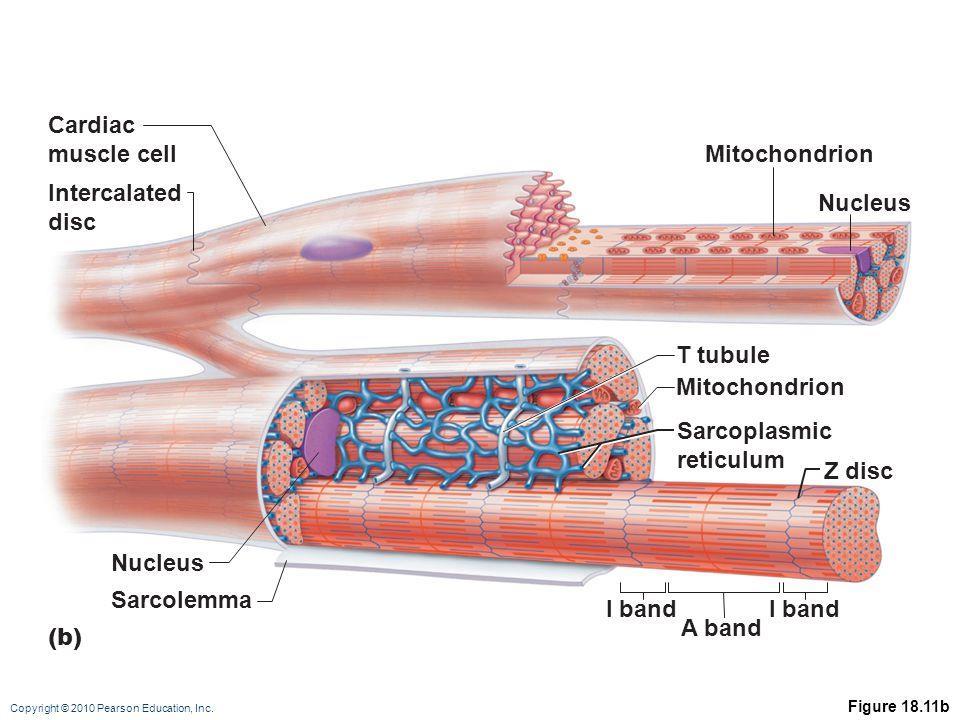
2 The Heart Wall

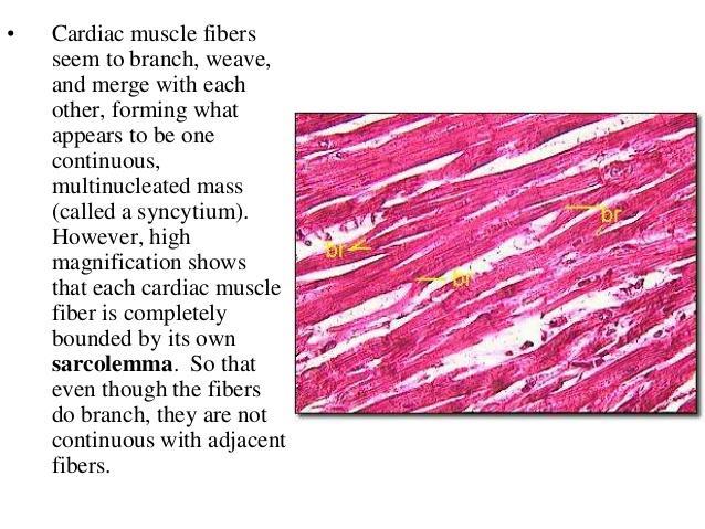
3 Contract as a single unit Cardiac Muscle Simultaneous contraction due to depolarizing at the same time Intercalated disk to speed depolarization automaticity

4 M -myocardium; E - endocardium; En -endothelium; S -ubendothelial layer

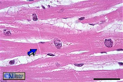
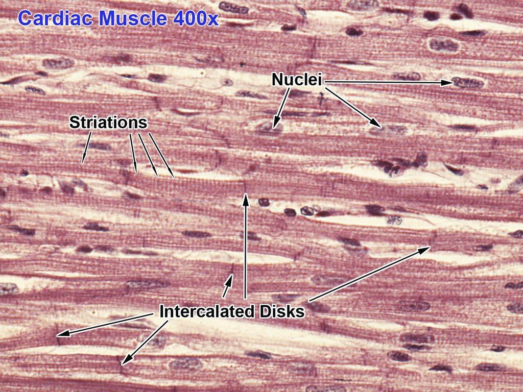
5 Cardiac Muscle Longitudinal Section Cardiac muscle consists of muscle cells mononucleated with centrally placed nucleus. Nuclei are oval, rather pale and which is µm wide. Cardiac muscle is innervated by the autonomic nervous system. Cardiac muscle exhibits crossstriations. Cardiac muscle is for these reasons also called involuntary striated muscle. cell nucleus One cell Intercalated Discs X40 Magnification
6
7
8

9 The Cardiac Muscle Cells
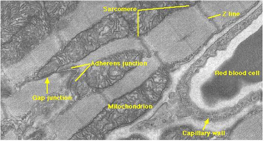
 transverse and lateral components. Bind individual myocytes to one another.")
10 Adherens Junction Desmosome Gap junction Fascia adherens major portion of transverse component. Anchoring sites for actin, and connect to the closest sarcomere. Macula adherens (desmosomes) transverse and lateral components. Bind individual myocytes to one another. stop separation during contraction by binding intermediate filaments, joining the cells together. Macula adherens junctions are also called desmosomes. Gap junctions - lateral component. Allow action potentials to spread between cardiac cells by passage of ions between cells, producing depolarization of the heart muscle. Allows muscle to act as syncytium.
11

12 Cardiac Muscle Tissue Cardiac cells are connected by intercalated discs Intercalated discs house desmosomes and gap junction. Desmosomes provide strength so that the cell do not get ripped apart during contraction Gap junctions are made of the connexin proteins and form a pore through which the cells can communicate.

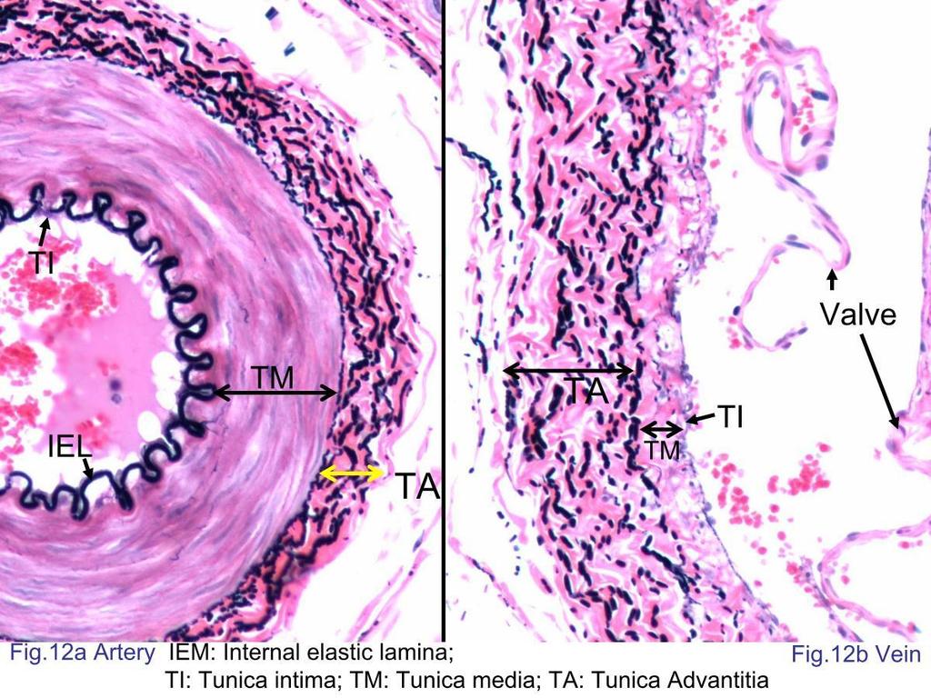
13 Cardiac Muscle Cross section X40 Magnification
14
15
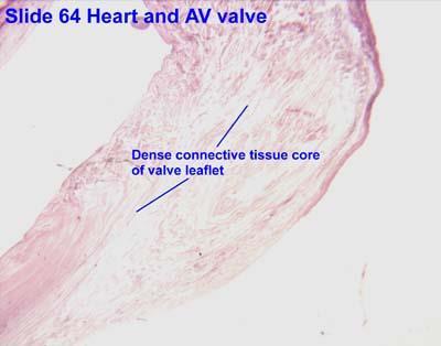
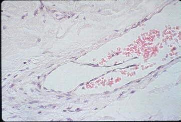
 shows that valves are largely dense connective tissue (C) covered with a thin layer of endothelium.")
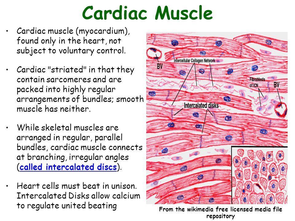
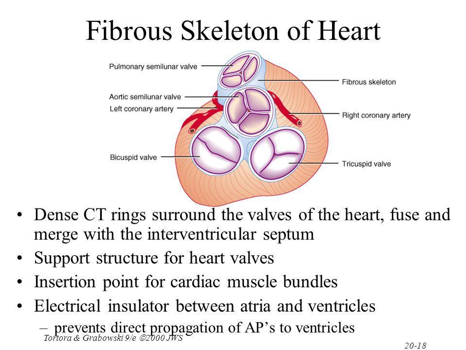
16 The fibrous skeleton of the heart consists of masses of dense connective tissue in the endocardium which anchors the valves and surrounds the two atrioventricular canals, maintaining their proper shape. Section through a leaflet of the left atrioventricular valve (arrows) shows that valves are largely dense connective tissue (C) covered with a thin layer of endothelium. The collagen-rich connective tissue of the valves is stained pale green here and is continuous with the fibrous ring of connective tissue at the base of the valves, which fills the endocardium (En) of this area between the atrium (A) and ventricle (V). The chordae tendinae (CT), small strands of connective tissue which bind distal parts of valve leaflets, can also be seen here. The interwoven nature of the cardiac muscle fibers, with many small fascicles, in the myocardium (M) is also shown.
17


18 Purkinje fibers 40X Are modified cardiac muscle cells. Compared to ordinary cardiac muscle thicker cells: Contain large amounts of glycogen fewer myofibrils.
19 Blood Vessels histology Blood is carried in a closed system of vessels that begins and ends at the heart The three major types of vessels are arteries, capillaries, and veins Arteries carry blood away from the heart, veins carry blood toward the heart Capillaries contact tissue cells and directly serve cellular needs
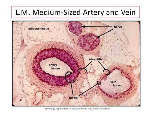
20 General Structure of Blood Vessels
21 Structure of blood vessel (Tunics) Tunica interna (tunica intima) Endothelial layer that lines the lumen of all vessels In vessels larger than 1 mm, a subendothelial connective tissue basement membrane is present Tunica media Smooth muscle and elastic fiber layer, regulated by sympathetic nervous system Controls vasoconstriction/vasodilation of vessels Tunica externa (tunica adventitia) Collagen fibers that protect and reinforce vessels Larger vessels contain vasa vasorum

22 General Histology Structure of Blood Vessels

23 A Comparison of a Typical Artery and a Typical Vein

24 Histological Structure of Blood Vessels
 allow low-resistance conduction of blood and act as conduits Contain elastin in all three tunics Withstand and smooth out large blood pressure fluctuations")
25 Elastic (Conducting) Arteries Thick-walled arteries near the heart; the aorta and its major branches Large lumen (2.5-1 cm diameter) allow low-resistance conduction of blood and act as conduits Contain elastin in all three tunics Withstand and smooth out large blood pressure fluctuations Allow blood to flow fairly continuously through the body
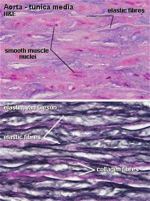
26 Large (Elastic) artery. Elastic Arteries are classified by: The tunica intimae consists of a lining of endothelial cells that rest on a thin layer of connective tissue. The tunica media arranged as lamellae, interspersed with the smooth muscle cells of the tunica media and collagen fibers are found between the layers of elastic fibers There are no elastic lamellae in the adventitia, but elastic fibers are present, though relatively few in number and can not be observed by H&E stain. Brown adipose tissue is one of the two types of adipose tissue. Its primary purpose is to generate body heat. In contrast to white adipocytes (fat cells) which contain a single, large fat vacuole, brown adipocytes contain several smaller vacuoles and centrally located nuclei.
")
27 Elastic (Conducting) Arteries
28
29 Muscular arteries The tunica intimae consists of an endothelial lining and a small amount of connective tissue. The muscular arteries are characterized by a layer of internal elastic lamina separating the tunica intima from the tunica media. The artery has a thicker tunica media, a narrower lumen than the similarly sized vein, and thickened elastic laminae that are not present in the vein. Muscular arteries have more smooth muscle and less elastin in the tunica media than elastic arteries. The less prominent and more variable external elastic lamina lies between the tunica media and the adventitia. The tunica adventitia is composed of collagen fibers (pink), elastic fibers (black) and vasa vasorum.

30 Muscular arteries Are called distributing arteries Middle sized.3mm-1cm Changes diameter to differentially regulate flow to organs as needed Internal as well as external elastic lamina Most of what we see as arteries Tunica media larger in proportion to the lumen, thus muscular 30

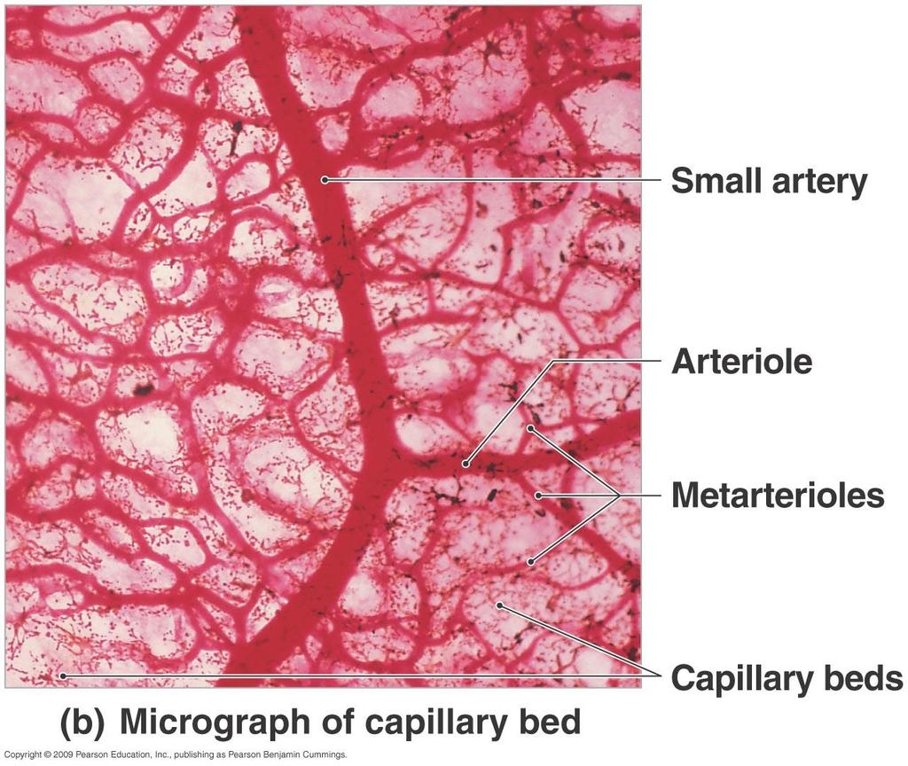
31 Muscular artery This slide is stained with Verhoeff's stain to visualize the elastic fibers, and with eosin to show the cellular structures.
32
33
34
35

36 Arterioles Smallest:.3mm- 10um Only larger ones have all 3 layers Regulated 2 ways: Locally in the tissues Sympathetic control Systemic blood pressure can be regulated through them Deliver blood into capillaries Tunica media has only a few layers of smooth muscle cells 36

37 Arterioles smallest arteries; lead to capillary beds Control flow into capillary beds via vasodilation and constriction

38 muscular middle sized artery
39 Smallest ARTERIOLE Endothelial cell For fast flow & non-stick, until clotting is needed Controls passage through the wall Helps control blood flow Smooth muscle cell SMC/ VSMC Contraction regulates flow by need Vasoconstriction Reticular fibers Mechanical support Smallest arteriole, in essence, is a capillary with smooth muscle cells wrapped around it, with modifications to the endothelial cells - less transport, more interaction with SMCs.
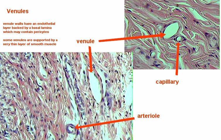
40
41 Capillaries Heart to arteries to capillaries to veins to heart Capillaries are smallest 8-10um Just big enough for single file erythrocytes Composed of: single layer of endothelial cells surrounded by basement membrane Universal function Oxygen and nutrient delivery to tissues CO2 and nitrogenous waste (protein break-down product) removal Some also have tissue specific functions

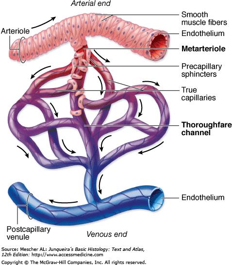
42 The Organization of a Capillary Bed
43 Capillary Beds A microcirculation of interwoven networks of capillaries, consisting of: Vascular shunts metarteriole thoroughfare channel connecting an arteriole directly with a postcapillary venule True capillaries 10 to 100 per capillary bed, capillaries branch off the metarteriole and return to the thoroughfare channel at the distal end of the bed
44 44

45 Capillary Structure Figure 21.4

46 Continuous capillaries are abundant in the skin and muscles, and have: Endothelial cells that provide an uninterrupted lining Adjacent cells that are held together with tight junctions Intercellular clefts of unjoined membranes that allow the passage of fluids Continuous capillaries of the brain: Have tight junctions completely around the endothelium Constitute the blood-brain barrier Continuous Capillaries
 Characterized by: An endothelium riddled with pores (fenestrations) Greater permeability to")
47 Fenestrated Capillaries Found wherever active capillary absorption or filtrate formation occurs (e.g., small intestines, endocrine glands, and kidneys) Characterized by: An endothelium riddled with pores (fenestrations) Greater permeability to solutes and fluids than other capillaries
 to pass between the blood and surrounding tissues Blood flows sluggishly, allowing for modification in various ways")
48 Highly modified, leaky, fenestrated capillaries with large lumens Found in the liver, bone marrow, lymphoid tissue, and in some endocrine organs Allow large molecules (proteins and blood cells) to pass between the blood and surrounding tissues Blood flows sluggishly, allowing for modification in various ways Sinusoids
49 Veins Collect blood from all tissues and organs and return it to the heart Are classified according to size Venules Medium-sized veins Large veins
50
51 The transition from capillaries to venules occurs gradually The immediate postcapillary venules are similar structurally to capillaries, with pericytes, but range in diameter from 15 to 20 m. A. Postcapillary venules participate in the exchanges between the blood and the tissues and, are the primary site at which white blood cells leave the circulation at sites of infection or tissue damage. B. Venules converge into larger collecting venules which have more contractile cells. With greater size the venules become surrounded by recognizable tunica media with two or three smooth muscle layers and are called C. Muscular venules. A characteristic feature of all venules is the large diameter of the lumen compared to the overall thinness of the wall

52 Venules Venules collect blood from capillary networks and gradually merge to form veins.


53 PCV: Postcapillary venules. CV: Capillary venules. MV Musculsr venules
54 Veins Blood entering veins is under very low pressure and moves toward the heart by contraction of the tunica media and external compressions from surrounding muscles and other organs. Valves project from the tunica intima to prevent back-flow of blood. Most veins are small or medium veins with diameters less than one centimeter. Located in parallel with corresponding muscular arteries. The intima usually has a thin subendothelial layer The media consists of small bundles of smooth muscle cells intermixed with reticular fibers and a delicate network of elastic fibers. The collagenous adventitial layer is well-developed.
: Micrograph of small vein (V) shows a relatively large lumen compared to the small muscular artery (A) with its thick media (M)")
55 Small Veins. (a): Micrograph of small vein (V) shows a relatively large lumen compared to the small muscular artery (A) with its thick media (M) and adventitia (Ad). The wall of a small vein is very thin, containing only two or three layers of smooth muscle. X200. H&E.
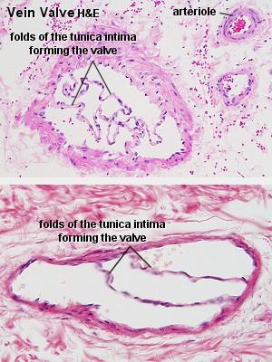
: Micrograph of a convergence between two small veins showing valves (arrow).")
56 Medium sized vines Vein with a much less compact muscle layer than you saw in the preceding arteries. unica media and adventitia, which is at least as wide as the media, and often even wider. There is no evident inner elastic membrane (b): Micrograph of a convergence between two small veins showing valves (arrow). Valves are thin folds of tunica intima projecting well into the lumen which act to prevent backflow of blood. X200. H&E.
. Both the media and adventitia are better developed, but the wall is often folded around the relatively large lumen. X100. H&E.")
57 (c): Micrograph of a medium vein (MV) showing a thicker wall, but still less prominent than that of the accompanying muscular artery (MA). Both the media and adventitia are better developed, but the wall is often folded around the relatively large lumen. X100. H&E.
. X200.")
58 (d): Micrograp h of a medium vein containing blood and showing valve folds (arrows). X200. Masson trichrome.
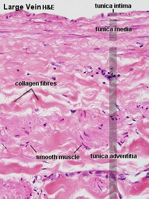
59 LARGE VEIN Details Intima { Adventitia { Occasional circular SMC Numerous elastic fibers Bundles of longitudinal smooth muscle


60 The big venous trunks, Paired with elastic arteries close to the heart, are large veins Large veins have a well-developed tunica intima, but the tunica media is relatively thin, with few layers of smooth muscle and abundant connective tissue. The adventitial layer is thick in large veins and frequently contains longitudinal bundles of smooth muscle. Both the media and adventitia contain elastic fibers, but elastic laminae like those of arteries are not present. Most veins have valves, but these are most prominent in large veins.
61
 Muscular contraction Aids the return of blood to heart in conjunction with valves Mechanical issues (really good to")
62 Special features of veins veins contain valves Prevent backflow of blood Valves Prevent backflow Most abundant in legs (where blood has to travel against gravity) Muscular contraction Aids the return of blood to heart in conjunction with valves Mechanical issues (really good to know)
63
Histology of the Cardiac System. Dr. Nabil Khoury Anatomy Department
 Histology of the Cardiac System Dr. Nabil Khoury Anatomy Department Objectives 1. Identify the 3 layers of the heart endocardium, myocardium, epicardium 2. Differentiate cardiacmuscle 3. Define intercalated
UNIT 4: BLOOD VESSELS
 UNIT 4: BLOOD VESSELS Dr. Moattar Raza Rizvi NRS237, Physiology Generalized Structure of Blood Vessels 1 Tunica interna (tunica intima) Endothelial layer that lines the lumen of all vessels In vessels
Copyright 2010 Pearson Education, Inc. Blood Vessel Structure
 Blood Vessel Structure Structure of Blood Vessel Walls Arteries and veins Tunica intima, tunica media, and tunica externa Lumen Central blood-containing space Capillaries Endothelium with sparse basal
Blood Vessels. Types of Blood Vessels Arteries carry blood away from the heart Capillaries smallest blood vessels. Veins carry blood toward the heart
 C H A P T E R Blood Vessels 20 Types of Blood Vessels Arteries carry blood away from the heart Capillaries smallest blood vessels The site of exchange of molecules between blood and tissue fluid Veins
Cardiovascular System Blood Vessels
 Cardiovascular System Blood Vessels Structure of Blood Vessels The three layers (tunics) Tunica intima composed of simple squamous epithelium Tunica media sheets of smooth muscle Contraction vasoconstriction
The cardiovascular system
 The cardiovascular system Components of the Cardiovascular system Heart Vessels: Arteries Capillaries Veins Functions of CVS: Transportation system where blood is the transporting vehicle Carries oxygen,
Histology of the myocardium and blood vessels. Prof. Abdulameer Al-Nuaimi
 Histology of the myocardium and blood vessels Prof. Abdulameer Al-Nuaimi E-mail: a.al-nuaimi@sheffield.ac.uk E-mail: abdulameerh@yahoo.com Histology of blood vessels The walls of arteries and veins are
2. capillaries - allow exchange of materials between blood and tissue fluid
 Chapter 19 - Vascular System A. categories and general functions: 1. arteries - carry blood away from heart 2. capillaries - allow exchange of materials between blood and tissue fluid 3. veins - return
Extra notes for lab- 1 histology. Slide 1 : cross section in the elastic artery ( aortic arch, ascending aorta, descending aorta )
") Extra notes for lab- 1 histology Slide 1 : cross section in the elastic artery ( aortic arch, ascending aorta, descending aorta ) - twin of ascending aorta is the pulmonary trunk. Ascending aorta represents
Extra notes for lab- 1 histology Slide 1 : cross section in the elastic artery ( aortic arch, ascending aorta, descending aorta ) - twin of ascending aorta is the pulmonary trunk. Ascending aorta represents
Cardivascular System Module 5: Structure and Function of Blood Vessels *
 OpenStax-CNX module: m49689 1 Cardivascular System Module 5: Structure and Function of Blood Vessels * Donna Browne Based on Structure and Function of Blood Vessels by OpenStax This work is produced by
Practical Histology. Cardiovascular System. Dr Narmeen S. Ahmad
 Practical Histology Cardiovascular System Dr Narmeen S. Ahmad The Cardiovascular System A closed system of the heart and blood vessels Functions of cardiovascular system: Transport nutrients, hormones
Cardiovascular (Circulatory) System
 System") Cardiovascular (Circulatory) System Piryaei May 2011 Circulatory System Heart Blood Vessels Macrovasculature (More than 0.1mm) Elastic Artery Muscular (Distributing) Artery Large Arteriol Small Vein Muscular
Cardiovascular (Circulatory) System Piryaei May 2011 Circulatory System Heart Blood Vessels Macrovasculature (More than 0.1mm) Elastic Artery Muscular (Distributing) Artery Large Arteriol Small Vein Muscular
The Circulatory System
 The Circulatory System Dr. Sami Zaqout The circulatory system Circulatory system Blood vascular systems Lymphatic vascular systems Blood vascular systems Blood vascular systems The circulatory system Circulatory
SCPA602 Cardiovascular System
 SCPA602 Cardiovascular System Associate Professor Dr. Wannee Jiraungkoorskul Department of Pathobiology, Faculty of Science, Mahidol University Tel: 02-201-5563, E-mail: wannee.jir@mahidol.ac.th 1 Objectives
Microscopic Anatomy CARDIOVASCULAR SYSTEM
 Microscopic Anatomy CARDIOVASCULAR SYSTEM I. Introduction The cardiovascular system is a closed system consisting of a pump, the heart, and a series of tubular blood vessels that interconnect all body
Lecture name: blood 2 & The Circulatory System Edited by: Buthainah Al masaeed & Yousef Qandeel
 Lecture name: blood 2 & The Circulatory System Edited by: Buthainah Al masaeed & Yousef Qandeel Now we will take about A granulocytes : Lymphocyte Monocytes 1- Lymphocyte - The second major type of presence
a) Endocardium The endocardium is the innermost layer of the heart wall. It forms the internal lining of the atria and ventricles.
 Endocardium The endocardium is the innermost layer of the heart wall. It forms the internal lining of the atria and ventricles.") Chapter 11 Circulatory System The circulatory system consists of a muscular, pulsing heart and a system of blood vessels.the blood vessels include: Arteries which will carry the blood and it's dissolved
Chapter 11 Circulatory System The circulatory system consists of a muscular, pulsing heart and a system of blood vessels.the blood vessels include: Arteries which will carry the blood and it's dissolved
Cardiac Conduction System
 Cardiac Conduction System What causes the Heart to Beat? Heart contracts by electrical signals! Cardiac muscle tissue contracts on its own an electrical signal is sent out by the heart so that all cells
Ch. 12 The Circulatory System. The heart. The heart is a double pump. A quick note on arteries vs. veins. = the muscular pump of the CV system
 Ch. 12 The Circulatory System The heart A.k.a. the cardiovascular system Blood was discussed in Ch. 11 Focus of Ch. 12: heart and blood vessels = the muscular pump of the CV system ~ 100,000 heartbeats/day!
Chapter 21. Blood Vessels and Circulation
 Chapter 21 Openstax: Chapter 20 Blood Vessels and Circulation Chapter 21 Learning Outcomes After completing Chapter 21, you will be able to: 1. Distinguish among the types of blood vessels based on their
1. What kind of blood is found in the rt. atrium? (oxygenated or deoxygenated)
") Carl Christennsen, PhD Chap. 19, 20, & 21 - Circulatory System Bio. 2304 Human Anatomy HEART 1. What kind of blood is found in the rt. atrium? (oxygenated or deoxygenated) Where does this blood come from?
Carl Christennsen, PhD Chap. 19, 20, & 21 - Circulatory System Bio. 2304 Human Anatomy HEART 1. What kind of blood is found in the rt. atrium? (oxygenated or deoxygenated) Where does this blood come from?
Cardiovascular System
 Cardiovascular System Purpose Transport oxygen and nutrients Take waste products away from tissues & organs Things we learned Blood pressure: the force of blood pushing against the walls of blood vessels
Collin County Community College
 Collin County Community College BIOL. 2402 Anatomy & Physiology WEEK 6 Blood Vessels 1 Anatomy of Blood Vessels Walls of blood vessels contain 3 distinct layers : Tunica intima innermost layer includes
The Cardiovascular and Lymphatic Systems Cardiovascular System Blood Vessels Blood Vessels Arteries Arteries Arteries
 CH 12 The Cardiovascular and s The Cardiovascular and s OUTLINE: Cardiovascular System Blood Vessels Blood Pressure Cardiovascular System The cardiovascular system is composed of Blood vessels This system
Lab Activity 25. Blood Vessels & Circulation. Portland Community College BI 232
 Lab Activity 25 Blood Vessels & Circulation Portland Community College BI 232 Artery and Vein Histology Walls have 3 layers: Tunica intima Tunica media Tunica externa 2 Tunica Intima Is the innermost layer
Muscle Tissue. General concepts. Classification of muscle. I. Functional classification is based on the type of neural control.
 Muscle Tissue LEARNING OBJECTIVES 1. Identify the three types of muscle tissue at the light microscopic level. 2. List and compare the structural and functional features of each of the three muscle fiber
The Cardiovascular and Lymphatic Systems
 BIOLOGY OF HUMANS Concepts, Applications, and Issues Fifth Edition Judith Goodenough Betty McGuire 12 The Cardiovascular and Lymphatic Systems Lecture Presentation Anne Gasc Hawaii Pacific University and
Anatomy Review: The Heart Graphics are used with permission of A.D.A.M. Software, Inc. and Benjamin/Cummings Publishing Co.
 Anatomy Review: The Heart Graphics are used with permission of A.D.A.M. Software, Inc. and Benjamin/Cummings Publishing Co. Anatomy Views Label the diagrams of the heart below: Interactive Physiology Study
Circulatory System Function Move circulatory fluid (blood) around body Gas Transport Nutrient Transport Excretory Product Transport
 around body Gas Transport Nutrient Transport Excretory Product Transport") Lecture 37 Introduction to Circulation BY DR QAZI IMTIAZ RASOOL OBJECTIVES Functions of the Heart Generating blood pressure Routing blood: separates pulmonary and systemic circulations Ensuring one-way
Lecture 37 Introduction to Circulation BY DR QAZI IMTIAZ RASOOL OBJECTIVES Functions of the Heart Generating blood pressure Routing blood: separates pulmonary and systemic circulations Ensuring one-way
Derived copy of Structure and Function of Blood Vessels *
 OpenStax-CNX module: m56696 1 Derived copy of Structure and Function of Blood Vessels * Stephanie Fretham Based on Structure and Function of Blood Vessels by OpenStax This work is produced by OpenStax-CNX
2402 : Anatomy/Physiology
 Dr. Chris Doumen Lecture 1 2402 : Anatomy/Physiology Hemo Dynamics and Blood Vessels I nt r oduc t i on TextBook Readings Pages 721 through 734. Make use of the figures in your textbook ; a picture is
組織學 Historlogy 台北醫學大學 / 解剖學科教授 : 邱瑞珍分機號碼 :3261. 電子郵件信箱
 組織學 Historlogy 台北醫學大學 / 解剖學科教授 : 邱瑞珍分機號碼 :3261 電子郵件信箱 :rueijen@tmu.edu.tw 1 The Circulatory System 台北醫學大學 / 解剖學科教授 : 邱瑞珍分機號碼 :3261 電子郵件信箱 :rueijen@tmu.edu.tw 2 學習目的 The structure of the arteries The structure
Chapter 21 (1) An Introduction to Blood Vessels and Circulation
 An Introduction to Blood Vessels and Circulation") Chapter 21 (1) An Introduction to Blood Vessels and Circulation Lecture Objectives Compare and contrast the structure of an artery, arteriole, vein, venule, and capillary Discuss the structure and function
Chapter 21 (1) An Introduction to Blood Vessels and Circulation Lecture Objectives Compare and contrast the structure of an artery, arteriole, vein, venule, and capillary Discuss the structure and function
Any of these questions could be asked as open question or lab question, thus study them well
 Any of these questions could be asked as open question or lab question, thus study them well describe the factors which regulate cardiac output describe the sympathetic and parasympathetic control of heart
Muscle tissues. Dr. Hersh Abdul Ham-Karim BVM&S, PG Dip, MSc and PhD
 Muscle tissues Dr. Hersh Abdul Ham-Karim BVM&S, PG Dip, MSc and PhD Muscle tissue is a soft tissue that composes muscles in animal bodies, and gives rise to muscles' ability to contract. Muscle tissue
Blood Vessels. Over view. We have about 60,000 miles of blood vessels!
 Blood Vessels Over view 3 types of blood vessels arteries - carry blood away from heart "branch", "diverge", and "fork" veins - carry blood toward heart "join", "merge", and "converge" capillaries - site
Human Anatomy, First Edition
 Human Anatomy, First Edition McKinley & O'Loughlin Chapter 23 : Vessels and Circulation 23-1 Blood Vessels An efficient style of transport for oxygen, nutrients, and waste products to and from body tissues.
Blood flows away from the heart in arteries, to the capillaries and back to the heart in the veins
 Cardiovascular System Summary Notes The cardiovascular system includes: The heart, a muscular pump The blood, a fluid connective tissue The blood vessels, arteries, veins and capillaries Blood flows away
Cardiovascular system
 BIO 301 Human Physiology Cardiovascular system The Cardiovascular System: consists of the heart plus all the blood vessels transports blood to all parts of the body in two 'circulations': pulmonary (lungs)
Heart. Heart 2-Tunica media: middle layer (media ='middle') muscle fibers (smooth or cardiac).
 muscle fibers (smooth or cardiac).") t. innermost lumenal General Circulatory system heart and blood vessels walls have 3 layers (inside to outside) 1-Tunica interna: aka tunica intima layer--lumenal layer epithelium--endothelium simple squamous
t. innermost lumenal General Circulatory system heart and blood vessels walls have 3 layers (inside to outside) 1-Tunica interna: aka tunica intima layer--lumenal layer epithelium--endothelium simple squamous
Major Function of the Cardiovascular System. Transportation. Structures of the Cardiovascular System. Heart - muscular pump
 Structures of the Cardiovascular System Heart - muscular pump Blood vessels - network of tubes Blood - liquid transport vehicle brachiocephalic trunk superior vena cava right pulmonary arteries right pulmonary
Remember: the CVS system is deriving from the mesenchyma and is lined by simple squamous epithelium called the endothelium
 Lecture 10 General med_2nd semester Microscopic anatomy and embryology of cardiovascular system Microscopic structure of the heart, excitomotoric system - its structural peculiarities Blood vessels - arteries
Circulation. Sinoatrial (SA) Node. Atrioventricular (AV) Node. Cardiac Conduction System. Cardiac Conduction System. Linked to the nervous system
 Node. Atrioventricular (AV) Node. Cardiac Conduction System. Cardiac Conduction System. Linked to the nervous system") Circulation Cardiac Conduction System AHS A H S Your body resembles a large roadmap. There are routes or arteries that take you downtown to the heart of the city and veins that take you to the outskirts
Circulation Cardiac Conduction System AHS A H S Your body resembles a large roadmap. There are routes or arteries that take you downtown to the heart of the city and veins that take you to the outskirts
Cardiovascular System. Blood Vessel anatomy Physiology & regulation
 Cardiovascular System Blood Vessel anatomy Physiology & regulation Path of blood flow Aorta Arteries Arterioles Capillaries Venules Veins Vena cava Vessel anatomy: 3 layers Tunica externa (adventitia):
Microcirculation. Lecture Block 11 (contributions from Brett Burton)
") Lecture Block 11 (contributions from Brett Burton) Elements of Arterioles, capillaries, venules Structure and function: transport Fluid balance Lymph system Vessels of the Circulatory System Diameter Aorta
Lecture Block 11 (contributions from Brett Burton) Elements of Arterioles, capillaries, venules Structure and function: transport Fluid balance Lymph system Vessels of the Circulatory System Diameter Aorta
Chapter 21 Peripheral circulation and Regulation
 Chapter 21 Peripheral circulation and Regulation I. Blood vessel structure A. Blood flows from large arteries to small capillaries 1. Large arteries contain large amounts of elastic tissue and little smooth
The Circulatory System. The Heart, Blood Vessels, Blood Types
 The Circulatory System The Heart, Blood Vessels, Blood Types The Closed Circulatory System Humans have a closed circulatory system, typical of all vertebrates, in which blood is confined to vessels and
The Cardiovascular System: Vessels and Routes. Pulmonary Circulation H E A R T. Systemic Circulation
 The Cardiovascular System: Vessels and Routes 1. Overview of Blood Circulation A. Pulmonary Circulation Lung Arterioles Pulmonary Artery Capillaries Pulmonary Circulation Venules Pulmonary Veins H E A
Cardiovascular Anatomy Dr. Gary Mumaugh
 Cardiovascular Anatomy Dr. Gary Mumaugh Location of Heart Approximately the size of your fist Location o Superior surface of diaphragm o Left of the midline in mediastinum o Anterior to the vertebral column,
Integrated Muscle. Red: important. Black: in male female slides. Gray: notes extra. Editing File
 Integrated Muscle Red: important. Black: in male female slides. Gray: notes extra. Editing File OBJECTIVES Identify and describe the histological structure of the three types of muscle cells and list the
Heart. Large lymphatic vessels Lymph node. Lymphatic. system Arteriovenous anastomosis. (exchange vessels)
") Venous system Large veins (capacitance vessels) Small veins (capacitance vessels) Postcapillary venule Thoroughfare channel Heart Large lymphatic vessels Lymph node Lymphatic system Arteriovenous anastomosis
Venous system Large veins (capacitance vessels) Small veins (capacitance vessels) Postcapillary venule Thoroughfare channel Heart Large lymphatic vessels Lymph node Lymphatic system Arteriovenous anastomosis
The Cardiovascular System
 The Cardiovascular System The Cardiovascular System A closed system of the heart and blood vessels The heart pumps blood Blood vessels allow blood to circulate to all parts of the body The function of
Approximately the size of your fist Location Superior surface of diaphragm Left of the midline in mediastinum Anterior to the vertebral column,
 Dr. Gary Mumaugh Approximately the size of your fist Location Superior surface of diaphragm Left of the midline in mediastinum Anterior to the vertebral column, posterior to the sternum Posteriorly the
Chapter 14. The Cardiovascular System
 Chapter 14 The Cardiovascular System Introduction Cardiovascular system - heart, blood and blood vessels Cardiac muscle makes up bulk of heart provides force to pump blood Function - transports blood 2
Topic 6: Human Physiology
 Topic 6: Human Physiology 6.2 The Blood System D.4 The Heart Essential Questions: 6.2 The blood system continuously transports substances to cells and simultaneously collects waste products. D.3 The chemical
Vessels by Design: Basic Vessel Anatomy. Student Information Page 3A
 Vessels by Design: Basic Vessel Anatomy Student Information Page 3A Activity Introduction: Once you get home from running around all day, your throat is probably a little dry. You go to your kitchen, get
Cardiovascular Physiology
 Cardiovascular Physiology Lecture 1 objectives Explain the basic anatomy of the heart and its arrangement into 4 chambers. Appreciate that blood flows in series through the systemic and pulmonary circulations.
Blood Vessels. Chapter 20
 Blood Vessels Chapter 20 Summary of the Characteristics of Arteries and Veins Characteristic Artery Vein Wall thickness thick thin Shape in cross section round flattened Thickest tunic media externa Collagen
GENERAL HISTOLOGY 4. Muscular Tissue
 Biology-232 GENERAL HISTOLOGY 4. Muscular Tissue Dr. Manal Othman Anatomy Department CMMS, AGU Responsible for MOST types of BODY MOVEMENT Made up of groups of elongated MUSCLE cells with contractile filaments
BIOH122 Human Biological Science 2
 BIOH122 Human Biological Science 2 Session 5 Cardiovascular System 3 Vasculature and Capillary Exchange Bioscience Department Endeavour College of Natural Health endeavour.edu.au Session Plan o Structure
STRUCTURES OF THE CARDIOVASCULAR SYSTEM
 STRUCTURES OF THE CARDIOVASCULAR SYSTEM CARDIOVASCULAR SYSTEM Also called the circulatory system Consists of the heart, arteries, veins, and capillaries Main function is to pump/circulate oxygenated blood
Muscle tissue. 1) Striated skeletal muscle tissue. 2) Striated cardiac muscle tissue. 3) Smooth muscle tissue.
 Striated skeletal muscle tissue. 2) Striated cardiac muscle tissue. 3) Smooth muscle tissue.") Muscle tissue 1) Striated skeletal muscle tissue. 2) Striated cardiac muscle tissue. 3) Smooth muscle tissue. General characteristic of muscle tissue Origin: mesoderm and mesenchyme Excitability Contraction
Muscle tissue 1) Striated skeletal muscle tissue. 2) Striated cardiac muscle tissue. 3) Smooth muscle tissue. General characteristic of muscle tissue Origin: mesoderm and mesenchyme Excitability Contraction
The Circulatory System
 The Circulatory System This system comprises both the blood and lymphatic vascular system. Blood vascular system is composed from; The heart: an organ whose function is to pump the blood. The arteries:
Basic Histology. By Mrs. Bailey
 Basic Histology By Mrs. Bailey Primary Tissues 1. Epithelial Tissue 2. Connective Tissue 3. Muscle Tissue 4. Nervous Tissue Very cellular Supported by underlying connective tissue Epithelial & connective
Microanatomy of Muscles. Anatomy & Physiology Class
 Microanatomy of Muscles Anatomy & Physiology Class Three Main Muscle Types Objectives: By the end of this presentation you will have the information to: 1. 2. 3. 4. 5. 6. Describe the 3 main types of muscles.
Skeletal muscle. General features :
 Muscular tissues In the first embryonic life the muscular tissues arise from mesoderm, The function of movement in multicellular organisms is usually assumed by specialized cells called muscle fibers which
Vascular System Part One
 Vascular System Part One Objectives Trace the route taken by blood as it leaves, and then returns to the heart. Describe the structure of the walls of arteries and veins. Discuss the structure and function
10. Thick deposits of lipids on the walls of blood vessels, called, can lead to serious circulatory issues. A. aneurysm B. atherosclerosis C.
 Heart Student: 1. carry blood away from the heart. A. Arteries B. Veins C. Capillaries 2. What is the leading cause of heart attack and stroke in North America? A. alcohol B. smoking C. arteriosclerosis
The Circulatory System. Lesson Overview. Lesson Overview The Circulatory System
 33.1 THINK ABOUT IT More than one-third of the 1.2 million Americans who suffer a heart attack each year die. This grim evidence shows that the heart and the circulatory system it powers are vital to life.
Lecture 9A. Muscle structure. Outline
 Lecture 9A Muscle structure Outline Smooth, skeletal, and cardiac muscle tissues Structure and function of skeletal muscle cells. Sarcomeres structure and contraction Actin-myosin interaction and sliding
Cardiovascular System, Blood, and Blood Cell Formation
 Cardiovascular System, Blood, and Blood Cell Formation 7 CONTENTS OVERVIEW OF THE CARDIOVASCULAR SYSTEM GENERAL STRUCTURE OF THE HEART Heart Valves HEART AS A PUMP Contractile Properties of Cardiac Myocytes
Tissues. tissue = many cells w/ same structure and function. cell shape aids its function tissue shape aids its function
 Tissues tissue = many cells w/ same structure and function cell shape aids its function tissue shape aids its function Histology = study of tissues 4 types of tissues Epithelial coverings contact openings
Peripheral Circulation and Regulation
 Peripheral Circulation and Regulation Functions of Peripheral Circulation 1. Contain the blood 2. Exchange nutrients, waste products, and gases with tissues 3. Transport 4. Regulate blood pressure, along
Anatomy & Physiology of Cardiovascular System. Chapter 18 & 19
 Anatomy & Physiology of Cardiovascular System Chapter 18 & 19 Objectives..cont 1. Discuss the physiological stages of cardiac muscle contraction. 2. Trace a typical ECG and label each wave or complex 3.
Warm Up- Monday -AND- Setup Cornell Notes.
 Warm Up- Monday Brainstorm in your notebook: If the heart sends blood to all organs, how and where does the heart get blood to provide oxygen for its muscles? -AND- Setup Cornell Notes. Announcements Unit
Chapter 19: Blood Vessels. 63 slides
 Chapter 19: Blood Vessels 63 slides 1 Blood Vessels The Blood Vessels are essentially a series of tubes. Three types of blood vessel tubes: Arteries (carry blood away from heart) Arterioles are the smallest
Structure. Arteries. 21_01d 4/18/12. The Cardiovascular System: Blood Vessels and Hemodynamics. Dr Badri Paudel GMC
 Goal of the Cardiovascular System: deliver blood to all parts of the body The Cardiovascular System: Blood Vessels and Hemodynamics Dr Badri Paudel GMC Does so by using different types of tubing, attached
Cardiovascular System. I. Structures of the heart A. : Pericardium sack that surrounds the heart
 Cardiovascular System I. Structures of the heart A. : Pericardium sack that surrounds the heart 1. : Pericardial Cavity serous fluid filled space between the heart and the pericardium B. Heart Wall 1.
Muscle Tissue. Xie Fenfen. Department of Histology and Embryology School of Basic Medicine Anhui Medical University
 Muscle Tissue Xie Fenfen Email:xff2005024@126.com Department of Histology and Embryology School of Basic Medicine Key points The structural differences (LM) of 3 types of muscle fibers Molecular structure
Connective tissue MUSCLE TISSUE
 Connective tissue MUSCLE TISSUE Part 1 General features of MT Develop from mesoderm Many cells, less intercellular matrix Function contraction (shortening) Skeletal (striated, voluntary) Types of MT Cardiac
Muscle Tissue. Dr. Heba Kalbouneh Associate Professor of Anatomy and Histology
 Muscle Tissue Dr. Heba Kalbouneh Associate Professor of Anatomy and Histology Functions of muscle tissue Movement Maintenance of posture Joint stabilization Heat generation Tendon Belly Tendon Types of
CIRCULATORY SYSTEM BLOOD VESSELS
 Name: Block: CIRCULATORY SYSTEM Multicellular organisms (above the level of roundworms) rely on a circulatory system to bring nutrients to, and take wastes away from, cells. In higher organisms such as
How many skeletal muscles are present in our body? Muscles are excitable & contractile, extensible and elastic to some extent.
 Muscles How many skeletal muscles are present in our body? -646 muscles The functions of the muscles are: Movement Maintenance of posture Generation of heat Stabilization of joints : amount of muscle surrounding
1. General characteristics of muscle tissues: 2. A. Skeletal muscle tissue ("striated muscle tissue")
") 1. General characteristics of muscle tissues: Muscle fibers, AKA, muscle cells Vascularized. Other tissues dense and loose C.T. nerves and nerve fibers Muscle fibers (muscle cells) close together. From
1. General characteristics of muscle tissues: Muscle fibers, AKA, muscle cells Vascularized. Other tissues dense and loose C.T. nerves and nerve fibers Muscle fibers (muscle cells) close together. From
Tissues 10/21/2016. Epithelial Tissue
 Tissues This is a generalized cell diagram. It shows the anatomy of a cell, but most cells do not actually look like this. Cells can have a wide variety of shapes and sizes, depending on their function.
Anatomy and Physiology, Spring 2015 Exam II: Form A April 9, Name Student Number
 Anatomy and Physiology, Spring 2015 Exam II: Form A April 9, 2015 Name Student Number For Questions 1 2 refer to the following table. 1 Ventricular pressure is greater than aortic 6 AV valve is open 2
Ch 19: Cardiovascular System - The Heart -
 Ch 19: Cardiovascular System - The Heart - Give a detailed description of the superficial and internal anatomy of the heart, including the pericardium, the myocardium, and the cardiac muscle. Trace the
Health Science 20 Circulatory System Notes
 Health Science 20 Circulatory System Notes Functions of the Circulatory System The circulatory system functions mainly as the body s transport system. It transports: o Oxygen o Nutrients o Cell waste o
The HEART. What is it???? Pericardium. Heart Facts. This muscle never stops working It works when you are asleep
 This muscle never stops working It works when you are asleep The HEART It works when you eat It really works when you exercise. What is it???? Located between the lungs in the mid thoracic region Apex
Cardiac Muscle Tissue. Cardiac Muscle Tissue
 Walls of the heart (cardia: heart); myocardium. Cardiac muscle fibers not as densely packed as skeletal cardiac muscle tissue is highly vascularized Other components; dense C.T. septa, larger blood vessels,
Kidney Functions Removal of toxins, metabolic wastes, and excess ions from the blood Regulation of blood volume, chemical composition, and ph
 The Urinary System Urinary System Organs Kidneys are major excretory organs Urinary bladder is the temporary storage reservoir for urine Ureters transport urine from the kidneys to the bladder Urethra
Physiology of Circulation
 Physiology of Circulation Dr. Ali Ebneshahidi Blood vessels Arteries: Blood vessels that carry blood away from the heart to the lungs and tissues. Arterioles are small arteries that deliver blood to the
Equine biological systems
 Equine biological systems Pack 3 Circulation Pack Code: EBS3 This pack you will help you to: describe how blood circulates round the horse's body. www.lbcnc.org.uk About this pack Objectives When you have
Muscle Histology. Dr. Heba Kalbouneh Assistant Professor of Anatomy and Histology
 Muscle Histology Dr. Heba Kalbouneh Assistant Professor of Anatomy and Histology Functions of muscle tissue Movement Maintenance of posture Joint stabilization Heat generation Types of Muscle Tissue Skeletal
Muscle Tissue.
 Muscle Tissue Email:lizhongjie@zju.edu.cn General description 1) components: ---cell: muscle cell - myofiber ---extracellular ground substance: CT with BV, LV and nerve Nomenclature in muscular cell Muscular
Animal Physiology Prof. Mainak Das Department of Biological Sciences and Bioengineering Indian Institute of Technology, Kanpur. Module - 1 Lecture 7
 Animal Physiology Prof. Mainak Das Department of Biological Sciences and Bioengineering Indian Institute of Technology, Kanpur Module - 1 Lecture 7 Welcome back to the lectures in Animal Physiology in
(b) Stomach s function 1. Dilution of food materials 2. Acidification of food (absorption of dietary Fe in small intestine) 3. Partial chemical digest
 Stomach s function 1. Dilution of food materials 2. Acidification of food (absorption of dietary Fe in small intestine) 3. Partial chemical digest") (1) General features a) Stomach is widened portion of gut-tube: between tubular and spherical; Note arranged of smooth muscle tissue in muscularis externa. 1 (b) Stomach s function 1. Dilution of food
(1) General features a) Stomach is widened portion of gut-tube: between tubular and spherical; Note arranged of smooth muscle tissue in muscularis externa. 1 (b) Stomach s function 1. Dilution of food
Cardiovascular. Function of the cardiovascular system is to transport blood containing: Nutrients Waste Hormones Immune cells Oxygen
 Cardiovascular The Cardiovascular System - Arteries Arteries Cardiovascular System Function of the cardiovascular system is to transport blood containing: Carry blood away from heart Carotid arteries Deliver
Muscle and Neuromuscular Junction. Peter Takizawa Department of Cell Biology
 Muscle and Neuromuscular Junction Peter Takizawa Department of Cell Biology Types and structure of muscle cells Structural basis of contraction Triggering muscle contraction Skeletal muscle consists of
Cardiovascular System
 Cardiovascular System I. Structure of the Heart A. Average adult heart is 14 cm long and 9 cm wide. B. Lies in the mediastinum. C. Enclosed in the pericardium. 1. Fibrous pericardium- Outer, tough connective