Atrioventricular Canal (Septal) Defects. Norman H Silverman MD. D Sc (Med),FACC, FAHA
|
|
|
- Alberta Carter
- 6 years ago
- Views:
Transcription


1 Atrioventricular Canal (Septal) Defects Norman H Silverman MD. D Sc (Med),FACC, FAHA


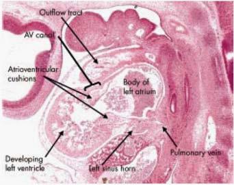
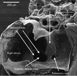
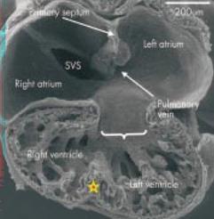
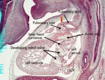
2 Embryology of the A-V Canal Looping NHS. Formation of the Atrial Septum


3 Embryology of the A-V Canal NHS. Development of the A-V Canal Cono-ventricular development

4 Moorman and Anderson
5


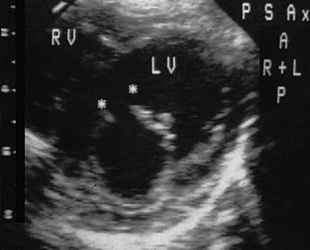
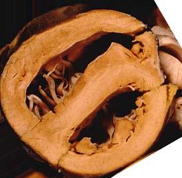
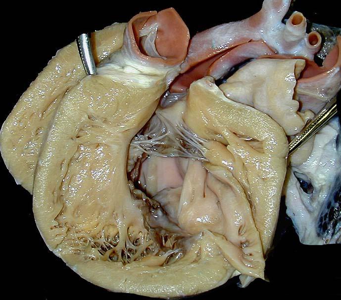
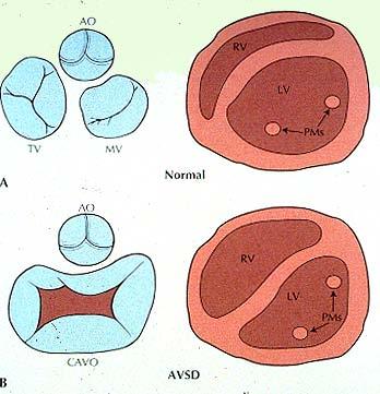
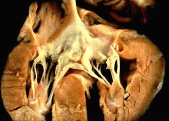
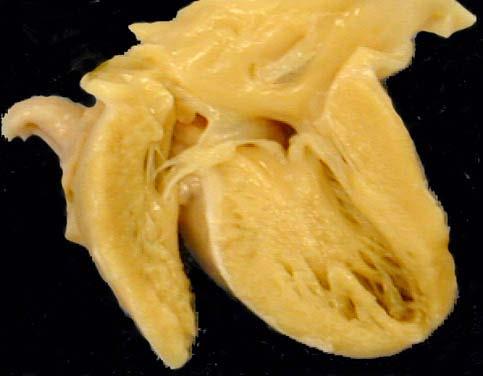
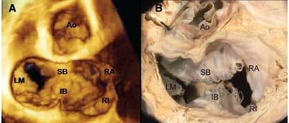
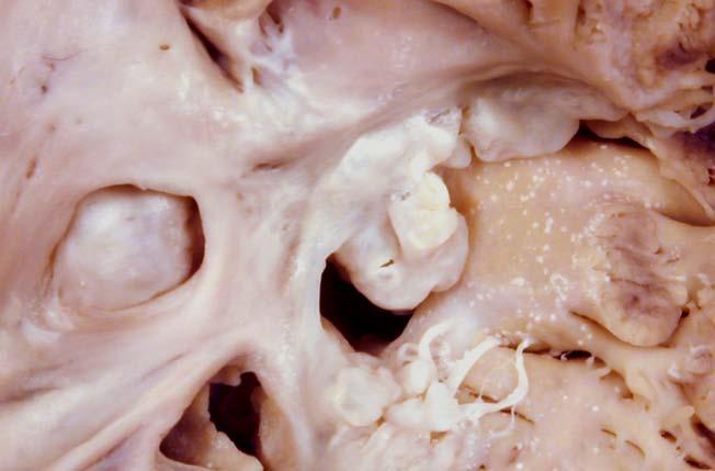
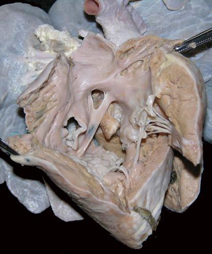
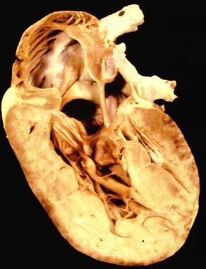
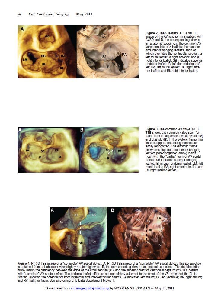
6 Pathology of AVSD PA The Atrioventricular Junction is Common in all cases of AV Canal Defects AO Courtesy of Robert Anderson The aorta is "Sprung" out of its usual position because of the common AV Junction. NHS. ML SBL IBL CS Ant.TL IL

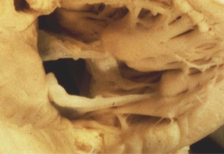
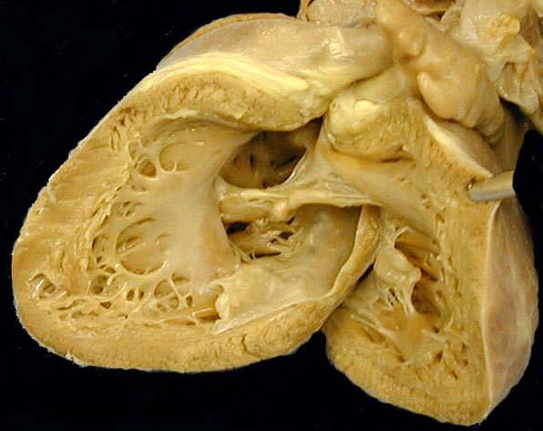
7 Separate valvar orifices For RV & LV Features of AV Canal Courtesy of Diane Spicer.


8 Features of AV Canal The phenotypic feature is the common atrioventricular junction Courtesy of Diane Spicer.


9 Features of AV Canal The phenotypic feature is the common atrioventricular junction Courtesy of Diane Spicer.


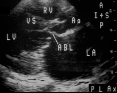
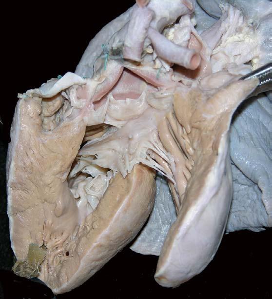
10 The Gooseneck Deformity Inlet-outlet disproportion is a feature of all AV Canal Defecgts


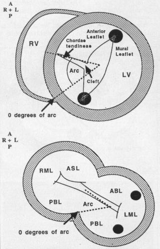
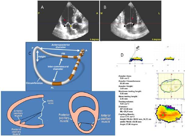
11 Cleft Valve and Papillary Muscle Position As papillary muscles support commisures
12 Goals of Ultrasound 1. Define the extent of the atrial communication. 2. Define the type and extent of the ventricular communications. 3. Demonstrate the valve morphology attachments and function. 4. Display the shunting patterns, the magnitude of the shunt. 5. Type of atrioventricular valve regurgitation, magnitude position and direction. 6. Assess the commitment of the atrioventricular junction to the underlying ventricular mass and the size of the underlying ventricle (balance). 7. Recognize associated anomalies.


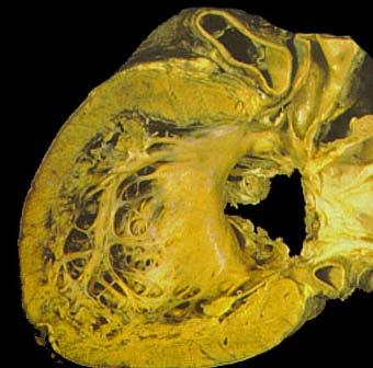
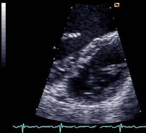
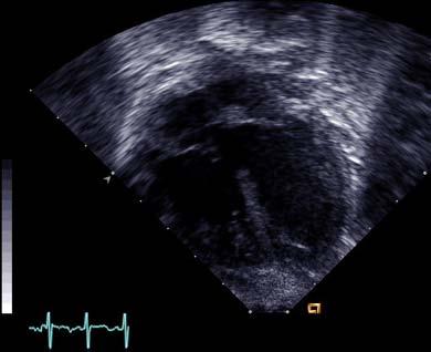
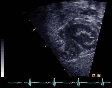
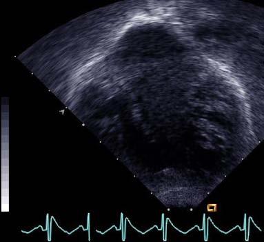
13 Type C AVSD - UCSF


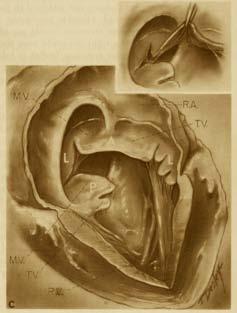
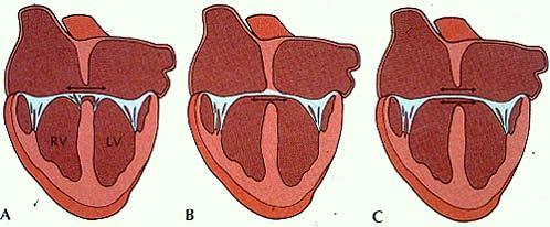
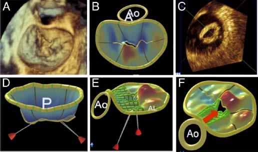
14 Mayo Clinic Drawings Rastelli A Rastelli B Rastelli C

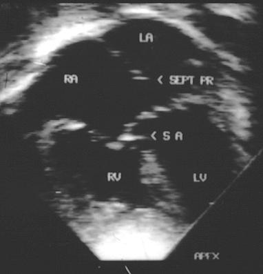
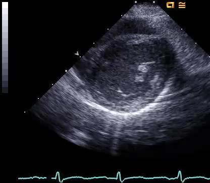
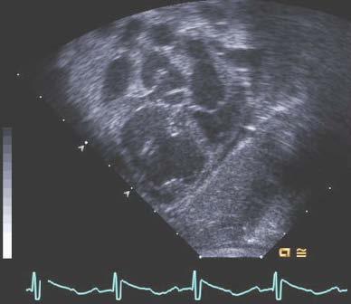
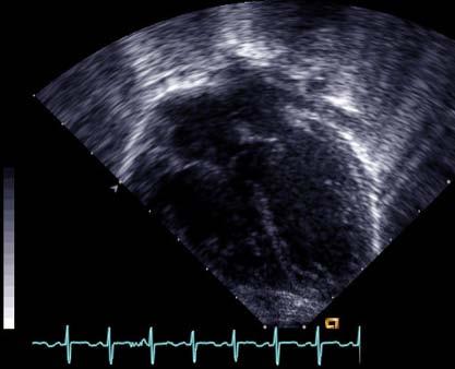
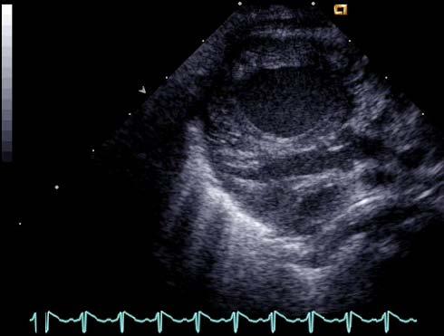
15 AVSD Concept by Echo


16 Rastelli Classification- AVSD B A C


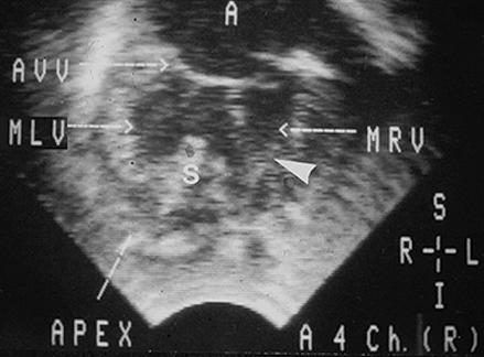
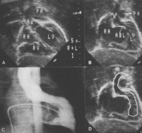
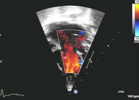
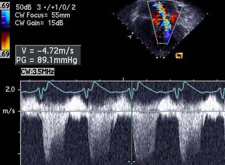
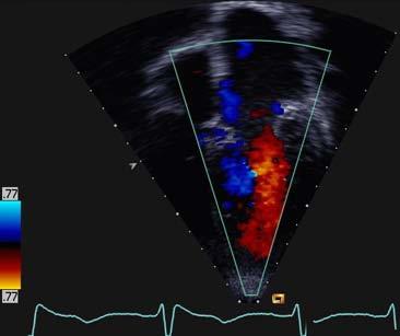
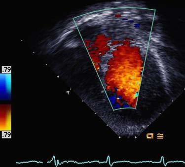
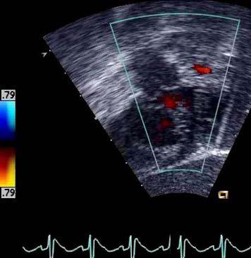
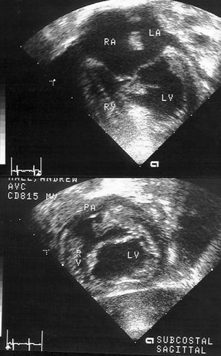
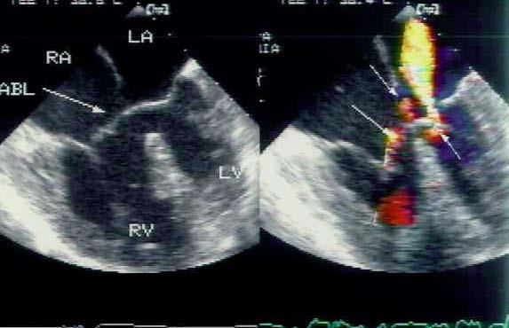
17 Rastelli Type A PA LV RV Subcostal Oblique


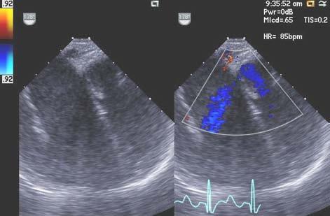
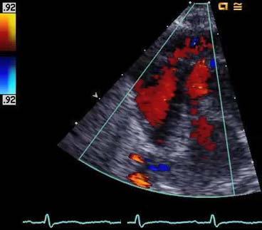
18 Rastelli Type C AO PA LV RV Subcostal Oblique

19 LV Outflow Obstruction

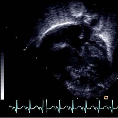
20 The Gooseneck Deformity

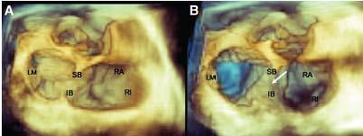
21 AV Septal Defect 3-D Left Side

22 AV Septal Defect 3-D Right Side

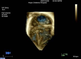
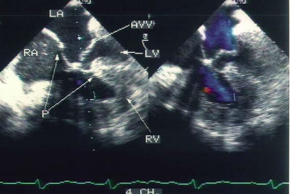
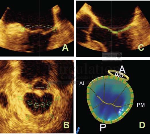
23 3 D Zoom Technique

24 TYPE A, AVSD: A V Valvar Regurgitation ASD2 ASD1 RA LA A 4 Ch. RV LV PV

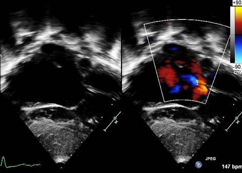
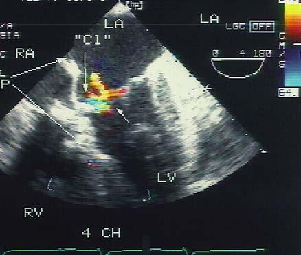
25 AVSD Type C : AV valve regurgitation RA LA RV LV A 4 Ch.
26 Entrainment
27 Left Ventricular -Right Atrial Shunt
28 Atrioventricular Septal Defects: Left AV Valvar Leakage

29 Atrioventricular Septal Defects:Associations- Sub AS


30 Ostium Primum ASD. (Same Patient)

31 Ostium Primum ASD.


32 The connecting tongue and the Partial Canal


33 Partial Atrioventricular Septal Defects:Associations - The Tricuspid Pouch Lesion

34 Atrioventricular Septal Defects:Associations - The Tricuspid Pouch Lesion

35 Cleft in the Left Atrioventricular Valve


36 NHS. The So-Called Cleft Mitral Valve


37 The so-called Cleft Mitral Valve


38 Left Atrioventricular Valve & Regurgitation 3D Normal Mitral Valve 3D Cleft Left Atrioventricular Valve Surgical View of Cleft Courtesy of Jeff. Smallhorn
39 New Contributions of 3 D Echocardiography.


40 The So-called Cleft Mitral Valve
41 3 D Zoom Technique


42 Single papillary muscle as a risk factor


43 Single left ventricular papillary muscle Parachute MV RV LV Subcostal Sagittal View
44 Isolated ventricular component of an AVSD
45 Isolated ventricular component of an AVSD

46 Isolated ventricular component of an AVSD
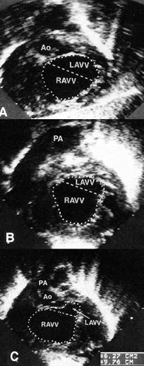
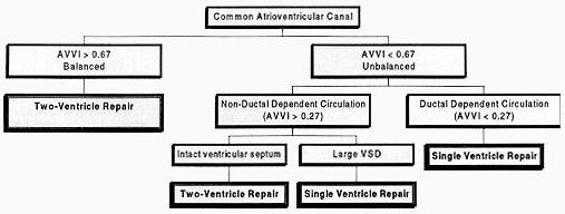
47 Isolated ventricular component of an AVSD

48 AVSD :Secundum ASD Type A. AVSD LA RA LV RV Subcostal Coronal

49 AVSD: Parasternal Short Axis of VSD VSD RV PA RA LA AO
50 Atrioventricular Septal Defects: Associations- DORV
51 Atrioventricular Septal Defects:Associations- Tetralogy of Fallot


52 Dextrocardia, Right Isomerism and R Dom. AVSD ASD2 ASD1 RA LA RV LV R Apex

53 Type A AVSD. Left Isomerism A MRV MLV Apex Rt
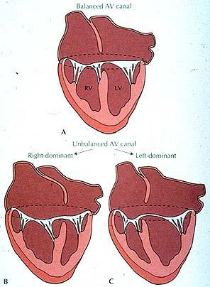

54 Unbalanced Atrioventricular Septal Defects

55 Left Dominant AVSD When is the right ventricle too small? This is less clear than for right ventricular dominance. Options include: cheating on the patch a one and a half ventricle repair single ventricle repair
56 Atrioventricular Septal Defects:Right Dominant Associations
57 Atrioventricular Septal Defects:Right Dominant Associations

58 Left Dominant AVSD

59 L Dominant AVSD RA LA EFF RV LV

60 Unbalanced Atrioventricular Canal Defects Repair related to:- Size of the ventricle Volume Length Size of the amount of tissue over the valve Area Quality of the valvar tissue The presence of associated defects Outflow tract Vascular abnormalities NHS.

61 Philosophy for Repair of Unbalanced AVSD s Relative advantages of a one vs. a two ventricle repair. Is a prosthetic valve worse or better than a one ventricle repair. Are there extraneous factors which would prevent a two ventricle repair.
62 Left Dominant AVSD When is the right ventricle too small? This is less clear than for right ventricular dominance. Options include: cheating on the patch a one and a half ventricle repair single ventricle repair
 RA LA CS RV A 4")
63 R. DOM. AVSD ( L. ISOMERISM) RA LA CS RV A 4 Ch. (L) LV

64 Left Isomerism, AVSD, (Rt. Dom.) PA AVVO LV RV
65 NHS. Questionable LV Size? Cor Triatriatum


66 Short axis change pre- and postop. TAPVR

67 Preoperative/postoperative echo in unbalanced Van Son & Phoon


68 Indexed LVEDV= 14.8±9.1 ml /m 2 Potential Indexed LV EDV= 32.2±18.8 Dimension Measurements Van Son & Phoon Indexed LV long axis dimension =8.9±1.4 cm/m 2 RV/LV Long axis ratio= 0.65±0.1 L AV Valve/Total Valve Ratio = 0.30±0.06 LV/RV Area Ratio= 0.27±0.01 NHS.
69 Postoperative echocardiographic dimensions Postoperative dimensions LV EDV Index 35.6± 3.9 ml. /m 2 LV/RV long ratio 0.88±0.11 L AV Valve / total Valve ratio = 0.42± 0.03 LV/RV area ratio= 0.88 ± 0.18 /m 2 Van Son, Phoon et al. Ann Thorac Surg. 1997;63:1567
70 NHS. LV Volume change real and calculated
71 NHS. Unbalanced AVSD. Cohen et al.

72 Balance :Summary! Balance is a complex issue! Treatment is based on several factors:! morphological factors! direct vs. indirect! surgical preference! philosophical factors! Echocardiography provides one of many determinants! Inspection of the morphology at surgery is the final point of arbitration. NHS

73 Conclusion Balance is a complex issue Treatment is based on several factors: morphological factors direct vs. indirect surgical preference philosophical factors Echocardiography provides one of many determinants Inspection of the morphology at surgery is the final point of arbitration. NHS
74 Postoperative Findings

75 Atrioventricular Septal Defects: Postop. L AV Valve
76 AV Valve Regurgitation- Pre- and Post Op


77 Tetralogy and AVSD ( Type C ): Pre & Post Repair


78 AVSD Postop. Contrast & Color Doppler Studies Subcostal Coronal View DAO RA LA LV RV


79 Postoperative breakdown of left AV valve repair Subcostal coronal view RA LA RV LV
 P Lax RV LV Cl")
80 Dehisced Sutured Commisure ( Cleft ) P Lax RV LV Cl LA

81 Postop AVVR

82 Postop AVVR

83 NHS. AV Regurgitation & Stenosis

84 NHS. Postoperative Pearls- The AV Septal Defect
85 Annuloplasty

86 Postop AVVR

87 The Normal and Abnormal (Mitral) Annulus Effect of Annular Shape on Leaflet Curvature in Reducing Mitral Leaflet Stress Ivan S. Salgo,
88 Changes in Areal Strain in 3 Shape Valriations.
89 The End
90 .
91 The End

92 Goals of Ultrasound 1. Define the extent of the atrial communication. 2. Define the type and extent of the ventricular communications. 3. Demonstrate the valve morphology attachments and function. 4. Display the shunting patterns, the magnitude of the shunt. 5. Type of atrioventricular valve regurgitation, magnitude position and direction. 6. Assess the commitment of the atrioventricular junction to the underlying ventricular mass and the size of the underlying ventricle (balance). 7. Recognize associated anomalies.
93 Thank You!
Anatomy of Atrioventricular Septal Defect (AVSD)
") Surgical challenges in atrio-ventricular septal defect in grown-up congenital heart disease Anatomy of Atrioventricular Septal Defect (AVSD) S. Yen Ho Professor of Cardiac Morphology Royal Brompton and
Surgical challenges in atrio-ventricular septal defect in grown-up congenital heart disease Anatomy of Atrioventricular Septal Defect (AVSD) S. Yen Ho Professor of Cardiac Morphology Royal Brompton and
Most common fetal cardiac anomalies
 Most common fetal cardiac anomalies Common congenital heart defects CHD % of cardiac defects Chromosomal Infants Fetuses anomaly (%) 22q11 deletion (%) VSD 30 5~10 20~40 10 PS 9 5 (PA w/ VSD) HLHS 7~9
ECHOCARDIOGRAPHIC APPROACH TO CONGENITAL HEART DISEASE: THE UNOPERATED ADULT
 ECHOCARDIOGRAPHIC APPROACH TO CONGENITAL HEART DISEASE: THE UNOPERATED ADULT Karen Stout, MD, FACC Divisions of Cardiology University of Washington Medical Center Seattle Children s Hospital NO DISCLOSURES
Cardiac Catheterization Cases Primary Cardiac Diagnoses Facility 12 month period from to PRIMARY DIAGNOSES (one per patient)
") PRIMARY DIAGNOSES (one per patient) Septal Defects ASD (Atrial Septal Defect) PFO (Patent Foramen Ovale) ASD, Secundum ASD, Sinus venosus ASD, Coronary sinus ASD, Common atrium (single atrium) VSD (Ventricular
PRIMARY DIAGNOSES (one per patient) Septal Defects ASD (Atrial Septal Defect) PFO (Patent Foramen Ovale) ASD, Secundum ASD, Sinus venosus ASD, Coronary sinus ASD, Common atrium (single atrium) VSD (Ventricular
Adult Congenital Heart Disease: What All Echocardiographers Should Know Sharon L. Roble, MD, FACC Echo Hawaii 2016
 1 Adult Congenital Heart Disease: What All Echocardiographers Should Know Sharon L. Roble, MD, FACC Echo Hawaii 2016 DISCLOSURES I have no disclosures relevant to today s talk 2 Why should all echocardiographers
Giovanni Di Salvo MD, PhD, FESC Second University of Naples Monaldi Hospital
 Giovanni Di Salvo MD, PhD, FESC Second University of Naples Monaldi Hospital VSD is one of the most common congenital cardiac abnormalities in the newborn. It can occur as an isolated finding or in combination
Double outlet right ventricle: navigation of surgeon to chose best treatment strategy
 Double outlet right ventricle: navigation of surgeon to chose best treatment strategy Jan Marek Great Ormond Street Hospital & Institute of Cardiovascular Sciences, University College London Double outlet
Congenital Heart Disease An Approach for Simple and Complex Anomalies
 Congenital Heart Disease An Approach for Simple and Complex Anomalies Michael D. Pettersen, MD Director, Echocardiography Rocky Mountain Hospital for Children Denver, CO None Disclosures 1 ASCeXAM Contains
Atrial Septal Defects
 Supplementary ACHD Echo Acquisition Protocol for Atrial Septal Defects The following protocol for echo in adult patients with atrial septal defects (ASDs) is a guide for performing a comprehensive assessment
Anomalous Systemic Venous Connection Systemic venous anomaly
 World Database for Pediatric and Congenital Heart Surgery Appendix B: Diagnosis (International Paediatric and Congenital Cardiac Codes (IPCCC) and definitions) Anomalous Systemic Venous Connection Systemic
Characteristics and Management of Cleft Mitral Valve
 Journal of the American College of Cardiology Vol. 42, No. 11, 2003 2003 by the American College of Cardiology Foundation ISSN 0735-1097/03/$30.00 Published by Elsevier Inc. doi:10.1016/j.jacc.2003.07.019
Cardiac ultrasound protocols
 Cardiac ultrasound protocols IDEXX Telemedicine Consultants Two-dimensional and M-mode imaging planes Right parasternal long axis four chamber Obtained from the right side Displays the relative proportions
"Lecture Index. 1) Heart Progenitors. 2) Cardiac Tube Formation. 3) Valvulogenesis and Chamber Formation. 4) Epicardium Development.
 Heart Progenitors. 2) Cardiac Tube Formation. 3) Valvulogenesis and Chamber Formation. 4) Epicardium Development.") "Lecture Index 1) Heart Progenitors. 2) Cardiac Tube Formation. 3) Valvulogenesis and Chamber Formation. 4) Epicardium Development. 5) Septation and Maturation. 6) Changes in Blood Flow during Development.
"Lecture Index 1) Heart Progenitors. 2) Cardiac Tube Formation. 3) Valvulogenesis and Chamber Formation. 4) Epicardium Development. 5) Septation and Maturation. 6) Changes in Blood Flow during Development.
Techniques for repair of complete atrioventricular septal
 No Ventricular Septal Defect Patch Atrioventricular Septal Defect Repair Carl L. Backer, MD *, Osama Eltayeb, MD *, Michael C. Mongé, MD *, and John M. Costello, MD For the past 10 years, our center has
Transposition of the Great Arteries Preoperative Diagnostic Considerations. John Simpson Evelina Children s Hospital London, UK
 Transposition of the Great Arteries Preoperative Diagnostic Considerations John Simpson Evelina Children s Hospital London, UK Areas to be covered Definitions Scope of occurrence of transposition of the
What is the Definition of Small Systemic Ventricle. Hong Ryang Kil, MD Department of Pediatrics, College of Medicine, Chungnam National University
 What is the Definition of Small Systemic Ventricle Hong Ryang Kil, MD Department of Pediatrics, College of Medicine, Chungnam National University Contents Introduction Aortic valve stenosis Aortic coarctation
ISUOG Basic Training. Obtaining & Interpreting Heart Views Correctly Alfred Abuhamad, USA. Basic training. Editable text here
 ISUOG Basic Training Obtaining & Interpreting Heart Views Correctly Alfred Abuhamad, USA Learning Objectives 6, 7 & 8 At the end of the lecture you will be able to: describe how to assess cardiac situs
Congenital Heart Defects
 Normal Heart Congenital Heart Defects 1. Patent Ductus Arteriosus The ductus arteriosus connects the main pulmonary artery to the aorta. In utero, it allows the blood leaving the right ventricle to bypass
Segmental approach to normal and abnormal situs arrangement - Echocardiography -
 Segmental approach to normal and abnormal situs arrangement - Echocardiography - Jan Marek Great Ormond Street Hospital & Institute of Cardiovascular Sciences, University College London No disclosures
List of Videos. Video 1.1
 Video 1.1 Video 1.2 Video 1.3 Video 1.4 Video 1.5 Video 1.6 Video 1.7 Video 1.8 The parasternal long-axis view of the left ventricle shows the left ventricular inflow and outflow tract. The left atrium
Pediatric Echocardiography Examination Content Outline
 Pediatric Echocardiography Examination Content Outline (Outline Summary) # Domain Subdomain Percentage 1 Anatomy and Physiology Normal Anatomy and Physiology 10% 2 Abnormal Pathology and Pathophysiology
Atrioventricular valve repair: The limits of operability
 Atrioventricular valve repair: The limits of operability Francis Fynn-Thompson, MD Co-Director, Center for Airway Disorders Surgical Director, Pediatric Mechanical Support Program Surgical Director, Heart
ADULT CONGENITAL HEART DISEASE. Stuart Lilley
 ADULT CONGENITAL HEART DISEASE Stuart Lilley More adults than children have congenital heart disease Huge variety of congenital lesions from minor to major Heart failure, re-operation and arrhythmia are
Spectrum of Cardiac Lesions Associated with Isolated Cleft Mitral Valve and their Impact on Therapeutic Choices
 Spectrum of Cardiac Lesions Associated with Isolated Cleft Mitral Valve and their Impact on Therapeutic Choices Ayoub El hammiri, Abdenasser Drighil, Sanaa Benhaourech Cardiology Department, Ibn Rochd
Heart and Lungs. LUNG Coronal section demonstrates relationship of pulmonary parenchyma to heart and chest wall.
 Heart and Lungs Normal Sonographic Anatomy THORAX Axial and coronal sections demonstrate integrity of thorax, fetal breathing movements, and overall size and shape. LUNG Coronal section demonstrates relationship
Tetralogy of Fallot (TOF) with atrioventricular (AV)
 with atrioventricular (AV)") Tetralogy of Fallot with Atrioventricular Canal Defect: Two Patch Repair Sitaram M. Emani, MD, and Pedro J. del Nido, MD Tetralogy of Fallot (TOF) with atrioventricular (AV) canal defect is classified
Tetralogy of Fallot with Atrioventricular Canal Defect: Two Patch Repair Sitaram M. Emani, MD, and Pedro J. del Nido, MD Tetralogy of Fallot (TOF) with atrioventricular (AV) canal defect is classified
Segmental Analysis. Gautam K. Singh, M.D. Washington University School of Medicine St. Louis
 Segmental Analysis Gautam K. Singh, M.D. Washington University School of Medicine St. Louis Segmental Analysis Segmental Analysis: From Veins to Ventricles Segmental Approach to Evaluation of Congenital
Appendix A.1: Tier 1 Surgical Procedure Terms and Definitions
 Appendix A.1: Tier 1 Surgical Procedure Terms and Definitions Tier 1 surgeries AV Canal Atrioventricular Septal Repair, Complete Repair of complete AV canal (AVSD) using one- or two-patch or other technique,
Echocardiography in Adult Congenital Heart Disease
 Echocardiography in Adult Congenital Heart Disease Michael Vogel Kinderherz-Praxis München CHD missed in childhood Subsequent lesions after repaired CHD Follow-up of cyanotic heart disease CHD missed in
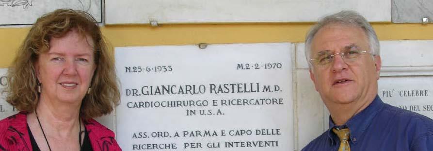
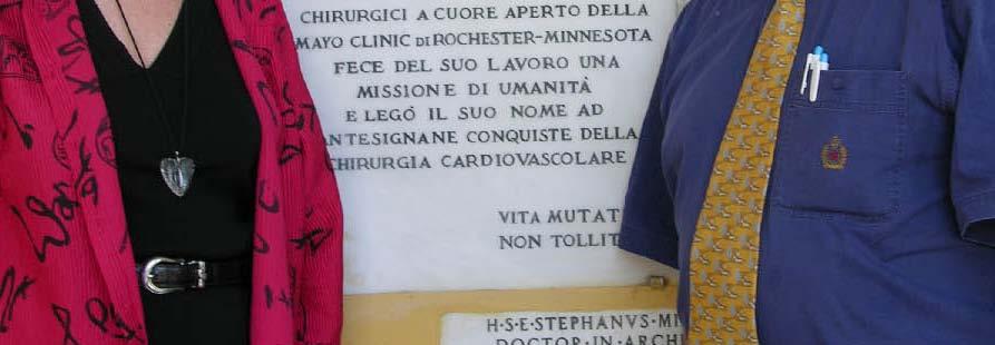
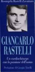
"Giancarlo Rastelli Lecture"
 "Giancarlo Rastelli Lecture" Surgical treatment of Malpositions of the Great Arteries Pascal Vouhé Giancarlo Rastelli (1933 1970) Cliquez pour modifier les styles du texte du masque Deuxième niveau Troisième
Surgical Treatment for Atrioventricular Septal Defect. Masakazu Nakao Consultant, Paediatric Cardiothoracic Surgery
 Surgical Treatment for Atrioventricular Septal Defect Masakazu Nakao Consultant, Paediatric Cardiothoracic Surgery 1 History Rastelli classification (Rastelli) Pulmonary artery banding (Muller & Dammann)
NASCI 2012 Segmental Analysis
 NASCI 2012 Segmental Analysis Frandics Chan, M.D., Ph.D. Stanford University Medical Center Lucile Packard Department Children s of Radiology Hospital Menagerie of Congenital Cardiac Lesions 1. Absent
The role of intraoperative TOE in congenital cardiac surgery
 The role of intraoperative TOE in congenital cardiac surgery Justiaan Swanevelder Dept of Anaesthesia Groote Schuur and Red Cross War Memorial Children s Hospitals University of Cape Town, South Africa
FUNCTIONALLY SINGLE VENTRICLE
 MORPHOLOGICAL DETERMINANTS VI TRAN EuroEcho, Budapest, 7 th December 2011 DECLARATION OF CONFLICT OF INTEREST: I have nothing to declare What is the functionally single ventricle? The heart that is incapable
Congenital Heart Disease: Physiology and Common Defects
 Congenital Heart Disease: Physiology and Common Defects Jamie S. Sutherell, M.D, M.Ed. Associate Professor, Pediatrics Division of Cardiology Director, Medical Student Education in Pediatrics Director,
CASE REPORT: DOUBLE ORIFICE MITRAL VALVE WITH CLEFT IN ANTERIOR LEAFLET OF DOMINANT VALVE IN AN AFRO-CARIBBEAN
 CASE REPORT: DOUBLE ORIFICE MITL VAE WITH CLEFT IN ANTERIOR LEAFLET OF DOMINANT VAE IN AN AFRO-CARIBBEAN Disclosure: No potential conflict of interest. Received: 27.08.13 Accepted: 23.06.14 Citation: EMJ
after AV Canal Repair: When and How To Intervene
 Left Atrioventricular Valve Regurgitation after AV Canal Repair: When and How To Intervene Thomas L Spray, M.D. Chief, Cardiothoracic Surgery Alice Langdon Warner Endowed Chair The Children s Hospital
LEFT VENTRICULAR OUTFLOW OBSTRUCTION WITH A VSD: OPTIONS FOR SURGICAL MANAGEMENT
 LEFT VENTRICULAR OUTFLOW OBSTRUCTION WITH A VSD: OPTIONS FOR SURGICAL MANAGEMENT 10-13 March 2017 Ritz Carlton, Riyadh, Saudi Arabia Zohair AlHalees, MD Consultant, Cardiac Surgery Heart Centre LEFT VENTRICULAR
Tetralogy of Fallot (TOF) repair, Ventriculotomy Coarctation repair, Other
 repair, Ventriculotomy Coarctation repair, Other") Tier 1 Surgery Form Date of Surgery DD/MM/YYYY Primary Cardiac Procedure Select the patient's primary surgical procedure. If the patient has multiple operating room visits, these should be reported on
Tier 1 Surgery Form Date of Surgery DD/MM/YYYY Primary Cardiac Procedure Select the patient's primary surgical procedure. If the patient has multiple operating room visits, these should be reported on
CARDIAC DEVELOPMENT CARDIAC DEVELOPMENT
 CARDIAC DEVELOPMENT CARDIAC DEVELOPMENT Diane E. Spicer, BS, PA(ASCP) University of Florida Dept. of Pediatric Cardiology Curator Van Mierop Cardiac Archive This lecture is given with special thanks to
Complete atrioventricular septal defect identified during routine 19 week fetal anomaly ultrasound
 CASE REPORT Complete atrioventricular septal defect identified during routine Mark Hare Royal Brisbane and Women s Hospital, Herston, QLD, Australia doi:10.1002/sono.12014 Introduction Complete atrioventricular
Preoperative Echocardiographic Assessment of Uni-ventricular Repair
 Preoperative Echocardiographic Assessment of Uni-ventricular Repair Salem Deraz, MD Pediatric Cardiologist, Aswan Heart Centre Magdi Yacoub Heart Foundation Uni-ventricular repair A single or series of
Yuichi Shiokawa, MD Anton E. Becker, MD, PhD
 THE LEFT VENTRICULAR OUTFLOW TRACT IN ATRIOVENTRICULAR SEPTAL DEFECT REVISITED: SURGICAL CONSIDERATIONS REGARDING PRESERVATION OF AORTIC VALVE INTEGRITY IN THE PERSPECTIVE OF ANATOMIC OBSERVATIONS Yuichi
September 26, 2012 Philip Stockwell, MD Lifespan CVI Assistant Professor of Medicine (Clinical)
") September 26, 2012 Philip Stockwell, MD Lifespan CVI Assistant Professor of Medicine (Clinical) Advances in cardiac surgery have created a new population of adult patients with repaired congenital heart
September 26, 2012 Philip Stockwell, MD Lifespan CVI Assistant Professor of Medicine (Clinical) Advances in cardiac surgery have created a new population of adult patients with repaired congenital heart
Repair of Complete Atrioventricular Septal Defects Single Patch Technique
 Repair of Complete Atrioventricular Septal Defects Single Patch Technique Fred A. Crawford, Jr., MD The first repair of a complete atrioventricular septal defect was performed in 1954 by Lillehei using
I have nothing to disclose.
 I have nothing to disclose. New approaches in tricuspid valve repair Christian Schreiber ..more than a simple displacement.., the valvar orifice is formed within the ventricular cavity.. Ebstein Historical
Morphological evaluation of atrioventricular septal defects by magnetic resonance imaging
 138 Br Heart J 1990;64:138-45 Morphological evaluation of atrioventricular septal defects by magnetic resonance imaging Department of Paediatric Cardiology, Guy's Hospital,. London J M Parsons E J Baker
Echocardiographic assessment in Adult Patients with Congenital Heart Diseases
 Echocardiographic assessment in Adult Patients with Congenital Heart Diseases Athanasios Koutsakis Cardiologist, Cl. Research Fellow George Giannakoulas Ass. Professor in Cardiology 1st Cardiology Department,
ULTRASOUND OF THE FETAL HEART
 ULTRASOUND OF THE FETAL HEART Cameron A. Manbeian, MD Disclosure Statement Today s faculty: Cameron Manbeian, MD does not have any relevant financial relationships with commercial interests or affiliations
Aortic Arch Abnormalities
 Aortic Arch Abnormalities IPOK Norman H Silverman MD, D Sc (Med.). FACC, FAHA. Stanford University & Lucile Packard Children s Hospital E mail: norm.silverman@stanford.edu. NHS. www.md1world.com Abnormalities
Common Defects With Expected Adult Survival:
 Common Defects With Expected Adult Survival: Bicuspid aortic valve :Acyanotic Mitral valve prolapse Coarctation of aorta Pulmonary valve stenosis Atrial septal defect Patent ductus arteriosus (V.S.D.)
Unbalanced AVC: When is it Time to Bail?
 Unbalanced AVC: When is it Time to Bail? David M. Overman Division of Pediatric Cardiac Surgery The Children s Heart Clinic Chief, Division of Cardiovascular Surgery Children s Hospitals and Clinics of
Cases in Adult Congenital Heart Disease
 Cases in Adult Congenital Heart Disease Sabrina Phillips, MD FACC FASE Associate Professor of Medicine The University of Oklahoma Health Sciences Center No Disclosures I Have Palpitations 18 Year old Man
What is Ebstein Anomaly?
 Echocardiograpnhic Evaluation of : Definition, Detection and Determinants of Outcome P. W. O Leary, M.D. Division of Pediatric Cardiology Mayo Clinic No Conflicts to Disclose What is? Failure of the TV
cardiac imaging planes planning basic cardiac & aortic views for MR
 cardiac imaging planes planning basic cardiac & aortic views for MR Dianna M. E. Bardo, M. D. Assistant Professor of Radiology & Cardiovascular Medicine Director of Cardiac Imaging cardiac imaging planes
가천의대길병원소아심장과최덕영 PA C IVS THE EVALUATION AND PRINCIPLES OF TREATMENT STRATEGY
 가천의대길병원소아심장과최덕영 PA C IVS THE EVALUATION AND PRINCIPLES OF TREATMENT STRATEGY PA c IVS (not only pulmonary valve disease) Edwards JE. Pathologic Alteration of the right heart. In: Konstam MA, Isner M, eds.
When to close an Atrial Septal Defect (ASD) in adulthood?
 in adulthood?") When to close an Atrial Septal Defect (ASD) in adulthood? Philippe ALDEBERT Hôpital de la Timone, CHU Marseille Département de cardiologie pédiatrique et congénitale médico-chirurgical Abbott Incidence
When to close an Atrial Septal Defect (ASD) in adulthood? Philippe ALDEBERT Hôpital de la Timone, CHU Marseille Département de cardiologie pédiatrique et congénitale médico-chirurgical Abbott Incidence
TGA Surgical techniques: tips & tricks (Arterial switch operation)
") TGA Surgical techniques: tips & tricks (Arterial switch operation) Seoul National University Children s Hospital Woong-Han Kim Surgical History 1951 Blalock and Hanlon, atrial septectomy 1954 Mustard et
TGA Surgical techniques: tips & tricks (Arterial switch operation) Seoul National University Children s Hospital Woong-Han Kim Surgical History 1951 Blalock and Hanlon, atrial septectomy 1954 Mustard et
pulmonary valve on, 107 pulmonary valve vegetations on, 113
 INDEX Adriamycin-induced cardiomyopathy, 176 Amyloidosis, 160-161 echocardiographic abnormalities in, 160 intra-mural tumors similar to, 294 myocardial involvement in, 160-161 two-dimensional echocardiography
Echocardiography in adult congenital heart disease
 S12 Department of Cardiology, Royal Hospital for Sick Children, Glasgow G3 8SJ, UK A Houston S Lilley T Richens University Department of Medicine and Therapeutics, Western Infirmary, Glasgow G11 6NT, UK
The background of the Cardiac Sonographer Network News masthead is a diagnostic image:
 Number 5 Welcome Number 5 Welcome to the newsletter created just for you: sonographers who perform pediatric echocardiograms in primarily adult echo labs. Each issue features tips on echocardiography of
Fetal Echocardiography and the Routine Obstetric Sonogram
 JDMS 23:143 149 May/June 2007 143 Fetal Echocardiography and the Routine Obstetric Sonogram SHELLY ZIMBELMAN, RT(R)(CT), RDMS, RDCS ASAD SHEIKH, MD, RDCS Congenital heart disease (CHD) is the most common
An anterior aortoventriculoplasty, known as the Konno-
 The Konno-Rastan Procedure for Anterior Aortic Annular Enlargement Mark E. Roeser, MD An anterior aortoventriculoplasty, known as the Konno-Rastan procedure, is a useful tool for the cardiac surgeon. Originally,
Double Outlet Right Ventricle with Anterior and Left-Sided Aorta and Subpulmonary Ventricular Septal Defect
 Case Report Double Outlet Right Ventricle with Anterior and Left-Sided rta and Subpulmonary Ventricular Septal Defect Luciana Braz Peixoto, Samira Morhy Borges Leal, Carlos Eduardo Suaide Silva, Sandra
Recently, real-time 3-dimensional (3D) echocardiography (RT-
 echocardiography (RT-") CONGENTIAL HEART DISEASE Value of Real-Time 3-Dimensional Echocardiography Sectional Diagnosis in Complex Congenital Heart Disease Evaluated by Receiver Operating Characteristic Analysis Guo-zhen Chen,
CONGENTIAL HEART DISEASE Value of Real-Time 3-Dimensional Echocardiography Sectional Diagnosis in Complex Congenital Heart Disease Evaluated by Receiver Operating Characteristic Analysis Guo-zhen Chen,
Mitral Valve Disease, When to Intervene
 Mitral Valve Disease, When to Intervene Swedish Heart and Vascular Institute Ming Zhang MD PhD Interventional Cardiology Structure Heart Disease Conflict of Interest None Current ACC/AHA guideline Stages
Imaging Assessment of Aortic Stenosis/Aortic Regurgitation
 Imaging Assessment of Aortic Stenosis/Aortic Regurgitation Craig E Fleishman, MD FACC FASE The Heart Center at Arnold Palmer Hospital for Children, Orlando SCAI Fall Fellows Course 2014 Las Vegas Disclosure
Adult Echocardiography Examination Content Outline
 Adult Echocardiography Examination Content Outline (Outline Summary) # Domain Subdomain Percentage 1 2 3 4 5 Anatomy and Physiology Pathology Clinical Care and Safety Measurement Techniques, Maneuvers,
By Dickens ATURWANAHO & ORIBA DAN LANGOYA MAKchs, MBchB CONGENTAL HEART DISEASE
 By Dickens ATURWANAHO & ORIBA DAN LANGOYA MAKchs, MBchB CONGENTAL HEART DISEASE Introduction CHDs are abnormalities of the heart or great vessels that are present at birth. Common type of heart disease
Complex Congenital Heart Disease in Adults
 Complex Congenital Heart Disease in Adults Linda B. Haramati, MD Disclosures Complex Congenital Heart Disease in Adults Linda B. Haramati MD, MS Jeffrey M. Levsky MD, PhD Meir Scheinfeld MD, PhD Department
Hypoplastic Left Heart Syndrome: Echocardiographic Assessment
 Hypoplastic Left Heart Syndrome: Echocardiographic Assessment Craig E Fleishman, MD, FACC, FASE Director, Non-invasive Cardiac Imaging The Hear Center at Arnold Palmer Hospital for Children, Orlando SCAI
Journal of American Science 2015;11(4)
") Atrioventricular Canal Anatomy and Differences from Normal Hearts, a Systemic Review of Literatures Walid AE Hammad* 1, M-Wael Badawi 1, Ahmet Arnaz 2, Ahmed Allam 3, Mohamed Elghanam 3, Ahmed Mostafa
Atrioventricular Canal Anatomy and Differences from Normal Hearts, a Systemic Review of Literatures Walid AE Hammad* 1, M-Wael Badawi 1, Ahmet Arnaz 2, Ahmed Allam 3, Mohamed Elghanam 3, Ahmed Mostafa
We present the case of an asymptomatic, 75-year-old
 Images in Cardiovascular Medicine Asymptomatic Rupture of the Left Ventricle Lech Paluszkiewicz, MD; Stefan Ożegowski, MD; Mohammad Amin Parsa, MD; Jan Gummert, PhD, MD We present the case of an asymptomatic,
CMR for Congenital Heart Disease
 CMR for Congenital Heart Disease * Second-line tool after TTE * Strengths of CMR : tissue characterisation, comprehensive access and coverage, relatively accurate measurements of biventricular function/
Unbalanced Atrioventricular Septal Defect A Congenital Heart Surgeons Society Inception Cohort Study
 Unbalanced Atrioventricular Septal Defect A Congenital Heart Surgeons Society Inception Cohort Study Table of Contents 1. Abstract 2. Specific Aims a. Objectives b. Hypothesis 3. Background and Rationale
Echocardiography in Congenital Heart Disease
 Chapter 44 Echocardiography in Congenital Heart Disease John L. Cotton and G. William Henry Multiple-plane cardiac imaging by echocardiography can noninvasively define the anatomy of the heart and the
Atrioventricular Septal Defect Echocardiographic Imaging
 Atrioventricular Septal Defect Echocardiographic Imaging Dr Choo Tze Liang Jonathan MBBS(S pore), FRCPCH (UK), DRCOG (UK), FRCP (Edin), FAMS (Paeds) Senior Consultant Cardiology Service KK Women s and
Anatomically Sound, Simplified Approach to Repair of "Complete" Atrioventricular Septal Defect
 Anatomically Sound, Simplified Approach to Repair of "Complete" Atrioventricular Septal Defect Benson R. Wilcox, MD, David R. Jones, MD, Elman G. Frantz, MD, Lela W. Brink, MD, G. William Henry, MD, Michael
3/14/2011 MANAGEMENT OF NEWBORNS CARDIAC INTENSIVE CARE CONFERENCE FOR HEALTH PROFESSIONALS IRVINE, CA. MARCH 7, 2011 WITH HEART DEFECTS
 CONFERENCE FOR HEALTH PROFESSIONALS IRVINE, CA. MARCH 7, 2011 MANAGEMENT OF NEWBORNS WITH HEART DEFECTS A NTHONY C. CHANG, MD, MBA, MPH M E D I C AL D I RE C T OR, HEART I N S T I T U T E C H I LDRE N
Outflow Tracts Anomalies
 Diagnosis of Outflow Tract Anomalies in the Fetus General Framing D.Paladini Fetal Medicine & Surgery Unit Gasllini Children s Hospital - Genoa dariopaladini@ospedale-gaslini.ge.it Outflow Tracts Anomalies
Data Collected: June 17, Reported: June 30, Survey Dates 05/24/ /07/2010
 Job Task Analysis for ARDMS Pediatric Echocardiography Data Collected: June 17, 2010 Reported: Analysis Summary For: Pediatric Echocardiography Exam Survey Dates 05/24/2010-06/07/2010 Invited Respondents
Heart and Soul Evaluation of the Fetal Heart
 Heart and Soul Evaluation of the Fetal Heart Ivana M. Vettraino, M.D., M.B.A. Clinical Associate Professor, Michigan State University College of Human Medicine Objectives Review the embryology of the formation
A New Radiopaque Surgical Suture* Juro WADA, M.D. and Masahiro ENDO, M.D.
 A New Radiopaque Surgical Suture* Juro WADA, M.D. and Masahiro ENDO, M.D. SUMMARY We have developed a new X-ray visible suture. It is a polyester suture containing platinum wires. The radiopaque suture
Surgical Treatment for Double Outlet Right Ventricle. Masakazu Nakao Consultant, Paediatric Cardiothoracic Surgery
 for Double Outlet Right Ventricle Masakazu Nakao Consultant, Paediatric Cardiothoracic Surgery 1 History Intraventricular tunnel (Kawashima) First repair of Taussig-Bing anomaly (Kirklin) Taussig-Bing
Adel Hasanin Ahmed 1 ASD
 Adel Hasanin Ahmed 1 ASD Atrial septal defect (ASD) is the commonest form of congenital heart disease seen in adults. The commonest form of defect is the secundum ASD, accounting for two thirds of cases,
MEDICAL MANAGEMENT WITH CAVEATS 1. In one study of 50 CHARGE patients with CHD, 75% required surgery. 2. Children with CHARGE may be resistant to chlo
 CARDIOLOGY IN CHARGE SYNDROME: FOR THE PHYSICIAN Angela E. Lin, M.D. Teratology Program/Active Malformation Surveillance, Brigham and Women's Hospital, Old PBBH-B501, 75 Francis St., Boston, MA 02115 alin@partners.org
M/3, cc-tga, PS, BCPC(+) Double Switch Operation
 Double Switch Operation") 2005 < Pros & Cons > M/3, cc-tga, PS, BCPC(+) Double Switch Operation Congenitally corrected TGA Atrio-Ventricular & Ventriculo-Arterial discordance Physiologically corrected circulation with the morphologic
2005 < Pros & Cons > M/3, cc-tga, PS, BCPC(+) Double Switch Operation Congenitally corrected TGA Atrio-Ventricular & Ventriculo-Arterial discordance Physiologically corrected circulation with the morphologic
Making Sense of Cardiac Views and Imaging Characteristics for 13 Congenital Heart Defects (CHDs)
") Making Sense of Cardiac Views and Imaging Characteristics for 13 Congenital Heart Defects (CHDs) Manny Gaziano, MD, FACOG obimages.net obimages.net@gmail.com Acknowledgements: Krista Wald, RDMS, sonographer,
Making Sense of Cardiac Views and Imaging Characteristics for 13 Congenital Heart Defects (CHDs) Manny Gaziano, MD, FACOG obimages.net obimages.net@gmail.com Acknowledgements: Krista Wald, RDMS, sonographer,
A brief history of valvular surgery
 Cardiac surgery Valvular heart disease University of Pecs, Medical Faculty Heart Institute A brief history of valvular surgery 1925. Souttar closed mitral commissurotomy 1960. McGoon plasty for mitral
Down Syndrome Medical Interest Group Friday, 12 June Cardiac Surgery in patients with Down Syndrome
 Down Syndrome Medical Interest Group Friday, 12 June 2015 Cardiac Surgery in patients with Down Syndrome Mr. Attilio Lotto, FRCS CTh Congenital Cardiac Surgeon Cardiac surgery in patients with Down syndrome
Late secondary TR after left sided heart disease correction: is it predictibale and preventable
 Late secondary TR after left sided heart disease correction: is it predictibale and preventable Gilles D. Dreyfus Professor of Cardiothoracic surgery Nath J, et al. JACC 2004 PREDICT Incidence of secondary
Notes by Sandra Dankwa 2009 HF- Heart Failure DS- Down Syndrome IE- Infective Endocarditis ET- Exercise Tolerance. Small VSD Symptoms -asymptomatic
 Congenital Heart Disease: Notes. Condition Pathology PC Ix Rx Ventricular septal defect (VSD) L R shuntsdefect anywhere in the ventricle, usually perimembranous (next to the tricuspid valve) 30% 1)small
Repair of very severe tricuspid regurgitation following detachment of the tricuspid valve
 OPEN ACCESS Images in cardiology Repair of very severe tricuspid regurgitation following detachment of the tricuspid valve Ahmed Mahgoub 1, Hassan Kamel 2, Walid Simry 1, Hatem Hosny 1, * 1 Aswan Heart
Congenital Heart Disease Cases
 Congenital Heart Disease Cases Sabrina Phillips, MD FACC FASE Mayo Clinic Congenital Heart Disease Center 2013 MFMER slide-1 No Disclosures 2013 MFMER slide-2 1 CASE 1 2013 MFMER slide-3 63 year old Woman
When should we intervene surgically in pediatric patient with MR?
 When should we intervene surgically in pediatric patient with MR? DR.SAUD A. BAHAIDARAH CONSULTANT, PEDIATRIC CARDIOLOGY ASSISTANT PROFESSOR OF PEDIATRICS HEAD OF CARDIOLOGY AND CARDIAC SURGERY UNIT KAUH
Children with Single Ventricle Physiology: The Possibilities
 Children with Single Ventricle Physiology: The Possibilities William I. Douglas, M.D. Pediatric Cardiovascular Surgery Children s Memorial Hermann Hospital The University of Texas Health Science Center
September 28-30, 2018
 September 28-30, 2018 Course Director Optimizing Detection of Congenital Heart Disease: Important Anatomic Cardiac Regions The Top 5 Critical Anatomic Regions in Fetal Cardiac Imaging Alfred Abuhamad,
Accessory mitral valve tissue causing left ventricular outflow tract obstruction
 Br Heart 1986; 55: 376-80 Accessory mitral valve tissue causing left ventricular outflow tract obstruction W G MELDRUM-HANNA, T B CARTMILL, R E HAWKER, J M CELERMAJER, C M WRIGHT From the Basser Institute
found that some patients without stenotic lesions had blood velocity or pressure measurement across the
 Br Heart J 1985; 53: 640-4 Increased blood velocities in the heart and great vessels of patients with congenital heart disease An assessment of their significance in the absence of valvar stenosis STANLEY
Appendix II: ECHOCARDIOGRAPHY ANALYSIS
 Appendix II: ECHOCARDIOGRAPHY ANALYSIS Two-Dimensional (2D) imaging was performed using the Vivid 7 Advantage cardiovascular ultrasound system (GE Medical Systems, Milwaukee) with a frame rate of 400 frames