Vascular Ultrasound: Current state, current needs, future directions
|
|
|
- Curtis Hamilton
- 6 years ago
- Views:
Transcription
1 Vascular Ultrasound: Current state, current needs, future directions Laurence Needleman, MD Thomas Jefferson University Hospitals Sidney Kimmel Medical College of Thomas Jefferson University
2 Disclosures Member, Intersocietal Accreditation Commission Vascular Testing (unpaid)
3 Overview What tests are done for vascular disease? What is the natural history of vascular disease using ultrasound? What shortcomings exist for US vascular diagnosis? What is the future direction of ultrasound? of vascular ultrasound?

4 Gray scale ultrasound Carotid stent

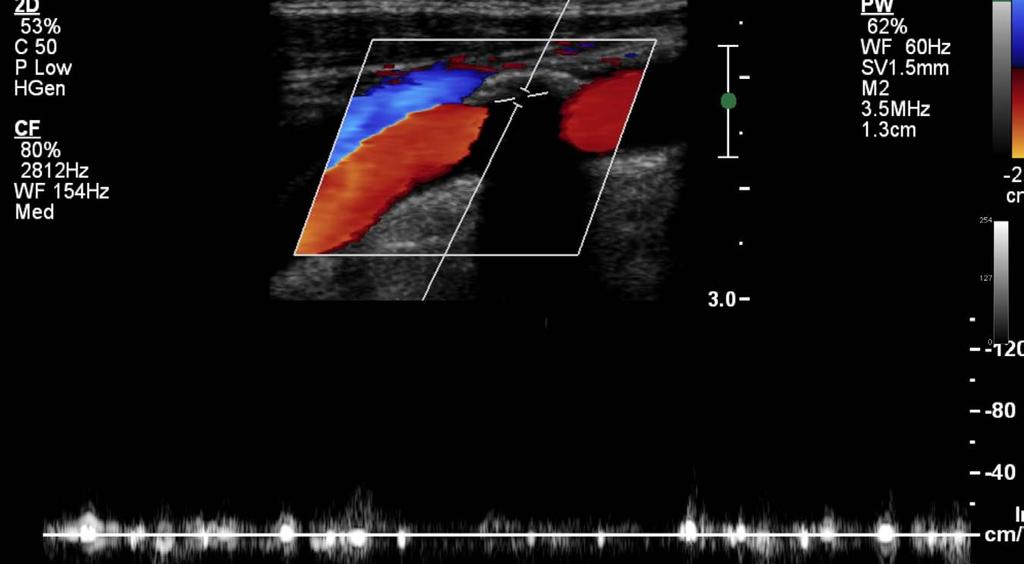
5 Graphical representation of Doppler Makeup Doppler frequency or velocity - y axis Time - x axis Strength of signal - gray scale Number of reflectors Spectral Doppler

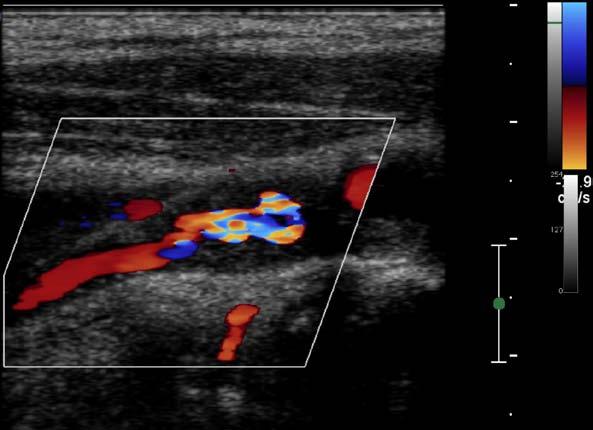
6 Color Doppler Carotid stenosis

7 Color Doppler Artifacts

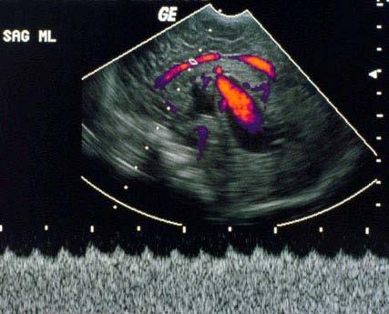
8 Vein of Galen Aneurysm
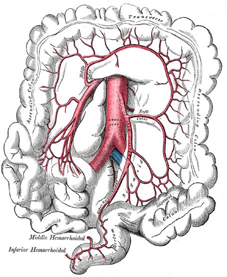
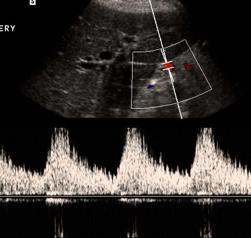
9 Inferior Mesenteric Artery
10 Current state
11 Tests Venous ultrasound obstruction DVT Carotid duplex ultrasound - stenosis Aortic ultrasound - aneurysm Abdominal Doppler Renal arteries - stenosis Liver (portal hypertension) hypertension, flow direction Ovaries and testes increased or decreaed flow, tumors
12 Acting Surgeon General Issues Ca ll to Action to Prevent Deep Vein Th rom b os is a n d Pu lm on a ry Em b olis m FOR IMMEDIATE RELEASE Monday September 15, 2008 Contact: Office of Public Health and Science (202) Acting Surgeon General Steven K. Galson, M.D., M.P.H., today issued a Ca ll to Action to reduce the number of cases of deep vein thrombosis and pulmonary embolism in the United States. Galson urged all Americans to learn about and prevent these treatable conditions. Deep vein thrombosis and pulmonary embolism affect an estimated 350,000 to 600,000 Americans each year, and the numbers are expected to increase as the U.S. population ages. Together, deep vein thrombosis and pulmonary embolism contribute to at least 100,000 deaths each
13 Venous thromboembolic disease Ultrasound is the gold standard to diagnose deep venous thrombosis in the legs CT and NM are the major tests to diagnose its major complication, pulmonary embolism DVT and PE are associated with mortality, diagnosis of cancer, and chronic diseases

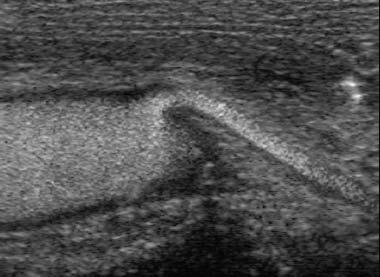
14 Venous US Normal Compression A A Vein
 Scarring Inadequate compression")

15 Noncompressible Vein: Causes Acute venous thrombosis (DVT) Scarring Inadequate compression Noncompressible Deep venous thrombosis Without compression With compression Acute Deep Venous Thrombus

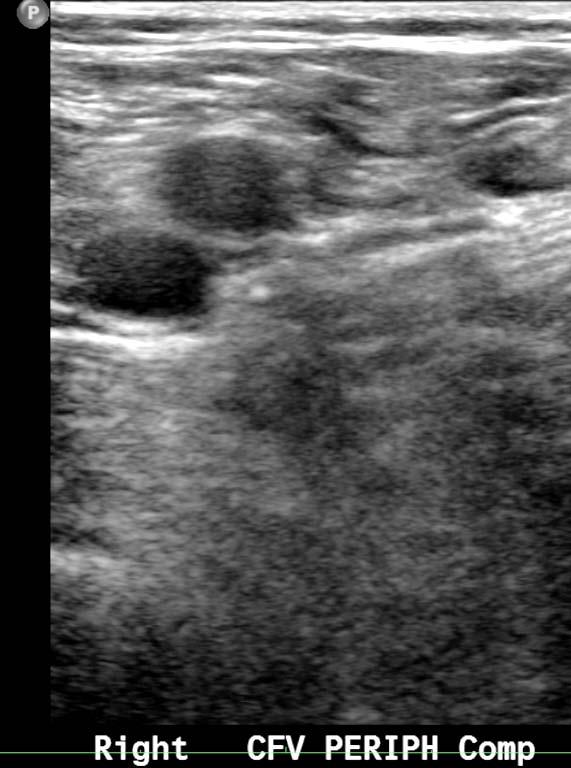
16 Acute Venous Thrombosis Soft, deformable with compression Enlarges vein Smooth Free floating CFV Great saphenous vein Femoral vein


17 Initial -8mm +7 mos 4mm +11 mos 4mm +13 mos 9mm Popliteal vein recurrent thrombosis +45 mos 4mm


18 Virchow s Triad


19 Development of DVT depends on baseline risk and risk events Normal Thrombophilia
20
21 Duplex Doppler ultrasound is used to diagnose and grade stenoses Gray scale narrowing Color narrowing and color changes of elevated velocity Spectral Doppler in and beyond stenosis


22


23 Bournoulli and Stenosis Pre-stenosis Stenosis Post-stenosis Total energy = Kinetic + potential Potential energy very decreased Total energy lower Potential energy decreased Kinetic energy very increased


24 Increased Velocity in Stenosis

25 Pre and In the stenosis


26 Beyond the stenosis Change from small lumen to large lumen destabilizes flow Jet spreads out High velocity also destabilizing Frank breakdown of regular flow disturbed flow (and evenually turbulence)


27 Post Stenotic Disturbed Flow
28 Criteria for Stenoses Some circulations use absolute velocity Internal carotid artery Most circulations do not have standard velocities - Need ratios Some circulations use downstream effects in addition Peak systolic velocity ratio (velocity ratio) Highest velocity in stenosis divided by velocity proximal to stenosis (in normal vessel) IC:CC ratio PSV ratio in arteries Renal aortic ratio Intrarenal criteria
29 Abdominal aortic aneursym Abnormal dilatation of aorta If enlarges over 5 cm and is untreated, rupture may occur High mortality if rupture Approved for Medicare screening

")
30 Abdominal Aortic Aneurysm (AAA)

31 AAA gray scale ultrasound


32 AAA- Easy to measure, hard to acquire
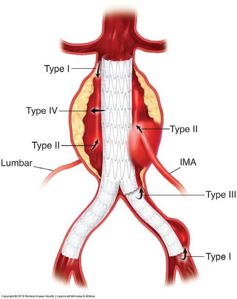
33 Endoleaks
34
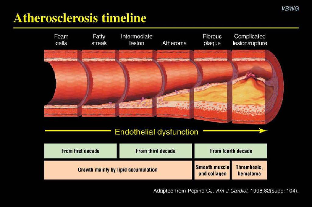
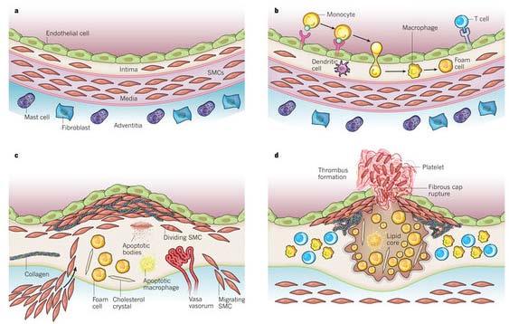
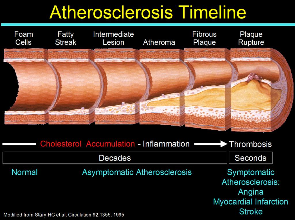
35 Natural history of atherosclerosis Preclinical disease Flow mediated dilatation, intima media thickness Location of plaque Clinical disease Degree of stenosis Plaque characterization Prediction of disease
36
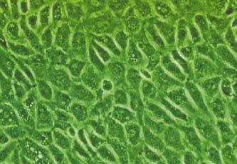
37 Drawing of cross-sections of identical, most proximal part of six left anterior descending coronary arteries. Stary H C et al. Circulation. 1995;92: Copyright American Heart Association, Inc. All rights reserved.

38 Intima Media Thickness Used in epidemiological studies Strong predictor of future cardiovascular events Additive to some traditional cardiovascular risk factors Used in pharmaceutical studies How can it be applied to the individual? Reproducible clinically? How does it compare to other tests?e.g. history, lipids, CRP, CT coronary calcium scoring? One time or serial test?
39 O Leary et al NEJM 1999:340:14-22


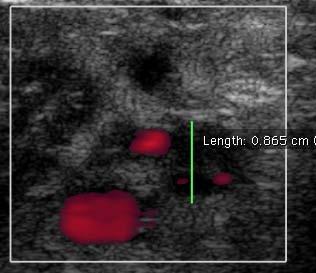
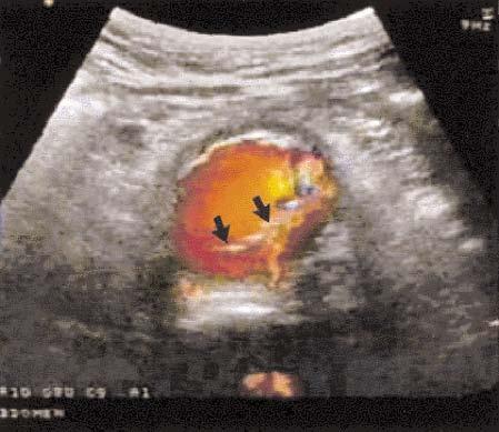
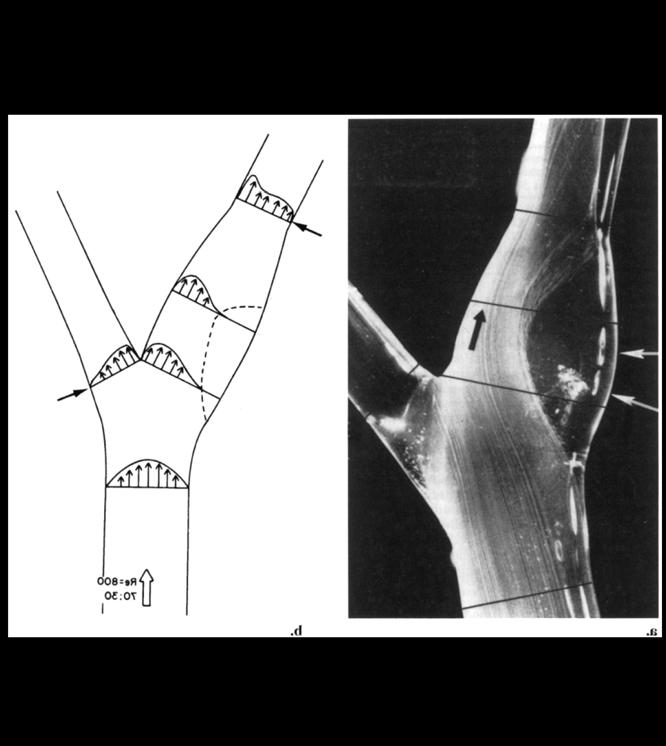
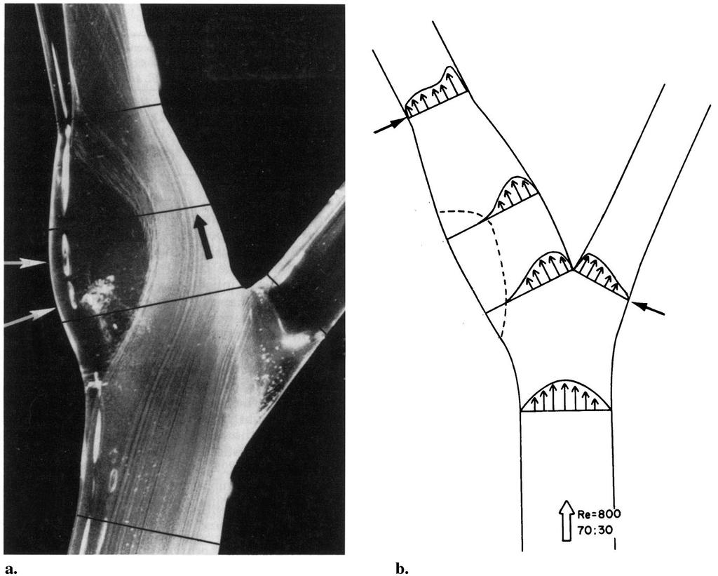
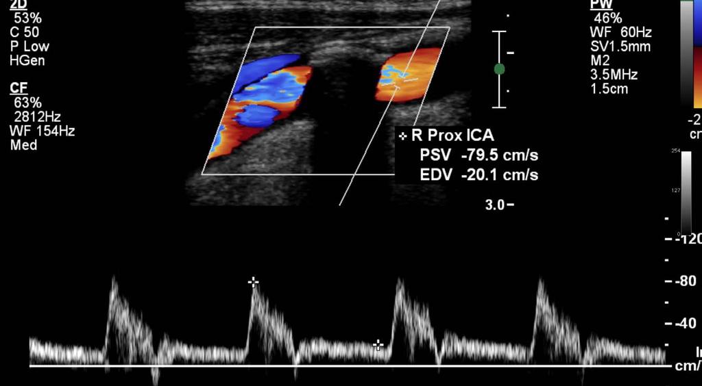
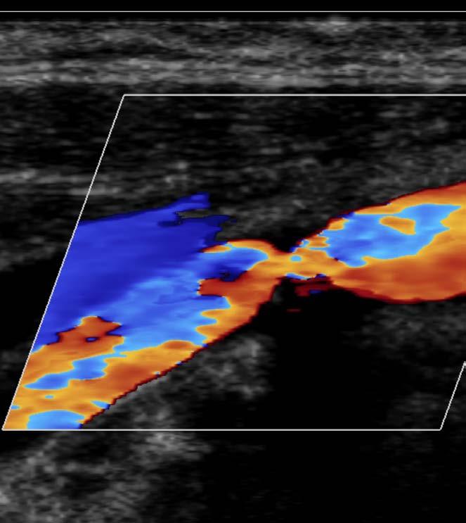
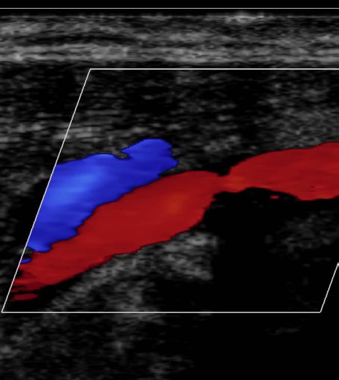
40 Weird Doppler from the bulb
41 Flow separation
42

43
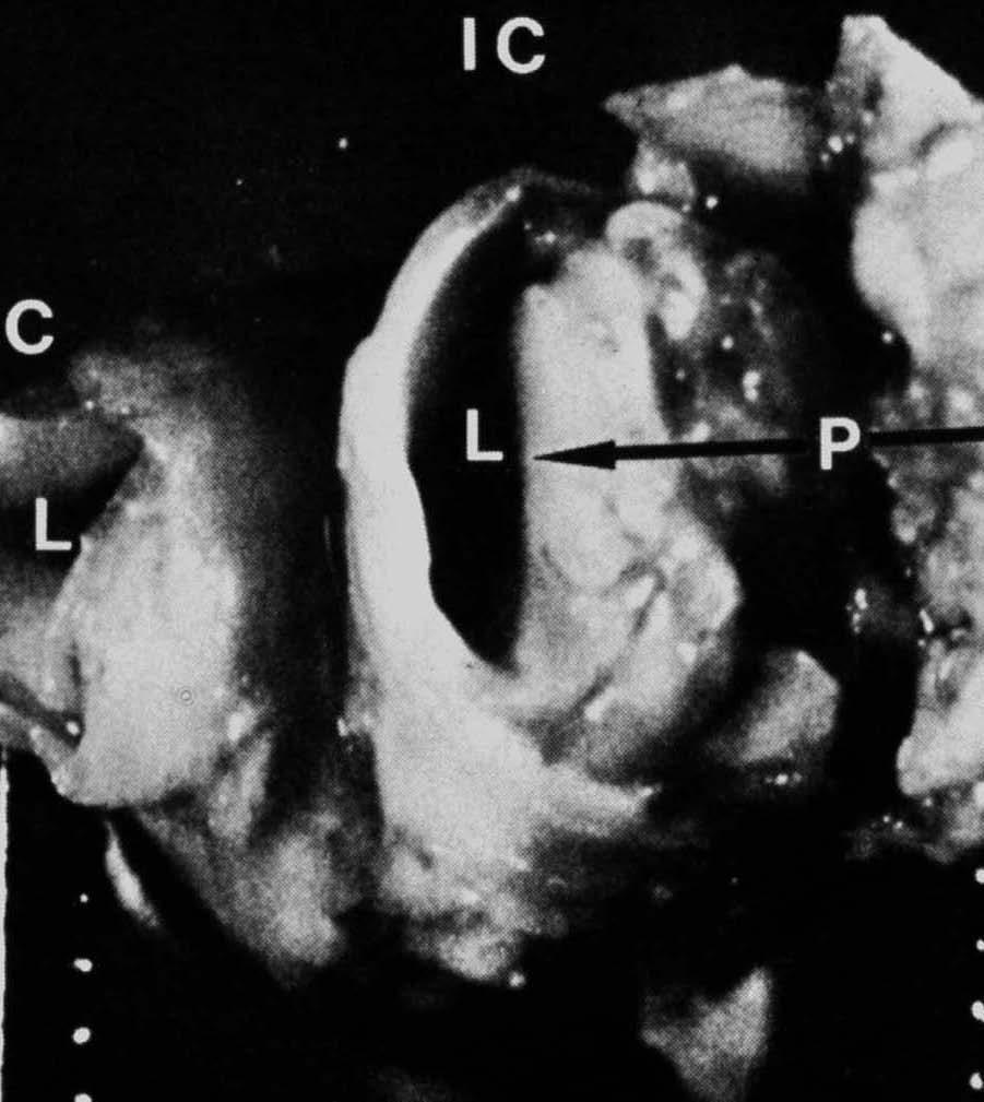
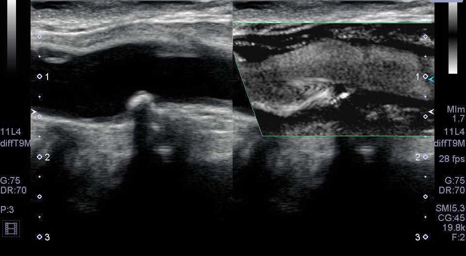
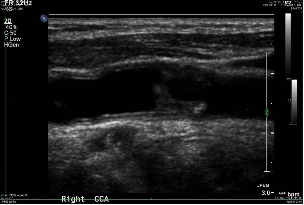
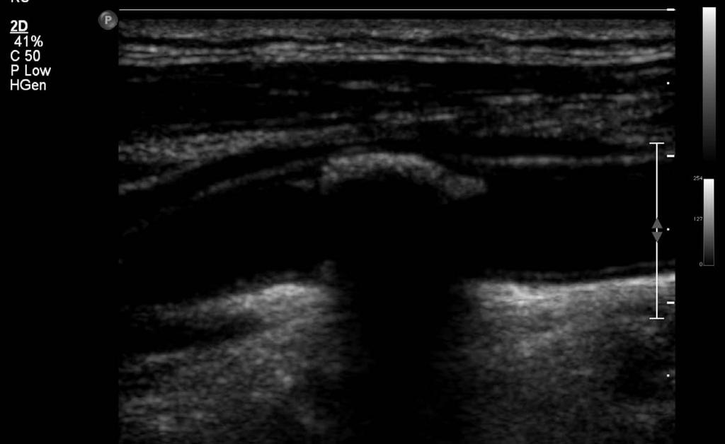
44 Plaque
45
46

47
48


49

50

51
52
53

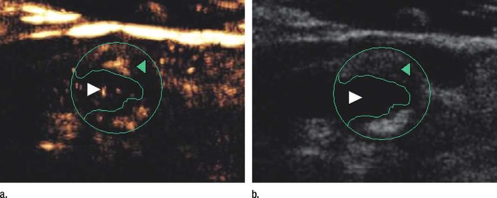
54 This plaque is different
55
56 Current needs and problems
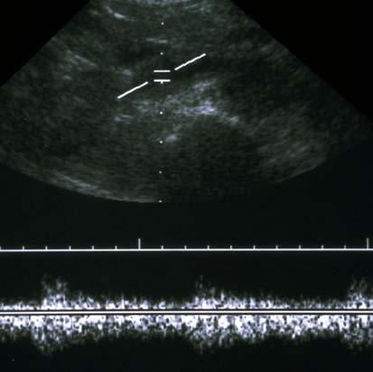
57 Calcifications and depth
58 PICA

59 PICA color
60 Before calcification
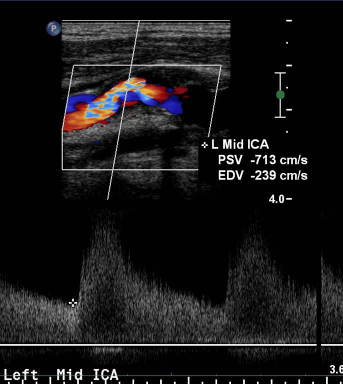
61 In

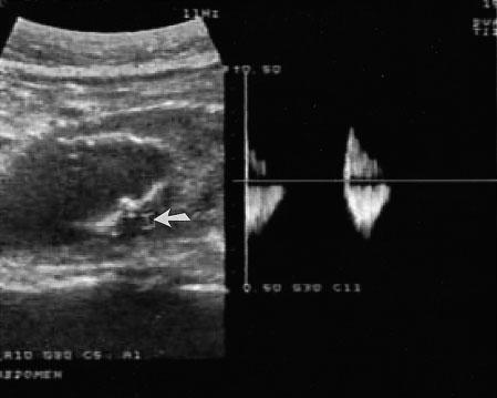
62 Distal 79 cm/s
63 Additional scanning

64 PSV 159, EDV 20, IC:CC 2
65 Duplex DSA Correlation Duplex Success Dark green best Light green Yellow Red worst Eiberg Eur J Endovasc Surg
66 Duplex Arteriography 1020 scans Not well visualized Iliac 73 Femoral 26 Popliteal 17 Infrapopliteal 221 Arterial wall calcifications 64 Poor runoff 18 Hingorani AP et al. Vascular 2008;16(3)
67 Solutions Better sonographers to find best direction CTA Future Multiplanar ultrasound Volume acquisition Volume flow (?) Sensitive techniques to low flow
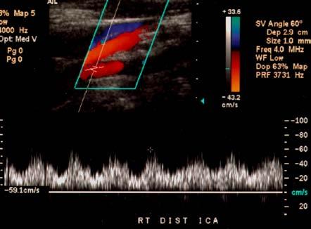
68 Angle

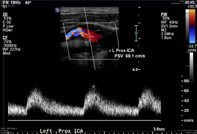
69 Velocity requires angle correction


70 60 o Angle Correction 60 o 92.6cm/s 60 o 60 o 83.2cm/s 69.1cm/s

71 Angle errors worse for higher angles Table courtesy of Frank Miele

72 Angle Correction 60 o 92.6cm/s 50 o 90.9cm/s 70 o 122cm/s 88 o 4cm/s

73 What is angle?
74 Solutions Angle dependent scanning Multidirectional acquisition Less reliance on Doppler Gray scale modes
75 Is carotid ultrasound a public health problem? Same results in different laboratories give different degrees of stenosis
76 SRU Carotid Consensus Grant EG, et al. Radiology 2003;229:340.
77 World Federation Neurology

78 2002 ICAVL Survey 100 Vascular Labs 11 Different Criteria Unpublished data courtesy Ms. Sandra Katanick, Intersocietal Accreditation Commission

79 2010 ICAVL Survey 152 Vascular Labs; >16 Diagnostic Criteria
80 Future directions
81 Trifurcation of Ultrasound Point of care Traditional Ultrasound Personalized ultrasound (Specialized care) ER, primary care, some specialists Radiologists, some specialists Ultrasound specialist and team Problem centered, short duration Performed by doctor Protocol driven, more complete, Variable duration Performed by sonographer/vascular technologist, presented to doctor Detailed diagnostic or therapeutic scans, time depends on study but generally long (e.g. biopsy, contrast injection, therapy) Performed by doctor, usually with assistance (nurse, technologist)
82 Trifurcation of Ultrasound Future trends Point of care Traditional Ultrasound Personalized ultrasound Prettier pictures Few or no buttons Wireless acquisition and storage Simpler controls Volumetric acquisition Post processing Less dependency on sonographer skill (e.g. smart Doppler, protocols, angle independence, automatic measurements) Flow and pressure Contrast approval by FDA Drug delivery New modes with better macro and micro vasculature
Transducer Selection. Renal Artery Duplex Exam. Renal Scan. Renal Scan Echogenicity. How to Perform an Optimal Renal Artery Doppler Examination
 How to Perform an Optimal Renal Artery Doppler Examination Director of Ultrasound Education & Quality Assurance Baylor College of Medicine Division of Maternal-Fetal Medicine Maternal Fetal Center Imaging
Disclosure Statement:
 Marsha M. Neumyer, BS, RVT, FSVU, FSDMS, FAIUM International Director Vascular Diagnostic Educational Services Vascular Resource Associates Harrisburg, PA Disclosure Statement: CME Calendar QR Code Marsha
Duplex Ultrasound of the Renal Arteries. Duplex Ultrasound. In the Beginning
 Duplex Ultrasound of the Renal Arteries DIMENSIONS IN HEART AND VASCULAR CARE 2013 PENN STATE HEART AND VASCULAR INSTITUTE ROBERT G. ATNIP MD PROFESSOR OF SURGERY AND RADIOLOGY Duplex Ultrasound Developed
11 TH ANNUAL VASCULAR NONINVASIVE TESTING SYMPOSIUM NOVEMBER 10, 2018
 11 TH ANNUAL VASCULAR NONINVASIVE TESTING SYMPOSIUM NOVEMBER 10, 2018 RENAL ARTERY DISEASE AND RENOVASCULAR HYPERTENSION 1 WHAT IS RENOVASCULAR HYPERTENSION? https://my.clevelandclinic.org/health/diseases/16459-renovascular-hypertension
Recommendations for Follow-up After Vascular Surgery Arterial Procedures SVS Practice Guidelines
 Recommendations for Follow-up After Vascular Surgery Arterial Procedures 2018 SVS Practice Guidelines vsweb.org/svsguidelines About the guidelines Published in the July 2018 issue of Journal of Vascular
Carotid Doppler: Doppler wave forms obtained from the common, external and internal carotid arteries. As well as the vertebral and subclavian
 Competency Carotid Doppler: Doppler wave forms obtained from the common, external and internal carotid arteries. As well as the vertebral and subclavian arteries. Preferred angle is 60 degrees or less.
Imaging Strategy For Claudication
 Who are the Debators? Imaging Strategy For Claudication Duplex Ultrasound Alone is Adequate to Select Patients for Endovascular Intervention - Pro: Dennis Bandyk MD No Disclosures PRO - Vascular Surgeon
Peripheral Vascular Disease
 Peripheral artery disease (PAD) results from the buildup of plaque (atherosclerosis) in the arteries of the legs. For people with PAD, symptoms may be mild, requiring no treatment except modification of
What effects will proximal or distal disease have on a waveform?
 Spectral Doppler Interpretation Director of Ultrasound Education & Quality Assurance Baylor College of Medicine Division of Maternal-Fetal Medicine Maternal Fetal Center Imaging Manager Texas Children
Deb Coghlan AMS (Vascular and General ) Brisbane, Australia
 Brisbane, Australia") Deb Coghlan AMS (Vascular and General ) Brisbane, Australia ANEURYSMAL DIISEASE The infrarenal aorta enlarges with age, and is the commonest site for arterial aneurysms. An aneurysm is a permanent focal
Deb Coghlan AMS (Vascular and General ) Brisbane, Australia ANEURYSMAL DIISEASE The infrarenal aorta enlarges with age, and is the commonest site for arterial aneurysms. An aneurysm is a permanent focal
Pre-and Post Procedure Non-Invasive Evaluation of the Patient with Carotid Disease
 Pre-and Post Procedure Non-Invasive Evaluation of the Patient with Carotid Disease Michael R. Jaff, D.O., F.A.C.P., F.A.C.C. Assistant Professor of Medicine Harvard Medical School Director, Vascular Medicine
Imaging for Peripheral Vascular Disease
 Imaging for Peripheral Vascular Disease James G. Jollis, MD Director, Rex Hospital Cardiovascular Imaging Imaging for Peripheral Vascular Disease 54 year old male with exertional calf pain in his right
INTRODUCTORY TEXT BOX
 INTRODUCTORY TEXT BOX Diagnostic Partners provides a range of onsite, in-office cardiac and vascular diagnostic testing services. From Resting Echocardiograms to Abdominal Aorta scans, each study is performed
STRUCTURED EDUCATION REQUIREMENTS IMPLEMENTATION DATE: JULY 1, 2016
 STRUCTURED EDUCATION REQUIREMENTS Vascular Sonography The purpose of structured education is to provide the opportunity for individuals to develop mastery of discipline-specific knowledge that, when coupled
Experience of endovascular procedures on abdominal and thoracic aorta in CA region
 Experience of endovascular procedures on abdominal and thoracic aorta in CA region May 14-15, 2015, Dubai Dr. Viktor Zemlyanskiy National Research Center of Emergency Care Astana, Kazakhstan Region Characteristics
Vascular Sonography Examination
 Vascular Sonography Examination The purpose of The American Registry of Radiologic Technologists (ARRT ) Vascular Sonography Examination is to assess the knowledge and cognitive skills underlying the intelligent
Carotid Abnormalities Coils, Kinks and Tortuosity David Lorelli M.D., RVT, FACS Michigan Vascular Association Conference Saturday, October 20, 2012
 Carotid Abnormalities Coils, Kinks and Tortuosity David Lorelli M.D., RVT, FACS Michigan Vascular Association Conference Saturday, October 20, 2012 Page 1 Table of Contents Carotid Anatomy Carotid Duplex
2015 ARDMS Physicians Vascular Interpretation Job Task Analysis Summary Report
 P a g e 1 2015 ARDMS Physicians Vascular Interpretation Job Task Analysis Summary Report American Registry for Diagnostic Medical Sonography (ARDMS) P a g e 2 Table of Contents ABOUT THE REPORT... 3 METHODOLOGY...
What effects will proximal or distal disease have on an waveform?
 Spectral Doppler Interpretation Director Director of of Ultrasound Ultrasound Education Education & & Quality Quality Assurance Assurance Baylor Baylor College College of of Medicine Medicine Division
Duplex ultrasound is first-line imaging for all
 Our Protocol for Transabdominal Pelvic Vein Duplex Ultrasound A summary of s protocol for pelvic vein duplex ultrasonography, including equipment, patient positioning, ultrasound settings, and technique.
An Overview of Post-EVAR Endoleaks: Imaging Findings and Management. Ravi Shergill BSc Sean A. Kennedy MD Mark O. Baerlocher MD FRCPC
 An Overview of Post-EVAR Endoleaks: Imaging Findings and Management Ravi Shergill BSc Sean A. Kennedy MD Mark O. Baerlocher MD FRCPC Disclosure Slide Mark O. Baerlocher: Current: Consultant for Boston
Visceral Vascular Ultrasound. Joel Thompson, MD, MPH Borg & Ide Imaging
 Visceral Vascular Ultrasound Joel Thompson, MD, MPH Borg & Ide Imaging Objectives: Review major abdominal vascular structures Identify normal peak systolic velocity (PSV) for major abdominal arteries.
Image Formation (10) 2 Evaluation and Selection of Representative Images (10)
 2 Evaluation and Selection of Representative Images (10)") STRUCTURED SELF ASSESSMENT CONTENT SPECIFICATIONS SSA LAUNCH DATE: JANUARY 1, 2018 Vascular Sonography The purpose of continuing qualifications requirements (CQR) is to assist registered technologists
STRUCTURED SELF ASSESSMENT CONTENT SPECIFICATIONS SSA LAUNCH DATE: JANUARY 1, 2018 Vascular Sonography The purpose of continuing qualifications requirements (CQR) is to assist registered technologists
MESENTERIC ISCHEMIA. Phillip J Bendick, PhD
 MESENTERIC ISCHEMIA Phillip J Bendick, PhD Arterial Celiac - Hepatic - Splenic Superior Mesenteric Artery Inferior Mesenteric Artery Venous Mesenteric system Porto - hepatic system Inferior Vena Cava Acute
Case Report 1. CTA head. (c) Tele3D Advantage, LLC
 Tele3D Advantage, LLC") Case Report 1 CTA head 1 History 82 YEAR OLD woman with signs and symptoms of increased intra cranial pressure in setting of SAH. CT Brain was performed followed by CT Angiography of head. 2 CT brain Extensive
Case Report 1 CTA head 1 History 82 YEAR OLD woman with signs and symptoms of increased intra cranial pressure in setting of SAH. CT Brain was performed followed by CT Angiography of head. 2 CT brain Extensive
Physician s Vascular Interpretation Examination Content Outline
 Physician s Vascular Interpretation Examination Content Outline (Outline Summary) # Domain Subdomain Percentage 1 2 3 4 5 6 Cerebrovascular Abdominal Peripheral Arterial - Duplex Imaging Peripheral Arterial
Imaging, it s central role in planning and guiding intervention. Prof. Luis Izquierdo. MD, PhD, FEBVS
 Imaging, it s central role in planning and guiding intervention Prof. Luis Izquierdo. MD, PhD, FEBVS IMPORTANT INFORMATION: These materials are intended to describe common clinical considerations and procedural
LOWER EXTREMITY VENOUS COMPRESSION ULTRASOUND. CPT Stacey Good, DO Emergency Medicine Ultrasound Fellow Madigan Army Medical Center
 LOWER EXTREMITY VENOUS COMPRESSION ULTRASOUND CPT Stacey Good, DO Emergency Medicine Ultrasound Fellow Madigan Army Medical Center Learning Objectives Setup and patient positioning for optimizing success
No financial or commercial relationships to disclose
 Deanna New, RVT No financial or commercial relationships to disclose IAC REQUIREMENTS: The main duty of a sonographer is to make the physician or radiologists job easier by capturing images and doing
Abdominal Aortic Aneurysms. A Surgeons Perspective Dr. Derek D. Muehrcke
 Abdominal Aortic Aneurysms A Surgeons Perspective Dr. Derek D. Muehrcke Aneurysm Definition The abnormal enlargement or bulging of an artery caused by an injury or weakness in the blood vessel wall A localized
Case 8038 Renal allograft complicated with renal artery stenosis
 Case 8038 Renal allograft complicated with renal artery stenosis Santiago I, Canelas A, Pinto AP Section: Cardiovascular Published: 2009, Nov. 30 Patient: 61 year(s), male Clinical History A 61-year-old
Certificate in Clinician Performed Ultrasound (CCPU) Syllabus
 Syllabus") Certificate in Clinician Performed Ultrasound (CCPU) Syllabus Proximal Deep Vein Thrombosis (DVT) Page 1 of 6 03/17 Deep Vein Thrombosis (DVT) Syllabus Purpose: This unit is designed to cover the theoretical
Certificate in Clinician Performed Ultrasound (CCPU) Syllabus Proximal Deep Vein Thrombosis (DVT) Page 1 of 6 03/17 Deep Vein Thrombosis (DVT) Syllabus Purpose: This unit is designed to cover the theoretical
8/20/18. The Doppler Effect. Objectives. What is the Doppler Effect. Doppler principles. Spectral Waveform. Image recognition. Vascular Ultrasound
 Vascular Ultrasound: Physics and Haemodynamics Objectives Doppler principles Spectral Waveform Key factors Haemodynamics: Stenosis Waveforms Image recognition Vascular Ultrasound: A flawed paradigm What
Abdominal Aortic Aneurysm (AAA)
") Abdominal Aortic Aneurysm (AAA) Vascular Workshop: Objectives Anatomy Keith VanHaltren Indications Technique Cases Abdominal Aorta: Normal Size Abdominal aortic aneurysm: Definition Normal diameter of
Abdominal Aortic Aneurysm (AAA) Vascular Workshop: Objectives Anatomy Keith VanHaltren Indications Technique Cases Abdominal Aorta: Normal Size Abdominal aortic aneurysm: Definition Normal diameter of
RENAL AND MESENTERIC ARTERY STENTS Are There Standard Velocity Criteria for Restenosis?
 RENAL AND MESENTERIC ARTERY STENTS Are There Standard Velocity Criteria for Restenosis? R. Eugene Zierler, M.D. The D. E. Strandness, Jr. Vascular Laboratory University of Washington Medical Center Division
Beyond Stenosis Severity: Top 5 Important Duplex Characteristics to Identify in a Patient with Carotid Disease
 Beyond Stenosis Severity: Top 5 Important Duplex Characteristics to Identify in a Patient with Carotid Disease Jan M. Sloves RVT, RCS, FASE Technical Director New York Cardiovascular Associates Disclosures
Bedside Ultrasound for DVT. Linear Probe. Leg Veins
 Bedside Ultrasound for DVT J. Christian Fox, MD, RDMS, FAAEM, FAIUM Director of Emergency Ultrasound Fellowship University of California, Irvine Jchrsitianfox@gmail.com Linear Probe High frequency transducer
Bedside Emergency Ultrasound For Deep Venous Thrombosis
 Bedside Emergency Ultrasound For Deep Venous Thrombosis Michael Blaivas, MD, MBA(candidate) FACEP, FAIUM Professor of Medicine University of South Carolina School of Medicine AIUM Third Vice President
What is the real place of venous echo Doppler in aircrew member flying rehabilitation after a thromboembolism event?
 89 th ASMA ANNUAL SCIENTIFIC MEETING DALLAS- May 6-10, 2018 What is the real place of venous echo Doppler in aircrew member flying rehabilitation after a thromboembolism event? S BISCONTE (1), V MARICOURT
Lab CT scan. Murad Kharabsheh Yaman Alali
 Lab CT scan Murad Kharabsheh Yaman Alali Some rules to read The CT Scan : 1. Remember that it s a transverse section across the body and we are looking at the inferior part of the section (not the superior),
Indications: following: embolization. artery that has diseases 5. The evaluation. of suspected. such entities. a cold hand. biopsy
 Peripheral Arterial Ultrasound Protocol Using Color and Spectral Doppler Reviewed by: Mark Yuhasz, MD Last Review Date: January 2015 Contact: (866) 761 4200, Option 1 Indications: The indications for peripheral
Fig MHz cm/s. Table 1 Fig. 2. Fig. 3, 4. Fig. 5
 GE Fig. 1 3. 5 MHz 7 10 MHz 3. 5 5. 0 MHz B 10 20 cm/s Table 1 Fig. 2 Fig. 1 1 2 3 3 3 : 1 2 3 Fig. 3, 4 Fig. 5 Table 1 a b c Fig. 2 a B b B c Fig. 6 Table 1 Fig. 7 a b c Fig. 3 a AV b A VV c 1 cm 2 1
Case 9799 Stanford type A aortic dissection: US and CT findings
 Case 9799 Stanford type A aortic dissection: US and CT findings Accogli S, Aringhieri G, Scalise P, Angelini G, Pancrazi F, Bemi P, Bartolozzi C Department of Diagnostic and Interventional Radiology, University
VFI Technology to Change the Way You Work
 Analogic Ultrasound VFI Technology to Change the Way You Work Vascular Ultrasound Made Easier Vector Flow Imaging VFI VFI is a ground-breaking technology that can revolutionize the workflow for many Doppler
Prof. Nabil CHAKFE et coll.
 Prof. Nabil CHAKFE et coll. For the Department of Vascular Surgery and Kidney Transplantation University Hospital of Strasbourg, FRANCE Popliteal artery entrapment: misdiagnosed Epidemiology Prevalence:
Ultrasound Evaluation after EVAR: (Trying to) Let the CAT Scan Out of the Bag
 Let the CAT Scan Out of the Bag") Ultrasound Evaluation after EVAR: (Trying to) Let the CAT Scan Out of the Bag Joseph-Vincent V. Blas, MD Division of Vascular Surgery Department of Surgery Greenville Health System University of South
Ultrasound Evaluation after EVAR: (Trying to) Let the CAT Scan Out of the Bag Joseph-Vincent V. Blas, MD Division of Vascular Surgery Department of Surgery Greenville Health System University of South
Step by step ultrasound examination of varicose veins. Dr. Özgün Sensebat Vascular Surgeon Private Vascular Clinic Dorsten & Borken, Germany
 Step by step ultrasound examination of varicose Dr. Özgün Sensebat Vascular Surgeon Private Vascular Clinic Dorsten & Borken, Germany Required technical setup: B-mode vessel imaging combined with color
Guidelines, Policies and Statements D20 Statement on Peripheral Venous Ultrasound
 Guidelines, Policies and Statements D20 Statement on Peripheral Venous Ultrasound Disclaimer and Copyright The ASUM Standards of Practice Board have made every effort to ensure that this Guideline/Policy/Statement
ATHEROSCLEROSIS. Secondary changes are found in other coats of the vessel wall.
 ATHEROSCLEROSIS Atherosclerosis Atherosclerosis is a disease process affecting the intima of the aorta and large and medium arteries, taking the form of focal thickening or plaques of fibrous tissue and
HD Scanning: Velocities and Volume Flow
 HD Scanning: Velocities and Volume Flow Non-Invasive Lab Symposium West Orange, NJ April 27, 2018 Volume Flow Cindy Sturt, MD, FACS, RVT 500,000 Americans on dialysis 20-25% annual mortality 65% 5 year
Ultrasound Imaging of The Posterior Circulation
 Ultrasound Imaging of The Posterior Circulation Michigan Sonographers Society 2 Nd Annual Fall Vascular Conference Larry N. Raber RDMS-RVT Clinical Manager General Ultrasound/Neurovascular Laboratory Cleveland
Brain Atrophy. Brain Atrophy
 Aging Central Nervous System Processes Age related brain atrophy Non-age related brain atrophy Cerebrovascular disease Cerebral infarction Hypertensive hemorrhage Carotid artery stenosis and occlusion
Quality ID #195 (NQF 0507): Radiology: Stenosis Measurement in Carotid Imaging Reports National Quality Strategy Domain: Effective Clinical Care
: Radiology: Stenosis Measurement in Carotid Imaging Reports National Quality Strategy Domain: Effective Clinical Care") Quality ID #195 (NQF 0507): Radiology: Stenosis Measurement in Carotid Imaging Reports National Quality Strategy Domain: Effective Clinical Care 2018 OPTIONS FOR INDIVIDUAL MEASURES: REGISTRY ONLY MEASURE
Quality ID #195 (NQF 0507): Radiology: Stenosis Measurement in Carotid Imaging Reports National Quality Strategy Domain: Effective Clinical Care 2018 OPTIONS FOR INDIVIDUAL MEASURES: REGISTRY ONLY MEASURE
Certificate in Clinician Performed Ultrasound (CCPU) Syllabus. Above Knee Deep Vein Thrombosis (DVT)
 Syllabus. Above Knee Deep Vein Thrombosis (DVT)") Certificate in Clinician Performed Ultrasound (CCPU) Syllabus Above Knee Deep Vein Thrombosis (DVT) Deep Vein Thrombosis (DVT) Purpose: Prerequisites: Training: Assessments: This unit is designed to cover
Certificate in Clinician Performed Ultrasound (CCPU) Syllabus Above Knee Deep Vein Thrombosis (DVT) Deep Vein Thrombosis (DVT) Purpose: Prerequisites: Training: Assessments: This unit is designed to cover
What Do We Know? Disclosure Statement: 3/11/2015. Deep abdominal imaging
 Marsha M. Neumyer, BS, RVT, FSVU, FSDMS, FAIUM International Director Vascular Diagnostic Educational Services Vascular Resource Associates Harrisburg, PA Disclosure Statement: CME Calendar QR Code Marsha
Endovascular treatment of acquired arteriovenous fistula with severe hemodynamic effects: a case report
 Endovascular treatment of acquired arteriovenous fistula with severe hemodynamic effects: a case report The Leipzig Interventional Course, January 24 27, 2017 El Samman K., Šedivý P., Šnajdrová A., Přindišová
Schedule of Benefits. for Professional Fees Vascular Procedures
 Schedule of Benefits for Professional Fees 2018 Vascular Procedures ANASTOMOSIS RULES 820 Arteriovenous anastomosis in arm 1453 Arteriovenous anastomosis, open by basilic vein transposition 1465 Splenorenal
Popliteal Aneurysm: When is surgical therapy indicated? PROF. GRZEGORZ OSZKINIS
 Popliteal Aneurysm: When is surgical therapy indicated? PROF. GRZEGORZ OSZKINIS Asymptomatic mass - 38-40%will develop symptoms at a rate of 14%/yr Intermittent claudic ation (chronic ischemia) - 25%-40%
Subclavian artery Stenting
 Subclavian artery Stenting Etiology Atherosclerosis Takayasu s arteritis Fibromuscular dysplasia Giant Cell Arteritis Radiation-induced Vascular Injury Thoracic Outlet Syndrome Neurofibromatosis Incidence
Multislice CT. - fast scanning - submilimeter slices
 CT angiography Multislice CT - fast scanning - submilimeter slices CT angiography - Minimal invasivity - High resolution (similar to DSA, higher than MRI) - Cannot assess hemodynamics (contrary to DSA)
Doppler ultrasound as noninvasive diagnosis of peripheral arterial disease
 Doppler ultrasound as noninvasive diagnosis of peripheral arterial disease Poster No.: C-0246 Congress: ECR 2012 Type: Scientific Exhibit Authors: C. Ballester Valles, F. Aparici-Robles; Valencia/ES Keywords:
GUNDERSEN/LUTHERAN ULTRASOUND DEPARTMENT POLICY AND PROCEDURE MANUAL
 GUNDERSEN/LUTHERAN ULTRASOUND DEPARTMENT POLICY AND PROCEDURE MANUAL SUBJECT: Carotid Duplex Ultrasound SECTION: Vascular Ultrasound ORIGINATOR: Deborah L. Richert, BSVT, RDMS, RVT DATE: October 15, 2015
Measure #195 (NQF 0507): Radiology: Stenosis Measurement in Carotid Imaging Reports National Quality Strategy Domain: Effective Clinical Care
: Radiology: Stenosis Measurement in Carotid Imaging Reports National Quality Strategy Domain: Effective Clinical Care") Measure #195 (NQF 0507): Radiology: Stenosis Measurement in Carotid Imaging Reports National Quality Strategy Domain: Effective Clinical Care 2017 OPTIONS FOR INDIVIDUAL MEASURES: CLAIMS ONLY MEASURE TYPE:
Measure #195 (NQF 0507): Radiology: Stenosis Measurement in Carotid Imaging Reports National Quality Strategy Domain: Effective Clinical Care 2017 OPTIONS FOR INDIVIDUAL MEASURES: CLAIMS ONLY MEASURE TYPE:
Carotid Duplex: Beyond Stenosis Ido Weinberg, MD Vascular Medicine Massachusetts General Hospital Assistant Professor of Medicine Harvard Medical
 Carotid Duplex: Beyond Stenosis Ido Weinberg, MD Vascular Medicine Massachusetts General Hospital Assistant Professor of Medicine Harvard Medical School Boston, Massachusetts Disclosures I do not have
Subject Area Modules. Subject Area Modules. Subject Area Modules Published on Society for Vascular Surgery (
 Purchasing the VESAP3 comprehensive package provides access to all 10 VESAP3 modules, in learning and exam mode, for $151 less than the cost of purchasing all modules individually. Click here to learn
BC Vascular Surgery Day
 BC Vascular Surgery Day November 4, 2017 1 Table of Contents Abdominal Aortic Aneurysm 3 4 Acute DVT 5 6 Peripheral Arterial Disease 7 9 Varicose Veins 10 11 Diabetic Foot Ulcers 12 13 Carotid Stenosis
Abdominal Exam: The examination of the abdomen used by physicians to detect an abdominal aortic aneurysm.
 Glossary of Terms Abdominal Exam: The examination of the abdomen used by physicians to detect an abdominal aortic aneurysm. Angiogram: A diagnostic test requiring the insertion of a catheter into an artery
Vascular Surgery Cases: Detours. Brian F. Stull, RDMS, RVT UNC REX Healthcare Vascular Specialists
 Vascular Surgery Cases: Detours Brian F. Stull, RDMS, RVT UNC REX Healthcare Vascular Specialists Brian.Stull@Unchealth.unc.edu Objectives Anatomy of a bypass graft Where does it connect, where does it
Guidelines, Policies and Statements D5 Statement on Abdominal Scanning
 Guidelines, Policies and Statements D5 Statement on Abdominal Scanning Disclaimer and Copyright The ASUM Standards of Practice Board have made every effort to ensure that this Guideline/Policy/Statement
MINIMALLY-INVASIVE TREATMENT OF VASCULAR DISEASE
 CLINIC PROFILE MINIMALLY-INVASIVE TREATMENT OF VASCULAR DISEASE The Section of Vascular Surgery at Dartmouth- Hitchcock Medical Center is committed to outstanding care of patients with vascular disease,
Artery 1 Head and Thoracic Arteries. Arrange the parts in the order blood flows through them.
 Artery 1 Head and Thoracic Arteries 1. Given the following parts of the aorta: 1. abdominal aorta 2. aortic arch 3. ascending aorta 4. thoracic aorta Arrange the parts in the order blood flows through
BC Vascular Day. Contents. November 3, Abdominal Aortic Aneurysm 2 3. Peripheral Arterial Disease 4 6. Deep Venous Thrombosis 7 8
 BC Vascular Day Contents Abdominal Aortic Aneurysm 2 3 November 3, 2018 Peripheral Arterial Disease 4 6 Deep Venous Thrombosis 7 8 Abdominal Aortic Aneurysm Conservative Management Risk factor modification
The role of ultrasound duplex in endovenous procedures
 The role of ultrasound duplex in endovenous procedures Neophytos A. Zambas MD, PhD Vascular Surgeon Polyclinic Ygia, Limassol, Cyprus ΚΕΑΕΧ ΚΥΠΡΙΑΚΗ ΕΤΑΙΡΕΙΑ ΑΓΓΕΙΑΚΗΣ ΚΑΙ ΕΝΔΑΓΓΕΙΑΚΗΣ ΧΕΙΡΟΥΡΓΙΚΗΣ Pre
NCVH. Ultrasongraphy: State of the Art Vein Forum 2015 A Multidisciplinary Approach to Otptimizing Venous Circulation From Wounds to WOW
 Ultrasongraphy: State of the Art 2015 NCVH New Cardiovascular Horizons Vein Forum 2015 A Multidisciplinary Approach to Otptimizing Venous Circulation From Wounds to WOW Anil K. Chagarlamudi, M.D. Cardiovascular
Acute Versus Chronic DVT Imaging in the Vascular Lab Heather Gornik, MD, RVT, RPVI
 Acute Versus Chronic DVT Imaging in the Vascular Lab Heather Gornik, MD, RVT, RPVI Cleveland Clinic Heart and Vascular Institute Heather L. Gornik, MD has the following relationships to disclose: CVR Global
BEDSIDE ULTRASOUND BEDSIDE ULTRASOUND. Deep Vein Thrombosis. Probe used
 BEDSIDE ULTRASOUND Part 2 Diagnosis of deep vein thrombosis Kishore Kumar Pichamuthu, Professor, Department of Critical Care, CMC, Vellore Summary: Deep vein thrombosis (DVT) is a problem encountered in
Case 37 Clinical Presentation
 Case 37 73 Clinical Presentation The patient is a 62-year-old woman with gastrointestinal (GI) bleeding. 74 RadCases Interventional Radiology Imaging Findings () Image from a selective digital subtraction
(Department of Radiology, Beylikdüzü State Hospital, İstanbul, Turkey) Corresponding Author: Dr. Mete Özdikici
 Corresponding Author: Dr. Mete Özdikici") Quest Journals Journal of Medical and Dental Science Research Volume 5~ Issue 6 (2018) pp: 61-65 ISSN(Online) : 2394-076X ISSN (Print):2394-0751 www.questjournals.org Research Paper Quantitative Measurements
Quest Journals Journal of Medical and Dental Science Research Volume 5~ Issue 6 (2018) pp: 61-65 ISSN(Online) : 2394-076X ISSN (Print):2394-0751 www.questjournals.org Research Paper Quantitative Measurements
Non-invasive examination
 Non-invasive examination Segmental pressure and Ankle-Brachial Index (ABI) The segmental blood pressure (SBP) examination is a simple, noninvasive method for diagnosing and localizing arterial disease.
DISCLOSURE TEST YOUR WAVEFORM IQ. Partial volume artifact. 86 yo female with right arm swelling, picc line. AVF on left? Dx?
 Deborah Rubens University of Rochester Rochester, NY DISCLOSURE Neither I nor my immediate family have a financial relationship with a commercial organization that may have a direct or indirect interest
The Role of US in Chronic Mesenteric Ischemia. Sagar S. Gandhi, MD Vascular Health Alliance Greenville Health System
 The Role of US in Chronic Mesenteric Ischemia Sagar S. Gandhi, MD Vascular Health Alliance Greenville Health System No Disclosures Mesenteric Ischemia Anatomy Presentation Diagnostic tools Treatment Celiac
Report For Center Created Gender D.O.B Page 1 Sean Breen HeartSmart IMT plus 3/29/2012 Male 11/26/1973 B C D E
 Report For Center Created Gender D.O.B Page 1 Carotid Assessment A B C D E Good Satisfactory Concern Serious Highest Risk Intima-Media Thickness Additional Findings Plaque Character Percent Stenosis Comments:
US of Renovascular Hypertension. Jonathan R. Dillman, MD, MSc Associate Professor Director, Thoracoabdominal Imaging
 US of Renovascular Hypertension Jonathan R. Dillman, MD, MSc Associate Professor Director, Thoracoabdominal Imaging Disclosures Nothing Relevant Unrelated grant funding Siemens US Toshiba US Objectives
Pulmonary Embolism. Thoracic radiologist Helena Lauri
 Pulmonary Embolism Thoracic radiologist Helena Lauri 8.5.2017 Statistics 1-2 out of 1000 adults annually are diagnosed with deep vein thrombosis (DVT) and/or pulmonary embolism (PE) About half of patients
POINT OF CARE ULTRASOUND - Venous US for DVT
 POINT OF CARE ULTRASOUND - Venous US for DVT The diagnosis of deep venous thrombosis (DVT) using ultrasound in the emergency department. DVT US is easy to perform and can be usually be completed in less
Section II: Patient Interview Grade: 5
 Only written competency completed with this EXACT form will be accepted for grading. No modifications to the LAYOUT of the form will be accepted for a written competency. Failure to comply will result
Optimising your Doppler settings for an accurate PI. Alison McGuinness Mid Yorks Hospitals
 Optimising your Doppler settings for an accurate PI Alison McGuinness Mid Yorks Hospitals Applications Both maternal uterine and fetal circulations can be studied with doppler sonography Uterine arteries
Reimbursement Guide Zenith Fenestrated AAA Endovascular Graft
 MEDICAL Reimbursement Guide Zenith Fenestrated AAA Endovascular Graft Disclaimer: The information provided herein reflects Cook s understanding of the procedure(s) and/or device(s) from sources that may
DOPPLER ULTRASOUND OF DEEP VENOUS THROMBOSIS
 TOKUDA HOSPITAL SOFIA DOPPLER ULTRASOUND OF DEEP VENOUS THROMBOSIS MILENA STANEVA, MD, PhD Department of vascular surgery and angiology Venous thromboembolic disease continues to cause significant morbidity
Vascular Technology Examination Content Outline
 Vascular Technology Examination Content Outline (Outline Summary) # Domain Subdomain Percentage 1 Normal Anatomy, Perfusion, and Function Evaluate normal anatomy, perfusion, function 2 Pathology, Perfusion,
Radiologic Evaluation of Peripheral Arterial Disease
 January 2003 Radiologic Evaluation of Peripheral Arterial Disease Grace Tye, Harvard Medical School Year III Patient D.M. CC: 44 y/o male with pain in his buttocks Occurs after walking 2 blocks. Pain is
ACUTE AORTIC SYNDROMES
 ACUTE AORTIC SYNDROMES AGNETA FLINCK MD, PhD Dept. of Thoracic Radiology Sahlgrenska University Hospital ACUTE AORTIC SYNDROMES Aortic dissection Intramural hematoma (IMH) 5-20% Penetrating atherosclerotic
Abdominal Aortic Aneurysm - Part 1. Learning Objectives. Disclosure. University of Toronto Division of Vascular Surgery
 University of Toronto Division of Vascular Surgery Abdominal Aortic Aneurysm - Part 1 Dr Mark Wheatcroft & Dr Elisa Greco Vascular Surgeon, St Michael s Hospital, Toronto & University of Toronto Disclosure
Optimizing Accuracy of Aortic Stent Grafts in Short Necks
 Optimizing Accuracy of Aortic Stent Grafts in Short Necks Venkatesh Ramaiah, MD, FACS Medical Director Arizona Heart Hospital Director Peripheral Vascular and Endovascular Research Arizona Heart Institute
B-Flow, Power Doppler and Color Doppler Ultrasound in the Assessment of Carotid Stenosis: Comparison with 64-MD-CT Angiography
 Med. J. Cairo Univ., Vol. 85, No. 2, March: 805-809, 2017 www.medicaljournalofcairouniversity.net B-Flow, Power Doppler and Color Doppler Ultrasound in the Assessment of Carotid Stenosis: Comparison with
ACCF/ACR/AIUM/ASE/ASN/ICAVL/SCAI/SCCT/SIR/SVM/SVS 2012 Appropriate Use Criteria. Indications A _ appropriate; I _ inappropriate; U _ uncertain
 National Imaging Associates, Inc. Clinical guidelines ABDOMINAL, PELVIS, SCROTAL, RETROPERITONEAL ORGANS DUPLEX SCAN (US) CPT Codes: 93975 Bilateral or Complete 93976 - Unilateral or Limited Guideline
TO CATCH A THIEF: IMAGING OF SUBCLAVIAN STEAL
 October 2013 TO CATCH A THIEF: IMAGING OF SUBCLAVIAN STEAL Sumir Pandit, Harvard Medical School, Year III 1 AGENDA Introduction to our patient A.B. Anatomy review of aorta and branches CT imaging of our
UNDERSTANDING ATHEROSCLEROSIS
 UNDERSTANDING ATHEROSCLEROSIS UNDERSTANDING ATHEROSCLEROSIS ARTERIES Arteries are blood vessels that carry oxygenated blood to all the organs of the body. Arteries are made up of three important layers:
NON-ATHEROSCLEROTIC PATHOLOGY OF THE CAROTID ARTERIES
 NON-ATHEROSCLEROTIC PATHOLOGY OF THE CAROTID ARTERIES Leslie M. Scoutt, MD, FACR Professor of Diagnostic Radiology & Surgery Vice Chair, Dept of Radiology & Biomedical Imaging Chief, Ultrasound Section