A Comparative Study on Calcification of Aortic Valves
|
|
|
- Jonas Skinner
- 6 years ago
- Views:
Transcription
1 University of Tennessee, Knoxville Trace: Tennessee Research and Creative Exchange Doctoral Dissertations Graduate School A Comparative Study on Calcification of Aortic Valves Shirin Masjedi University of Tennessee - Knoxville, smasjedi@vols.utk.edu Recommended Citation Masjedi, Shirin, "A Comparative Study on Calcification of Aortic Valves. " PhD diss., University of Tennessee, This Dissertation is brought to you for free and open access by the Graduate School at Trace: Tennessee Research and Creative Exchange. It has been accepted for inclusion in Doctoral Dissertations by an authorized administrator of Trace: Tennessee Research and Creative Exchange. For more information, please contact trace@utk.edu.
2 To the Graduate Council: I am submitting herewith a dissertation written by Shirin Masjedi entitled "A Comparative Study on Calcification of Aortic Valves." I have examined the final electronic copy of this dissertation for form and content and recommend that it be accepted in partial fulfillment of the requirements for the degree of Doctor of Philosophy, with a major in Biomedical Engineering. We have read this dissertation and recommend its acceptance: Roberto S. Benson, Deidra J. Mountain, Shanfeng Wang (Original signatures are on file with official student records.) Zannatul Ferdous, Major Professor Accepted for the Council: Dixie L. Thompson Vice Provost and Dean of the Graduate School
3 A Comparative Study on Calcification of Aortic Valves A Dissertation Presented for the Doctor of Philosophy Degree The University of Tennessee, Knoxville Shirin Masjedi May 2016
4 Copyright 2016 by Shirin Masjedi All rights reserved. ii
5 ACKNOWLEDGEMENTS To my parents, Ahlam Ahangar and Mostafa Masjedi iii
6 ABSTRACT Calcific Aortic Valve Disease (CAVD) is a major disorder in the developed countries among elderly. It is characterized by calcific deposition and stiffening of the aortic valve cusps. CAVD is a highly cell-mediated condition where valvular interstitial cells (VICs) become activated and differentiate into osteoblast-like cells. This is associated with upregulation of calcific markers like Alkaline phosphatase (ALP) and Runx2. ECM remodeling in another characteristic of stenotic aortic valves due to VIC activation. Reports show that CAVD initiates majorly on the noncoronary side of the aortic valves. Additionally, male sex is a significant risk factor of CAVD. Aortic valves are surrounded by a complicated hemodynamic environment. Pathological levels of mechanical forces applied to the aortic cusps interact with the VICs remodeling and differentiation and lead to abnormalities within the aortic cusps. In this dissertation, comparative studies were performed among VICs isolated from individual aortic cusps or between male and female to verify whether the differences in aortic calcification are observed at a cellular level. The calcification of VICs isolated from coronary and noncoronary aortic cusps was investigated in osteogenic culture condition to identify whether VICs show different amounts of calcific markers. Moreover, the osteogenic differentiation of VICs isolated separately from male and female aortic valves was analyzed to find if there are sex-related differences in their proliferation and calcification. Finally, both hypotheses were tested under cyclic strain to iv
7 determine the impact of pathological vs. physiological strain on enhancing the VICs calcification among coronary and noncoronary cusps or between male and female valves. It was found that VICs isolated from noncoronary cusps demonstrated greater calcification potential than coronary cusps. Likewise, male VICs showed increased ECM remodeling compared to female. Pathological cyclic strain applied to VICs cultured in osteogenic condition enhanced their calcification, however VICs cultured in normal condition seem not to be affected by pathological cyclic strain. The results from this dissertation provide a new perspective on the biomolecular events associated with VICs calcification. It shows that VICs in different cusps of an aortic valve have heterogeneous populations. Furthermore, it confirms that sex of the cells matter in aortic valve calcification. v
8 There is still need to think and plan, but on a different scale, and along different lines. C.S Forester ( ), The Hornblower companion vi
9 TABLE OF CONTENTS Chapter One Introduction... 1 Significance: Aortic valve Valvular interstitial cells (VICs) Origin of the aortic valve Calcific aortic valve disease (CAVD) Sex/gender as a risk factor Mechanical forces on valve calcification... 8 Chapter Two A Review of Sex-related Studies on Major Heart Valve Diseases and Atherosclerosis Introduction Aortic Stenosis Mitral Valve Prolapse Atherosclerosis Summary Chapter Three Comparison of Calcification Potential of Valvular Interstitial Cells Isolated from Individual Aortic Cusps Abstract: Introduction Materials and Methods vii
10 3.2.1 VIC Isolation and Culture Hematoxylin & Eosin (H&E) Staining of Tissue Sections DNA Content Quantitation Von Kossa and Alizarin Red Staining of Cultured VICs Calcium Assay Alkaline Phosphatase (ALP) Staining and Enzyme Assay Glycosaminoglycan (GAG) Content Collagen Content Gelatin Zymography Western Blotting Statistical Analysis Results Comparable Cell Density in VICs Isolated from Coronary and Noncoronary Aortic Cusps Elevated Osteogenic Marker Expression in VICs Isolated from Noncoronary Aortic Cusps Elevated Matrix Remodeling in the Calcified Cells Discussion Conclusion Chapter Four Sex-related Differences in Matrix Remodeling and Early Calcific Markers in Rat Aortic Valvular Interstitial Cells viii
11 Abstract: Introduction Materials and methods Primary VIC isolation and culture DNA content Cell growth assay GAG content Collagen type I content Gelatin zymography ALP staining and enzyme activity β-estradiol treatment Statistical analysis Results Comparable cell density between male and female RAVICs Greater proliferation in female RAVICs compared to male Elevated matrix remodeling in calcified male RAVICs compared with female Elevated early calcific marker expression in male RAVICs as compared to female Diminished proliferation of the female VICs due to β-estradiol treatment ix
12 4.4 Discussion Conclusion Chapter Five Calcification Potential of Aortic Valvular Interstitial Cells under Cyclc Strain Introduction Materials and Methods VIC isolation Culture under cyclic strain Alizarin red S staining ALP enzyme activity assay statistical analysis Results Greater nodule formation and nodule size in male RAVICs than female under pathological cyclic strain Elevated early calcific marker expression in male RAVICs compared to female under pathological cyclic strain Greater calcification in PAVICs isolated from noncoronary cusps under pathological cyclic strain Discussion Chapter Six Conclusions and Summary List of References x
13 Appendix Vita xi
14 LIST OF TABLES Table 2.1 Prevalence of Cardiovascular Diseases in Men and Women Table 3.1 Tissue section thickness of the porcine aortic cusps xii
15 LIST OF FIGURES Figure 1.1 Aortic cusp layers, rat aortic valve H&E staining... 4 Figure 1.2 Different stages of CAVD: sclerosis (left), stenosis (right) [2]... 6 Figure 1.3 Aortic valve cusps subjected to different mechanical forces at each systolic cycle [1]... 9 Figure 1.4 Flexcell system components Figure 2.1 (A) aortic cusp layers, (B) mitral leaflet layers, porcine aortic valves H&E.. 15 Figure 2.2 (A) Aorta cross-section, (B) vascular layers from aorta, rat H&E Figure 3.1 H&E staining of paraffin-embedded healthy porcine aortic valve cusp sections: (A) left, (B) right, and (C) noncoronary cusp. Parts D and E shows collagen and GAG content for the three cusps measured using a collagen and a GAG assay. No significant difference was observed in the thickness, collagen, and GAG content among the cusps. The data represent mean ± std. error, n=3 porcine aortic valves. Scale bar is 250 μm Figure 3.2 DNA content measured using a DNA assay: (A) DNA concentration for VICs isolated from left, right, and noncoronary aortic cusps at day 14 of culture in regular and osteogenic media, and (B) fold change for the above VICs in osteogenic culture normalized to their respective controls. The data represent mean ± std. error, n=4 porcine aortic valves xiii
16 Figure 3.3 Images of Von Kossa stained VIC samples isolated from (A) left coronary, (B) right coronary, and (C) noncoronary aortic cusps cultured in osteogenic media for 14 days; scale bar is 250 μm. (D) shows mineral deposition for a representative nodule from left, right and noncoronary cusps (left to right); scale bar is 100 μm. Image analysis results show (E) the area fraction and (F) integrated density of mineral deposition in VICs isolated from each aortic cusp after 4, 7, 10, and 14 days of culture in osteogenic media. The data represent mean ± std. error, n=4 porcine aortic valves. indicates p<0.05 compared to left coronary and # indicates p<0.05 compared to right coronary at the same time point Figure 3.4 Nodule formation measured by alizarin red staining in VIC samples isolated from (A, D) left coronary, (B, E) right coronary, and (C, F) noncoronary aortic cusps cultured in osteogenic media for 10 and 14 days respectively. Scale bar is 250 μm. Image analysis results show (G) the nodule area fraction, (H) nodule perimeter, and (I) nodule integrated density in VICs isolated from each aortic cusp after 4, 7, 10, and 14 days of culture in osteogenic media. The data represent mean ± std. error, n=4 porcine aortic valves. indicates p<0.05 compared to left coronary and # indicates p<0.05 compared to right coronary at the same time point Figure 3.5 Fold change in the calcium, GAG, and collagen I content for VICs isolated from left, right, and noncoronary cusps normalized to their respective controls at 14 days of culture in osteogenic media. Noncoronary VICs showed significantly greater calcium, GAG, and collagen I content compared to the coronary VICs. The data xiv
17 represents mean ± std. error, n=4 porcine aortic valves. indicates p<0.05 compared to left coronary and # indicates p<0.05 compared to right coronary Figure 3.6 ALP staining and enzyme activity measurement in VICs: (A) Images of ALP positive VIC samples isolated from left coronary, right coronary, and noncoronary aortic cusps cultured in osteogenic media for 7,10,14 days; scale bar is 200 μm (B) Representative image of stained cells for image analysis; scale bar is 50 μm (C) Percent of ALP/TNAP positive VICs at days 7, 10, and 14 of osteogenic culture. (D) Fold change in the ALP enzyme activity at day 14 of osteogenic culture normalized to respective controls. The data represents mean ± std. error, n=4 porcine aortic valves. indicates p<0.05 compared to left coronary and # indicates p<0.05 compared to right coronary Figure 3.7 Runx2 expression in VICs isolated from left, right, and noncoronary aortic cusps at 14 days of culture. (A) Representative image of bands for Runx2 and β- actin (control) for VICs isolated from one porcine aortic valve. For each cusp, samples 1 and 2 were cultured in osteogenic media and control was cultured in regular media. (B) Image analysis of the bands at day 14 of culture. The data represent mean ± std. error, n=2 porcine aortic valves. indicates p<0.05 compared to left coronary, indicates p=0.05 compared to right coronary Figure 3.8 MMP-2 expression (active, inactive, total) detected by gelatin zymography for VICs isolated from left, right, and noncoronary at 14 days of culture. (A) Representative gel image for VICs isolated from left, right, and noncoronary cusps xv
18 from the same porcine aortic valve. (B) Image analysis of the bands for VICs isolated from each aortic cusp at day 14 of culture. The data represent mean ± std. deviation, n=4 porcine aortic valves. # indicates P<0.05 compared to right coronary Figure 4.1 DNA content of VICs isolated from (A) male and (B) female rat aortic valves after 10, 15 and 20 days of culture in regular and osteogenic media. (C) comparison of DNA fold change for VICs cultured in osteogenic media normalized to their respective controls among male and female at all time points. Data represents mean ± std.error, n=4 rat pairs Figure 4.2 (A) representative images of female and male VICs cultured for 2 and 12 days in regular media. Scale bar is 250μm. (B) Number of cells measured every 2 day for female and male VICs in both regular and osteogenic media with day 0 being the first day of osteogenic culture. The data represent mean ± std.error, n=2 rat pairs, *P < 0.05 compared to male VICs in regular media at the same time point Figure 4.3 Fold change in (A) GAG and (B) collagen type I content for VICs isolated from male and female rat aortic valves normalized to their respective controls after 10, 15 and 20 days of osteogenic culture. The data represent mean ± std.error, n=4 rat pairs for GAG, n=3 rat pairs for collagen I, *P < 0.05 and P=0.07 compared to female VICs at the same time point Figure 4.4 MMP-2 expression in VICs isolated from male and female rat aortic valves after 15 days of culture; (A) representative gel image of control and calcified VICs, xvi
19 and (B) MMP-2 fold change (inactive, active, total) of VICs cultured in osteogenic media normalized to their respective controls. The data represent mean ± std.error, n=2 rat pairs, *P < 0.05 compared to female VICs at day Figure 4.5 ALP expression measurements in female and male VICs after 10 (A,B), 15 (C,D) and 20 (E,F) days of culture in osteogenic media. Scale bar is 100 μm (G) Image analysis show percent of ALP positive male and female VICs for day 10, 15 and 20, (H) Fold change in ALP enzyme activity of RAVICs cultured in osteogenic media for 10, 15 and 20 days normalized to their respective controls measured by absorbance. The data represent mean ± std.error, n=2 rat pairs, *P < 0.05 compared to female RAVICs at the same time point Figure 4.6 (A) Images of male and female VICs treated with 0 and 20 ng/ml β-estradiol in regular culture media for 2 days. Scale bar is 250μm. (B) number of male and female VICs treated with different concentrations of β-estradiol ranging from 0 to 40 ng/ml for 2 days. The data represent mean ± std.error, n=2 rat pairs Figure 5.1 (A) Calcific nodules in male and female VICs as measured using alizarin red staining. Scale bar is 100 μm. greater normalized nodule (B)area percentage(c) integrated density and (D) size in male RAVICs compared to female under 15% cyclic strain. data represent mean ± std. error, n=2 rat pairs. indicates p<0.05 compared to female xvii
20 Figure 5.2 Fold change in ALP enzyme activity under 10 and 15% cyclic strain normalized to respective controls in static condition after 7 days in osteogenic media. The data represents mean ± std. error Figure 5.3 (A) Representative images of stained nodules in VICs isolated from left, right and noncoronary aortic cusps under 15% cyclic strain for 7 days in osteogenic media. Scale bar is 250μm. (B) single nodules of left, right and noncoronary VICs under 15% cyclic strain. scale bar is 100μm. (C) Fold change in area percentage and (D) nodule size o VICs from left, right and noncoroanry aortic cusps cultured under 10 and 15% cyclic strain in osteogenic media normalize to respective static control. The data represent mean ± std. error Figure Ap.1 Mineral deposition among male and female VICs isolated from rat and porcine aortic valves after 15 days of culture in osteogenic media. Scale bar is 250μm Figure Ap.2 Calcific nodules among male and female VICs isolated from rat and porcine aortic valves after 15 days of culture in osteogenic media. Scale bar is 250μm Figure Ap.3 Representative images of VICs isolated from (A) female and (B) male aged rat aortic valves after 2 passages. Scale bar is 250μm xviii
21 LIST OF SYMBOLS AND ABBREVIATIONS ALP= Alkaline phosphatase BMP-2= Bone morphogenic protein-2 CAVD= Calcific Aortic valve disease CT= Computer tomography DMMB= Dymethylmethylene blue ECM= Extracellular matrix EMT= Epithelial-to-mesenchymal transition α-er= Estrogen receptor α β-er= Estrogen receptor β GAG= Glycosaminoglycan HVD= Heart valve disease LDL= Low density lipoprotein LV= Left ventricular MGP= Matrix gla protein MMP= Matrix metalloproteinase MR= Mitral regurgitation MVP= Mitral valve prolapse NCC= Neural crest cells NO= Nitric oxide OPG= Osteoprotegerin xix
22 OPN= Osteopontin oxldl= oxidatively modified low density lipoprotein PAVIC= Porcine aortic valvular interstitial cell RAVIC= Rat aortic valve interstitial cell ROS= Reactive oxidative species Runx2= Runt-related transcription factor 2 SMA= Smooth muscle actin SSHR= Steroid sex hormone receptor TGF- β1= Transforming growth factor β1 TIMP= Tissue inhibitors of matrix metalloproteinase UV= Ultra violet VEC= Valvular endothelial cell VIC= Valvular interstitial cell VSMC= Vascular smooth muscle cell xx
23 CHAPTER ONE INTRODUCTION 1
24 Significance: For decades, Calcific Aortic Valve Disease (CAVD) was believed to be a passive degenerative condition. However, it is now well-accepted that CAVD is an active and highly cell-mediated process [1, 2]. This disease affects approximately 26% of the population over 65 years old and results in enormous cost for surgeries and medical care [3]. CAVD is a general term addressing all forms of aortic valve calcification including aortic sclerosis and stenosis. Aortic stenosis is the most prevalent heart valve disorder which in advance stages contributes to more severe heart disorders. Currently the only viable treatment for patients with major stenosis conditions is aortic valve replacement. Also, there is no method for early diagnosis and prevention of the disease progression. Every year 275,000 valve replacements are performed due to heart valve diseases worldwide with 82,000 replacements in only US [4]. With increasing number of patients with CAVD and complications associated with valve replacement and implant failure, there is a critical need to find an alternate treatment for CAVD. 1.1 Aortic valve The aortic valve is located between the left ventricle and the ascending aorta. It prevents the blood backflow into the left ventricle during diastole. The aortic valve opens and closes about 40 million times per year and 3 billion times in an average lifetime. In a normal heart, the configuration of an aortic valve includes three cusps that attach to the aortic wall in a semilunar form, ascending to their commissures. Behind each cusp there 2
25 is a pocket called sinus of Valsalva. The aortic cusps are named with respect to their position toward the coronary arteries. The left and right coronary cusps face coronary inlets within their respective sinus of Valsalva whereas non-coronary faces no inlet. The microstructure of aortic cusps displays three different layers: Fibrosa on the aorta side in is the load-bearing component of the aortic valve with a network of collagenous crimps that enables valve opening in stress free condition; Spongiosa in the middle functions as a damper mostly consists of Glycosaminoglycans (GAGs), and Ventricularis facing the left ventricle with arranged elastin fibers with a small amount of collagen fibers (Figure 1.1) [5]. 1.2 Valvular interstitial cells (VICs) The dominant cell population within the aortic heart valves is valvular interstitial cells (VICs). They are distributed non-uniformly among the aortic cusp layers with the highest density in fibrosa and the lowest in spongiosa. VICs are a heterogeneous population of fibroblasts with different sub-phenotypes. Overall, five distinct VIC phenotypes have been identified at different s conditions: evic (Embryonic Progenitor endothelial/ Mesenchymal Cells), qvics (quiescent), pvics (mesenchymal progenitor), avics (activated) and obvics (osteoblastic) [5]. Adult VICs are majorly quiescent fibroblasts with a limited population of mesenchymal progenitor cells that have the capability of multiple lineage differentiation, most likely into myofibroblasts (activated) and osteoblast-like (osteoblastic) cells [6]. These two cell types are extensively seen within the diseased aortic valve transplants. Myofibroblasts are characterized by 3
![significant expression of α-smooth muscle actin (α-sma). Likewise, osteoblast-like VICs are characterized by osteogenic markers expression like alkaline phosphatase (ALP) [7]. Figure 1.](/docs-images/77/74994775/images/26-0.jpg "1 Aortic cusp layers, rat aortic valve H&E staining 1.")
26 significant expression of α-smooth muscle actin (α-sma). Likewise, osteoblast-like VICs are characterized by osteogenic markers expression like alkaline phosphatase (ALP) [7]. Figure 1.1 Aortic cusp layers, rat aortic valve H&E staining 1.3 Origin of the aortic valve During cardiogenesis, valves are formed in four stages: endocardial cushion formation, epithelial-to-mesenchymal transition (EMT), valve primordia, and diversification of cell types/maturation. Endocardial cushion is a mesenchymal outgrowth 4
27 that makes the aortic valve cusps, after the heart looping is completed. Notch1 signaling pathway is required for the endocardiac cushion development [8]. Neural crest cells (NCCs) in the endocardiac cushion regulate the outflow septation, the stage after which the aorta, pulmonary artery and the valves are formed [9]. NCCs are distributed among the aortic valve cusps during and after the septation. At the maturation stage of the valves that will result in diverse phenotypes of the valve cells, NNCs differentiate into mesenchymal cells via a complex pathway that will ultimately lead to VICs within the aortic valves. However, a study showed that majority of NCCs are exclusively present in the aortic valve septal cusps which corresponds to left and non-coronaty cusps, suggesting that VICs in distal cusp which corresponds to right coronary might originate from another migratory embryonic cell type [10]. 1.4 Calcific aortic valve disease (CAVD) CAVD is a highly cell-mediated disorder in which VICs become activated and contribute to the disease progression. CAVD is marked by calcific deposits within the aortic valve cusps. At the early stages of the disease or aortic sclerosis, inflammation and thickening of the cusps is noticed, whereas in advanced stage of the disease or aortic stenosis, large calcium nodules can be seen within the cusps (Figure 1.2). Although much remains to be learned about the etiology of CAVD, analyses of calcified human valve explants have identified number of effective characteristics of CAVD, such as: presence of inflammatory cells/molecules, angiogenesis, lipid deposition, extracellular matrix 5
![disarray, elevated cellular migration, proliferation and apoptosis, and increased transforming growth factor- β (TGF- β1) expression [1, 11, 12]. Figure 1.](/docs-images/77/74994775/images/28-0.jpg "2 Different stages of CAVD: sclerosis (left), stenosis (right) [2] Initiation of CAVD is related to trapping of low density lipoprotein (LDL) from the blood.")
28 disarray, elevated cellular migration, proliferation and apoptosis, and increased transforming growth factor- β (TGF- β1) expression [1, 11, 12]. Figure 1.2 Different stages of CAVD: sclerosis (left), stenosis (right) [2] Initiation of CAVD is related to trapping of low density lipoprotein (LDL) from the blood. Studies have shown that the proteoglycans within the aortic cusps are preferred sites for LDL accumulation. The entrapped LDLs transform into oxidatively modified LDL (oxldl) and induce oxidative stress to the Valvular endothelial cells (VECs). This is followed by macrophage infiltration and activation [12] and release of matrix degrading enzymes such as matrix metalloproteinase (MMPs) and their tissue inhibitors- tissue inhibitors of metalloproteinase (TIMPs)- and Cathepsin S [11, 13]. Abnormal extracellular matrix (ECM) degradation and synthesis activates the residing VICs which 6
29 is associated with increased secretion of osteogenic cytokines, most importantly ALP, TGF-β1 [14, 15] and bone morphogenetic protein-2 (BMP-2) [16, 17]. 1.5 Sex/gender as a risk factor CAVD has number of identified risk factors such as age, male sex, smoking, hyperglycemia. Male sex is one of the most important risk factors for CAVDs; however, the molecular mechanisms that make male more susceptible to aortic valve complications are under investigated. Men have two fold excess risk of CAVD compared to women. Reports have shown that women, in general, develop CAVD 7-10 years later than men [18]. However, prevalence of CAVD increases drastically after menopause. It can be hypothesized that the loss of estrogen, which is an aspect of postmenopausal ovarian function, leads to cardiovascular dysfunction in older women [19, 20]. The sex difference in vascular disease is determined by the steroidal hormones and their cell receptors SSHRs. The biological process of steroidal hormones involvement in CVDs in general is complicated; both estrogen and testosterone receptors exist in cardiovascular cells. These receptors are members of the nuclear hormone receptor superfamily and can directly bind to DNA and regulate the expression of some adjacent genes, thus categorized as transcription factors [21]. Not much is known about the pathological events of CAVD related to the sex at the cellular level. To our knowledge, sex steroid hormones effect on valvular tissue and cells in both sexes has not been reported. 7
30 1.6 Mechanical forces on valve calcification Mechanical forces are an important component in the physiological microstructure. Aortic valve faces complex mechanical forces in each systolic cycle, including tensile stretch, bending stretch, fluid shear stress and varying pressures (Figure 1.3) [5]. During diastole, the tissue of the cusps is stretched via a backpressure. During systole, the cusp tissue becomes relaxed and shortens as a result of recoil of the elastin in ventricularis, which was elongated and taut during diastole. Consequently, the resident VICs are exposed to various mechanical forces exerted on valve tissue, most importantly tensile stretch [22]. Part of the tensile stretch enforced on VICs is due to the high blood overpressure in diastole, the other is due to outflow in systole when the cusps are subjected to high pulsatile shear stress. The VICs are designed to function under physiological mechanical forces. However, any abnormal changes in the mechanical forces experienced by the VICs will result in pathological condition, which leads to ECM remodeling and ultimately calcification [23]. Calcific deposits in aortic valve occur in the regions with the highest functional valve stresses [24]. The asymmetric configuration of a normal tricuspid aortic valve with coronary inlets in two of the three cusps, result in variation in mechanical forces experienced by different parts of 8
31 Figure 1.3 Aortic valve cusps subjected to different mechanical forces at each systolic cycle [1] 9
32 one aortic valve. Studies have confirmed that different parts of the aortic valve experience different amounts of mechanical force at each cycle [25, 26]. Studying the VICs behavior is essential for achieving a thorough understanding of the underlying events in stenotic conditions; however, this requires mimicking the mechanical forces experienced by VICs in vitro. This has been accomplished by commercial and custom-made bioreactors that can apply predefined physiological forces to cells and tissues in a laboratory culture environment. Flexcell system is a commercially available bioreactor designed to apply strain on the cultured cells in a sterile chamber to simulate the stretch. The system comprises of a base plate, pump, controller, computer and monitor, and culture plates (Figure 1.4). The culture plates are exclusive for this bioreactor, named Bioflex plates made of flexible bottom in their well. Various culture plate designs are available including uniaxial and circumferential strain plates. The software provided in the system is capable of applying different forms of strain on the well plates at varying extents. Moreover, the culture plates can be coated with different ECM proteins such as collagen I. Flexcell bioreactor functions based on a simple model that incorporates applying a vaccum. The plates are fixed on their baseplates. A vacuum is attached to the baseplate pulling the outer ring of the cultured well and applying strecth to the inside area. 10


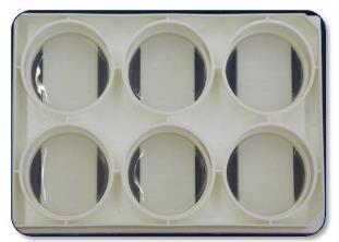
33 Figure 1.4 Flexcell system components 11
34 CHAPTER TWO A REVIEW OF SEX-RELATED STUDIES ON MAJOR HEART VALVE DISEASES AND ATHEROSCLEROSIS 12
35 A version of this chapter has been published as: Masjedi S, Ferdous Z. Understanding the Role of Sex in Heart Valve and Major Vascular Diseases. Cardiovascular engineering and technology. 2015;6(3): Springer 2.1 Introduction It is well accepted that there is difference in the prevalence and severity of major cardiovascular disorders among men and women. Male has been classically considered a risk factor in cardiac disorders. The molecular mechanism behind sex s role in the initiation and progression of heart valve diseases and atherosclerosis has barely been investigated. In chapter 1, I presented the microstructure of the aortic valve. Three thin layers are in each aortic cusp (Figure 2.1A): Fibrosa in the aortic side consists of densely oriented fibers of type I and type III collagens; Spongiosa in the middle mainly consists of proteoglycans and glycosaminoglycans with some collagen fibers; and Ventricularis on the ventricular side consists of a network of radially oriented elastin fibers [27]. In the mitral valve, the two leaflets are attached to the myocardial membrane by a series of tendon-like structures, called chordae tendinae (Figure 2.1B). The normal mitral leaflet layers are: Fibrosa, Spongiosa, and Atrialis facing the left atrium; whereas the chordae tendinae contains large number of longitudinally oriented collagen fibers [28]. 13
36 The blood vessel structure consists of three layers: Intima, a layer of endothelial cells in direct contact with the blood flow and an underlying connective tissue; Media, sheets of smooth muscles with connective tissues in between; and Adventitia, a layer of fibrous connective tissue covering the outermost part of the vessels (Figure 2.2 A,B) [29]. Previously, aortic valve stenosis was believed to be a passive condition resulted from atherosclerosis. This statement was due to numerous similarities between CAVD and atherosclerosis. The major similar characteristics of the two conditions include but not limited to: - Shared risk factors such as male sex, smoking, diabetes, hyperglycemia,.. - Calcium deposition within the tissue - Expression of similar enzymes and growth factors like ALP, BMPs, Osteopontin Previous studies compared the cultured VICs and vascular smooth muscle cells (VSMCs) differentiation and deformation in pathological conditions [30]. However, the sex-related cellular mechanism which expose male to excess risk have not been investigated in VICs and barely investigated in VSMCs. Similarly, no comparative sex-related study has been performed among heart valve disorders and atherosclerosis. The similar pathophysiology of these disorders motivated us to review previous sex-related studies on aortic stenosis, mitral valve prolapse and atherosclerosis. 2.2 Aortic Stenosis Aortic stenosis refers to the deposition of calcium within the aortic valve tissue [5, 6, 31]. Majority of early sex/gender-difference studies on aortic stenosis focused on the 14
 aortic cusp")

37 Figure 2.1 (A) aortic cusp layers, (B) mitral leaflet layers, porcine aortic valves H&E 15
 Aorta cross-section, (B)")
38 Figure 2.2 (A) Aorta cross-section, (B) vascular layers from aorta, rat H&E 16
39 effect of aortic stenosis on the myocardium, most importantly on left ventricular (LV) hypertrophy and LV degeneration, but not on the diseased aortic valve. For example, in a study by Carroll et al. on patients over 60 years with aortic stenosis, women s LV response was more adaptive, while men s cardiac performance (quantified by defined cardiac function indexes) was more frequently depressed [32]. Nordmeyer et al. investigated sex-dependent alterations in myocardial estrogen receptor and hypertrophyrelated gene expression in human aortic valve stenosis [33]. Estrogen receptors (ER) are a subgroup of Sex Steroid Hormone Receptors (SSHRs) expressed in mouse myocytes, fibroblasts, and human heart cells. The authors showed that α-er and β-er mrna increased in human hearts with aortic stenosis. While α-er mrna content among men and women were comparable, β -ER mrna content was significantly greater in women than men (47% vs. 28%) with respect to controls. This suggested a link between β-er expression and greater aortic stenosis in women [33]. Other studies measured sex difference in the biomolecular turnover in LV myocardium during aortic stenosis. Regitz-Zagrosek et al. reported that the overload pressure in left ventricle led to increased extracellular matrix (ECM) remodeling and collagen I, III, and fibronectin deposition in early stages of aortic stenosis [34]. Similarly, Skavdhal et al. studied the role of ER subtypes by inducing aortic stenosis condition in male and female mouse models [35]. They found that the female wild type mice had significantly lower induced hypertrophy in response to pressure overload as compared to male mice. Additionally, β-er attenuated response to pressure overload in female mice. 17
40 [36]. More comprehensive discussion on LV pathophysiology and changes in myocytes due to hypertrophy and role of sex hormones can be found in excellent review by Regitz- Zagrosek and Seeland [37]. Even though majority of sex studies on aortic stenosis focused on the heart, Aggarwal et al. used multidetector CT imaging to evaluate the severity of aortic stenosis among men and women. They found that aortic valve calcification is significantly lower in women compared to men at all stages even after accounting for their smaller body size. This work, however, did not offer any information on impact of sex/gender on the pathogenesis of aortic stenosis [38]. Another study by McCoy et al. on aortic valvular interstitial cells identified 183 genes to be significantly different in male vs. female healthy porcine aortic valves, many of them relevant to pathways activated in HVD. They concluded that the greater occurrence of HVD in male may originate from their genetic propensity to develop HVD at the cellular scale [39]. Similarly, data from our laboratory (in chapter 4) indicate a difference in ECM remodeling and early calcific marker expression such as alkaline phosphatase, between male and female valvular cells derived from rats. With growing awareness on the role of sex in HVDs, we anticipate more research will elaborate on the sex-difference in aortic stenosis in the near future. 2.3 Mitral Valve Prolapse Mitral valve prolapse (MVP) is often associated with a number of ruptured chordae tendineae, myxomatous thickening and tissue remodeling [40]. In advanced stages of 18
41 MVP, mitral regurgitation (MR) occurs with high possibility of heart failure. In fact, according to epidemiological studies, MR is the most common HVD [41, 42]. MR frequently occurs due to progression of myxomatous changes in valves with chronic prolapse conditions [43, 44]. The major risk factor for myxomatous mitral valve disease is age; however, a few sex/gender-difference studies in humans or dogs are available. In one of the early studies, Lax et al. reported that mitral valve prolapse occurs more frequently in women by testing a hypothesis that mild dehydration induces mitral valve prolapse [45, 46]. The interesting observation in these studies was that 50% of the women showed MVP symptoms, whereas only 10% men were affected. Even after considering the anatomical differences between female and male mitral valve leaflets and annulus, it could be concluded that MVP induced by dehydration is more likely to happen in healthy women than in men. In contrast, a separate study performed on a population with different body habitus, Savage et al. showed no significant difference in mitral valve prolapse between female and male in asthenic patients [47]. In another study, Avierinos et al. used a population of male and female patients with different stages of MVP: no regurgitation, with mild prolapse, and moderate or severe regurgitation [48]. They also accounted for the number of surgeries and the mortality rate in male and female. They found that women had significantly greater number of anterior and bileaflet prolapse, leaflet thickening (32% vs. 28%), lower posterior prolapse (22% vs. 31%), flail (2% vs. 8%), and severe regurgitation (10% vs. 19
42 23%) than men (p<0.001). Therefore, they concluded that women had a higher likelihood of getting MVP and at a younger age than men. Sex and gender difference studies on MR have also been very limited; however, a few clinical studies have identified male [43, 49] or, female [50-53] as risk factors for MR. Singh et al. designed a clinical study with two groups of patients: one with severe MR due to MVP and a group of randomly selected patients with MVP [44]. Their findings showed a lower cumulative risk of surgery for women with MVP than men (0.8% vs. 2.6%) before age 65 and (1.4% vs. 5.5%) by age 75. In another populationbased study, Jones et al. studied various MR risk factors in American Indian population [54]. They divided the participants by MR severity level and concluded that MR is independently associated with female sex, lower body mass index, older age and renal dysfunction. Likewise, Wilcken et al. performed a cohort study on Australian men and women that were at risk of MVP [55], and combined that with a clinical study on patients that had mitral valve surgery due to myxomatous mitral disease. The goal of this study was to estimate the cumulative risk of developing severe MR due to MVP in men and women. Their result showed that before age 50, the risk was low in general; however, men over 50 were at an increased risk of MR. In all age groups (50s, 60s and 70s), the risk in women was less than half of that in men [43]. The contradicting results on the above population-based studies on MVP/MR are not surprising, since it is difficult to isolate other variables and risk factors in clinical studies. Perhaps studies on the cellular/molecular mechanisms in MVP could shed light 20
43 on the sex-difference in this disease. Unfortunately, the authors were surprised to see the lack of literature in this area. In fact, we found only one report by Grande-Allen et al. that studied the difference in the abundance of a major proteoglycan versican among the male and female patients with myxomatous disease. [56]. Sex-difference was apparent in both normal and myxomatous valves: in normal valves, versican amount was higher in female than in male; in myxomatous samples, versican was higher in male. In addition, when the abundance of total proteoglycan (Decorin, Biglycan and Versican) was measured in myxomatous valves, male showed a trend of greater total proteoglycan than female valves. 2.4 Atherosclerosis Atherosclerosis is a chronic inflammatory disease with a similar mineralization process as aortic stenosis [57-59]. In later stages of atherosclerosis, vascular smooth muscle cells (SMCs) differentiate into osteoblast-like cells, and express proteins involved in bone formation, such as bone morphogenic protein (BMP)-2, osteopontin (OPN), matrix gla protein (MGP), and osteoprotegerin (OPG) [57]. A number of studies have reported some differences in the manifestations of atherosclerosis in male vs. female [58, 60-62]. Even though majority of these studies were based on clinical or epidemiological data, some recent reports investigated role of biomolecular factors in sex-dependent behavior of SMCs in atherosclerotic condition. For example, reactive oxidative species (ROS) contributes to atherosclerosis by enhancing oxidative stress and apoptosis [63-65]. However, only a few studies have investigated the effects of sex on the oxidative stress in 21
44 isolated vascular SMCs. Malorni et al. exposed freshly isolated male and female vascular SMCs to UV radiation as oxidative stress inducer. They reported that male cells produced significantly greater amounts of hydrogen peroxide and superoxide anion, both important ROS molecules. Additionally, they showed that male cells had more cytoskeleton damage due to UV radiation, which might lead to their increased apoptosis [66]. In their recent study, they further analyzed female cells apoptotic resistance by studying their behavior in anoikis, a type of cell apoptosis, via detachment from substrate. Their results confirmed that number of detached cells in female were lower both prior to UV radiation and after radiation [67]. A series of studies have shown sex-dependent interactions between SMCs and Nitric Oxide (NO), a molecule known to inhibit the development of neointimal hyperplasia [68, 69]. Hogg et al. evaluated the efficacy of NO at inhibiting the vascular SMC proliferation, migration and cell cycle progression in vitro, as well as neointimal hyperplasia in vivo. They reported that NO effectively inhibited proliferation, migration and cell cycle arrest in both sexes; however, male SMCs showed stronger response in both hormone-replete and deplete conditions than females. Similarly, their rat models treated with weight-dependent dosage of NO showed reduced neointimal hyperplasia in males than females. Furthermore, their rat models with carotid artery injury in hormonally intact or, castrated rats were in continuance with the in vitro study [68]. To determine the underlying causes for these sex differences, the authors further studied the role of ER on proliferation of male and female vascular SMCs following exposure to NO. 22
45 They observed that both α-er and β-er repressed proliferation in males, whereas only β- ER was effective in females. Since NO signals through ERs, it was less effective in reducing neointimal hyperplasia in female [69]. Another marker OPN, a non-collagenous adhesive protein in bone matrix [57, 70], promotes atherosclerotic lesions in early stages, but inhibits mineral deposition in later stages by attaching to the apatite crystal and preventing its growth [58, 71, 72]. Using mice deficient in both OPN and apolipoprotein E, Matsui et al. showed that female wild type mice had significantly larger atherosclerotic lesions compared to the knockout mice, suggesting they are at greater risk for atherosclerosis. No notable difference among male mice genotypes was observed in their study [58]. Other studies have focused on lipoprotein(a) [73, 74], a major risk factor for atherosclerosis and coronary heart disease [75]. High levels of plasma lipoprotein(a) correlate to early stages of coronary atherosclerosis predominantly in men [73, 74]. In a population-based study, Shreiner et al. compared plasma lipoprotein(a) levels to preclinical carotid atherosclerosis in middle-aged (45 to 64) white and black male and female patients by measuring their carotid wall thickness [73]. In this study, other risk factors such as smoking, diabetes and menopause were also considered. They reported that white men on average had the greatest mean carotid intimal-medial far-wall thickness among the four race/sex groups (0.798 mm compared to 0.779, 0.718, and mm in black men, black women, and white women, respectively). In Black women with diabetes, the lipoprotein(a) was significantly associated with wall thickness; 23
46 however, this association was not observed for non-diabetics. For white women, the association between lipoprotein(a) and wall thickness depended mostly on smoking status with nearly statistical significance. In general, they reported a correlation between lipoprotein(a) and carotid wall thickness in the preclinical atherosclerotic disease; however, this association was heavily affected by other risk factors. Nonetheless, in the absence of major risk factors, women had lower carotid wall thicknesses and lower amount of lipoprotein(a) in their blood compared to men. Stensland-Bugge et al. similarly employed ultrasonographic scanning to determine sex and age differences in carotid atherosclerosis by measuring carotid artery intima-media thickness [76]. Their results showed that in all age groups (25-40, 40-60, >60), men had a thicker mean intima-media layer than women, and this difference increased significantly with age. In addition, this study suggested fibrinogen as a strong risk factor in men than women. Another risk factor, physical activity, reduced the risk of cardiovascular disease in men, but had no significant effect on women [77]. When low, medium and high level of physical activity in each group was compared, medium physical activity had the most significant protective effect against CVD in men. Alternatively, a different study showed that low physical activity at work led to elevated incidents of stroke and death in women [78]. In another study by Stensland-Bugge et al., triglycerides strongly correlated with subclinical atherosclerosis in women than in men [76]. While previous studies have linked serum triglycerides levels and high-density lipoprotein cholesterol [79-81], this study reported 24
47 that intima-media thickness and triglycerides in women was independent of high-density lipoprotein cholesterol levels [76]. 2.5 Summary As reviewed in this chapter, male is a significant risk factor for CAVD and atherosclerosis. Comparatively, the sex-related investigations on the pathology of CAVD and atherosclerosis suggest similarities in the molecular events in the diseases initiation (endothelia cell activation) and progression (osteogenic differentiation and presence of osteoblast-like cells in calcific lesions). However, in mitral valve disorders females were reported to be at a higher risk. Table 2.1 summarizes the prevalence of the reviewed cardiovascular disorders among men and women. Table 2.1 Prevalence of Cardiovascular Diseases in Men and Women Disorder Male Female Aortic Stenosis A recognized risk factor Shows increased left ventricular hypertrophy after menopause Mitral Valve prolapse Increased ECM production Clinically at higher risk Atherosclerosis Greater risk in men Larger atherosclerotic lesion in OPN deficient female mice 25
48 CHAPTER THREE COMPARISON OF CALCIFICATION POTENTIAL OF VALVULAR INTERSTITIAL CELLS ISOLATED FROM INDIVIDUAL AORTIC CUSPS 26
49 A version of this chapter has been published as: Masjedi S, Amarnath A, Baily KM, Ferdous Z. Comparison of calcification potential of valvular interstitial cells isolated from individual aortic valve cusps. cardiovascular pathology. 2016;25: Elsevier Abstract: Calcific aortic valve disease (CAVD) is one of the most prevalent disorders among the elderly in the developed countries. CAVD develops via cell-mediated processes, and clinical data show that CAVD initiates mostly in the noncoronary cusp of the aortic valve. Valvular interstitial cells (VICs) populate the inside of heart valves and are a heterogeneous cell population. The goal of this study is to elucidate the difference in calcification potential among VICs isolated from the left, right, and noncoronary cusps of porcine aortic valves. VICs were isolated from each of the aortic valve cusps and cultured in calcifying medium for 14 days to induce calcification. The samples were assessed for calcium deposits, nodule formation, and calcific markers using alizarin red and Von Kossa staining, alkaline phosphatase (ALP) staining, ALP enzyme activity assay, and western blot. Extracellular matrix (ECM) production and degradation was measured using collagen and glycosaminoglycan (GAG) assay and gelatin zymography. We observed that VICs isolated from the noncoronary cusp expressed greatest amount of the above calcific markers as compared to the coronary cusps. Also, collagen and GAG content was 27
50 the greatest in noncoronary VICs. However, our zymography results showed significant difference only for active Matrix metalloproteinase-2 (MMP-2) expression between right and noncoronary VICs. Our results suggest that VICs among the three cusps within aortic valve might be inherently different, where a subpopulation of VICs might be predisposed to calcification. Keywords: Calcific aortic valve disease, coronary and noncoronary cusps, valvular interstitial cells, calcification 3.1 Introduction Calcific aortic valve disease (CAVD) refers to a major disorder ranging from mild inflammation within the aortic valve cusps (aortic sclerosis) to severe calcific nodule formation in the aortic valve tissue (aortic stenosis) [5]. These two conditions affect 26% and 2% of the population over 65 years in the developed countries, respectively [3, 4, 82]. End-stage aortic stenosis is associated with life-threatening outcomes, such as left ventricular hypertrophy [83, 84]. Unfortunately, the underlying events associated with the initiation and progression of CAVD is not fully understood. It has been reported that CAVD is a highly active condition mediated by valvular interstitial cells (VICs), the dominant cell population in heart valve tissue. In the aortic valve, VICs are distributed non-uniformly among the three valve layers the ventricularis, fibrosa, and spongiosa the fibrosa layer has the highest and spongiosa 28
51 layer the lowest cell density [1]. Numerous studies have shown that VICs have a heterogeneous cell population among the four heart valves and also in different locations within each valve [5, 7, 85, 86]. VICs were initially believed to mainly consist of fibroblast and myofibroblast characteristics [7, 87]. Recently, VICs have been categorized into a major population of quiescent VICs with a small subpopulation of progenitor VICs and osteoblast-like VICs [7]. The mesenchymal progenitor VICs have limited self-renewal ability when activated (avics) that may contribute to valve repair in injuries [6]. On the other hand, in calcifying conditions, VICs differentiate into osteoblastic VICs (obvic). avics display features of myofibroblasts, while obvics show characteristics of osteoblasts, or bone-like cells [88, 89]. The calcification of VICs in the presence of organic phosphate in culture occurs due to the upregulation of alkaline phosphatase enzyme. VICs also express hydroxyapatite in calcified nodules similar to the bone formation process [90]. The normal tricuspid aortic valve has two cusps with coronary inlets left and right coronary cusps that are connected to the left and right coronary arteries and are responsible for the blood supply to the heart muscle. The other cusp with no coronary inlet is referred as noncoronary cusp. This asymmetric configuration of the tricuspid aortic valve incurs different blood backflow pressure or possibly localized turbulence on the coronary and noncoronary cusps during the end diastolic cycle. In CAVD, the noncoronary cusp is often the first cusp affected with aortic lesions [2], and its stenotic area is larger than that of the other coronary cusps [91]. While the reason behind this 29
52 difference in calcification is mostly unknown, since VICs are sensitive to their mechanical environment, the non-uniform mechanical forces suggest that a heterogeneous cell population would exist in these cusps. The goal of this study is to assess the inherent cellular heterogeneity among the aortic valve cusps by quantifying VICs osteogenic properties in response to biomolecular factors in culture. We hypothesized that VICs residing in each of the aortic cusps have intrinsically different characteristics, where VICs isolated from the noncoronary cusp would calcify more than the coronary cusps. 3.2 Materials and Methods VIC Isolation and Culture Aortic valves were removed from freshly sacrificed four to six year old female porcine hearts obtained from Wampler s Farm Sausage, Inc., Lenoir City, TN. Each of the aortic valve cusps were removed, finely chopped, and incubated in collagenase II digestion solution (15000 U/ml) for 4 hours. The cells were cultured in growth media consisting of DMEM, 10% FBS, 1% L-glutamine, and 1% Penicillin/Streptomycin (all purchased from Mediatech, Inc., Manassas, VA) at 37 o C and 5% CO 2. All cells were passaged at 90% confluency and used for experiments at 4 passages. VICs were isolated from four different porcine valves (n=4). Unless otherwise specified, the VICs isolated from each of the left, right and noncoronary cusp were seeded onto 6-well plates at a density of 250,000 cells/well. 30
53 Osteogenic media consisted of 2.18 g/l beta-glycerophosphate, 50 mg/l L-ascorbic acid 2-phosphate and 1mM dexamethasone (all purchased from Sigma, St. Louis, MO) added to regular media. Experimental samples were cultured in osteogenic media, where the control wells were cultured in regular media. Culture duration was determined by preliminary studies in which the VICs were cultured in osteogenic media for different time points ranging from 4 to 20 days. We noticed the VICs expressed strongest osteogenic markers at day 14, whereas by 20 days, we observed increased cell monolayer detachment possibly due to cell apoptosis. As a result, we selected day 14 as the longest calcification duration for this study. Further experiments were performed at 4, 7, 10, and 14 day time-points Hematoxylin & Eosin (H&E) Staining of Tissue Sections Freshly isolated aortic valve cusps were fixed in 10% formalin, paraffin embedded, and sectioned using established histology procedure [92]. The tissue sections were stained with Hematoxylin 2 for 5 minutes and Eosin Y for 1 minute, both purchased from Richard-Allen Scientific, Kalamazoo, MI. The stained sections were imaged using a brightfield microscope, and the width of the imaged sections was measured using ImageJ software (NIH) DNA Content Quantitation The cell density for VICs isolated from the coronary and noncoronary cusps were measured using a DNA assay [92]. Briefly, the cell monolayers were scraped from each 31
54 of the wells of 6-well plate and washed with phosphate buffered saline (PBS) and digested in Proteinase-K solution (1mg/mL) (Sigma). The cell lysates were vortexed and then incubated at a temperature between 60 o C and 70 o C for 90 minutes in a water bath and then heated at >70 o C for 30 minutes to denature the Proteinase-K. Released DNA was tagged with Hoechst dye (Sigma) and fluorescence emission was measured using a BioTek H1 flourocolorimeter at 458 nm. Calf thymus DNA standards (Sigma) were used in each assay to calculate DNA density Von Kossa and Alizarin Red Staining of Cultured VICs The amount of mineral deposition of cultured VICs isolated from each aortic valve cusp was determined using Von Kossa staining [30]. The cell monolayers in each well were fixed in 10% formalin (Fisher Scientific, Fair Lawn, NJ). A silver nitrate (Ricca Chemical Co., Arlington, TX) solution (3%) was added to the fixed cells, incubated and then exposed to UV until the calcium salts turned dark brown or black. The undissolved salts were removed using a 5% sodium thiosulfate (Sigma) solution and dehydrated with Flex before imaging. The calcium precipitation and nodule formation of the calcified VICs were assessed using alizarin red S staining [93]. First, the VICs were fixed with 10% formalin and treated with alizarin red solutions at room temperature for 4 to 5 minutes. The excess dye was washed off, and then the VICs were dehydrated with 100% acetone for less than 1 minute. The stained samples were finally imaged at 10X magnification; image analysis was performed using ImageJ software. 32
55 For image analysis, a number of 30 to 40 images per well were imported into Image J. The color channel on each image was split, and the threshold was adjusted to form a black and white image. Finally, the area, area fraction, integrated density, and perimeter of each image were measured Calcium Assay To measure the calcium content [30], the VICs were homogenized in 1N acetic acid, and arsenazo dye was added to the samples. The calcium content was measured against calcium standard (Ricca chemical, Arlington, TX), and the resulting absorbance was detected at 650 nm using a BioTek H1 plate reader. The calcium content was normalized to total DNA content Alkaline Phosphatase (ALP) Staining and Enzyme Assay The VICs were stained for ALP using a kit (Sigma) following manufacturer s instruction. Briefly, the VICs were fixed with citric acid, 99% methanol and 10% formalin. The VICs were then incubated in the FRV-alkaline dye and hematoxylin for 15 minutes each. The number of stained cells and total number of cells were counted using the particles analyzing tool in Image J software. To measure the ALP enzyme activity [93], the cultured VICs were rinsed with PBS, then each well was scrapped into 700μL RIPA lysis buffer and sonicated. The ALP enzyme in the samples was measured against standards prepared from 50 μm p- Nitrophenol solution. A solution of 221 alkaline buffer (2-Amino-2-methyl-1-propanol, 33
56 1.5mol/L, Sigma) and phosphatase substrate (p-nitrophenyl phosphate, disodium, Sigma) was added to the samples and incubated for 30 minutes at 37 o C. The reaction was stopped using 1mM sodium hydroxide solution and the absorbance was measured at 405 nm using a BioTek H1 plate reader. The ALP amount in each sample was normalized to the total protein content. The total protein content was determined using Pierce BCA Protein assay kit (Thermo Scientific) Glycosaminoglycan (GAG) Content The VICs and lyophilized aortic valve cusp tissue samples digested in Proteinase-K solution were used to measure GAG content [30]. Dymethylmethylene blue (DMMB) dye was added to the samples, and the resulting absorbance was measured at 525 nm. The total GAG content in each sample was determined against chondroitin sulfate standards and normalized to total DNA content Collagen Content The collagen type I content was measured using a sirius red assay. The VICs were digested in proteinase-k, and the lyophilized cusp tissues were digested in pepsin (Fisher Scientific). The samples were incubated with a sirius red dye (TCI, Portland, OR) solution (0.3 mg/ml, in 0.5M acetic acid) and then centrifuged at 6000 rpm for 30 minutes. The supernatant was carefully collected from each sample, and the absorbance was measured at 540 nm using a BioTek H1 plate reader. The collagen content was 34
57 measured against standards prepared from bovine dermal collagen type I (MP Biomedicals, Solon, OH) and normalized to total DNA content Gelatin Zymography Gelatin zymography was used to determine the amount of matrix metalloproteinase (MMP)-2, 9 enzyme produced within the cultured cell monolayers [30]. Briefly, equal amounts (5 mg of protein) of non-reduced samples were loaded into 10% zymogram gels. After electrophoresis, the gels were shaken in a renaturing buffer for 30 minutes and cleared overnight in developing buffer at 37 o C (Life Technologies, Grand Island, NY). The gels were then stained using colloidal blue staining kit (Life Technologies) and destained in 30% ethanol/10% acetic acid for 30 minutes, followed by a rehydration cycle in 2% acetic acid for at least 15 minutes. The gels were imaged using a Biorad ChemiDoc XRS imager, and the band densities were calculated using gel analysis tool in Image J software Western Blotting The cell pellet was collected in a similar manner as described in DNA assay and digested in RIPA buffer containing protease inhibitor. The protein concentrations were measured using Pierce BCA protein assay kit (Thermo Scientific). Equal amounts (5 mg of protein) of samples were run on the acrylamide gels and transferred into nitrocellulose membranes (A&B biosystems). The protein bands were detected using Runx2 antibody (Santa Cruz Biotechnology Inc., Santa Cruz, CA) and loading control actin (1-19) (sc- 35
58 1616) (Santa Cruz Biotechnology Inc.). The secondary antibody was donkey polyclonal secondary antibody to rabbit IgG- HPR (ab6802) (Abcam Inc., Cambridge, MA). The bands were detected using a western blot chemiluminescence detection kit (Fisher Scientific). The membranes were imaged using a Biorad ChemiDoc XRS imager, and the band densities were calculated using gel analysis tool in Image J software Statistical Analysis Each experiment was repeated for four different cell isolations (n=4). The most significant difference was seen after 14 days of culture in almost all of the experiments; therefore, the plots are presented only for day 14 for the majority of the measurements. Statistical significance for the different experiments was obtained using single-factor ANOVA. A p-value of 0.05 was considered significant. 3.3 Results H&E images of healthy porcine aortic valve tissue sections did not show any visible difference in composition among the three cusps (Fig. 3.1A,B,C). Similarly, no significant difference was observed in the thickness of the cusps (0.1579, , cm for left, right and noncoronary respectively, Table 3.1) measured using Image J software. Furthermore, no significant difference was observed in collagen and GAG content among the healthy porcine aortic valve cusps (Fig. 3.1D,E). 36
59 Table 2.1 Tissue section thickness of the porcine aortic cusps Aortic valve cusp Left Right Noncoronary Average thickness (cm) n= Comparable Cell Density in VICs Isolated from Coronary and Noncoronary Aortic Cusps There was no significant difference in DNA content for VICs isolated from the cusps at the different time points. As shown in Fig.3.2, DNA concentration and fold change normalized to respective controls was comparable after 14 days of osteogenic culture for the VICs isolated from each of the aortic cusps Elevated Osteogenic Marker Expression in VICs Isolated from Noncoronary Aortic Cusps Von Kossa staining of the VICs isolated from all three cusps demonstrated calcification in the cell monolayers under osteogenic condition compared to respective controls. The calcified area percentage and integrated density were significantly greater for the VICs isolated from the noncoronary cusp than for the left or right coronary cusps after 14 days of culture (Figure 3.3e,f). Furthermore, the amount of calcification increased with culture duration in osteogenic media in VICs isolated from all aortic cusps. 37

60 Figure 3.1 H&E staining of paraffin-embedded healthy porcine aortic valve cusp sections: (A) left, (B) right, and (C) noncoronary cusp. Parts D and E shows collagen and GAG content for the three cusps measured using a collagen and a GAG assay. No significant difference was observed in the thickness, collagen, and GAG content among the cusps. The data represent mean ± std. error, n=3 porcine aortic valves. Scale bar is 250 μm. 38
 DNA concentration for VICs isolated from")
 fold change for the above VICs in osteogenic culture normalized to their")
61 Figure 3.2 DNA content measured using a DNA assay: (A) DNA concentration for VICs isolated from left, right, and noncoronary aortic cusps at day 14 of culture in regular and osteogenic media, and (B) fold change for the above VICs in osteogenic culture normalized to their respective controls. The data represent mean ± std. error, n=4 porcine aortic valves. 39
 shows mineral deposition for a representative nodule from left, right and noncoronary cusps (left to right); scale bar is 100 μm.")
62 Figure 3.3 Images of Von Kossa stained VIC samples isolated from (A) left coronary, (B) right coronary, and (C) noncoronary aortic cusps cultured in osteogenic media for 14 days; scale bar is 250 μm. (D) shows mineral deposition for a representative nodule from left, right and noncoronary cusps (left to right); scale bar is 100 μm. Image analysis results show (E) the area fraction and (F) integrated density of mineral deposition in VICs isolated from each aortic cusp after 4, 7, 10, and 14 days of culture in osteogenic media. The data represent mean ± std. error, n=4 porcine aortic valves. indicates p<0.05 compared to left coronary and # indicates p<0.05 compared to right coronary at the same time point. 40
63 Alizarin red S staining of the calcific nodules in VIC monolayers demonstrated that there were differences in the area percentage, integrated density, and size of the calcific nodules in the VICs isolated from each aortic cusp. The noncoronary and right coronary cusps showed significantly greater area fraction and nodule size compared to the left coronary after 14 days of culture in osteogenic condition (Figure 3.4g,h).For integrated density, the only significant difference was for noncoronary cusp when compared to the left coronary (Figure 3.4i). Furthermore, the size of the calcific nodules increased with culture duration for all of the aortic cusps. Not unexpectedly, as the nodules fused with each other and grew bigger, their number went down in most cases. The calcium content normalized to respective controls was significantly greater for VICs isolated from noncoronary aortic cusp than left and right coronary cusps after 14 days of culture in osteogenic condition (Figure 3.5). ALP staining of the VICs in osteogenic culture showed positive staining from day 7 onwards, and maximum number of ALP positive cells was observed by day 14 (Figure 3.6 A,B). No ALP positive staining was detected in the control wells at any time point. At day 14, the % ALP positive cell was significantly greater for VICs from noncoronary cusp than those from either left or right coronary cusp (Figure 3.6C). Similarly, the ALP enzyme activity increased in all VICs when cultured for longer than 7 days in osteogenic condition as compared to their respective controls (data not shown). Additionally, the 41
 left coronary, (B, E) right coronary, and (C, F) noncoronary aortic cusps cultured in osteogenic media for 10")
64 Figure 3.4 Nodule formation measured by alizarin red staining in VIC samples isolated from (A, D) left coronary, (B, E) right coronary, and (C, F) noncoronary aortic cusps cultured in osteogenic media for 10 and 14 days respectively. Scale bar is 250 μm. Image analysis results show (G) the nodule area fraction, (H) nodule perimeter, and (I) nodule integrated density in VICs isolated from each aortic cusp after 4, 7, 10, and 14 days of culture in osteogenic media. The data represent mean ± std. error, n=4 porcine aortic valves. indicates p<0.05 compared to left coronary and # indicates p<0.05 compared to right coronary at the same time point. 42
65 Figure 3.5 Fold change in the calcium, GAG, and collagen I content for VICs isolated from left, right, and noncoronary cusps normalized to their respective controls at 14 days of culture in osteogenic media. Noncoronary VICs showed significantly greater calcium, GAG, and collagen I content compared to the coronary VICs. The data represents mean ± std. error, n=4 porcine aortic valves. indicates p<0.05 compared to left coronary and # indicates p<0.05 compared to right coronary. normalized ALP activity was significantly elevated for VICs isolated from noncoronary cusps with respect to the left and right coronary cusps at day 14 (Figure 3.6D). Similar to ALP, Runx-2 expression, measured using western blot and normalized to housekeeping marker β-actin and then to respective controls, was significantly greater in VICs from the noncoronary cusp than from the coronary cusps after 14 days of culture (Figure 3.7A,B). 43
 Images of ALP positive VIC samples isolated from left coronary, right coronary, and noncoronary aortic cusps cultured in osteogenic media")
66 Figure 3.6 ALP staining and enzyme activity measurement in VICs: (A) Images of ALP positive VIC samples isolated from left coronary, right coronary, and noncoronary aortic cusps cultured in osteogenic media for 7,10,14 days; scale bar is 200 μm (B) Representative image of stained cells for image analysis; scale bar is 50 μm (C) Percent of ALP/TNAP positive VICs at days 7, 10, and 14 of osteogenic culture. (D) Fold change in the ALP enzyme activity at day 14 of osteogenic culture normalized to respective controls. The data represents mean ± std. error, n=4 porcine aortic valves. indicates p<0.05 compared to left coronary and # indicates p<0.05 compared to right coronary. 44
 Representative image of bands for Runx2 and β-actin (control) for VICs isolated from one porcine aortic valve.")
67 Figure 3.7 Runx2 expression in VICs isolated from left, right, and noncoronary aortic cusps at 14 days of culture. (A) Representative image of bands for Runx2 and β-actin (control) for VICs isolated from one porcine aortic valve. For each cusp, samples 1 and 2 were cultured in osteogenic media and control was cultured in regular media. (B) Image analysis of the bands at day 14 of culture. The data represent mean ± std. error, n=2 porcine aortic valves. indicates p<0.05 compared to left coronary, indicates p=0.05 compared to right coronary. 45
68 3.3.3 Elevated Matrix Remodeling in the Calcified Cells The total GAG content normalized to respective controls was significantly greater for VICs isolated from noncoronary aortic cusp than left and right coronary cusps after 14 days of culture in osteogenic condition (Figure 3.5). The difference in normalized GAG content was greatest at day 14 than for earlier time points for the noncoronary cusp as compared to the coronary cusps (data not shown for earlier time points). Similarly, collagen I content normalized to respective controls at day 14 was significantly greater for the noncoronary cusp as compared to the coronary cusps. Gelatin zymography showed that VICs in both osteogenic and regular media expressed inactive (pro) MMP-2; however, activated MMP-2 was observed only in VICs cultured in osteogenic condition. Figure 3.8A shows a representative image of the active (bottom white lines) and inactive (top lines) MMP-2. Image analysis of the bands showed that the VICs isolated from noncoronary aortic cusps demonstrated significantly elevated amount of active MMP-2 enzyme as compared to the right coronary cusp; however, active MMP-2 amount in the noncoronary cusps was not significantly elevated when compared to the left coronary cusp (Figure 3.8B). Similarly, no significant difference was obtained for the inactive and total MMP-2 content among the VICs from any of the groups. The bands for MMP-9 were not detectable in this study. 46
 Representative gel image for VICs isolated from left, right, and noncoronary cusps from the same porcine aortic valve.")
69 Figure 3.8 MMP-2 expression (active, inactive, total) detected by gelatin zymography for VICs isolated from left, right, and noncoronary at 14 days of culture. (A) Representative gel image for VICs isolated from left, right, and noncoronary cusps from the same porcine aortic valve. (B) Image analysis of the bands for VICs isolated from each aortic cusp at day 14 of culture. The data represent mean ± std. deviation, n=4 porcine aortic valves. # indicates P<0.05 compared to right coronary. 47
70 3.4 Discussion In the present work, we observed a correlation between the location of the VICs within aortic valve cusps and the events associated with the initiation of calcification when cultured in pathological conditions. There was no significant difference in tissue thickness, collagen, and GAG content among the three cusps of healthy porcine aortic valves used for VIC isolation. The primary outcomes of this research are that VICs isolated from the noncoronary aortic cusp expressed a greater amount of calcific markers (ALP), osteogenic differentiation (Runx2), and mineralization when compared to left and right coronary cusps after 14 days of culture in osteogenic condition. Moreover, no significant difference in the normalized DNA content was noticed among the VICs, inferring that the observed difference in calcific markers and mineralization among the cusps was not due to a larger cell density. Our results suggest that there might be an inherent difference in VICs characteristics isolated from noncoronary and coronary cusps that initiates calcification earlier than other cell population. Numerous reports suggest that the noncoronary cusp is affected first during CAVD. The difference in calcification among aortic cusps has been studied clinically using echocardiography [94, 95] or CT imaging [26, 96] on stenotic patients. It was reported that the location of noncoronary cusp commissure correlates to the highest pressure overload or calcium deposition. An attempt to detect difference in early-stage calcification between the coronary and noncoronary cusps has been performed by Cujec and Pollick [95]. They reported that isolated thickening of a single aortic cusp was 48
71 frequently seen in the noncoronary cusp and rarely seen in the left coronary cusp. In another work, the calcified aortic cusps were weighted separately in valve transplantation patients, and the most frequent case reported was that one cusp was heavier with the other two having similar weights. However, they did not clarify which cusp had more weight [97]. Recently, a new study measured the calcific volume using the CT method in severely calcified cusps and showed that on average, the noncoronary cusps had greater volume of calcification than the left and right coronary cusps [96]. Ectopic aortic calcification is associated with expression of osteogenic and matrix remodeling markers. In pathological conditions, VICs form osteogenic nodules with increased mineralization and ALP enzyme activity [31, 90, 98]. Our study showed that the area covered and average size of the calcific nodules were not similar for VICs isolated from all cusps the noncoronary VICs demonstrated the greatest % area and perimeter of nodules. Similarly, the integrated density analysis of the nodules demonstrated the greatest mineralization in noncoronary cusps. To support these observations, the calcium content was greatest for the VICs from noncoronary cusp by 14 days of osteogenic culture. Aortic calcification is associated with bone formation via osteogenesis [99]. The VICs under osteogenic culture condition differentiated into osteoblast-like cells [6], as is found in human calcified aortic valve tissues [100]. Since calcific nodules contain osteoblast-like VICs, it can be inferred that noncoronary cusps has an elevated number of differentiated VICs and a propensity for active bone formation. Osteogenic nodule formation is marked with significant amount of ALP 49
72 enzyme within the calcific deposits [13]. In our study, we observed that the amount of ALP and percent ALP positive cells were significantly greater in noncoronary VICs than in the left and right coronary cusps by day 14 of culture. Likewise, Runx-2 expression was significantly enhanced in VICs from noncoronary than for coronary cusps at 14 days of culture, suggesting osteoblast-like differentiation is ongoing at different levels in the VICs. Interestingly, we noticed positive Runx-2 band in a few of the control western blot samples, even though the control samples did not stain positive in von kossa or alizarin red. Since VICs have been reported to spontaneously calcify [101, 102], it is possible that some of the VICs in control samples were also undergoing osteogenic differentiation. Calcific aortic valves show major ECM remodeling and abnormal activation of MMPs, a family of zinc-containing matrix enzymes that cleave various ECM components. An abundance of active MMP-2 enzyme among VICs is an early marker of matrix remodeling in aortic valve cusps [13, 103]. In our study, increased collagen I was synthesized by noncoronary VICs in osteogenic culture compared with left and right coronary. Furthermore, we observed a trend of increased active MMP-2 in VICs isolated from the noncoronary cusps in osteogenic culture after 14 days compared to left and right coronary cusps, indicating elevated matrix remodeling in noncoronary cusps. However, the trend for total active and inactive MMP-2 was not consistent, and no significance was noted among the groups. It might be that a longer culture or greater cell density, which was not selected in this study due to increased apoptosis, is needed to obtain a stronger difference in MMP-2 enzyme expression for the VICs. It should be noted that other 50
73 MMPs, such as MMP-1, 9, and 13 have been detected in diseased valve tissues [104], but only MMP-2 was detected in the VICs in this study. Our results are consistent with the literature since majority of VIC calcification studies measured only MMP-2 enzyme expression in VICs [103, 105, 106]. In fact, MMP-2 is the predominant ECM degrading enzyme expressed by VICs under most conditions [7], and expression of other MMPs in VICs in culture often requires their isolation from diseased tissues [107] or stimulation by growth factors and cytokines. Since no growth factor or cytokines were used in the osteogenic media, we could only detect the heavily expressed MMP-2 in this study. ECM remodeling is also associated with excessive synthesis of ECM proteins like GAGs [108]. Our results also showed increased GAG amount in noncoronary VICs compared with left and right coronary, confirming excessive matrix synthesis in the noncoronary cusp. VICs have been identified as a heterogeneous population in numerous cell culture and tissue-based studies. The mesenchymal progenitor VICs subpopulation has the ability to differentiate into osteoblast- or myofibroblast-like cells based on environmental cues or local mechanical stimuli [6]. In the present study, we showed for the first time that the osteogenic marker expression of the VICs and their ECM remodeling differ among the coronary and noncoronary aortic cusps. Heterogeneity of VICs according to their location within the aortic valve has been reported previously showing that VICs from fibrosa layer demonstrated increased matrix remodeling and osteogenic markers compared to the ventricularis layers [ ]. It was suggested that this is due to the different levels of cellular level strain experienced by VICs residing in each side of the cusp. VIC 51
74 phenotype analysis further showed that VICs in the fibrosa layer have a higher tendency to appear as myofibroblast- or osteoblast-like cells. On the other hand, VICs from the ventricularis layer showed greater potential for myofibroblast differentiation than those from the fibrosa layer in response to profibrotic stimuli in vitro suggesting that fibrosa VICs are inherently less sensitive to profibrotic signals [11]. Based on the present study, VICs from coronary cusps are inherently less responsive to osteogenic condition than noncoronary VICs. This study was limited by the fact that only early calcification markers, such as ALP and Runx2, could be detected in the osteogenic cultures. Since we chose not to use any calcification enhancing growth factors and cytokines, such as TGF-β, bone morphogenic proteins (BMPs), tumor necrosis factors (TNF)-α in our osteogenic media, a culture of minimum 21 days [31, 114] would likely be required to detect late calcification markers (osteocalcin, osteopontin etc.). Unfortunately, we had to limit our culture to 14 days to avoid excessive apoptosis in the VICs. This study also opens up questions regarding size of different cell population within each cusp. In addition to the local difference in mechanical forces due to the coronary inlets and mitral backflow, the embryonic origin of aortic valve cusps could predispose these VICs differently to calcification. For example, cardiac neural crest cells (NCC) are uniquely distributed within the aortic valve during development, where majority of NCCs are present in left and noncoronary cusp [115]. Additionally, selective knockout of certain NCC markers reportedly results in abnormal aortic valve formation similar to bicuspid aortic valves [116]. It is possible that due their 52
75 embryonic origin, VICs from the left and noncoronary cusp will have elevated response to procalcific conditions than the right cusp. Unfortunately, the measurements reported in this study did not show any consistent trend between the left and right cusp. Further investigation is thus needed to discern the relationship between the VICs propensity towards osteogenic differentiation and the contributions of cellular embryonic origin and local mechanical stimuli. 3.5 Conclusion In this study, we report a difference in the initiation of calcification of the VICs isolated from the coronary or noncoronary cusps. VICs isolated from noncoronary aortic cusps expressed elevated amount of osteogenic markers and greater matrix remodeling than VICs isolated from left and right coronary cusps. We also observed a positive correlation between the duration of osteogenic culture and calcification of the isolated VICs for all three aortic cusps. Furthermore, the above differences among the VICs were not due to an increased cell density in culture. Our results suggest that the VICs distributed among all three aortic valve cusps might be inherently different, or that a subpopulation of VICs predisposed to calcification might be present at varying density among the cusps. Further investigation is required to characterize the VICs different subphenotypes throughout the aortic valve and to determine the role of local mechanical stimuli and cellular embryonic origin on VIC differentiation. 53
76 CHAPTER FOUR SEX-RELATED DIFFERENCES IN MATRIX REMODELING AND EARLY CALCIFIC MARKERS IN RAT AORTIC VALVULAR INTERSTITIAL CELLS 54
77 A version of this chapter is to be published as: Masjedi S, Lei Y, Ferdous Z. Sex-related differences in matrix remodeling and early calcific markers in rat aortic valvular interstitial cells, in preparation. Abstract: Calcific Aortic Valve Disease (CAVD) is a major cardiovascular disorder in the developed countries. Male is a known risk factor in this disease; unfortunately, how sex contributes to CAVD is mostly unknown. The objective of this study was to determine whether valvular interstitial cells (VICs) isolated from male versus female aortic valves demonstrate difference in calcification and/or extracellular matrix (ECM) remodeling. VICs were isolated from male and female rat aortic valves and cultured in osteogenic media for 10, 15 and 20 days. The proliferation among male and female VICs was assessed by a cell growth assay. The matrix remodeling of the VIC samples was quantified using glycosaminoglycan (GAG), collagen type I and gelatin zymography assays. Early calcific marker expression was assessed using alkaline phosphatase (ALP) staining and enzyme activity assay. Our result showed that proliferation of VICs was significantly greater in female than male after 12 days of culture in regular media. Additionally, male VICs showed greater amount of normalized GAG, collagen I, and activated matrix metallopreoteniase-2 expression compared to female. Similarly, ALP content was greater in male VICs than female at all time points. 55
78 The results from this research suggest that there is sex-related difference in the events associated with osteogenic differentiation of the aortic VICs, where male VICs are more prone to calcification. Keywords: Calcific aortic valve disease, valvular interstitial cells, sex-related differences, osteogenic differentiation 4.1 Introduction Aortic valve stenosis is the most common cause of valve replacement surgery in the US [12] and occurs at the advanced stage of calcific aortic valve disease (CAVD). CAVD is an active cell-mediated disorder mostly caused by ectopic calcification in the aortic valves [15]. Unfortunately, the underlying mechanisms of initiation and progression of CAVD are not fully understood. Furthermore, since sex is a major risk factor for CAVD, there is an urgency for a better understanding of this diseases pathology and role of key risk factors. Male sex has been identified as an independent risk factor for CAVD. Clinical reports have shown that male gender has two fold excess risk of aortic valve calcification [82]. Similarly, based on the Society of Thoracic Surgery risk scores, men have 19% higher relative rate of undergoing aortic valve replacement surgery due to aortic valve stenosis than women [117]. Several population-based studies highlighted differences in aortic valve calcification between male and female indirectly by concentrating on left ventricular hypertrophy due to pressure overload caused by aortic valve stenosis [118-56
79 121]. Furthermore, men appear to have more aortic valve calcium score and fibrosis thickening compared to women with aortic valve stenosis measured by Computer- Tomography [38, 122]. Analysis of the longitudinal sections of the aortic valves in aged mice showed significantly thicker leaflets and higher degree of aortic regurgitation in male mice compared to female [123]. The impact of sex on pathogenesis and progression of aortic valve stenosis at cellular and molecular levels thus needs to be investigated. Aortic valve calcification involves various mechanisms to interact with the residing cells within the aortic valve. There are two main cell types in the aortic valve: valvular endothelial cells (VECs), a cellular monolayer lining the outer surface of the valve cusps; and valvular interstitial cells (VICs), the dominant valve cell population that are distributed heterogeneously within the interstitial matrix [7]. At the initial stage of calcification, inflammation occurs due to VEC activation which triggers the extracellular matrix (ECM) remodeling and VIC differentiation. This is followed by expression of calcific markers such as alkaline phosphatase (ALP), Runx2, osteocalcin, and osteopontin, change in ECM proteins and enzymes such as glycosaminoglycan (GAGs) and matrix metalloproteinases (MMPs). The abnormal amount of calcific deposition within the ECM results in tissue stiffness and nodule formation as seen in stenotic valves. The role of sex/gender on non-valvular cardiovascular diseases has been contributed to the sex steroid hormones- mainly estrogen- and their receptors in the cardiovascular cells. β-estradiol, the natural form of estrogen in human, was shown to have a protective effect 57
80 in women in heart valve diseases. Molecular mechanism of β-estradiol contribution to VICs fate still remains unknown. In this study, we investigated whether the rat aortic valvular interstitial cells VICs isolated from male and female rat aortic valves show differences in matrix remodeling and early calcific markers when exposed to osteogenic culture conditions. We also assessed the VICs proliferation in the presence of β-estradiol. We hypothesized that VICs isolated from each of the sexes would demonstrate different characteristics, where male VICs would demonstrate greater matrix remodeling and calcification compared to female VICs. 4.2 Materials and methods Primary VIC isolation and culture Rat aortic valvular interstitial cells (RAVICs) were isolated and cultured from the aortic valves of freshly sacrificed 9-week-old male and female Sprague-Dawley rats (Envigo, formerly Harlan laboratory, Indianapolis, IN) using explant technique [17]. Briefly, the aortic valves were gently cut into 4-5 pieces and placed into a T25 culture plate and covered with a growth media consisting of DMEM, 10% FBS, 1% L-glutamine, and 1% Penicillin/Streptomycin (all purchased from Mediatech, Inc., Manassas, VA). The culture plates were then placed in cell culture incubators at 37 o C and 5% CO 2. At 50% confluency, the valve pieces were removed, and the RAVICs were split into T75 culture dishes. All cells were passaged at 90% confluency and used for experiments 58
81 between 6-7 passages. RAVICs were isolated from four different pairs of rat valves (n=4 for each sex). Unless otherwise specified, the male and female RAVICs were seeded onto 6-well plates at a density of 250,000 cells/well. Osteogenic media consisted of 2.18 g/l betaglycerophosphate, 50 mg/l L-ascorbic acid 2-phosphate and 1mM dexamethasone (all purchased from Sigma) added to regular media. Experimental samples were cultured in osteogenic media, where the control wells were cultured in regular media. Culture duration was determined by preliminary studies where the RAVICs were cultured in osteogenic media for different time points ranging from 10 to 25 days. We noticed the RAVICs expressed strongest osteogenic markers at day 15 whereas by 21 days, we observed increased cell monolayer detachment possibly due to cell apoptosis. As a result, we selected day 20 as the longest calcification duration for this study. Further experiments were performed at 10, 15 and 20 day time-points DNA content A colorimetric DNA assay was used to quantify the cell density of the male and female RAVICs [124]. Briefly, the cell pellets were digested in Proteinase-K solution (1mg/mL) (Sigma), followed by incubation of the cell lysates at a temperature between 60 o C and 70 o C for 90 minutes in a water bath and then heating at >70 o C for 30 minutes to denature the Proteinase-K. Hoechst dye (Sigma) was used to tag the released DNA. The fluorescence emission was measured using a BioTek H1 Fluoro-colorimeter at 458 nm. Calf thymus DNA standards (Sigma) were used to calculate the DNA density. 59
82 4.2.3 Cell growth assay The rates of RAVICs growth for each sex were quantitatively determined as previously described [125]. Briefly, male and female RAVICs were seeded in separate 12-well plates at a density of 10,000 cells/well and cultured for up to14 days in both regular and osteogenic culture media. The media was changed every 2 days. Every 2 days, the cell populations were counted from triplicate wells using a hemocytometer (trypan blue exclusion) GAG content The GAG content was measured by adding a solution of Dymethylmethylene blue (DMMB) dye (Sigma) to the male and female RAVICs samples digested in Proteinase-K solution. The resulting absorbance was measured at 525 nm using a BioTeck H1 plate reader. The total GAG content in each sample was determined against chondroitin sulfate standards and normalized to total DNA content Collagen type I content The collagen type I content was measured using a sirius red assay. The Proteinase- K digested RAVICs samples were incubated with a sirius red dye (TCI) solution (0.3 mg/ml, in 0.5M acetic acid) and then the supernatant was carefully collected from each sample after centrifugation at 6000 rpm for 30 minutes. The absorbance was measured at 540 nm using a BioTek H1 plate reader. The collagen standards were prepared using 60
83 bovine dermal collagen type I (MP Biomedicals). The total collagen type I content was normalized to total DNA content Gelatin zymography The amount of matrix metalloproteinase (MMP)-2, 9 enzyme produced by the RAVICs from each sex was determined using gelatin zymography. Briefly, equal amounts (5 mg of protein) of samples were ran through 10% zymogram gels, then renatured for 30 minutes and rinsed overnight in developing buffer at 37 o C (Life Technologies, Grand Island, NY). A colloidal blue staining kit (Life Technologies) was used to stain the gels followed by destaining in 30% ethanol/10% acetic acid for 10 minutes and rehydration in 2% acetic acid for at least 15 minutes. The gels were imaged using a Bio-Rad ChemiDoc XRS imager, and the band densities were determined using Image J software ALP staining and enzyme activity For ALP staining, the male and female RAVICs were fixed by a previously optimized mixture of citric acid, methanol and formalin [124]. Next, the fixed cell monolayers were stained with FRV-alkaline dye according to manufacturer s protocol (Sigma 86R kit), followed by counter staining with diluted hematoxylin (1:1). The percentage of positively stained and total cells were calculated using the particles analyzing tool in Image J software. 61
84 To measure the ALP enzyme activity, the cultured RAVICs were homogenized in RIPA lysis buffer. 50 μm p-nitrophenol solution (Sigma) was used to prepare standards. The samples were incubated with alkaline buffer (2-Amino-2-methyl-1-propanol, 1.5mol/L, Sigma) and phosphatase substrate (p-nitrophenyl phosphate, disodium, Sigma) for 30 minutes at 37 o C. The reaction was stopped using 1mM sodium hydroxide solution, and the absorbance was measured at 405 nm using a BioTek H1 plate reader. The ALP amount in each sample was normalized to the total protein content determined using Pierce BCA Protein assay kit (Thermo Scientific) β-estradiol treatment The male and female RAVICs were seeded separately in 12-well culture plates at a density of 10,000 cells/well and cultured in regular media prepared with phenol red-free DMEM (Life Technologies) for 24 hours. Next, the VICs were made quiescent by serum starvation for 48 hours in the above mentioned media without FBS. Lastly, the VICs were treated with media supplemented with different concentrations of β-estradiol (0-40 ng/ml) (Sigma) for 48hours. The cell numbers were counted in triplicate for each concentration of β-estradiol by trypan blue exclusion using a hemocytometer Statistical analysis Each experiment was repeated for three different cell isolations from each pair of rats (n=4). Statistical significance for the different experiments was obtained using single-factor ANOVA. A p-value of 0.05 was considered significant. 62
85 4.3 Results Comparable cell density between male and female RAVICs The DNA content of the cell lysates is an indicator of the cellular density. For each sex, there was no significant difference in the DNA content between calcified VICs in osteogenic media and their respective controls cultured in regular media for all time points (Figure 4.1 A and B for male and female VICs, respectively). Likewise, the DNA fold change of the RAVICs normalized to their respective controls was comparable among male and female for all time points (Figure 4.1C) Greater proliferation in female RAVICs compared to male Visual comparison of the VICs proliferation indicated similar confluency at the initial stage of culture (day 2) in female and male cells; however, after 12 days of regular culture female cells showed more confluency (Figure 4.2A). The cell growth measurements in regular and osteogenic media showed that osteogenic condition reduced cell growth for both male and female RAVICs as compared to regular media over time. Furthermore, female RAVICs showed significantly greater proliferation at 12 days in regular media as compared to male. No significant difference was seen among male and female RAVICs in osteogenic media (Figure 4.2B). 63
 comparison of DNA fold change for VICs cultured in osteogenic media normalized to their respective controls among male and female at all time points. Data represents mean ± std.")
86 Figure 4.1 DNA content of VICs isolated from (A) male and (B) female rat aortic valves after 10, 15 and 20 days of culture in regular and osteogenic media. (C) comparison of DNA fold change for VICs cultured in osteogenic media normalized to their respective controls among male and female at all time points. Data represents mean ± std.error, n=4 rat pairs. 64
 Number of cells measured every 2 day for female and male VICs in both regular and osteogenic media with day 0 being the first day of osteogenic culture. The data represent mean ± std.")
87 Figure 4.2 (A) representative images of female and male VICs cultured for 2 and 12 days in regular media. Scale bar is 250μm. (B) Number of cells measured every 2 day for female and male VICs in both regular and osteogenic media with day 0 being the first day of osteogenic culture. The data represent mean ± std.error, n=2 rat pairs, *P < 0.05 compared to male VICs in regular media at the same time point. 65
88 4.3.3 Elevated matrix remodeling in calcified male RAVICs compared with female The male RAVICs demonstrated significantly greater amount of total GAG content when normalized to their respective controls as compared with female after 10 and 15 days of culture in osteogenic media. No significance was observed after 20 days of osteogenic culture although a difference was noticed (Figure 4.3A). The collagen type I content normalized to respective controls was significantly greater in male VICs than female at 15 days of culture in osteogenic media (Figure 4.3B). Evaluation of MMPs by gelatin zymography showed that VICs cultured in both regular and osteogenic media expressed pro-mmp-2 (inactive). Activated MMP-2, however, was present only in VICs cultured in osteogenic media. Expressed MMP-2 bands for VICs isolated from male and female rat aortic valves after 15 days of culture is shown in Figure 4.4A, where top white lines correspond to pro-mmp-2 and bottom white lines to active MMP-2. After image analysis of the bands and normalizing to their respective controls, the amount of activated MMP-2 in male RAVICs was significantly elevated compared to female after 15 days of culture in osteogenic media (Figure 4.4B). However, no significance was seen in the inactive and total MMP-2 amounts between male and female VICs cultured in osteogenic media for 15 days. Similarly, no significance was seen in the active, inactive and total MMP-2 contents at 10 and 20 days of culture between male and female VICs (data not shown). MMP-9 bands were not detectable in this study. 66
 GAG and (B) collagen type I content for VICs isolated from male and female rat aortic valves normalized to their respective controls after 10, 15 and 20 days of")
89 Figure 4.3 Fold change in (A) GAG and (B) collagen type I content for VICs isolated from male and female rat aortic valves normalized to their respective controls after 10, 15 and 20 days of osteogenic culture. The data represent mean ± std.error, n=4 rat pairs for GAG, n=3 rat pairs for collagen I, *P < 0.05 and P=0.07 compared to female VICs at the same time point. 67
 representative gel image of control and calcified VICs, and (B) MMP-2 fold change (inactive,")
90 Figure 4.4 MMP-2 expression in VICs isolated from male and female rat aortic valves after 15 days of culture; (A) representative gel image of control and calcified VICs, and (B) MMP-2 fold change (inactive, active, total) of VICs cultured in osteogenic media normalized to their respective controls. The data represent mean ± std.error, n=2 rat pairs, *P < 0.05 compared to female VICs at day
91 4.3.4 Elevated early calcific marker expression in male RAVICs as compared to female ALP staining of the VICs cultured in osteogenic condition showed that female cells did not stain positive for ALP at day 10 (Figure 4.5A); however, after 15 and 20 days of culture, the number of positive stained female cells increased (Figure 4.5B,C). On the other hand, male VICs stained positive at all time points (Figure 4.5D, E, F). The image analysis of ALP positive cells showed that the maximum percentage of positive stained cells occurred at day 15 of osteogenic culture for male VICs. Furthermore, the % ALP positive cells were significantly greater for male VICs than female at all time points (Figure 4.5G). The control VICs did not stain positive for ALP. Similarly, ALP enzyme activity measurement showed significantly greater amounts of ALP enzyme in male VICs in osteogenic media normalized to respective controls compared to female VICs at all time points; although the difference in ALP enzyme activity between male and female VICs was largest at day 10 of osteogenic culture (Figure 4.5H) Diminished proliferation of the female VICs due to β-estradiol treatment Images of RAVICs showed that for both male and female, less cell growth was noticeable when β-estradiol was added to the media after 2 days (Figure 4.6A). The cell number of female RAVICs decreased when treated with β-estradiol for 2 days, and cell number decreased further in more concentrated β-estradiol media. However, male RAVICs proliferation rate was not influenced by β-estradiol (Figure 4.6B). 69
 Image analysis show percent of ALP positive male and female VICs for day 10, 15 and 20, (H) Fold change in ALP enzyme activity of RAVICs cultured in osteogenic media for 10,")
92 Figure 4.5 ALP expression measurements in female and male VICs after 10 (A,B), 15 (C,D) and 20 (E,F) days of culture in osteogenic media. Scale bar is 100 μm (G) Image analysis show percent of ALP positive male and female VICs for day 10, 15 and 20, (H) Fold change in ALP enzyme activity of RAVICs cultured in osteogenic media for 10, 15 and 20 days normalized to their respective controls measured by absorbance. The data represent mean ± std.error, n=2 rat pairs, *P < 0.05 compared to female RAVICs at the same time point. 70
 number of male and female VICs treated with different concentrations of β-estradiol ranging from 0 to 40 ng/ml for 2 days. The data represent mean ± std.")
93 Figure 4.6 (A) Images of male and female VICs treated with 0 and 20 ng/ml β-estradiol in regular culture media for 2 days. Scale bar is 250μm. (B) number of male and female VICs treated with different concentrations of β-estradiol ranging from 0 to 40 ng/ml for 2 days. The data represent mean ± std.error, n=2 rat pairs. 71
94 4.4 Discussion In the present study, we showed that sex-related differences exist in the events associated with the osteogenic differentiation of VICs cultured in pathological condition. The highlights of this research are that VICs isolated from male aortic valves demonstrated greater amounts of matrix remodeling proteins (MMP-2) and excessive matrix synthesis (GAG, collagen I) after 15 days of culture in osteogenic media compared to female. Similarly, male VICs showed significantly elevated early calcific marker expression (ALP) compared to female VICs when cultured in osteogenic condition for 10, 15 and 20 days. Moreover, VICs isolated from female aortic valves showed greater proliferation than male when cultured for 12 days in regular media. Osteogenic culture condition seems to hinder proliferation for both male and female VICs. No significant difference was observed in DNA content of VICs cultured in osteogenic vs. regular media and male vs. female for each time point. This indicates that the observed differences in matrix remolding and calcific marker expression between the male and female aortic valves are not due to larger cell population. These results suggest that cellular-level differences among male and female VICs exist and may contribute to the excess risk of aortic valve calcification in male. It is well accepted that male is a significant risk factor for CAVD. Calcium deposits within the stenotic aortic cusps have been measured by computer-tomography calcium scoring, showing that men have greater aortic valve calcification score than women with similar aortic stenosis severity [38, 122]. This may be related to the more adaptive left 72
95 ventricular response in women to the hypertrophy caused by aortic stenosis, which is an index for measuring the severity of aortic stenosis [32]. Similarly, calcium volume of the stenotic aortic valves was greater in aged men than women patients [126]. A study on the influence of sex on pathological changes performed on ApoE Knock-out aged mice reported increased thickness, acellularity, GAG transformation and fibrosis of the spongiosa layer in male aortic leaflets compared to aged female mice; however, no valvular calcification in ApoE or wildtype mice was observed [123]. Another work on genetic evaluation of the sex-related differences in aortic valves has identified 183 genes to be significantly different among male and female VICs isolated from healthy porcine aortic valves, where few of these genes contribute to the pathways associated with CAVD [39]. Our study, for the first time, showed that there are sex-related differences in early osteogenic differentiation of the aortic valves at the cellular level. In a normal aortic valve cusp, majority of VICs are a quiescent sub-phenotype of fibroblasts [11, 127]. A small population of mesenchymal progenitor VICs exists with differentiation capability into myofibroblasts or osteoblast-like cells. In aortic valve injuries, the mesenchymal progenitor VICs become activated and proliferate to repair the valve [6, 7]. Our results show that in regular culture condition, female RAVICs proliferate faster than male. It can be inferred that male VICs are inherently less prone to proliferation. Previous studies associated VICs proliferation to aortic valve calcification; however, VICs used in these studies were isolated from stenotic or aged patients [128], or cytokines like Interleukin-1 beta (IL-1β) [129] were added to the VICs. Our results 73
96 confirmed that osteogenic culture inhibited VICs proliferation regardless of the sex of the cells. Matrix remodeling, excessive ECM protein synthesis and calcific markers expression are hallmarks of calcific aortic valves. MMP-2 is the major matrix degrading enzyme that is abundantly expressed among calcified VICs [13]. In our study, VICs isolated from male rat aortic valves showed significantly elevated expression of activated MMP-2 compared to female after 15 days of culture in osteogenic media. However, the amounts of inactive and total MMP-2 among male and female VICs were comparable. Furthermore, longer osteogenic culture duration of 20 days did not show significant difference in any type of MMP-2 (active, inactive, total) among male and female VICs possibly due to apoptosis. Larger cell density might be required to obtain greater difference in MMP-2 expression among male and female VICs. In calcific valves, matrix deposition and fibrosis have also been reported where activated VICs produce excessive collagen I, proteoglycans and other matrix proteins [130]. Our results showed greater amounts of normalized GAG and collagen I contents in male RAVICs compared to female cultured in osteogenic media, indicating elevated matrix remodeling in activated male RAVICs. Osteogenic differentiation of aortic VICs is associated with calcific marker upregulation. ALP is often detected in VICs isolated from diseased aortic valves [14] or, when cultured in pathological conditions [124]. Our study showed that amounts of ALP enzyme and percent ALP positive cells were significantly greater in VICs isolated from 74
97 male rat aortic valves than female for all time points of osteogenic culture. This suggests that male VICs might be more prone to osteogenic differentiation in pathological condition. At the cellular level, every cell in the body has a sex due to chromosomal complements that define the sex. Estrogen Receptors (ERs) subtypes are a member of intracellular receptor family known as the nuclear ligand-activated transcription factors. ER-α and -β and GPER (G-protein coupled estrogen receptor) located on the plasma membrane are present in both human vascular smooth muscle cells [131, 132] and endothelial cells [133, 134]. Through binding to ERs, the natural estrogen β-estradiol demonstrated regulatory effects in the vascular cells, as previously reviewed [135]. Examples include rapid generation of Nitric Oxide (NO) in the endothelial cells that has anti-inflammatory and vasodilatory effects in vasculature [136], or modulation of Ca 2+ ion channel mobilization on smooth muscle cells [137]. Sex-related differences of cultured vascular smooth muscle cells have been studied in response to NO exposure [68] or UV radiation to stimulate atherosclerotic condition [138]; likewise, sex-related differences in their ability of ECM production and remodeling and proliferation have been tested when treated with β-estradiol [139]. In this study, we measured sex-related difference of β-estradiol treatment on RAVICs. Our results showed that β-estradiol decreased the cell proliferation in female RAVICs indicating an inhibitory effect on female VICs proliferation. However, β-estradiol appeared to have no effect on male VICs proliferation. 75
98 Limitations of this study include VICs from aged rats were not used for sex-related difference assessment. In fact, we isolated VICs from aged male and female rats (Appendix, Figure Ap.3A,B); however, we were not able to grow the aged cells to a large population possibly due to low proliferation ability of the older cells and the small size of the aortic valve in a rat heart. Coating of the culture plates with gelatin or collagen I and using growth factors are potential solutions that we did not use in this study to be coherent with the results from young VICs; however, can be used for future investigations. Another limitation is the inability to detect late calcific markers. A larger cell density or adding of a calcification promoter like Tgf-β might be required to obtain late calcific markers. Unfortunately, we did not use lager cell density or growth factors to avoid apoptosis. Furthermore, no nodule formation or mineralization was observed within both male or female VICs at all times of osteogenic culture. The latter opens up possibilities for comparative studies on calcification among different species. Another limitation is that we did not test the effect of other hormones like dihydrotestosterone or a cocktail of hormones on VICs. Unfortunately, our laboratory has limitations regarding use of certain hormones like dihydrotestosterone. 4.4 Conclusion In this study, we report sex-related differences among VICs isolated from male versus female rat aortic valves in the events associated with osteogenic differentiation. Male VICs demonstrated greater matrix remodeling and elevated amounts of early calcific markers than female VICs when cultured in pathological condition. The above 76
99 differences were not due to differences in cell density among male and female. We also observed an inhibitory effect of β-estradiol on female VICs proliferation only. Our results suggest that there is cellular differences within the male and female aortic valves. A subpopulation of VICs that is predisposed to osteogenic differentiation might exist at varying density within male and female aortic valves that causes these differences, or the density of estrogen receptors in male and female VICs might be inherently different. Further investigations are required to assess the VICs inherent characteristics or to quantify different subpopulations within male and female aortic valves. 77
100 CHAPTER FIVE CALCIFICATION POTENTIAL OF AORTIC VALVULAR INTERSTITIAL CELLS UNDER CYCLIC STRAIN 78
101 In chapter 3, I presented a comparative study on calcification of the VICs isolated separately from coronary and noncoronary cusps of porcine aortic valves. I found that VICs isolated from noncoronary cusps demonstrate greater calcification potential compared to coronary cusps. In chapter 4, I investigated the sex-related differences in osteogenic differentiation among VICs isolated from male and female rat aortic valves. I identified that male VICs represent greater ECM remodeling and early calcification than female. In the present chapter, I tested both hypotheses under cyclic strain to verify whether the VICs calcification will be enhanced between male and female or among different aortic cusps. For evaluation the male and female RAVICs calcification under cyclic strain, I have primary contribution to all the steps of designing and performing the experiments, analyzing the results, and writing the manuscript. In the second part for testing PAVICs calcification from different aortic cusps under cyclic stretch, the experiments were performed in collaboration with another graduate student from our lab. 5.1 Introduction Aortic valve disease is a major cardiovascular disorder that affects approximately 28% of the elderly population. It has been well established that male sex is a risk factor for aortic valve disease [140]. However, the underlying mechanism relating male sex to increased aortic valve calcification remains unclear. In addition to sex, several studies have shown a direct relationship between hypertension and aortic valve disease. 79
102 It has been shown that normal aortic valves constantly interact with the enforced mechanical forces in each systolic cycle. The inhabitant aortic VICs characteristics vary due to hemodynamic changes. Previous studies on tissue [110, 141] or cultured VICs [30] have shown that cyclic strain contribute to the VIC differentiation and remodeling; where at abnormal levels VICs demonstrate elevated calcification. In the present study, we aim to determine if the aortic VICs isolated from male and female rats develop different amounts of calcification when cultured under representative physiological (10%) or pathological (15%) levels of cyclic strain. Additionally, we will test whether VICs isolated from different aortic cusps of porcine valves demonstrate different amount of calcification under cyclic strain in osteogenic media. The results from this study would provide crucial information at cellular level to understand if the VICs isolated from different sex or aortic cusps demonstrate different amounts of calcification under cyclic stretching. 5.2 Materials and Methods VIC isolation For RAVICs, 9 week old Male and Female Sprague-Dawley rats were purchased from Harlan laboratory. The cell isolation was identical to part Briefly, the rats were euthanized using CO 2 inhalation and the aortic valves were removed from the rat hearts using a dissecting microscope. Each aortic valve was carefully cut into 5-6 pieces, then placed into a culture plate and covered with media consisting of DMEM, 10% FBS, 80
103 1% l-glutamine and 1% penicillin/streptomycin (Mediatech). The VICs from each sex were used for experiments at passage 7. For PAVICs, aortic valves of 4-5years old pigs were used for cell isolation. PAVIC isolation process is identical to part Briefly, aortic valves were dissected and PAVICs were isolated by collagenase digestion from each cusp separately. The cells were passaged at 90% confluency and used for experiments at 4 passages Culture under cyclic strain In order to study the effect of cyclic strain on calcification, the RAVICs and PAVICs were plated in collagen-i coated Bioflex 6-well culture plates. First, the cells were cultured under static condition for two days for the cells to adhere to the plates. For experimental samples, calcifying culture media was then added consisting of betaglycerophosphate, ascorbic acid and dexamethasone in regular culture medium. The culture plates were then placed into a commercial Flexcell system (Flexcell International Inc.) and stretched under 10% and 15% cyclic stretching. The controls wells under stretch and static controls were cultured in regular media. All the samples were collected for analysis after 7 days of stretching Alizarin red S staining Calcific nodule formation and calcium precipitation was determined for each sample using Alizarin red S staining [124]. First, the RAVICs and PAVICs were fixed with 10% formalin and treated with alizarin red solutions at room temperature for 4 to 5 81
104 minutes. The excess dye was washed off, and then the VICs were dehydrated with 100% acetone for less than 1 minute. The stained samples were finally imaged at 10X magnification; image analysis was performed using ImageJ software. For image analysis, an average number of 36 images per well were imported into Image J. The color channel on each image was split, and the threshold was adjusted to form a black and white image. Finally, the area, area fraction, integrated density, and perimeter of each image were measured ALP enzyme activity assay The RAVICs were assessed for ALP upregulation using an ALP enzyme activity assay. The steps were similar to part Briefly, the cell layers were lysed in 400μm RIPA buffer, a dye solution of 221 Alkaline buffer and phosphatase substrate (both from Sigma) was added to the samples and the resulting absorbance was measured at 405 nm using a BioTek H1 plate reader. The total ALP enzyme was normalized to protein content measured using BCA assay (kit purchased from Fisher Scientific, data not shown here) statistical analysis Statistical significance was determined using one-way ANOVA. A p-value of 0.05 was considered significant. 82
105 5.3 Results Greater nodule formation and nodule size in male RAVICs than female under pathological cyclic strain The calcified nodules in RAVICs stained by alizarin red S show greater amount of calcific deposition in male than female. Figure 5.1A shows representative images of calcific nodules in male and female RAVICs cultured under 10% and 15% cyclic strain for 7 days in osteogenic media. We found that the nodule parameters were significantly greater for pathological strain (15%) only in male RAVICs. Moreover, the normalized percentage of area covered with calcific nodules and normalized integrated density to their respective static controls in osteogenic media were significantly greater for male RAVICs compared to female (Figure 5.1 B,C). Similarly, the normalized nodule size to static control in osteogenic media was greater in male RAVICs compared to female under pathological strain (15%) after 7 days in osteogenic media. Interestingly, the pathological strain increased the size of the nodules for both female and male RAVICs compared to physiological strain. We did not observe any nodule formation in the controls under all levels of strain in regular media Elevated early calcific marker expression in male RAVICs compared to female under pathological cyclic strain The ALP enzyme activity increased under pathological strain (15%) for both female and male RAVICs, however the normalized ALP content in RAVICs under 83
area percentage(c) integrated density and (D) size in male RAVICs compared to female under 15% cyclic strain. data represent mean ± std.")
106 Figure 5.1 (A) Calcific nodules in male and female VICs as measured using alizarin red staining. Scale bar is 100 μm. greater normalized nodule (B)area percentage(c) integrated density and (D) size in male RAVICs compared to female under 15% cyclic strain. data represent mean ± std. error, n=2 rat pairs. indicates p<0.05 compared to female. 84

107 pathological strain was greater in male RAVICs compared to female to respective controls in static condition after 7 days in osteogenic media (Figure 5.2). Moreover, our data for control samples under cyclic stretch cultured in regular media for 7 days show expression of ALP enzyme in all the samples, however male controls showed increased amounts of ALP enzyme compared to female at similar levels of strain (data not shown). Figure 5.2 Fold change in ALP enzyme activity under 10 and 15% cyclic strain normalized to respective controls in static condition after 7 days in osteogenic media. The data represents mean ± std. error Greater calcification in PAVICs isolated from noncoronary cusps under pathological cyclic strain Alizarin red S staining of the calcified nodules among PAVIC monolayers after being stretched for 7 days in osteogenic media showed that calcific nodules were formed 85
108 in all the PAVICs from left, right and non-coronary under both physiological and pathological levels of strain. Figure 5.3A and B show representative images of nodules in PAVICs isolated from each aortic cusp and cultured under 15% stretch in osteogenic media. Moreover, no nodules were formed in respective controls under strain in regular media; however, nodules were observed in controls in static condition. The noncoronary PAVICs showed increased nodule area percentage and integrated density normalized to their respective statin control in osteogenic media under both physiological (10%) and pathological cyclic strain (Figure 5.3C,D). 5.4 Discussion In this study, we observed that male VICs form greater amount of calcific nodules compared to female VICs under pathological levels of cyclic stretch. The nodule formation is an indicator of early cell accumulation, which triggers cell differentiation into diseased phenotypes. Similarly, male VICs demonstrated elevated ALP enzyme expression compared to female under pathological levels of strain. Aortic valve cusps are in direct contact with the high-pressure blood flow pumped out of the left ventricle; Additionally, after the valve closes that blocks the blood backflow into the ventricle, high pressure is applied to the aortic cusps. At both stages, aortic cusps are stretched which subjects residing VICs to cyclic strain. Our results support than male is at higher risk of calcification than female, and this risk is greater under pathological stretch as observed in hypertension. Further 86
 single nodules of left, right and noncoronary VICs under 15% cyclic strain. scale bar is 100μm.")
109 Figure 5.3 (A) Representative images of stained nodules in VICs isolated from left, right and noncoronary aortic cusps under 15% cyclic strain for 7 days in osteogenic media. Scale bar is 250μm. (B) single nodules of left, right and noncoronary VICs under 15% cyclic strain. scale bar is 100μm. (C) Fold change in area percentage and (D) nodule size o VICs from left, right and noncoroanry aortic cusps cultured under 10 and 15% cyclic strain in osteogenic media normalize to respective static control. The data represent mean ± std. error. 87
110 investigation on these cell types is required to fully understand the interplay between sex and cyclic stretch in aortic valve disease. 88
111 CHAPTER SIX CONCLUSIONS AND SUMMARY 89
112 Studying the calcific aortic valve disease (CAVD) on different levels has recently gained considerable attention. This is due to interesting characteristics of the aortic valves and CAVD s pathogenesis: first, the aortic valve cusps have a highly complicated structure within a very small thickness. Three thin layers- Fibrosa, Spongiosa, and Ventricularis- have varying amounts of collagen fibers and cell densities, each with a specific role in aortic valve function. Second, the VICs in the adult aortic valve have a heterogeneous population. A certain sub-phenotype (mesenchymal progenitor VICs) show differentiation capabilities into osteoblast-like cells under pathological condition. Third, CAVD has prominent risk factors with extensive prevalence like age, male sex, renal disease, etc. The knowledge on the pathological mechanism linking these risk factors to CAVD progression is very limited. Particularly the molecular biology behind contribution of male sex to greater risk of this disease remains unknown. Fourth, aortic valves are anatomically located in a very dynamic environment. The highest number of clinical heart valve disorders in human belongs to aortic valve. Aortic valve cusps are in direct contact with the high-pressure blood flow which subjects residing VICs to cyclic strain at each systole and diastole. Male is a risk factor in a number of major cardiovascular diseases; however, limited research have been performed to understand the molecular and cellular mechanisms exposing male to greater risk of cardiovascular disorders. In chapter 2, the previous studies on sex-related differences in aortic stenosis, mitral valve prolapse and atherosclerosis have been reviewed. We learned that aortic valve stenosis and 90
113 atherosclerosis share number of major characteristics such as activation of endothelial cells and macrophage infiltration at the initial stages, osteogenic differentiation into osteoblast-like cells, etc. In chapter 3, the difference in calcification among individual aortic cusps was determined at a cellular level. It is known that noncoronary cusp and commissures are the preferred site for CAVD initiation. VICs were isolated separately from porcine aortic cusps and cultured in osteogenic condition for up to 14 days, then calcific markers expression and ECM remodeling were measured for each group. We found that VICs isolated from noncoronary cusps demonstrated greater calcific markers expression and nodule formation. In addition, elevated ECM remodeling enzymes were present within noncoronary VICs. We suggest that there might be an inherent different in the VICs characteristics in each aortic cusp or a subpopulation of VICs that is predisposed to calcification might be present at different densities among coronary and noncoronary cusps. In chapter 4, the greater risk of aortic valve calcification in male sex was assessed at a cellular level. To our knowledge, this is the first study to assess the cellular differences between male and female in the events associated with aortic valve calcification. VICs were isolated from male and female rat aortic valves and cultured in osteogenic media for up to 20 days. Our results confirmed the hypothesis that sex-related differences exist between the male and female VICs cultured in pathological condition. The VICs isolated from male aortic valves showed greater amounts of matrix synthesis 91
114 and remodeling. Moreover, early calcific markers were upregulated in male VICs more significantly than female. Neither of the male or female VICs showed nodule formation or mineral deposition even after 20 days of osteogenic culture. We suggest that VICs in male or female aortic valves might be inherently different, or maybe the different heart sizes between male and female enforce greater mechanical forces to male and cause different cell densities of VIC sub-phenotypes between male and female. In chapter 5, we tested both studies of the previous chapters under cyclic strain to verify whether application of cyclic strain enhances the calcification of the VICs and the differences observed in the previous studies. VICs isolated from male and female rat and porcine aortic valves were cultured in collagen I-coated Bioflex culture plates and subjected to physiological (10%) and pathological (15%) cyclic stretch using a flexcell system. Our results showed increased calcific marker expression in male VICs after normalizing to respective controls cultured in static condition in osteogenic media for similar length of time (7 days). In addition, control samples that were under similar cyclic strain and in regular media showed no nodule formation. It is confirmative that pathological levels of cyclic stretch enhances the calcification of male VICs more significantly than female when cultured in osteogenic media. We suggest that pathological cyclic strain individually does not initiate the VICs osteogenic differentiation where there are no biochemical factors in the environment. We conclude that calcification of VICs in the native tissue might be caused by combination of pathological mechanical forces and inflammatory or degenerative enzymes in the ECM. 92
115 This dissertation provides comparative studies on VICs calcification potential from different aortic valve locations or sexes. Research presented in this dissertation could have great impact on analyzing the molecular events associated with aortic valve calcification, providing differences on the VICs characteristic where no growth factor was added to the cells to elevate the calcification. The results provided in this dissertation can change the experimental approaches in the studies on aortic valve calcification, and in the future, necessitate different treatments for CAVD patients based on their gender and clinical history. 93
116 LIST OF REFERENCES 94
117 1. Rajamannan NM, Evans FJ, Aikawa E, Grande-Allen KJ, Demer LL, Heistad DD, et al. Calcific aortic valve disease: not simply a degenerative process: A review and agenda for research from the National Heart and Lung and Blood Institute Aortic Stenosis Working Group. Executive summary: Calcific aortic valve disease-2011 update. Circulation. 2011;124(16): Freeman RV, Otto CM. Spectrum of calcific aortic valve disease: pathogenesis, disease progression, and treatment strategies. Circulation. 2005;111(24): Chen JH, Simmons CA. Cell-matrix interactions in the pathobiology of calcific aortic valve disease: critical roles for matricellular, matricrine, and matrix mechanics cues. Circulation research. 2011;108(12): Sellaro TL, Hildebrand D, Lu Q, Vyavahare N, Scott M, Sacks MS. Effects of collagen fiber orientation on the response of biologically derived soft tissue biomaterials to cyclic loading. Journal of biomedical materials research Part A. 2007;80(1): Schoen FJ. Evolving concepts of cardiac valve dynamics: the continuum of development, functional structure, pathobiology, and tissue engineering. Circulation. 2008;118(18): Chen JH, Yip CY, Sone ED, Simmons CA. Identification and characterization of aortic valve mesenchymal progenitor cells with robust osteogenic calcification potential. The American journal of pathology. 2009;174(3):
118 7. Liu AC, Joag VR, Gotlieb AI. The emerging role of valve interstitial cell phenotypes in regulating heart valve pathobiology. The American journal of pathology. 2007;171(5): Srivastava D. Making or breaking the heart: from lineage determination to morphogenesis. Cell. 2006;126(6): Hutson MR, Kirby ML. Neural crest and cardiovascular development: a 20-year perspective. Birth defects research Part C, Embryo today : reviews. 2003;69(1): Nakamura T, Colbert MC, Robbins J. Neural crest cells retain multipotential characteristics in the developing valves and label the cardiac conduction system. Circulation research. 2006;98(12): Yip CY, Simmons CA. The aortic valve microenvironment and its role in calcific aortic valve disease. Cardiovascular pathology : the official journal of the Society for Cardiovascular Pathology. 2011;20(3): Rajamannan NM. Mechanisms of aortic valve calcification: the LDL-densityradius theory: a translation from cell signaling to physiology. American journal of physiology Heart and circulatory physiology. 2010;298(1):H Jian B, Jones PL, Li Q, Mohler ER, 3rd, Schoen FJ, Levy RJ. Matrix metalloproteinase-2 is associated with tenascin-c in calcific aortic stenosis. The American journal of pathology. 2001;159(1): Jian B, Narula N, Li QY, Mohler ER, 3rd, Levy RJ. Progression of aortic valve stenosis: TGF-beta1 is present in calcified aortic valve cusps and promotes aortic valve 96
119 interstitial cell calcification via apoptosis. The Annals of thoracic surgery. 2003;75(2):457-65; discussion Mohler ER, 3rd. Mechanisms of aortic valve calcification. The American journal of cardiology. 2004;94(11): , A Wu B, Elmariah S, Kaplan FS, Cheng G, Mohler ER, 3rd. Paradoxical effects of statins on aortic valve myofibroblasts and osteoblasts: implications for end-stage valvular heart disease. Arteriosclerosis, thrombosis, and vascular biology. 2005;25(3): Seya K, Yu Z, Kanemaru K, Daitoku K, Akemoto Y, Shibuya H, et al. Contribution of bone morphogenetic protein-2 to aortic valve calcification in aged rat. Journal of pharmacological sciences. 2011;115(1): Hayward CS, Kelly RP, Collins P. The roles of gender, the menopause and hormone replacement on cardiovascular function. Cardiovascular research. 2000;46(1): Tanko LB, Christiansen C, Cox DA, Geiger MJ, McNabb MA, Cummings SR. Relationship between osteoporosis and cardiovascular disease in postmenopausal women. Journal of bone and mineral research : the official journal of the American Society for Bone and Mineral Research. 2005;20(11): Maas AH, Appelman YE. Gender differences in coronary heart disease. Netherlands heart journal : monthly journal of the Netherlands Society of Cardiology and the Netherlands Heart Foundation. 2010;18(12):
120 21. Mendelsohn ME, Karas RH. Molecular and cellular basis of cardiovascular gender differences. Science. 2005;308(5728): Balachandran K, Sucosky P, Yoganathan AP. Hemodynamics and mechanobiology of aortic valve inflammation and calcification. International journal of inflammation. 2011;2011: Sacks MS, Yoganathan AP. Heart valve function: a biomechanical perspective. Philosophical transactions of the Royal Society of London Series B, Biological sciences. 2007;362(1484): Thubrikar MJ, Aouad J, Nolan SP. Patterns of calcific deposits in operatively excised stenotic or purely regurgitant aortic valves and their relation to mechanical stress. The American journal of cardiology. 1986;58(3): Back M, Gasser TC, Michel JB, Caligiuri G. Biomechanical factors in the biology of aortic wall and aortic valve diseases. Cardiovascular research. 2013;99(2): Liu F, Coursey CA, Grahame-Clarke C, Sciacca RR, Rozenshtein A, Homma S, et al. Aortic valve calcification as an incidental finding at CT of the elderly: severity and location as predictors of aortic stenosis. AJR American journal of roentgenology. 2006;186(2): Mendelson K, Schoen FJ. Heart valve tissue engineering: concepts, approaches, progress, and challenges. Ann Biomed Eng. 2006;34(12): Ritchie J, Warnock JN, Yoganathan AP. Structural characterization of the chordae tendineae in native porcine mitral valves. Ann Thorac Surg. 2005;80(1):
121 29. Isenberg BC, Williams C, Tranquillo RT. Endothelialization and flow conditioning of fibrin-based media-equivalents. Ann Biomed Eng. 2006;34(6): Ferdous Z, Jo H, Nerem RM. Differences in valvular and vascular cell responses to strain in osteogenic media. Biomaterials. 2011;32(11): Osman L, Yacoub MH, Latif N, Amrani M, Chester AH. Role of human valve interstitial cells in valve calcification and their response to atorvastatin. Circulation. 2006;114(1 Suppl):I Carroll JD, Carroll EP, Feldman T, Ward DM, Lang RM, McGaughey D, et al. Sex-associated differences in left ventricular function in aortic stenosis of the elderly. Circulation. 1992;86(4): Nordmeyer J, Eder S, Mahmoodzadeh S, Martus P, Fielitz J, Bass J, et al. Upregulation of myocardial estrogen receptors in human aortic stenosis. Circulation. 2004;110(20): Fielitz J, Leuschner M, Zurbrugg HR, Hannack B, Pregla R, Hetzer R, et al. Regulation of matrix metalloproteinases and their inhibitors in the left ventricular myocardium of patients with aortic stenosis. Journal of molecular medicine. 2004;82(12): Skavdahl M, Steenbergen C, Clark J, Myers P, Demianenko T, Mao L, et al. Estrogen receptor-beta mediates male-female differences in the development of pressure overload hypertrophy. American journal of physiology Heart and circulatory physiology. 2005;288(2):H
122 36. Merkel M, Eckel RH, Goldberg IJ. Lipoprotein lipase: genetics, lipid uptake, and regulation. Journal of lipid research. 2002;43(12): Regitz-Zagrosek V, Seeland U. Sex and gender differences in myocardial hypertrophy and heart failure. Wiener medizinische Wochenschrift. 2011;161(5-6): Aggarwal SR, Clavel MA, Messika-Zeitoun D, Cueff C, Malouf J, Araoz PA, et al. Sex differences in aortic valve calcification measured by multidetector computed tomography in aortic stenosis. Circulation Cardiovascular imaging. 2013;6(1): McCoy CM, Nicholas DQ, Masters KS. Sex-related differences in gene expression by porcine aortic valvular interstitial cells. PloS one. 2012;7(7):e Shah PM. Current concepts in mitral valve prolapse--diagnosis and management. Journal of cardiology. 2010;56(2): Nkomo VT, Gardin JM, Skelton TN, Gottdiener JS, Scott CG, Enriquez-Sarano M. Burden of valvular heart diseases: a population-based study. Lancet. 2006;368(9540): Iung B, Baron G, Butchart EG, Delahaye F, Gohlke-Barwolf C, Levang OW, et al. A prospective survey of patients with valvular heart disease in Europe: The Euro Heart Survey on Valvular Heart Disease. European heart journal. 2003;24(13): Wilcken DE, Hickey AJ. Lifetime risk for patients with mitral valve prolapse of developing severe valve regurgitation requiring surgery. Circulation. 1988;78(1):
123 44. Singh RG, Cappucci R, Kramer-Fox R, Roman MJ, Kligfield P, Borer JS, et al. Severe mitral regurgitation due to mitral valve prolapse: Risk factors for development, progression, and need for mitral valve surgery. Am J Cardiol. 2000;85(2): Lax D, Eicher M, Goldberg SJ. Mild dehydration induces echocardiographic signs of mitral valve prolapse in healthy females with prior normal cardiac findings. American heart journal. 1992;124(6): Aufderheide S, Lax D, Goldberg SJ. Gender differences in dehydration-induced mitral valve prolapse. American heart journal. 1995;129(1): Savage DD, Garrison RJ, Devereux RB, Castelli WP, Anderson SJ, Levy D, et al. Mitral valve prolapse in the general population. 1. Epidemiologic features: the Framingham Study. American heart journal. 1983;106(3): Avierinos JF, Inamo J, Grigioni F, Gersh B, Shub C, Enriquez-Sarano M. Sex differences in morphology and outcomes of mitral valve prolapse. Annals of internal medicine. 2008;149(11): Devereux RB, Hawkins I, Kramer-Fox R, Lutas EM, Hammond IW, Spitzer MC, et al. Complications of mitral valve prolapse. Disproportionate occurrence in men and older patients. The American journal of medicine. 1986;81(5): Devereux RB, Brown WT, Kramer-Fox R, Sachs I. Inheritance of mitral valve prolapse: effect of age and sex on gene expression. Annals of internal medicine. 1982;97(6):
124 51. Sattur S, Bates S, Movahed MR. Prevalence of mitral valve prolapse and associated valvular regurgitations in healthy teenagers undergoing screening echocardiography. Experimental and clinical cardiology. 2010;15(1):e Freed LA, Levy D, Levine RA, Larson MG, Evans JC, Fuller DL, et al. Prevalence and clinical outcome of mitral-valve prolapse. The New England journal of medicine. 1999;341(1): Writing Group M, Lloyd-Jones D, Adams RJ, Brown TM, Carnethon M, Dai S, et al. Heart disease and stroke statistics update: a report from the American Heart Association. Circulation. 2010;121(7):e46-e Jones EC, Devereux RB, Roman MJ, Liu JE, Fishman D, Lee ET, et al. Prevalence and correlates of mitral regurgitation in a population-based sample (The Strong Heart Study). Am J Cardiol. 2001;87(3): Hickey AJ, Wolfers J, Wilcken DE. Mitral-valve prolapse: prevalence in an Australian population. The Medical journal of Australia. 1981;1(1): Gupta V, Barzilla JE, Mendez JS, Stephens EH, Lee EL, Collard CD, et al. Abundance and location of proteoglycans and hyaluronan within normal and myxomatous mitral valves. Cardiovascular pathology : the official journal of the Society for Cardiovascular Pathology. 2009;18(4): Abedin M, Tintut Y, Demer LL. Vascular calcification: mechanisms and clinical ramifications. Arteriosclerosis, thrombosis, and vascular biology. 2004;24(7):
125 58. Matsui Y, Rittling SR, Okamoto H, Inobe M, Jia N, Shimizu T, et al. Osteopontin deficiency attenuates atherosclerosis in female apolipoprotein E-deficient mice. Arteriosclerosis, thrombosis, and vascular biology. 2003;23(6): Mannarino E, Pirro M. Molecular biology of atherosclerosis. Clinical cases in mineral and bone metabolism : the official journal of the Italian Society of Osteoporosis, Mineral Metabolism, and Skeletal Diseases. 2008;5(1): Hanke H, Hanke S, Bruck B, Brehme U, Gugel N, Finking G, et al. Inhibition of the protective effect of estrogen by progesterone in experimental atherosclerosis. Atherosclerosis. 1996;121(1): Virdis A, Ghiadoni L, Pinto S, Lombardo M, Petraglia F, Gennazzani A, et al. Mechanisms responsible for endothelial dysfunction associated with acute estrogen deprivation in normotensive women. Circulation. 2000;101(19): Heiss G, Sharrett AR, Barnes R, Chambless LE, Szklo M, Alzola C. Carotid atherosclerosis measured by B-mode ultrasound in populations: associations with cardiovascular risk factors in the ARIC study. American journal of epidemiology. 1991;134(3): Maselli A, Matarrese P, Straface E, Canu S, Franconi F, Malorni W. Cell sex: a new look at cell fate studies. The FASEB Journal. 2009;23(4): Ott M, Gogvadze V, Orrenius S, Zhivotovsky B. Mitochondria, oxidative stress and cell death. Apoptosis : an international journal on programmed cell death. 2007;12(5):
126 65. Touyz RM, Schiffrin EL. Reactive oxygen species in vascular biology: implications in hypertension. Histochem Cell Biol. 2004;122(4): Malorni W, Straface E, Matarrese P, Ascione B, Coinu R, Canu S, et al. Redox state and gender differences in vascular smooth muscle cells. Febs Lett. 2008;582(5): Straface E, Vona R, Gambardella L, Ascione B, Marino M, Bulzomi P, et al. Cell sex determines anoikis resistance in vascular smooth muscle cells. Febs Lett. 2009;583(21): Hogg ME, Varu VN, Vavra AK, Popowich DA, Banerjee MN, Martinez J, et al. Effect of nitric oxide on neointimal hyperplasia based on sex and hormone status. Free radical biology & medicine. 2011;50(9): Hogg ME, Vavra AK, Banerjee MN, Martinez J, Jiang Q, Keefer LK, et al. The role of estrogen receptor alpha and beta in regulating vascular smooth muscle cell proliferation is based on sex. The Journal of surgical research. 2012;173(1):e Lund SA, Giachelli CM, Scatena M. The role of osteopontin in inflammatory processes. Journal of cell communication and signaling. 2009;3(3-4): Ikeda T, Shirasawa T, Esaki Y, Yoshiki S, Hirokawa K. Osteopontin mrna is expressed by smooth muscle-derived foam cells in human atherosclerotic lesions of the aorta. The Journal of clinical investigation. 1993;92(6): Tangirala RK, Rubin EM, Palinski W. Quantitation of atherosclerosis in murine models: correlation between lesions in the aortic origin and in the entire aorta, and 104
127 differences in the extent of lesions between sexes in LDL receptor-deficient and apolipoprotein E-deficient mice. Journal of lipid research. 1995;36(11): Schreiner PJ, Heiss G, Tyroler HA, Morrisett JD, Davis CE, Smith R. Race and gender differences in the association of Lp(a) with carotid artery wall thickness - The atherosclerosis risk in communities (ARIC) study. Arterioscl Throm Vas. 1996;16(3): Armstrong VW, Cremer P, Eberle E, Manke A, Schulze F, Wieland H, et al. The association between serum Lp(a) concentrations and angiographically assessed coronary atherosclerosis. Dependence on serum LDL levels. Atherosclerosis. 1986;62(3): Tayal D, Goswami B, Koner BC, Mallika V. Role of Homocysteine and Lipoprotein (A) in atherosclerosis: An Update. Biomed Res-India. 2011;22(4): Stensland-Bugge E, Bonaa KH, Joakimsen O. Age and sex differences in the relationship between inherited and lifestyle risk factors and subclinical carotid atherosclerosis: the Tromso study. Atherosclerosis. 2001;154(2): Kiely DK, Wolf PA, Cupples LA, Beiser AS, Kannel WB. Physical activity and stroke risk: the Framingham Study. American journal of epidemiology. 1994;140(7): Lapidus L, Bengtsson C. Socioeconomic factors and physical activity in relation to cardiovascular disease and death. A 12 year follow up of participants in a population study of women in Gothenburg, Sweden. British heart journal. 1986;55(3):
128 79. Ballantyne CM, Olsson AG, Cook TJ, Mercuri MF, Pedersen TR, Kjekshus J. Influence of low high-density lipoprotein cholesterol and elevated triglyceride on coronary heart disease events and response to simvastatin therapy in 4S. Circulation. 2001;104(25): Frontini MG, Srinivasan SR, Elkasabany A, Berenson GS. Distribution and cardiovascular risk correlates of serum triglycerides in young adults from a biracial community: the Bogalusa Heart Study. Atherosclerosis. 2001;155(1): Watts GF. Treating patients with low high-density lipoprotein cholesterol: choices, issues and opportunities. Current controlled trials in cardiovascular medicine. 2001;2(3): Stewart BF, Siscovick D, Lind BK, Gardin JM, Gottdiener JS, Smith VE, et al. Clinical factors associated with calcific aortic valve disease. Cardiovascular Health Study. Journal of the American College of Cardiology. 1997;29(3): Kupari M, Turto H, Lommi J. Left ventricular hypertrophy in aortic valve stenosis: preventive or promotive of systolic dysfunction and heart failure? European heart journal. 2005;26(17): Dweck MR, Joshi S, Murigu T, Gulati A, Alpendurada F, Jabbour A, et al. Left ventricular remodeling and hypertrophy in patients with aortic stenosis: insights from cardiovascular magnetic resonance. Journal of cardiovascular magnetic resonance : official journal of the Society for Cardiovascular Magnetic Resonance. 2012;14:
129 85. Blevins TL, Peterson SB, Lee EL, Bailey AM, Frederick JD, Huynh TN, et al. Mitral valvular interstitial cells demonstrate regional, adhesional, and synthetic heterogeneity. Cells, tissues, organs. 2008;187(2): Stephens EH, Shangkuan J, Kuo JJ, Carroll JL, Kearney DL, Carberry KE, et al. Extracellular matrix remodeling and cell phenotypic changes in dysplastic and hemodynamically altered semilunar human cardiac valves. Cardiovascular pathology : the official journal of the Society for Cardiovascular Pathology. 2011;20(5):e Taylor PM, Allen SP, Yacoub MH. Phenotypic and functional characterization of interstitial cells from human heart valves, pericardium and skin. The Journal of heart valve disease. 2000;9(1): Rajamannan NM, Nealis TB, Subramaniam M, Pandya S, Stock SR, Ignatiev CI, et al. Calcified rheumatic valve neoangiogenesis is associated with vascular endothelial growth factor expression and osteoblast-like bone formation. Circulation. 2005;111(24): Rodriguez KJ, Masters KS. Regulation of valvular interstitial cell calcification by components of the extracellular matrix. Journal of biomedical materials research Part A. 2009;90(4): Mohler ER, 3rd, Chawla MK, Chang AW, Vyavahare N, Levy RJ, Graham L, et al. Identification and characterization of calcifying valve cells from human and canine aortic valves. The Journal of heart valve disease. 1999;8(3):
130 91. Feuchtner GM, Dichtl W, Friedrich GJ, Frick M, Alber H, Schachner T, et al. Multislice computed tomography for detection of patients with aortic valve stenosis and quantification of severity. Journal of the American College of Cardiology. 2006;47(7): Ferdous Z, Wei VM, Iozzo R, Hook M, Grande-Allen KJ. Decorin-transforming growth factor- interaction regulates matrix organization and mechanical characteristics of three-dimensional collagen matrices. The Journal of biological chemistry. 2007;282(49): Ferdous Z, Jo H, Nerem RM. Strain magnitude-dependent calcific marker expression in valvular and vascular cells. Cells, tissues, organs. 2013;197(5): Ewe SH, Ng AC, Schuijf JD, van der Kley F, Colli A, Palmen M, et al. Location and severity of aortic valve calcium and implications for aortic regurgitation after transcatheter aortic valve implantation. The American journal of cardiology. 2011;108(10): Cujec B, Pollick C. Isolated thickening of one aortic cusp: preferential thickening of the noncoronary cusp. Journal of the American Society of Echocardiography : official publication of the American Society of Echocardiography. 1988;1(6): Halevi R, Hamdan A, Marom G, Mega M, Raanani E, Haj-Ali R. Progressive aortic valve calcification: three-dimensional visualization and biomechanical analysis. Journal of biomechanics. 2015;48(3):
131 97. Roberts WC, Ko JM. Weights of individual cusps in operatively-excised stenotic three-cuspid aortic valves. The American journal of cardiology. 2004;94(5): Clark-Greuel JN, Connolly JM, Sorichillo E, Narula NR, Rapoport HS, Mohler ER, 3rd, et al. Transforming growth factor-beta1 mechanisms in aortic valve calcification: increased alkaline phosphatase and related events. The Annals of thoracic surgery. 2007;83(3): Mohler ER, 3rd, Gannon F, Reynolds C, Zimmerman R, Keane MG, Kaplan FS. Bone formation and inflammation in cardiac valves. Circulation. 2001;103(11): Rajamannan NM, Subramaniam M, Rickard D, Stock SR, Donovan J, Springett M, et al. Human aortic valve calcification is associated with an osteoblast phenotype. Circulation. 2003;107(17): Mohler ER, 3rd. Mechanisms of aortic valve calcification. Am J Cardiol. 2004;94(11): , A Mohler ER, 3rd, Chawla MK, Chang AW, Vyavahare N, Levy RJ, Graham L, et al. Identification and characterization of calcifying valve cells from human and canine aortic valves. J Heart Valve Dis. 1999;8(3): Fondard O, Detaint D, Iung B, Choqueux C, Adle-Biassette H, Jarraya M, et al. Extracellular matrix remodelling in human aortic valve disease: the role of matrix metalloproteinases and their tissue inhibitors. European heart journal. 2005;26(13):
132 104. Rabkin E, Aikawa M, Stone JR, Fukumoto Y, Libby P, Schoen FJ. Activated interstitial myofibroblasts express catabolic enzymes and mediate matrix remodeling in myxomatous heart valves. Circulation. 2001;104(21): Clark-Greuel JN, Connolly JM, Sorichillo E, Narula NR, Rapoport HS, Mohler ER, 3rd, et al. Transforming growth factor-beta1 mechanisms in aortic valve calcification: increased alkaline phosphatase and related events. Ann Thorac Surg. 2007;83(3): Jian B, Jones PL, Li Q, Mohler ER, 3rd, Schoen FJ, Levy RJ. Matrix metalloproteinase-2 is associated with tenascin-c in calcific aortic stenosis. Am J Pathol. 2001;159(1): Kaden JJ, Dempfle CE, Grobholz R, Fischer CS, Vocke DC, Kilic R, et al. Inflammatory regulation of extracellular matrix remodeling in calcific aortic valve stenosis. Cardiovasc Pathol. 2005;14(2): Stephens EH, Saltarrelli JG, Baggett LS, Nandi I, Kuo JJ, Davis AR, et al. Differential proteoglycan and hyaluronan distribution in calcified aortic valves. Cardiovascular pathology : the official journal of the Society for Cardiovascular Pathology. 2011;20(6): Sucosky P, Balachandran K, Elhammali A, Jo H, Yoganathan AP. Altered shear stress stimulates upregulation of endothelial VCAM-1 and ICAM-1 in a BMP-4- and TGF-beta1-dependent pathway. Arteriosclerosis, thrombosis, and vascular biology. 2009;29(2):
133 110. Balachandran K, Konduri S, Sucosky P, Jo H, Yoganathan AP. An ex vivo study of the biological properties of porcine aortic valves in response to circumferential cyclic stretch. Annals of biomedical engineering. 2006;34(11): Yap CH, Saikrishnan N, Yoganathan AP. Experimental measurement of dynamic fluid shear stress on the ventricular surface of the aortic valve leaflet. Biomechanics and modeling in mechanobiology. 2012;11(1-2): Yap CH, Saikrishnan N, Tamilselvan G, Yoganathan AP. Experimental measurement of dynamic fluid shear stress on the aortic surface of the aortic valve leaflet. Biomechanics and modeling in mechanobiology. 2012;11(1-2): Sun L, Rajamannan NM, Sucosky P. Defining the role of fluid shear stress in the expression of early signaling markers for calcific aortic valve disease. PloS one. 2013;8(12):e Chen JH, Yip CY, Sone ED, Simmons CA. Identification and characterization of aortic valve mesenchymal progenitor cells with robust osteogenic calcification potential. Am J Pathol. 2009;174(3): Nakamura T, Colbert MC, Robbins J. Neural crest cells retain multipotential characteristics in the developing valves and label the cardiac conduction system. Circ Res. 2006;98(12): Jain R, Engleka KA, Rentschler SL, Manderfield LJ, Li L, Yuan L, et al. Cardiac neural crest orchestrates remodeling and functional maturation of mouse semilunar valves. The Journal of clinical investigation. 2011;121(1):
134 117. Hartzell M, Malhotra R, Yared K, Rosenfield HR, Walker JD, Wood MJ. Effect of gender on treatment and outcomes in severe aortic stenosis. The American journal of cardiology. 2011;107(11): Kostkiewicz M, Tracz W, Olszowska M, Podolec P, Drop D. Left ventricular geometry and function in patients with aortic stenosis: gender differences. International journal of cardiology. 1999;71(1): Legget ME, Kuusisto J, Healy NL, Fujioka M, Schwaegler RG, Otto CM. Gender differences in left ventricular function at rest and with exercise in asymptomatic aortic stenosis. American heart journal. 1996;131(1): Masjedi S, Ferdous Z. Understanding the Role of Sex in Heart Valve and Major Vascular Diseases. Cardiovascular engineering and technology. 2015;6(3): Cramariuc D, Rogge BP, Lonnebakken MT, Boman K, Bahlmann E, Gohlke- Barwolf C, et al. Sex differences in cardiovascular outcome during progression of aortic valve stenosis. Heart. 2015;101(3): Thaden JJ, Nkomo VT, Suri RM, Maleszewski JJ, Soderberg DJ, Clavel MA, et al. Sex-related differences in calcific aortic stenosis: correlating clinical and echocardiographic characteristics and computed tomography aortic valve calcium score to excised aortic valve weight. European heart journal Pereira TM, Nogueira BV, Lima LC, Porto ML, Arruda JA, Vasquez EC, et al. Cardiac and vascular changes in elderly atherosclerotic mice: the influence of gender. Lipids in health and disease. 2010;9:
135 124. Masjedi S, Amarnath A, Baily KM, Ferdous Z. Comparison of calcification potential of valvular interstitial cells isolated from individual aortic valve cusps. cardiovascular pathology. 2016;25: Ferdous Z, Peterson SB, Tseng H, Anderson DK, Iozzo RV, Grande-Allen KJ. A role for decorin in controlling proliferation, adhesion, and migration of murine embryonic fibroblasts. Journal of biomedical materials research Part A. 2010;93(2): Liyanage L, Lee NJ, Cook T, Herrmann HC, Jagasia D, Litt H, et al. The impact of gender on cardiovascular system calcification in very elderly patients with severe aortic stenosis. The international journal of cardiovascular imaging. 2016;32(1): Chester AH, Taylor PM. Molecular and functional characteristics of heart-valve interstitial cells. Philosophical transactions of the Royal Society of London Series B, Biological sciences. 2007;362(1484): Yang X, Meng X, Su X, Mauchley DC, Ao L, Cleveland JC, Jr., et al. Bone morphogenic protein 2 induces Runx2 and osteopontin expression in human aortic valve interstitial cells: role of Smad1 and extracellular signal-regulated kinase 1/2. The Journal of thoracic and cardiovascular surgery. 2009;138(4): Kaden JJ, Dempfle CE, Grobholz R, Tran HT, Kilic R, Sarikoc A, et al. Interleukin-1 beta promotes matrix metalloproteinase expression and cell proliferation in calcific aortic valve stenosis. Atherosclerosis. 2003;170(2): Latif N, Sarathchandra P, Thomas PS, Antoniw J, Batten P, Chester AH, et al. Characterization of structural and signaling molecules by human valve interstitial cells 113
136 and comparison to human mesenchymal stem cells. The Journal of heart valve disease. 2007;16(1): Karas RH, Patterson BL, Mendelsohn ME. Human vascular smooth muscle cells contain functional estrogen receptor. Circulation. 1994;89(5): Haas E, Meyer MR, Schurr U, Bhattacharya I, Minotti R, Nguyen HH, et al. Differential effects of 17beta-estradiol on function and expression of estrogen receptor alpha, estrogen receptor beta, and GPR30 in arteries and veins of patients with atherosclerosis. Hypertension. 2007;49(6): Venkov CD, Rankin AB, Vaughan DE. Identification of authentic estrogen receptor in cultured endothelial cells. A potential mechanism for steroid hormone regulation of endothelial function. Circulation. 1996;94(4): Russell KS, Haynes MP, Sinha D, Clerisme E, Bender JR. Human vascular endothelial cells contain membrane binding sites for estradiol, which mediate rapid intracellular signaling. Proceedings of the National Academy of Sciences of the United States of America. 2000;97(11): Chakrabarti S, Lekontseva O, Davidge ST. Estrogen is a modulator of vascular inflammation. IUBMB life. 2008;60(6): Haynes MP, Sinha D, Russell KS, Collinge M, Fulton D, Morales-Ruiz M, et al. Membrane estrogen receptor engagement activates endothelial nitric oxide synthase via the PI3-kinase-Akt pathway in human endothelial cells. Circulation research. 2000;87(8):
137 137. Zhang F, Ram JL, Standley PR, Sowers JR. 17 beta-estradiol attenuates voltagedependent Ca2+ currents in A7r5 vascular smooth muscle cell line. The American journal of physiology. 1994;266(4 Pt 1):C Malorni W, Straface E, Matarrese P, Ascione B, Coinu R, Canu S, et al. Redox state and gender differences in vascular smooth muscle cells. FEBS letters. 2008;582(5): Fitzpatrick LA. Gender-related differences in the development of atherosclerosis: studies at the cellular level. Clinical and experimental pharmacology & physiology. 1996;23(3): Ortlepp JR, Schmitz F, Bozoglu T, Hanrath P, Hoffmann R. Cardiovascular risk factors in patients with aortic stenosis predict prevalence of coronary artery disease but not of aortic stenosis: an angiographic pair matched case-control study. Heart. 2003;89(9): Balachandran K, Sucosky P, Jo H, Yoganathan AP. Elevated cyclic stretch induces aortic valve calcification in a bone morphogenic protein-dependent manner. The American journal of pathology. 2010;177(1):
138 APPENDIX 116
139 In chapter 4, it was mentioned that no nodule formation or mineral deposition were observed within the VICs isolated from male or female rat aortic valves even after up to 20 days of culture in osteogenic media. We decided to repeat the tests for VICs isolated from male and female porcine aortic valves. VICs isolation and culture methods were similar to part Next, the male and female PAVICs were stained for alizarin red S and Von Kossa using methods similar to part Our results showed nodule formation and mineral deposition in PAVICs from both sexes. Images of male and female VICs from rat and porcine aortic valves stained for Von Kossa indicated that mineral deposition occurs in porcine VICs and not in rat VICs (Figure Ap.1). Images of male and female rat and porcine VICs stained for alizarin red S demonstrated the abundance of calcific nodules within porcine VICs; however no nodules was formed in rat VICs (Figure Ap.2). This project is still ongoing; however the results we obtained so far suggest that there might be a difference among species in the events associated with aortic valve calcification. Further investigation is required to confirm this hypothesis. 117

140 Figure 6.1 Mineral deposition among male and female VICs isolated from rat and porcine aortic valves after 15 days of culture in osteogenic media. Scale bar is 250μm. 118

141 Figure 6.2 Calcific nodules among male and female VICs isolated from rat and porcine aortic valves after 15 days of culture in osteogenic media. Scale bar is 250μm. 119

142 In chapter 4, it was mentioned that VICs have also been isolated and cultured from aged rats (30 weeks); however, they did not grow to a large population and stopped growing after 3 passages. Therefore, we were not able to perform the experiments on aged VICs to determine their calcification. Figure Ap. 3A,B show representative images of the female and male aged rat VICs, respectively. Figure 6.3 Representative images of VICs isolated from (A) female and (B) male aged rat aortic valves after 2 passages. Scale bar is 250μm. 120
Contribution of Interstitial Valve Cells to Aortic Valve Calcification
 UNIVERSITÀ DEGLI STUDI DI PADOVA SEDE AMMINISTRATIVA: UNIVERSITÀ DEGLI STUDI DI PADOVA DIPARTIMENTO DI SCIENZE MEDICO-DIAGNOSTICHE E TERAPIE SPECIALI SCUOLA DI DOTTORATO DI RICERCA IN SCIENZE MEDICHE,
Calcification of Porcine Aortic Valvular Interstitial Cells
 Calcification of Porcine Aortic Valvular Interstitial Cells Liwen Gu 1,2* Supervisor: Craig A. Simmons 1 Department of Engineering Science, 2 Institute of Biomaterials and Biomedical Engineering, University
Two semilunar valves. Two atrioventricular valves. Valves of the heart. Left atrioventricular or bicuspid valve Mitral valve
 The Heart 3 Valves of the heart Two atrioventricular valves Two semilunar valves Right atrioventricular or tricuspid valve Left atrioventricular or bicuspid valve Mitral valve Aortic valve Pulmonary valve
By the end of this session, the student should be able to:
 Valvular Heart disease HVD By Dr. Ashraf Abdelfatah Deyab VHD- Objectives By the end of this session, the student should be able to: Define and classify valvular heart disease. Enlist the causes of acquired
Introduction to soft tissues
 Modelli Costitutivi dei Materiali Corso di Laurea in Ingegneria Biomedica Pavia, 2013 Introduction to soft tissues Ferdinando Auricchio 1 2 3 4 1 Dipartimento di Ingegneria Civile e Architettura, Università
Vavlular heart diseases result in deaths annually
 This article is part of a thematic series on Pathobiology of Calcific Vasculopathy and Valvulopathy, which includes the following articles: Thematic series on the pathobiology of vascular calcification:
Vavlular heart diseases result in deaths annually
 This article is part of a thematic series on Pathobiology of Calcific Vasculopathy and Valvulopathy, which includes the following articles: Thematic series on the pathobiology of vascular calcification:
Pathology of Coronary Artery Disease
 Pathology of Coronary Artery Disease Seth J. Kligerman, MD Pathology of Coronary Artery Disease Seth Kligerman, MD Assistant Professor Medical Director of MRI University of Maryland Department of Radiology
Sarah J. Miller, DVM, Diplomate ACVIM (Cardiology) Degenerative Valvular Disease What s New?
 Degenerative Valvular Disease What s New?") Sarah J. Miller, DVM, Diplomate ACVIM (Cardiology) Degenerative Valvular Disease What s New? Chronic degenerative valvular disease is the most common cardiovascular disease in small animals, and is also
Sarah J. Miller, DVM, Diplomate ACVIM (Cardiology) Degenerative Valvular Disease What s New? Chronic degenerative valvular disease is the most common cardiovascular disease in small animals, and is also
The Cardiovascular System Part I: Heart Outline of class lecture After studying part I of this chapter you should be able to:
 The Cardiovascular System Part I: Heart Outline of class lecture After studying part I of this chapter you should be able to: 1. Describe the functions of the heart 2. Describe the location of the heart,
THE HEART OBJECTIVES: LOCATION OF THE HEART IN THE THORACIC CAVITY CARDIOVASCULAR SYSTEM
 BIOLOGY II CARDIOVASCULAR SYSTEM ACTIVITY #3 NAME DATE HOUR THE HEART OBJECTIVES: Describe the anatomy of the heart and identify and give the functions of all parts. (pp. 356 363) Trace the flow of blood
CorMatrix ECM Bioscaffold
 CorMatrix ECM Bioscaffold REMODEL. REGROW. RESTORE. CorMatrix ECM Bioscaffold provides a natural bioscaffold matrix that enables the body s own cells to repair and remodel damaged cardio-vascular tissue.
Chapter 14. The Cardiovascular System
 Chapter 14 The Cardiovascular System Introduction Cardiovascular system - heart, blood and blood vessels Cardiac muscle makes up bulk of heart provides force to pump blood Function - transports blood 2
Arteriosclerosis & Atherosclerosis
 Arteriosclerosis & Atherosclerosis Arteriosclerosis = hardening of arteries = arterial wall thickening + loss of elasticity 3 types: -Arteriolosclerosis -Monckeberg medial sclerosis -Atherosclerosis Arteriosclerosis,
In Vivo Animal Models of Heart Disease. Why Animal Models of Disease? Timothy A Hacker, PhD Department of Medicine University of Wisconsin-Madison
 In Vivo Animal Models of Heart Disease Timothy A Hacker, PhD Department of Medicine University of Wisconsin-Madison Why Animal Models of Disease? Heart Failure (HF) Leading cause of morbidity and mortality
Outcomes of Mitral Valve Repair for Mitral Regurgitation Due to Degenerative Disease
 Outcomes of Mitral Valve Repair for Mitral Regurgitation Due to Degenerative Disease TIRONE E. DAVID, MD ; SEMIN THORAC CARDIOVASC SURG 19:116-120c 2007 ELSEVIER INC. PRESENTED BY INTERN 許士盟 Mitral valve
BONE TISSUE. Dr. Heba Kalbouneh Associate Professor of Anatomy and Histology
 BONE TISSUE Dr. Heba Kalbouneh Associate Professor of Anatomy and Histology BONE FUNCTION Support Protection (protect internal organs) Movement (provide leverage system for skeletal muscles, tendons, ligaments
Ch 19: Cardiovascular System - The Heart -
 Ch 19: Cardiovascular System - The Heart - Give a detailed description of the superficial and internal anatomy of the heart, including the pericardium, the myocardium, and the cardiac muscle. Trace the
the Cardiovascular System I
 the Cardiovascular System I By: Dr. Nabil A Khouri MD, MsC, Ph.D MEDIASTINUM 1. Superior Mediastinum 2. inferior Mediastinum Anterior mediastinum. Middle mediastinum. Posterior mediastinum Anatomy of
Cardiovascular System. Heart Anatomy
 Cardiovascular System Heart Anatomy 1 The Heart Location & general description: Atria vs. ventricles Pulmonary vs. systemic circulation Coverings Walls The heart is found in the mediastinum, the medial
Most abundant and widely distributed tissues in the body Binds, support, and strengthen body tissues, protect and insulate internal organ, serve as
 Connective tissue Most abundant and widely distributed tissues in the body Binds, support, and strengthen body tissues, protect and insulate internal organ, serve as major transport system, compartmentalizes
Danil Hammoudi.MD 1/12/2009
 Danil Hammoudi.MD Aorta the biggest and longest artery (a blood vessel carrying blood away from the heart) in the body. It carries oxygen rich blood from the left ventricle of the heart to the body.inferior
Tissue repair. (3&4 of 4)
") Tissue repair (3&4 of 4) What will we discuss today: Regeneration in tissue repair Scar formation Cutaneous wound healing Pathologic aspects of repair Regeneration in tissue repair Labile tissues rapid
Tissue repair (3&4 of 4) What will we discuss today: Regeneration in tissue repair Scar formation Cutaneous wound healing Pathologic aspects of repair Regeneration in tissue repair Labile tissues rapid
Image Analysis and Cytometry in Three-Dimensional Digital Reconstruction of Porcine Native Aortic Valve Leaflets
 Image Analysis and Cytometry in Three-Dimensional Digital Reconstruction of Porcine Native Aortic Valve Leaflets Introduction Chi Zheng, M1 - University of Pittsburgh; BSE - University of Michigan In association
PROSTHETIC VALVE BOARD REVIEW
 PROSTHETIC VALVE BOARD REVIEW The correct answer D This two chamber view shows a porcine mitral prosthesis with the typical appearance of the struts although the leaflets are not well seen. The valve
FOCUS ON CARDIOVASCULAR DISEASE
 The Consequences of Vitamin D Deficiency: FOCUS ON CARDIOVASCULAR DISEASE Vitamin D deficiency is a global health problem. With all the medical advances of the century, vitamin D deficiency is still epidemic.
Stefanos K. Roumeliotis. Department of Nephrology, Medical School Democritus University of Thrace, Alexandroupolis, Greece. Stefanos K.
 Department of Nephrology, Medical School Democritus University of Thrace, Alexandroupolis, Greece Passive, degenerative accumulation process of Ca ++ /P +++ without treatment options Active, complex, condition:
ATHEROSCLEROSIS زيد ثامر جابر. Zaid. Th. Jaber
 ATHEROSCLEROSIS زيد ثامر جابر Zaid. Th. Jaber Objectives 1- Review the normal histological features of blood vessels walls. 2-define the atherosclerosis. 3- display the risk factors of atherosclerosis.
Cardiac Surgery A Resource of Experimental Design
 Cardiac Surgery A Resource of Experimental Design Complete Transposition: a. Atrial switch the chronically systemic right ventricle b. Arterial switch the suddenly systemic left ventricle Fontan operation
Cardiovascular System. I. Structures of the heart A. : Pericardium sack that surrounds the heart
 Cardiovascular System I. Structures of the heart A. : Pericardium sack that surrounds the heart 1. : Pericardial Cavity serous fluid filled space between the heart and the pericardium B. Heart Wall 1.
IB TOPIC 6.2 THE BLOOD SYSTEM
 IB TOPIC 6.2 THE BLOOD SYSTEM TERMS TO KNOW circulation ventricle artery vein THE BLOOD SYSTEM 6.2.U1 - Arteries convey blood at high pressure from the ventricles to the tissues of the body Circulation
THE HEART. A. The Pericardium - a double sac of serous membrane surrounding the heart
 THE HEART I. Size and Location: A. Fist-size weighing less than a pound (250 to 350 grams). B. Located in the mediastinum between the 2 nd rib and the 5 th intercostal space. 1. Tipped to the left, resting
조현재 서울대학교병원순환기내과, 심혈관연구실,
 Molecular Mechanism of Vascular Calcification 2010. 4. 17. 조현재 서울대학교병원순환기내과, 심혈관연구실, 서울대학교병원심혈관센터 Vascular calcification : clinical hurdles Vascular calcification : clinical significance! Clinical consequences
Invited Review. Vascular smooth muscle cell proliferation in the pathogenesis of atherosclerotic cardiovascular diseases
 Histol Histopathol (2000) 15: 557-571 Histology and Histopathology Cellular and Molecular Biology Invited Review Vascular smooth muscle cell proliferation in the pathogenesis of atherosclerotic cardiovascular
LECTURE 5. Anatomy of the heart
 LECTURE 5. Anatomy of the heart Main components of the CVS: Heart Blood circulatory system arterial compartment haemomicrocirculatory (=microvascular) compartment venous compartment Lymphatic circulatory
Anatomy of left ventricular outflow tract'
 Anatomy of left ventricular outflow tract' ROBERT WALMSLEY British Heart Journal, 1979, 41, 263-267 From the Department of Anatomy and Experimental Pathology, The University, St Andrews, Scotland SUMMARY
Histology of the myocardium and blood vessels. Prof. Abdulameer Al-Nuaimi
 Histology of the myocardium and blood vessels Prof. Abdulameer Al-Nuaimi E-mail: a.al-nuaimi@sheffield.ac.uk E-mail: abdulameerh@yahoo.com Histology of blood vessels The walls of arteries and veins are
Chapter 6: Skeletal System: Bones and Bone Tissue
 Chapter 6: Skeletal System: Bones and Bone Tissue I. Functions A. List and describe the five major functions of the skeletal system: 1. 2. 3.. 4. 5.. II. Cartilage A. What do chondroblasts do? B. When
Adult Cardiac Surgery
 Adult Cardiac Surgery Mahmoud ABU-ABEELEH Associate Professor Department of Surgery Division of Cardiothoracic Surgery School of Medicine University Of Jordan Adult Cardiac Surgery: Ischemic Heart Disease
Tissue renewal and Repair. Nisamanee Charoenchon, PhD Department of Pathobiology, Faculty of Science
 Tissue renewal and Repair Nisamanee Charoenchon, PhD Email: nisamanee.cha@mahidol.ac.th Department of Pathobiology, Faculty of Science Topic Objectives 1. Describe processes of tissue repair, regeneration
This lab activity is aligned with Visible Body s A&P app. Learn more at visiblebody.com/professors
 1 This lab activity is aligned with Visible Body s A&P app. Learn more at visiblebody.com/professors 2 PRE-LAB EXERCISES: A. Watch the video 29.1 Heart Overview and make the following observations: 1.
Anatomy of the Heart. Figure 20 2c
 Anatomy of the Heart Figure 20 2c Pericardium & Myocardium Remember, the heart sits in it s own cavity, known as the mediastinum. The heart is surrounded by the Pericardium, a double lining of the pericardial
The Heart. Happy Friday! #takeoutyournotes #testnotgradedyet
 The Heart Happy Friday! #takeoutyournotes #testnotgradedyet Introduction Cardiovascular system distributes blood Pump (heart) Distribution areas (capillaries) Heart has 4 compartments 2 receive blood (atria)
8/31/2016. Mitraclip in Matthew Johnson, MD
 Mitraclip in 2016 Matthew Johnson, MD 1 Abnormal Valve Function Valve Stenosis Obstruction to valve flow during that phase of the cardiac cycle when the valve is normally open. Hemodynamic hallmark - pressure
Effects of Kidney Disease on Cardiovascular Morbidity and Mortality
 Effects of Kidney Disease on Cardiovascular Morbidity and Mortality Joachim H. Ix, MD, MAS Assistant Professor in Residence Division of Nephrology University of California San Diego, and Veterans Affairs
Histopathology: Vascular pathology
 Histopathology: Vascular pathology These presentations are to help you identify basic histopathological features. They do not contain the additional factual information that you need to learn about these
The Cardiovascular and Lymphatic Systems Cardiovascular System Blood Vessels Blood Vessels Arteries Arteries Arteries
 CH 12 The Cardiovascular and s The Cardiovascular and s OUTLINE: Cardiovascular System Blood Vessels Blood Pressure Cardiovascular System The cardiovascular system is composed of Blood vessels This system
Heart Dissection. 5. Locate the tip of the heart or the apex. Only the left ventricle extends all the way to the apex.
 Heart Dissection Page 1 of 6 Background: The heart is a four-chambered, hollow organ composed primarily of cardiac muscle tissue. It is located in the center of the chest in between the lungs. It is the
The Skeletal System:Bone Tissue
 The Skeletal System:Bone Tissue Dynamic and ever-changing throughout life Skeleton composed of many different tissues cartilage, bone tissue, epithelium, nerve, blood forming tissue, adipose, and dense
Chapter 20 (1) The Heart
 The Heart") Chapter 20 (1) The Heart Learning Objectives Describe the location and structure of the heart Describe the path of a drop of blood from the superior vena cava or inferior vena cava through the heart out
Chapter 20 (1) The Heart Learning Objectives Describe the location and structure of the heart Describe the path of a drop of blood from the superior vena cava or inferior vena cava through the heart out
The Heart and Cardiovascular System
 The Heart and Cardiovascular System What you will learn The location of the heart 3 layers and covering of the heart Explain the function of the heart as 2 separate pumps Identify the 4 chambers of the
THE MATERIAL PROPERTIES OF THE CHORDAE TENDINEAE OF THE MITRAL VALVE: AN IN VITRO INVESTIGATION. A Thesis Presented to The Academic Faculty
 THE MATERIAL PROPERTIES OF THE CHORDAE TENDINEAE OF THE MITRAL VALVE: AN IN VITRO INVESTIGATION A Thesis Presented to The Academic Faculty By Jennifer Lynn Ritchie In Partial Fulfillment Of the Requirements
Contents 1 Computational Haemodynamics An Introduction 2 The Human Cardiovascular System
 Contents 1 Computational Haemodynamics An Introduction... 1 1.1 What is Computational Haemodynamics (CHD)... 1 1.2 Advantages of CHD... 3 1.3 Applications in the Cardiovascular System... 4 1.3.1 CHD as
Ch.15 Cardiovascular System Pgs {15-12} {15-13}
 Ch.15 Cardiovascular System Pgs {15-12} {15-13} E. Skeleton of the Heart 1. The skeleton of the heart is composed of rings of dense connective tissue and other masses of connective tissue in the interventricular
Anatomy of the Heart
 Biology 212: Anatomy and Physiology II Anatomy of the Heart References: Saladin, KS: Anatomy and Physiology, The Unity of Form and Function 8 th (2018). Required reading before beginning this lab: Chapter
Practical Histology. Cardiovascular System. Dr Narmeen S. Ahmad
 Practical Histology Cardiovascular System Dr Narmeen S. Ahmad The Cardiovascular System A closed system of the heart and blood vessels Functions of cardiovascular system: Transport nutrients, hormones
1 Functions of endothelial cells include all the following EXCEPT. 2 Response to vascular injury is characterised by
 airns ase Hospital mergency epartment Part 1 FM MQs 1 Functions of endothelial cells include all the following XPT Formation of von-willebrand factor Formation of collagen and proteoglycans Formation of
14 Valvular Stenosis
 14 Valvular Stenosis 14-1. Valvular Stenosis unicuspid valve FIGUE 14-1. This photograph shows severe valvular stenosis as it occurs in a newborn. There is a unicuspid, horseshoe-shaped leaflet with a
Supplementary Figure 1:
 Supplementary Figure 1: (A) Whole aortic cross-sections stained with Hematoxylin and Eosin (H&E), 7 days after porcine-pancreatic-elastase (PPE)-induced AAA compared to untreated, healthy control aortas
Mitral Valve Disease, When to Intervene
 Mitral Valve Disease, When to Intervene Swedish Heart and Vascular Institute Ming Zhang MD PhD Interventional Cardiology Structure Heart Disease Conflict of Interest None Current ACC/AHA guideline Stages
Blood Vessels. Dr. Nabila Hamdi MD, PhD
 Blood Vessels Dr. Nabila Hamdi MD, PhD ILOs Understand the structure and function of blood vessels. Discuss the different mechanisms of blood pressure regulation. Compare and contrast the following types
New Insights into the Puzzling Pathogenesis of Calcific Aortic Stenosis
 New Insights into the Puzzling Pathogenesis of Calcific Aortic Stenosis Chen Li *, BSc., Songyi Xu *, MSc., Avrum I. Gotlieb, MDCM, FRCPC Departments of Pathology, Toronto General Research Institute, University
Cardiovascular System Note-Taking Guide
 FUNctions: Name: 3-27-14 Cardiovascular System Note-Taking Guide Heart: Pumps and delivers through the body Blood: Vessels: Delivers and to the body Carries waste and Maintains homeostasis - Carries blood
Degenerative Mitral Regurgitation: Etiology and Natural History of Disease and Triggers for Intervention
 Degenerative Mitral Regurgitation: Etiology and Natural History of Disease and Triggers for Intervention John N. Hamaty D.O. FACC, FACOI November 17 th 2017 I have no financial disclosures Primary Mitral
Nano-analytical electron microscopy reveals fundamental insights into human cardiovascular tissue calcification
 Nano-analytical electron microscopy reveals fundamental insights into human cardiovascular tissue calcification Supplementary table 1: Aortic valve donor information (n=32). Patient age (yr) 57±11 (range:
Cardiovascular System
 Cardiovascular System Purpose Transport oxygen and nutrients Take waste products away from tissues & organs Things we learned Blood pressure: the force of blood pushing against the walls of blood vessels
The Cardiovascular and Lymphatic Systems
 BIOLOGY OF HUMANS Concepts, Applications, and Issues Fifth Edition Judith Goodenough Betty McGuire 12 The Cardiovascular and Lymphatic Systems Lecture Presentation Anne Gasc Hawaii Pacific University and
Heart. Heart 2-Tunica media: middle layer (media ='middle') muscle fibers (smooth or cardiac).
 muscle fibers (smooth or cardiac).") t. innermost lumenal General Circulatory system heart and blood vessels walls have 3 layers (inside to outside) 1-Tunica interna: aka tunica intima layer--lumenal layer epithelium--endothelium simple squamous
t. innermost lumenal General Circulatory system heart and blood vessels walls have 3 layers (inside to outside) 1-Tunica interna: aka tunica intima layer--lumenal layer epithelium--endothelium simple squamous
Blood and Heart. Student Learning Objectives:
 Blood and Heart Student Learning Objectives: Identify the major components of the blood. Identify the primary structures associated with the heart Follow the blood through the path of the circulation.
Lecture Overview. Connective Tissues. Marieb s Human Anatomy and Physiology. Chapter 4 Tissues: The Living Fabric Connective Tissues Lecture 10
 Marieb s Human Anatomy and Physiology Marieb Hoehn Chapter 4 Tissues: The Living Fabric Connective Tissues Lecture 10 Lecture Overview General composition and function of connective tissue Components of
Chapter 18 - Heart. I. Heart Anatomy: size of your fist; located in mediastinum (medial cavity)
") Chapter 18 - Heart I. Heart Anatomy: size of your fist; located in mediastinum (medial cavity) A. Coverings: heart enclosed in double walled sac called the pericardium 1. Fibrous pericardium: dense connective
Chapter 18 - Heart I. Heart Anatomy: size of your fist; located in mediastinum (medial cavity) A. Coverings: heart enclosed in double walled sac called the pericardium 1. Fibrous pericardium: dense connective
Valve interstitial cell activation and proliferation are associated with changes in β-catenin
 Valve interstitial cell activation and proliferation are associated with changes in β-catenin by Songyi Xu A thesis submitted in conformity with the requirements for the degree of Master of Science Department
Abstract Clinical and paraclinical studies on myocardial and endocardial diseases in dog
 Abstract The doctoral thesis entitled Clinical and paraclinical studies on myocardial and endocardial diseases in dog was motivated by the study of the most frequent cardiopathies in dogs, which involves
Major Function of the Cardiovascular System. Transportation. Structures of the Cardiovascular System. Heart - muscular pump
 Structures of the Cardiovascular System Heart - muscular pump Blood vessels - network of tubes Blood - liquid transport vehicle brachiocephalic trunk superior vena cava right pulmonary arteries right pulmonary
Read Me. covering the Heart Anatomy. Labs. textbook. use. car: you
 Heart Anatomy Lab Pre-Lab Exercises Read Me These exercises should be done before coming to lab, after watching the videos covering the Heart Anatomy Labs. Answer the questions in this guide using the
Left ventricular hypertrophy: why does it happen?
 Nephrol Dial Transplant (2003) 18 [Suppl 8]: viii2 viii6 DOI: 10.1093/ndt/gfg1083 Left ventricular hypertrophy: why does it happen? Gerard M. London Department of Nephrology and Dialysis, Manhes Hospital,
PATIENTS AND METHODS:
 BACKGROUND: Rheumatoid arthritis (RA) is a chronic systemic inflammatory disease characterized by erosive synovitis that involves peripheral joints and implicates an important influence in the quality
IB TOPIC 6.2 THE BLOOD SYSTEM
 IB TOPIC 6.2 THE BLOOD SYSTEM THE BLOOD SYSTEM TERMS TO KNOW circulation ventricle artery vein 6.2.U1 - Arteries convey blood at high pressure from the ventricles to the tissues of the body Circulation
Pathophysiology of Cardiovascular System. Dr. Hemn Hassan Othman, PhD
 Pathophysiology of Cardiovascular System Dr. Hemn Hassan Othman, PhD hemn.othman@univsul.edu.iq What is the circulatory system? The circulatory system carries blood and dissolved substances to and from
10. Thick deposits of lipids on the walls of blood vessels, called, can lead to serious circulatory issues. A. aneurysm B. atherosclerosis C.
 Heart Student: 1. carry blood away from the heart. A. Arteries B. Veins C. Capillaries 2. What is the leading cause of heart attack and stroke in North America? A. alcohol B. smoking C. arteriosclerosis
AP2 Lab 3 Coronary Vessels, Valves, Sounds, and Dissection
 AP2 Lab 3 Coronary Vessels, Valves, Sounds, and Dissection Project 1 - BLOOD Supply to the Myocardium (Figs. 18.5 &18.10) The myocardium is not nourished by the blood while it is being pumped through the
Module 2:! Functional Musculoskeletal Anatomy A! Semester 1! !!! !!!! Hard Tissues, Distal Upper Limb & Neurovascular Supply of Upper Limb!
 Functional Musculoskeletal Anatomy A Module 2: Hard Tissues, Distal Upper Limb & Neurovascular Supply of Upper Limb Semester 1 1 18. Bone Tissue & Growth of Bones 18.1 Describe the structure of bone tissue
What are the parts of the skeletal system? Chapter 6- Part I Bones and Skeletal Tissues. Growth of Cartilage. Bones come in many shapes
 Chapter 6- Part I Bones and Skeletal Tissues Components of the skeletal system Classification of Bone (bone shapes) Functions of bone Bone structure Microscopic structure of bone and bone cells What are
LAB 12-1 HEART DISSECTION GROSS ANATOMY OF THE HEART
 LAB 12-1 HEART DISSECTION GROSS ANATOMY OF THE HEART Because mammals are warm-blooded and generally very active animals, they require high metabolic rates. One major requirement of a high metabolism is
Dyslipidemia Endothelial dysfunction Free radicals Immunologic
 ATHEROSCLEROSIS Hossein Mehrani Professor of Clinical Biochemistry Definition Atherosclerosis: Is a chronic inflammatory process characterized by plaque formation within the vessel wall of arteries and
VALVULO-METABOLIC RISK IN AORTIC STENOSIS
 January 2008 (Vol. 1, Issue 1, pages 21-25) VALVULO-METABOLIC RISK IN AORTIC STENOSIS By Philippe Pibarot, DVM, PhD, FACC, FAHA Groupe de Recherche en Valvulopathies (GRV), Hôpital Laval Research Centre
2. right heart = pulmonary pump takes blood to lungs to pick up oxygen and get rid of carbon dioxide
 A. location in thorax, in inferior mediastinum posterior to sternum medial to lungs superior to diaphragm anterior to vertebrae orientation - oblique apex points down and to the left 2/3 of mass on left
Diseases of the Conduction System
 4 CHAPTER 4 Diseases of the Conduction System Diseases of the conduction system are numerous and varied. The authors have selected a few representative entities for this section: complete heart block as
Aortic Valve Stenosis and TAVR: Putting it all together.
 Aortic Valve Stenosis and TAVR: Putting it all together. Maria L. Held, MSN CNS Valve Clinic Coordinator at The Cleveland Clinic Alliance of Cardiovascular Professionals April 14 th, 2018 Brief Anatomy
CIRCULATORY SYSTEM BLOOD VESSELS
 Name: Block: CIRCULATORY SYSTEM Multicellular organisms (above the level of roundworms) rely on a circulatory system to bring nutrients to, and take wastes away from, cells. In higher organisms such as
Valve Analysis and Pathoanatomy: THE MITRAL VALVE
 : THE MITRAL VALVE Marc R. Moon, M.D. John M. Shoenberg Chair in CV Disease Chief, Cardiac Surgery Washington University School of Medicine, St. Louis, MO Secretary, American Association for Thoracic Surgery
The Skeletal System:Bone Tissue
 The Skeletal System:Bone Tissue Dynamic and ever-changing throughout life Skeleton composed of many different tissues cartilage, bone tissue, epithelium, nerve, blood forming tissue, adipose, and dense
Figure ) The specific chamber of the heart that is indicated by letter A is called the. Diff: 1 Page Ref: 364
 The specific chamber of the heart that is indicated by letter A is called the. Diff: 1 Page Ref: 364") Essentials of Anatomy and Physiology, 9e (Marieb) Chapter 11 The Cardiovascular System Short Answer Figure 11.1 Using Figure 11.1, identify the following: 1) The Purkinje fibers are indicated by label.
Essentials of Anatomy and Physiology, 9e (Marieb) Chapter 11 The Cardiovascular System Short Answer Figure 11.1 Using Figure 11.1, identify the following: 1) The Purkinje fibers are indicated by label.
Functional anatomy of the aortic root. ΔΡΟΣΟΣ ΓΕΩΡΓΙΟΣ Διεσθσνηής Καρδιοθωρακοτειροσργικής Κλινικής Γ.Ν. «Γ. Παπανικολάοσ» Θεζζαλονίκη
 Functional anatomy of the aortic root ΔΡΟΣΟΣ ΓΕΩΡΓΙΟΣ Διεσθσνηής Καρδιοθωρακοτειροσργικής Κλινικής Γ.Ν. «Γ. Παπανικολάοσ» Θεζζαλονίκη What is the aortic root? represents the outflow tract from the LV provides
renoprotection therapy goals 208, 209
 Subject Index Aldosterone, plasminogen activator inhibitor-1 induction 163, 164, 168 Aminopeptidases angiotensin II processing 64 66, 214 diabetic expression 214, 215 Angiotensin I intrarenal compartmentalization
stem cell products Basement Membrane Matrix Products Rat Mesenchymal Stem Cell Growth and Differentiation Products
 stem cell products Basement Membrane Matrix Products Rat Mesenchymal Stem Cell Growth and Differentiation Products Stem Cell Qualified Extracellular Matrix Proteins Stem cell research requires the finest
Blood Vessel Mechanics
 Blood Vessel Mechanics Ying Zheng, Ph.D. Department of Bioengineering BIOEN 326 11/01/2013 Blood Vessel Structure A Typical Artery and a Typical Vein Pressure and Blood Flow Wall stress ~ pressure Poiseuille