Cat Dissection. Muscular Labs
|
|
|
- John Casey
- 6 years ago
- Views:
Transcription
1 Cat Dissection Muscular Labs

2 Tibialis anterior External oblique Pectroalis minor Gastrocnemius Sartorius Pectoralis major
3 Gastrocnemius Semitendinosis Sartorius External oblique Trapezius Latissimus dorsi Levator scapula Trapezius Gluteal muscles Biceps femoris Trapezius Deltoid Deltoid Lumbodorsal fascia
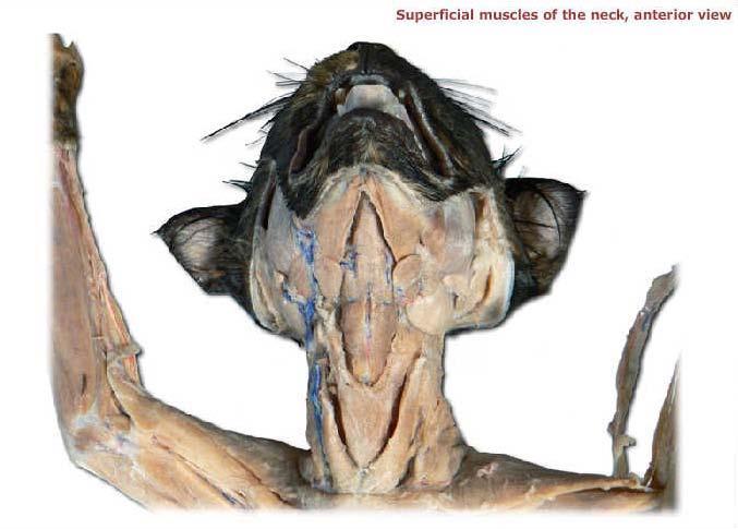
4 Sternohyoid Sternomastoid

5 Pectoralis minor Pectoralis major
6 External oblique Rectus abdominis Transverse abdominis External oblique (reflected) Internal oblique

7 Lumbodorsal fascia Latissimus dorsi Trapezius Trapezius Deltoid Trapezius Deltoid Levator scapula Deltoid

8 Trapezius Trapezius Trapezius Latissimus dorsi

9 Flexor carpi radialis Brachioradialis Extensor carpi radialis Flexor carpi ulnaris Biceps brachii Triceps brachii

10 Biceps brachii Flexor carpi radialis Flexor carpi ulnaris

11 Extensor carpi ulnaris Triceps brachii Extensor carpi radialis longus Deltoid Triceps brachii Deltoid Deltoid Trapezius
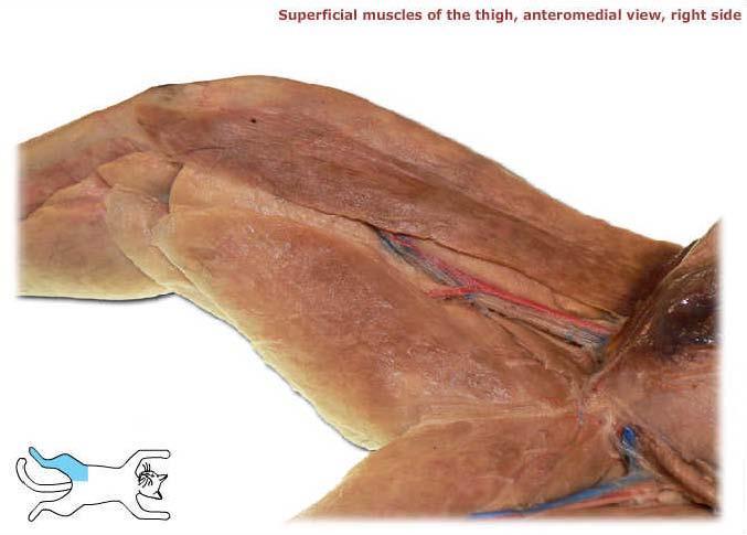
12 Sartorius

13 Adductor longus Semimembranosus Adductor femoris Vastus medialis Vastus lateralis Rectus femoris Tensor fasciae latae

14 Tibialis anterior Gastrocnemius Flexor digitorum longus

15 Biceps femoris Tensor fasciae latae Semimembranosus Semitendinosus Gluteus maximus Gluteus medius

16 Gastrocnemius Extensor digitorum longus Soleus Fibularis muscles
17 Brachioradiallis Triceps (lateral and long heads)

18 Brachioradialis Biceps brachii Triceps (medial head)

19 Trapezius Deltoid Levator scapula Trapezius Deltoid Deltoid Trapezius Latissimus dorsi
20 External oblique (right side cut and reflected) Rectus abdominis Transversus abdominis Internal oblique
21 Pectoralis major Pectoralis minor External oblique Rectus abdominis
22 Pectoralis major Serratus ventralis Rectus abdominis
23 Latissimus dorsi
24 Gastrocnemius Flexor digitorum longus Tibialis anterior

25 Rectus femoris Vastus lateralis Adductor longus Adductor femoris Vastus medialis Semimembranosus

26 Gluteus medius Gluteus maximus Biceps femoris Semitendinosus Gastrocnemius
27 Uinar nerve
28 Femoral nerve
29 Sciatic nerve

30 Parotid gland Masseter
31 Greater omentum Pancreas Cecum Small intestine Large intestine
32 Diaphragm Liver Pancreas Stomach Adrenal gland Intestine
33 Gall bladder Right lobes of liver Duodenum Pancreas Ileocecal junction Cecum Jejunum Falciform ligament Left lobes of liver Lesser omentum Stomach Ascending colon Spleen Greater omentum Small intestine Ileum Descending colon
34 Liver Bile duct Gall bladder Stomach Duodenum Pancreas

35 Trachea Esophagus
36 Diaphragm Falciform ligament Gallbladder Left medial lobe Right medial lobe Left lateral lobe Right lateral lobe Spleen Caudate lobe Stomach
37 Diaphragm Liver Gallbladder Stomach Greater omentum Spleen
38 Greater omentum Pancreas Cecum Small intestine Large intestine
39 Liver Lesser omentum Stomach Greater omentum Spleen Small intestine
40 Spleen Intestines Pancreas Stomach Liver Superior mesenteric artery Diaphragm
41 Colon
42 Pancreas Duodenum Cecum Small intestine
43 Ascending colon Ileum Colon Cecum Spleen Small intestine

44 Right common carotid artery Left common carotid artery Right brachial artery Left axillary artery Right axillary artery Right subclavian artery Left subclavian artery Brachiocephalic artery Arch of the aorta Heart

45 Left axillary artery Left brachial artery Left subclavian artery
46 Right common carotid artery Left common carotid artery Left subclavian artery Right subclavian artery Brachiocephalic artery Arch of aorta
47 Superior mesenteric artery Celiac trunk Right renal vein Descending aorta Inferior vena cava Inferior mesenteric artery

48 Right common carotid artery Right subclavian artery Left common carotid artery Left subclavian artery Brachiocephalic artery Arch of the aorta
49 Left external carotid artery Left internal carotid artery Left common carotid artery
50 Brachiocephalic artery Arch of the aorta Left subclavian artery Descending aorta (thoracic aorta) Heart
51 Intestines Superior mesenteric artery Inferior vena cava Abdominal aorta
52 Inferior vena cava Superior mesenteric artery Descending aorta Renal vein Renal arteries

53 Descending aorta Right internal iliac artery Right external iliac artery Left internal iliac artery Left external iliac artery Deep femoral artery Left femoral artery Right femoral artery
54 Left external jugular vein Left internal jugular vein Left brachiocephalic vein Right brachiocephalic vein Left axillary vein Superior vena cava Left subclavian vein
55 Hepatic portal vein Superior mesenteric vein
56 Inferior vena cava Right common iliac vein Right internal iliac vein Left common iliac vein Left internal iliac vein Right external iliac vein Left external iliac vein Femoral veins Greater saphenous veins Deep femoral veins Median sacral vein
57 Arch of the aorta Right atrium Right ventricle Pulmonary trunk Coronary vessels Left atrium Left ventricle Apex of the heart
58 Superior vena cava Brachiocephalic artery Arch of the aorta Pulmonary trunk Right atrium Left atrium Right ventricle Left ventricle Apex of the heart
59 Thyroid Right common carotid artery Trachea Right subclavian artery Brachiocephalic artery Lung (right anterior lobe) Heart (within pericardium) Larynx Left common carotid artery Esophagus Left subclavian artery Lung (left anterior lobe) Aortic arch Lung (left middle lobe) Lung (right middle lobe) Lung (left posterior lobe) Lung (right posterior lobe) Diaphragm Lung (right mediastinal lobe)
60 Right external jugular vein Right common carotid artery Left common carotid artery Right subclavian vein Right brachiocephalic vein Right subclavian artery Superior vena cava Left external jugular vein Left subclavian vein Left brachiocephalic vein Left subclavian artery Brachiocephalic artery Aortic arch
61 Inferior vena cava Superior mesenteric artery Celiac trunk Renal arteries Renal veins Abdominal aorta Common iliac veins External iliac arteries Internal iliac arteries Femoral vein Femoral arteries

62 Inferior vena cava Abdominal aorta Internal iliac artery External iliac artery Femoral vein Femoral artery
63 Brachiocephalic artery Left subclavian artery Ascending aorta Aortic arch Pulmonary trunk Descending aorta

64 Radial vein Radial artery Brachial artery Ulnar artery Brachial vein

65 Opening to esophagus Hyoid bone Epiglottis
66 Larynx Trachea Left anterior lobe of lung Right middle lobe of lung Right mediastinal lobe of lung Right inferior lobe of lung Diaphragm
67 Larynx Trachea Right anterior lobe of the lung Left anterior lobe of the lung Left middle lobe of the lung Right middle lobe of the lung Left posterior lobe of the lung Right posterior lobe of the lung Diaphragm Right mediastinal lobe of the lung

68 Trachea Right anterior lobe of lung Left anterior lobe of lung Left ventricle of heart Right middle lobe of lung Left middle lobe of lung Right posterior lobe of lung Diaphragm Left posterior lobe of lung

69 Right kidney Left kidney Right ureter Left ureter Urinary bladder Urethra
70 Right kidney Left kidney Right ureter Left ureter Urinary bladder Penis Urethra

71 Left kidney Right kidney Renal veins (right and left) Right ureter Left ureter Urinary bladder Urethra Prostate
72 Right kidney Right ureter Left kidney Renal artery Renal vein Left ureter Urinary bladder Urethra
73 Right ductus deferens Prostate gland Bulbourethral glands Urethra Left ductus deferens Right testis Epididymis Penis Left testis Scrotum
74 Prostate Right ductus deferens Urethra Epididymis Scrotum Left testis
75 Right ovary Left ovary Left uterine tube Right uterine tube Uterine body Vagina Urethra
76 Left uterine tube Left ovary Uterine body Vagina
77 Right ovary Left ovary Right uterine horn with fetus Left uterine horn with fetus Uterine body Urinary bladder Urethra
78 Placenta Fetus Uterine horns, opened Fetus Uterine body Urethra
79 Right testis Left testis Scrotu m
80 Ovary
Contents. Pig Dissection. Contents. External Features Sex Determination Mouth and Maxillary Nerve Muscles Index Internal Systems Index
 Pig Dissection External Features Sex Determination Mouth and Maxillary Nerve Muscles Index Internal Systems Index External features Sex determination Male Female Male to External anatomy 1. Pinna 2. External
Muscles of the Cat. N Deltoid MUSCLES OF THE CHEST. Pectoralis major. (This muscle is superior to Pectoralis minor) MUSCLES OF THE CHEST
 MUSCLES OF THE CHEST") MUSCLES OF THE CHEST Pectoralis major (This muscle is superior to Pectoralis minor) 1. MUSCLES OF THE CHEST Pectoralis minor (This muscle is inferior to Pectoralis major) 2. MUSCLES OF THE ARM Deltoid
MUSCLES OF THE CHEST Pectoralis major (This muscle is superior to Pectoralis minor) 1. MUSCLES OF THE CHEST Pectoralis minor (This muscle is inferior to Pectoralis major) 2. MUSCLES OF THE ARM Deltoid
Control center! (set point)! (Change is compared! to the set point.)!
! (Change is compared! to the set point.)!") Fig01.05 Control center! (set point)! Receptors (Change is compared! to the set point.)! Effectors! (muscles or glands)! Stimulus! (Change occurs! in internal! environment.)! Response! (Change is corrected.)
Fig01.05 Control center! (set point)! Receptors (Change is compared! to the set point.)! Effectors! (muscles or glands)! Stimulus! (Change occurs! in internal! environment.)! Response! (Change is corrected.)
Practical #1 Checklist Spring 2010
 External Features Caudal fin Clasper Dorsal fins External nares Gill slits Pectoral fin Rostrum Spiracle SHARK CHECK LIST STRUCTURE CHECK Internal Anatomy Digestive system Bile Duct Gall Bladder Rectal
Cat Muscles Flashcards Mt SAC
 1. MUSCLES OF THE CHEST Pectoralis major (This muscle is superior to Pectoralis minor) 2. MUSCLES OF THE CHEST Pectoralis minor (This muscle is inferior to Pectoralis major) 3. MUSCLES OF THE ARM AD CHEST
Name this muscle. Name this muscle
 this muscle this muscle Pectoralis Major Pectoralis Minor Serratus anterior Pectoralis minor Serratus anterior this muscle Deltoid: The major abductor of the upper limb this muscle this muscle this muscle
Muscle fiber (cell) Blood vessel. Perimysium. Epimysium. Fascicle (wrapped by perimysium) Endomysium (between fibers) Tendon. Bone
 Blood vessel. Perimysium. Epimysium. Fascicle (wrapped by perimysium) Endomysium (between fibers) Tendon. Bone") Figure 6.1 Connective tissue wrappings of skeletal muscle. Blood vessel Muscle fiber (cell) Perimysium Epimysium Fascicle (wrapped by perimysium) Tendon Endomysium (between fibers) Bone Figure 6.15 Superficial
Figure 6.1 Connective tissue wrappings of skeletal muscle. Blood vessel Muscle fiber (cell) Perimysium Epimysium Fascicle (wrapped by perimysium) Tendon Endomysium (between fibers) Bone Figure 6.15 Superficial
What you should do in labs 11 & 12
 Bio 101 Laboratories 11 & 12 Muscle Histology Gross Human Skeletal Muscle Cat Muscle Dissection 1 What you should do in labs 11 & 12 Lab 11 Muscle Histology (skeletal, smooth, cardiac) Human gross skeletal
Biology 218 Human Anatomy. Adapted from Martini Human Anatomy 7th ed. Chapter 12 Surface Anatomy and Cross-Sectional Anatomy
 Adapted from Martini Human Anatomy 7th ed. Chapter 12 Surface Anatomy and Introduction Surface anatomy is the study of anatomical landmarks on the exterior of the human body Knowledge of surface anatomy
What you should do in labs 11 & 12
 Bio 101 Laboratories 11 & 12 Muscle Histology Gross Human Skeletal Muscle Cat Muscle Dissection 1 What you should do in labs 11 & 12 Today (Lab 11) Muscle Histology (skeletal, smooth, cardiac) Human gross
Fetal Pig Visual Dissection Guide
 Fetal Pig Visual Dissection Guide WARD470156-776 Orientation Cranial Anterior Sagittal plane Frontal plane Ventral Dorsal Transverse plane Caudal Posterior 1 Incisions 1 Gender Key Male Female Both 4 3
OVARIES URETER FALLOPIAN TUBES BLADDER UROGENITAL OPENINGS (BOTH SEXES) PENIS VAGINA UTERUS
 PENIS VAGINA UTERUS") URETER OVARIES FALLOPIAN TUBES BLADDER UROGENITAL OPENINGS (BOTH SEXES) PENIS VAGINA UTERUS REPRODUCTIVE PRODUCE FEMALE HORMONES EXCRETORY FROM KIDNEY TO BLADDER EXCRETORY STORES URINE REPRODUCTIVE TRANSPORTS
URETER OVARIES FALLOPIAN TUBES BLADDER UROGENITAL OPENINGS (BOTH SEXES) PENIS VAGINA UTERUS REPRODUCTIVE PRODUCE FEMALE HORMONES EXCRETORY FROM KIDNEY TO BLADDER EXCRETORY STORES URINE REPRODUCTIVE TRANSPORTS
Honors Biology: Rat Dissection ONLINE ASSIGNMENT
 Name: Honors Biology: Rat Dissection ONLINE ASSIGNMENT You and your group members will use the Honors Biology WIKI to create an online dissection manual. The point of this assignment is to illustrate what
Dissection Lab Manuals: Required Content
 Dissection Lab Manuals: Required Content 1. Introduction a. Basic terminology (directions) b. External features of the cat c. Adaptations to predatory niche d. How to skin a cat e. How to make the incisions
Rat Dissection Lab. Background
 Anatomy & Physiology Corvallis High School Rat Dissection Lab Name: Class Period: Background Dissection of any organism or organ allows a 3-dimensional look at structural connections within the body. It
DISSECTION 1: SKELETAL MUSCLES
 8546d_c01_1-42 6/21/02 1:34 PM Page 4 mac62 mac62:1253_ge: 4 Cat Dissection DISSECTION 1: SKELETAL MUSCLES Many skeletal muscles of the cat are similar to human muscles. This dissection will reinforce
MicroAnatomy Muscle Fiber Model
 MicroAnatomy Muscle Fiber Model Muscle fiber whole model (but model is only a fraction of a fiber) Sarcolemma 14 Myofibril 1 Nucleus 8 Mitochondria 2 Triad 16 Sarcoplasmic reticulum 17 T tubule 15 Thin
Due in Lab weeks because of Thanksgiving Prelab #10. Homework #8. Both sides! Both sides!
 Lab 8 MUSCLES Due in Lab 10 2 weeks because of Thanksgiving Prelab #10 Both sides! Homework #8 Both sides! Refer to Muscles 22-23 Naming of muscles Origin Site of muscle attachment that doesn t move during
Temporalis Elevates & retracts mandible. Masseter Elevates mandible. Sternocleidomastoid Neck flexion. Trapezius Elevates & depresses shoulders
 Anterior Posterior Temporalis Elevates & retracts mandible Masseter Elevates mandible Sternocleidomastoid Neck flexion Trapezius Elevates & depresses shoulders Masseter Elevates mandible Temporalis Elevates
Biology 2401 Muscles List for CPC models
 Biology 2401 List for CPC models Italicized muscles are dissect and similar in the cat = Dissect and note the differences in human and cat Major of the Human Head Facial Expression Epicranius frontalis
The Muscular System Lab Power Point
 The Muscular System Lab Power Point Myoneural Junction Sarcoplasm Nucleus Myofibrils Sarcomere (black line to black line) Sarcolemma Myoneural space Nucleus Endomysium Motor Neuron Muscles of Facial Expression
8546d_fm_i-iv 6/26/02 3:51 PM Page 1 mac62 mac62:1253_ge: CAT DISSECTION A LABORATORY GUIDE
 8546d_fm_i-iv 6/26/02 3:51 PM Page 1 mac62 mac62:1253_ge: CAT DISSECTION A LABORATORY GUIDE 8546d_c01_1-42 6/21/02 1:34 PM Page 2 mac62 mac62:1253_ge: 2 Cat Dissection PREFACE A. Preparing the Cat 1. With
CARDIOVASCULAR DANIL HAMMOUDI.MD
 CARDIOVASCULAR DANIL HAMMOUDI.MD 18 Systemic Circulation Figure 19.19 Pulmonary Circulation Figure 19.18b 1. Thyroid gland 2. Trachea 3. Brachiocephalic 4. Common carotid 5. Internal jugular 6. Superior
Day 5 Respiratory & Cardiovascular: Respiratory System
 Day 5 Respiratory & Cardiovascular: Respiratory System Be very careful not to damage the heart and lungs while separating the ribs! Analysis Questions-Respiratory & Cardiovascular Log into QUIA using your
Human Muscles (Anterior View) Model 3-44
 Model 3-44") Human Muscles (Anterior View) Model 3-44 Temporalis Frontalis Orbicularis Occuli Orbicularis Oris Masseter Sternocleidomastoid Orbicularis Occuli Human Muscles (Anterior View) Model 3-65 Temporalis Masseter
Human Muscles (Anterior View) Model 3-44 Temporalis Frontalis Orbicularis Occuli Orbicularis Oris Masseter Sternocleidomastoid Orbicularis Occuli Human Muscles (Anterior View) Model 3-65 Temporalis Masseter
Human Anatomy Lab #7: Muscles of the Cadaver
 Human Anatomy Lab #7: Muscles of the Cadaver Table of Contents: Expected Learning Outcomes.... 1 Introduction...... 1 Identifying Muscles on Yourself.... 2 Muscles of the Anterior Trunk and Arm.. 2 Muscles
Dissections. Striated (Skeletal) Muscle Fibers (Longitudinal Section) Eder, et al.: Laboratory Atlas of Anatomy and Physiology, Third Edition
 Muscle Fibers (Longitudinal Section) Eder, et al.: Laboratory Atlas of Anatomy and Physiology, Third Edition") . Dissections Text The McGraw Hill Companies, 00 C H A P T E R Dissections Striated (Skeletal) Muscle Fibers (Longitudinal Section) . Dissections Text The McGraw Hill Companies, 00 0 CHAPTER Figure - The
. Dissections Text The McGraw Hill Companies, 00 C H A P T E R Dissections Striated (Skeletal) Muscle Fibers (Longitudinal Section) . Dissections Text The McGraw Hill Companies, 00 0 CHAPTER Figure - The
1 Right & left Hepatic ducts Gastric Impression of spleen
 Pancreatic Model 1 Right & left Hepatic ducts 14 Gastric Impression of spleen 2 Common hepatic duct 15 Renal Impression of spleen 3 Cystic Duct 16 Colic Impression of spleen 4 Common Bile Duct 17 Splenic
3 Circulatory Pathways
 40 Chapter 3 Circulatory Pathways Systemic Arteries -Arteries carry blood away from the heart to the various organs of the body. -The aorta is the longest artery in the body; it branches to give rise to
11/15/2018. Temporalis Elevates & retracts mandible. Masseter = Prime mover of jaw closure. Levator scapulae Supraspinatus Clavicle.
 Due in Lab 10 Lab 8 MUSCLES 2 weeks because of Thanksgiving Prelab #10 Both sides! Homework #8 Both sides! Refer to Muscles 22-23 Examples of Origin & Insertion Naming of muscles Origin Site of muscle
Lab Monitor Images Dissection of the Abdominal Vasculature + Lower Digestive System
 Lab Monitor Images Dissection of the Abdominal Vasculature + Lower Digestive System Stomach & Duodenum Frontal (AP) View Nasogastric tube 2 1 3 4 Stomach Pylorus Duodenum 1 Duodenum 2 Duodenum 3 Duodenum
Epicranius (frontal belly) Zygomaticus minor. Zygomaticus major Buccinator
 Zygomaticus minor. Zygomaticus major Buccinator") Epicranius (frontal belly) Zygomaticus minor Zygomaticus major Buccinator Masseter Digastric (posterior belly) Stylohyoid Sternocleidomastoid Trapezius Scalenus Omohyoid (inferior belly) Orbicularis oris
Epicranius (frontal belly) Zygomaticus minor Zygomaticus major Buccinator Masseter Digastric (posterior belly) Stylohyoid Sternocleidomastoid Trapezius Scalenus Omohyoid (inferior belly) Orbicularis oris
Muscle Anatomy Review Chart
 Muscle Anatomy Review Chart BACK Superficial (5) Trapezius Transverse cervical a. Latissimus dorsi Thoracodorsal a. Rhomboideus major Dorsal scapular a. Rhomboideus minor Levator scapulae Intermediate
Cadaver Muscular System Practice Practical
 Cadaver Muscular System Practice Practical Station 1 Station 1 1. Specific structure 1. Rectus sheath 2. Red line 2. Linea alba Station 2 Station 2 3. Red muscle 1. Rectus abdominis 4. Red muscle actions
In-Depth Foundations: Anatomy Terms to Know
 Be familiar with / able to identify and define all the following parts. The Spine Cranium Vertebrae Cervical, Thoracic, Lumbar Sacrum Coccyx Bones of Upper Body Cranium Mastoid process; Occipital condyle,
Artery 1 Head and Thoracic Arteries. Arrange the parts in the order blood flows through them.
 Artery 1 Head and Thoracic Arteries 1. Given the following parts of the aorta: 1. abdominal aorta 2. aortic arch 3. ascending aorta 4. thoracic aorta Arrange the parts in the order blood flows through
Monday, November 13, 2017 A & P 2401
 Monday, November 13, 2017 A & P 2401 Today you will complete the following handouts. Study the last part of the handout for this will be on your quiz, which will be on Wednesday. It is titled steps of
Anatomy. Contents Brain (Questions)
") Anatomy 12 Contents 12.1 Brain (Questions).................................................... 683 12.2 Head and Neck (Questions)............................................. 685 12.3 Thorax (Questions)...................................................
Anatomy 12 Contents 12.1 Brain (Questions).................................................... 683 12.2 Head and Neck (Questions)............................................. 685 12.3 Thorax (Questions)...................................................
Skeleton. The left forearm is in the position of supination, the right in pronation.
 S ystemic review A Skeleton A from the front B B from behind The left forearm is in the position of supination, the right in pronation. Skull Mandible Hyoid bone Cervical vertebrae Clavicle Sternum Costal
The Human Muscular System Required reading before beginning this lab: Saladin, KS: Human Anatomy 5th ed (2017) Chapters 10, 11, 12 INTRODUCTION
 Chapters 10, 11, 12 INTRODUCTION") Biology 322: Human Anatomy The Human Muscular System Required reading before beginning this lab: Saladin, KS: Human Anatomy 5 th ed (2017) Chapters 10, 11, 12 INTRODUCTION We will use a number of lab periods
Biology 322: Human Anatomy The Human Muscular System Required reading before beginning this lab: Saladin, KS: Human Anatomy 5 th ed (2017) Chapters 10, 11, 12 INTRODUCTION We will use a number of lab periods
2. Be able to define and give examples of each of the following planes:
 Anatomy Skeletal and Muscular Systems Part 1: Directional terms, Planes, Body Cavities Lab Materials: male & female surface landmarks models various models, textbook Lab Activities: Use models and charts
TRACE A DROP OF BLOOD FROM RIGHT EAR TO LEFT OCULOMOTOR NERVE
 TRACE A DROP OF BLOOD FROM RIGHT EAR TO LEFT OCULOMOTOR NERVE KEY: TRACE A DROP OF BLOOD FROM RIGHT EAR TO LEFT OCULOMOTOR NERVE RIGHT EAR RIGHT ATRIUM LEFT SUBCLAVIAN ARTERY RIGHT EXTERNAL JUGULAR VEIN
SUBJECTS 2nd year, 1st semester I. 1. Primitive gut - limits, derivatives 2. Foregut -limits, evolution, derivatives 3. Midgut -limits, evolution,
 SUBJECTS 2nd year, 1st semester I. 1. Primitive gut - limits, derivatives 2. Foregut -limits, evolution, derivatives 3. Midgut -limits, evolution, derivatives 4. Hindgut- limits, evolution, derivatives
First BHMS Anatomy Question Papers Calicut University
 First BHMS Anatomy Question Papers Calicut University 1996-2000 FIRST B.H.M.S. DEGREE EXAMINATION, DECEMBER 1996 Time: Three Hours Maximum: 100 Marks Answer any five questions. Draw diagrams wherever needed.
Synergist Muscles. Shoulder (glenohumeral joint) Flexion Deltoid (anterior fibers) Pectoralis major (upper fibers) Biceps Brachii Coracobrachialis
 Flexion Deltoid (anterior fibers) Pectoralis major (upper fibers) Biceps Brachii Coracobrachialis") Synergist Muscles Dr Gene Desepoli DrGeneLMT@gmail.com Shoulder (glenohumeral joint) Deltoid (anterior fibers) Pectoralis major (upper fibers) Biceps Brachii Coracobrachialis Deltoid (posterior fibers)
Synergist Muscles Dr Gene Desepoli DrGeneLMT@gmail.com Shoulder (glenohumeral joint) Deltoid (anterior fibers) Pectoralis major (upper fibers) Biceps Brachii Coracobrachialis Deltoid (posterior fibers)
BLOCK IV: OFFICIAL BODY PARTS LIST FOR ANTERIOR ABDOMINAL WALL AND ABDOMINAL CONTENTS
 BLOCK IV: OFFICIAL BODY PARTS LIST FOR ANTERIOR ABDOMINAL WALL AND ABDOMINAL CONTENTS External oblique muscle Muscular portion Aponeurotic portion Superficial inguinal ring Lateral (inferior) crus Medial
2/4/2018. Identify the two reasons why muscle cells may go through muscle fatigue. Ch.7 Review. Sternocleidomastoid.
 Ch.7 Review Identify the two reasons why muscle cells may go through muscle fatigue Temporalis Depressor anguli oris Sternocleidomastoid Tibialis anterior 1 Gluteus medius Deltoid Adducts & rotates scapula
Human Anatomy and Physiology I Laboratory
 Human Anatomy and Physiology I Laboratory Gross Anatomy of the Muscular System (Two weeks) 1 This lab involves study of the laboratory exercise Gross Anatomy of the Muscular System. Complete the Review
MINK DISSECTION LAB DAY 1 NECK
 MINK DISSECTION LAB PURPOSE: This lab dissection is designed to give you first hand experience with the organs (or their mink counterparts) that we have learned about all year long. After seeing the organs,
Fetal Pig Dissection: External Anatomy
 Name Fetal Pig Dissection: External Anatomy External Anatomy 1. Determine the sex of your pig by looking for the urogenital opening. On females, this opening is located near the anus. On males, the opening
Labs 1 and 2: MUSCULAR SYSTEM Cat Dissection: Photo Atlas, Chapter 19 Human Muscles: Unit 7, Muscle Tissue and Muscular System
 137 Lab manual: Exploring Anatomy & Physiology in the Laboratory Core Concepts by Erin Amerman, Morton Publishing, 2014, Photo Atlas (Cat Dissection Ch. 19, p. 169-184 ) Labs 1 and 2: MUSCULAR SYSTEM Cat
Lab 9 Abdomen MUSCLES
 Lab 9 Abdomen MUSCLES External abdominal oblique continuous with the external intercostal muscle; its fibers point in a caudal direction as it moves anteriorly until it inserts on the linea alba via its
LIST OF IMPORTED PORK PRODUCTS WHICH WILL BE EXEMPT FROM FURTHER PROCESSING ON ARRIVAL IN SOUTH AFRICA FROM BRAZIL
 LIST OF IMPORTED PORK PRODUCTS WHICH WILL BE EXEMPT FROM FURTHER PROCESSING ON ARRIVAL IN SOUTH AFRICA FROM BRAZIL 2015-12 (TO BE USED FOR PORK IMPORTED ON THE CERTIFICATE FOR UNRESTRICTED ENTRY) The products
List of Muscles and Function. Region View Muscle Function Facial Anterior/Oblique Occipitofrontalis front belly Raises eyebrows
 List of Muscles and Function Region View Muscle Function Facial Anterior/Oblique Occipitofrontalis front belly Raises eyebrows Orbicularis oculi Closes eye Orbicularis oris Purses lips Zygomaticus minor/major
NORTH CENTRAL HIGH SCHOOL NOTE & STUDY GUIDE. X Biology II
 Unit 2-5, Animal Biology & Organ Systems, FETAL PIG DISSECTION MANUAL X Biology II, Mr. Doc Miller, M.Ed. North Central High School Name: ID#: NORTH CENTRAL HIGH SCHOOL NOTE & STUDY GUIDE X Biology II
Scapula Spine Lateral edge of clavicle. Medial border Scapula. Medial border of Scapula, between superior angle and root of spine. Scapula.
 Muscle attachments and actions answer sheet Muscle Origins insertions Movements Joints crossed Trapezius Base of skull Spinous process of C7 Thoracic Spine Lateral edge of clavicle Elevation Retraction
VESSELS: GROSS ANATOMY
 ACTIVITY 10: VESSELS AND CIRCULATION OBJECTIVES: 1) How to get ready: Read Chapter 23, McKinley et al., Human Anatomy, 4e. All text references are for this textbook. 2) Observe and sketch histology slide
Match the types of muscle tissues with the words and phrases. 1) Skeletal 2) Smooth 3) Cardiac 2 Walls of blood vessels. 2 Walls of digestive tract
 Skeletal 2) Smooth 3) Cardiac 2 Walls of blood vessels. 2 Walls of digestive tract") S T U D Y G U I D E. Types of Muscle Tissues Match the types of muscle tissues with the words and phrases. ) Skeletal ) Smooth ) Cardiac, Striated Walls of blood vessels, Single nucleus Heart muscle, Involuntary
S T U D Y G U I D E. Types of Muscle Tissues Match the types of muscle tissues with the words and phrases. ) Skeletal ) Smooth ) Cardiac, Striated Walls of blood vessels, Single nucleus Heart muscle, Involuntary
Biology Human Anatomy Abdominal and Pelvic Cavities
 Biology 351 - Human Anatomy Abdominal and Pelvic Cavities Please place your name and I.D. number on the back of the last page of this exam. You must answer all questions on this exam. Because statistics
YOU MUST BRING GLOVES FOR THIS ACTIVITY
 ACTIVITY 10: VESSELS AND CIRCULATION OBJECTIVES: 1) How to get ready: Read Chapter 23, McKinley et al., Human Anatomy, 5e. All text references are for this textbook. 2) Observe and sketch histology slide
Contents. Preface xv. SECTION 1: Introduction to the Bodynamic System 1. SECTION 2: The Bodynamic Psycho-Motor Anatomy 29
 Contents Preface xv SECTION 1: Introduction to the Bodynamic System 1 Definitions in the Bodynamic System 3 Ego Formation through the Coding Elements 9 Examples of Formation of Coding 17 Using This Book
Bio 113 Anatomy and Physiology The Muscles. Muscles of the Head and Neck. Masseter. Orbicularis occuli. Orbicularis oris. Sternocleidomastoid
 Bio 113 Anatomy and Physiology The Muscles Muscles of the Head and Neck Masseter Orbicularis occuli Orbicularis oris Sternocleidomastoid Temporalis BIO 113 Fall 2011 Muscles Page 1 of 5 Muscles of the
A. All movements require muscle which are organs using chemical energy to contract.
 Ch 8 Muscles Introduction: A. All movements require muscle which are organs using chemical energy to contract. B. The three types of muscle in the body are skeletal, smooth, and cardiac muscle. C. This
5/21/2013. Muscle Anatomy. Thursday January, 24 th, Skeletal Muscle. Smooth Muscle. Cardiac Muscle
 Muscle Anatomy Thursday January, 24 th, 2013 Skeletal Muscle Cardiac Muscle Smooth Muscle 1 Smooth Muscle 1. Found in the walls of the digestive system, bladder, uterus and blood vessels 2. Involuntary
Chiropractic Technician Class
 Chiropractic Technician Class Presentation By: Dr. Kay Miller. The Role of Exercise as it Relates to Our Musculoskeletal System Introduction to the topic and Preliminary Physical exam Musculoskeletal anatomy:
Anatomical Considerations for Lab Practical II
 Anatomical Considerations for Lab Practical II For each of the following please be prepared to provide: Identification System Organ(s) or ducts to Function(s) location which it is attached Use your lecture
The Muscular System. Chapter 10 Part D. PowerPoint Lecture Slides prepared by Karen Dunbar Kareiva Ivy Tech Community College
 Chapter 10 Part D The Muscular System Annie Leibovitz/Contact Press Images PowerPoint Lecture Slides prepared by Karen Dunbar Kareiva Ivy Tech Community College Table 10.14: Muscles Crossing the Hip and
Chapter 9. The Muscular System
 1 Chapter 9 The Muscular System 2 Introduction Skeletal muscles: movement in environment Smooth muscles: intestines, ureters, veins and arteries Cardiac muscle: pumps blood through heart and blood vessels
Exercise Science Section 3: The Muscular System
 Exercise Science Section 3: The Muscular System An Introduction to Health and Physical Education Ted Temertzoglou Paul Challen ISBN 1-55077-132-9 Major Functions of Muscles Movement Includes: breathing,
HumAn: A Cat Dissection Tutorial
 University of Rhode Island DigitalCommons@URI Senior Honors Projects Honors Program at the University of Rhode Island 2015 HumAn: A Cat Dissection Tutorial Sean P. Pierel University of Rhode Island, spie129@my.uri.edu
SYLLABUS BDS I PROFESSIONAL GENERAL HUMAN ANATOMY INCLUDING EMBRYOLOGY AND HISTOLOGY
 GENERAL HUMAN ANATOMY INCLUDING EMBRYOLOGY AND HISTOLOGY I. General Anatomy 1. Anatomical terms 2. Skin, superficial fascia & deep fascia 3. Cardiovascular system, portal system, collateral circulation
Peritoneum: Def. : It is a thin serous membrane that lines the walls of the abdominal and pelvic cavities and clothes the viscera.
 Peritoneum: Def. : It is a thin serous membrane that lines the walls of the abdominal and pelvic cavities and clothes the viscera. Layers of the peritoneum: 1. Outer Layer ( Parietal Peritoneum) : lines
Atlas A. Lecture Outline A-1. Copyright (c) The McGraw-Hill Companies, Inc. Permission required for reproduction or display.
 The McGraw-Hill Companies, Inc. Permission required for reproduction or display.") Atlas A Lecture Outline Copyright (c) The McGraw-Hill Companies, Inc. Permission required for reproduction or display. A-1 Atlas A (Orientation to Anatomy) Anatomical position Anatomical planes Directional
Atlas A Lecture Outline Copyright (c) The McGraw-Hill Companies, Inc. Permission required for reproduction or display. A-1 Atlas A (Orientation to Anatomy) Anatomical position Anatomical planes Directional
Nasogastric tube. Stomach. Pylorus. Duodenum 1. Duodenum 2. Duodenum 3. Duodenum 4
 Esophagus Barium Swallow Stomach and Duodenum 4 year old Upper GI Nasogastric tube Stomach and Duodenum 4 year old Upper GI Nasogastric tube Stomach Pylorus Duodenum 1 Duodenum 2 Duodenum 3 Duodenum 4
This lab activity is aligned with Visible Body s Human Anatomy Atlas app. Learn more at visiblebody.com/professors
 1 This lab activity is aligned with Visible Body s Human Anatomy Atlas app. Learn more at visiblebody.com/professors 2 A. Digestive System Overview To Start: Go to the Views menu and scroll down to the
Human Anatomy Biology 351
 Human Anatomy Biology 351 Lower Limb Please place your name on the back of the last page of this exam. You must answer all questions on this exam. Because statistics demonstrate that, on average, between
Human Anatomy Biology 351
 Human Anatomy Biology 351 Lower Limb Please place your name on the back of the last page of this exam. You must answer all questions on this exam. Because statistics demonstrate that, on average, between
Veterinary Technician s Daily Reference Guide Canine and Feline. Third Edition COPYRIGHTED MATERIAL
 Veterinary Technician s Daily Reference Guide Canine and Feline Third Edition COPYRIGHTED MATERIAL Section One Anatomy Chapter 1 Anatomy Anatomy 6 Overall 6 Musculature 8 Skeletal 8 Internal Organs 9
The Clavicle Right clavicle Deltoid tubercle: Conoid tubercle, conoid ligamen Impression for the
 The Clavicle Muscle Attachment Sites in the Upper Limb Pectoralis major Right clavicle Smooth superior surface of the shaft, under the platysma muscle tubercle: attachment of the deltoid Acromial facet
Fetal Pig Dissection Packet
 Fetal Pig Dissection Packet Name Period * Each person will turn in his/her own packet You may use the Virtual Fetal Pig Dissection website from Whitman College as a visual reference for all stages of dissection.
3D Digital Human Information Modeling
 3D Digital Human Information Modeling SC24 WG9 & Web3D Meetings January 21-24, 2019 Myeong Won Lee (U. of Suwon) and Seung-Pyo Lee (Seoul National U.) Topics for 3D Digital Human Information Model A whole
A&P 1 Muscle In-Lab Guide
 A&P 1 Muscle In-Lab Guide This lab guide includes a table with all the muscles you need to ID, along with their origins, insertions and actions Dashed lines means ignore. If several actions are listed,
Anatomy Module Study Guide
 Anatomy Module Study Guide Please use this step-by-step study guide and checklist to ensure that you have covered all that you need to learn by the time you come in for the face-to-face Anatomy tutorial.
HUMAN BODY COURSE LOWER LIMB NERVES AND VESSELS
 HUMAN BODY COURSE LOWER LIMB NERVES AND VESSELS October 22, 2010 D. LOWER LIMB MUSCLES 2. Lower limb compartments ANTERIOR THIGH COMPARTMENT General lfunction: Hip flexion, knee extension, other motions
Violation of these rules, failure to participate, or unsafe behavior will result in the loss of participation points. THERE WILL BE NO WARNINGS!
 BIOLOGY II MINK DISSECTION ACTIVITY #1 NAME DATE HOUR MINK DISSECTION PART 1: CLASSROOM RULES 1. No food or drink in the lab. 2. Follow directions the first time they are given. 3. No unauthorized dissections.
Muscles of Lesson Five. Muscular Nomenclature and Kinesiology - Two. Muscles of Lesson Five, cont. Chapter 16
 Chapter 16 Muscular Nomenclature and Kinesiology - Two Lessons 5-6 Muscles of Lesson Five Iliopsoas (psoas major, iliacus) Hip outward rotators (piriformis, gemellus superior, gemellus inferior, obturator
Peripheral Nervous System: Lower Body
 Peripheral Nervous System: Lower Body MSTN121 - Neurophysiology Session 11 Department of Myotherapy Lumbar Plexus Iliohypogastric nerve (T12-L1) Motor: Transverse abdominis and internal obliques Sensory:
Course: Human Body Systems Year: Teacher: Meghann Redman
 Course: Human Body Systems Year: 2015 2016 Teacher: Meghann Redman Unit 1: The Human Body: An Orientation Approximate Time Frame: 2 Weeks The human body has its own set of medical terminology. What terms
ANATOMY COURSE STUDY GUIDE & CHECKLIST
 ANATOMY COURSE STUDY GUIDE & CHECKLIST 1 Anatomy Course Study Guide Please use this step-by-step study guide and checklist to ensure that you have covered all that you need to learn by the time you come
Dissection of the Rat
 Dissection of the Rat Introduction In this laboratory exercise, the anatomy of the rat will be examined in some detail. You may recall that in your first year biology course you dissected a grass frog
Lab Exercise 8. BIOPAC Exercise. Muscle Tissue. Muscles. What you need to be able to do on the exam after completing this lab exercise:
 Lab Exercise 8 BIOPAC Exercise Muscle Tissue Muscles Textbook Reference: See Chapters 9 & 10 What you need to be able to do on the exam after completing this lab exercise: Be able to answer questions covering
HUMAN HEART. Learn the following structures on the heart models.
 HUMAN HEART Learn the following structures on the heart models. The human heart has four chambers that consist of the right atrium, left atrium, right ventricle, and left ventricle. The atria are smaller
Lab 9: Learn origin and insertion for each of the listed muscles. For Exercise 15, do Activities 1-6 in 9 th edition, Activities 1-4 in 10 th edition
 The Muscular System Exercises 14, 15, and 16 (begins: page 187 in 9 th and 10 th editions) Exercises 12, 13, and 14 (begins: page 185 in 11 th edition, page 189 in 12 th edition) Lab 8 and 9 Objectives
Manual Muscle Testing, MMT
 Muscle Testing, MMT Manual This is the official list of names of all manual neuromuscular tests within Manual Muscle Testing MMT as used in Manual Kinesiology at the Swedish School of Kinesiology and the
ANATOMICAL GUIDE FOR THE ELECTROMYOGRAPHER
 ANATOMICAL GUIDE FOR THE ELECTROMYOGRAPHER ANATOMICAL GUIDE FOR THE ELECTROMYOGRAPHER The Limbs and Trunk By EDWARD F. DELAGI, M.D. JOHN IAZZETTI, M.D. ALDO O. PEROTTO, M.D. DANIEL MORRISON, M.D. Fourth
Musculoskeletal Anatomy Coloring Book
 Musculoskeletal Anatomy Coloring Book Muscolino, Joseph E. ISBN-13: 9780323057219 Table of Contents Introduction â How to Use This Book 1. The Skeletal System Bones of the Head â Anterior View Bones of
Electrode Placement. Skin Preparation. Frontalis (FRL) (Specific) Temporalis Anterior (TA) (Specific) Sternocleidomastoid (SCM) (Specific)
 (Specific) Temporalis Anterior (TA) (Specific) Sternocleidomastoid (SCM) (Specific)") Electrode Placement Skin Preparation 1) Removing the hair: Shave if necessary 2) Clean the skin: Use a towel or abrasive pad with conductive cleaning paste or alcohol to remove dead skin cells (high impedance)
Electrode Placement Skin Preparation 1) Removing the hair: Shave if necessary 2) Clean the skin: Use a towel or abrasive pad with conductive cleaning paste or alcohol to remove dead skin cells (high impedance)
Netter's Anatomy Flash Cards Section 4 List 4 th Edition
 Netter's Anatomy Flash Cards Section 4 List 4 th Edition https://www.memrise.com/course/1577335/ Section 4 Abdomen (31 cards) Plate 4-1 Bony Framework of Abdomen 1.1 Costal cartilages 1.2 Iliac crest 1.3
Biology Overview Dissection Assignment
 Biology Overview Dissection Assignment What is this assignment about? After learning several major systems of the human body, this overview dissection assignment is for you to review all of those major