THE VESSELS OF THE HEART
|
|
|
- Felicia Stone
- 6 years ago
- Views:
Transcription

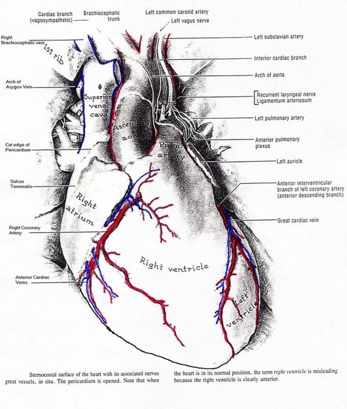
1 1 THE VESSELS OF THE HEART The vessels of the heart include the coronary arteries, which supply the heart and the veins and lymph vessels, which drain the heart. THE CORONARY ARTERIES These are the blood vessels responsible for taking blood to the all the tissues of the heart with the exception of the fibrous and the parietal serous layers of the pericardium. The coronary arteries are modified muscular arteries with poorly developed internal elastic lamina. SOURCES (Figure 1) The coronary arteries originate from the ascending aorta. They are commonly two in number (Right and left) but could be up to four in number. The left artery usually has a larger diameter than the right one.
2 Figure 2 2
3 3 THE RIGHT CORONARY ARTERY: This artery arises from the Right (Anterior) aortic sinus and passes between the right auricle and the root of the pulmonary trunk to enter the anterior part of the coronary sulcus (Groove). It runs towards the inferior border within the sulcus and curves over that border to enter the posterior part of the coronary sulcus, where it terminates just before the crux of the heart, at the crux or to the left of the crux. Its terminal twigs anastomose with terminal twigs of the Left coronary artery. Branches of the Right Coronary artery: 1. Right Conus artery (3 rd Coronary artery): This is usually the first branch of the right coronary artery but could arise directly from the right aortic sinus (36%). It supplies the infundibular part of the right ventricle and the proximal part of the pulmonary trunk. It terminates around the pulmonary trunk by anastomosing with a similar branch (Left Conus artery) of the left coronary artery. 2. The artery to the sinuatrial node: This artery is a branch of the right coronary artery in 60% of the population. This branch also supplies the right atrium, which accommodates the node. 3. Right anterior (atrial and ventricular) rami (Branches): These branches supply the sternocostal surface of the right atrium and right ventricle. 4. Right marginal artery: This artery runs towards the apex of the heart along the inferior border of the heart. 5. Right posterior (atrial and ventricular) rami: These arteries supply the diaphragmatic surface of the right ventricle and the right atrium. 6. Posterior interventricular artery: This artery runs in the posterior interventricular groove, sending short perforating septal branches to the interventricular septum. 7. Atrioventricular rami: These vessels supply adjacent areas of the atria and ventricle in the vicinity of the posterior aspect of the atrioventricular groove (Coronary sulcus). 8. The artery to the atrioventricular node. This artery arises in the region of the crux from the right coronary artery in 80% of the population. Summary of the distribution of the Right Coronary artery: In general, the right coronary artery supplies both right atrium and ventricle, parts of the left atrium and ventricle, part of the interventricular septum, SA and AV nodes. THE LEFT CORONARY ARTERY This artery has a short trunk and arises from the Left (Left posterior) aortic sinus. It emerges between the left auricle and the root of the pulmonary trunk and immediately bifurcates to form its circumflex and anterior interventricular branches. Anterior interventricular artery: This is the larger of the two main branches of the left coronary artery. It runs within the anterior interventricular groove up to the inferior margin of the heart around which it curves to reach the apical part of the posterior interventricular groove. Its terminal twigs anastomose within the groove with the terminal twigs of the posterior interventricular artery. Its branches include: a. Left conus artery: This branch anastomoses with a similar branch of the right coronary artery. b. Lateral diagonal branch. This is a large Left anterior ventricular branch.
4 4 c. Right and left anterior ventricular rami, which supply the sternocostal surface of the two ventricles. d. Anterior septal rami, which supply the anterior 2/3 of the interventricular septum. e. Posterior septal rami, which supply the apical part of the posterior 1/3 of the septum. Circumflex branch of Left Coronary artery This artery runs in the posterior part of the Coronary Groove (Sulcus) and terminates at or just before the crux of the heart by anastomosing with twigs of the right coronary artery. Its branches include: a. Left marginal branch, which supplies the left ventricle. b. Anterior and posterior ventricular branches c. Atrial branches, which supply the left atrium. d. The artery to the sinuatrial node: This artery arises from the left coronary artery in 40% of the population. e. The artery to the atrioventricular node. This artery arises from the Left coronary artery in 20% of the population. Summary of the distribution of the Left Coronary artery: In general, the left coronary artery supplies most of the left ventricle, a narrow strip of the right ventricle, the anterior 2/3 of the intereventricular septum and most of the left atrium. It also supplies the SA and AV nodes CORONARY ARTERIAL DOMINANCE Coronary arterial dominance is based on the pattern of distribution of the two coronary arteries and is determined by the artery, which gives rise to the posterior interventricular artery. The posterior interventricular artery supplies: a. The posterior part (1/3) of the interventricular septum. b. The posterolateral wall of the left ventricle. There are conflicting reports on the pattern of domination between the right and the left coronary arteries in the population. However, the popular view is that: 1. In 67% of the population, the right coronary artery is the dominant artery. (A) and (D) 2. In 15% of the population, the left coronary artery is the dominant artery. (B and C) 3. In 18% of the population, the distribution is shared equally.

5 5 NB: C in the diagram is an unusual case of a single coronary artery arising from the left aortic sinus. CORONARY ARTERIAL ANASTOMOSES In general, branches of the coronary arteries are referred to as End arteries (An artery whose area of distribution receives no supply from a branch or branches of another large artery). Nevertheless, some degree of anastomosis occurs between branches of the coronary arteries at various levels. Anastomotic Levels: a. Sub-endocardial In all the cavities. b. Myocardial- In the tunica media and the interventricular septum. c. Subepicardial In the coronary grooves d. Extracardial- Involving the pericardiacophrenic, bronchial and mediasternal branches. These anastomoses occur between branches of individual coronary arteries (Intracoronary) and between branches of the left and the right coronary arteries (Intercoronary). Extracardial and sub-epicardial amastomoses are usually arterial in nature while myocardial and sub-endocardial anastomoses are arteriolar in nature. The existence of cardiac arteriovenous shunts has also been suggested but yet to be substantiated. In cardiac anoxia and other obstructive coronary heart diseases, the anastomotic channels become pronounced but usually not enough to meet the need of the endangered heart. These situations usually result in Myocardial Infarction (MI).

6 VENOUS DRAINAGE OF THE HEART The heart is drained principally by the 2cm long Coronary sinus, which drains into the right atrium. It receives the following tributaries: 1. The great cardiac vein, which accompanies the anterior interventricular artery and enters the left end of the sinus. This vein usually has a valve at the point of entry into the coronary sinus 2. The left marginal vein also drains into the left end of the sinus. 3. The left posterior ventricular vein also drains into the left aspect of the sinus. 4. The middle cardiac vein, which accompanies the posterior interventricular artery drains into the right end of the sinus. 5. The small cardiac vein, which accompanies the right marginal artery drains into the right end of the sinus or drains directly into the right atrium. 6. The oblique vein of the left atrium drains the posterior wall of the left atrium into the left aspect of the sinus. The minor/accessory cardiac veins are: 6
7 7 1. Anterior cardiac veins, which cross the anterior part of the coronary groove and drain directly into the right atrium. Some of these veins drain into the small cardiac vein. 2. Venae cordis minimae: These are the smallest cardiac veins previously called Thebesian veins. They arise from the myocardium and drain directly into all four chambers of the heart but mainly into the right atrium. Blood could flow in the reverse direction in these veins thus; they could supply the myocardium with blood. LYMPHATIC DRAINAGE OF THE HEART The lymphatics of the heart consist of small lymph vessels in the connective tissues of the tunica intima and media, which drain into an extensive epicardial lymphatic plexus. Efferent vessels from the epicardial plexus converge in the cardiac sulci to form right and left cardiac collecting lymph trunks, which run along with branches of the coronary arteries towards the root of the ascending aorta and pulmonary trunk. Here they converge once again to form two lymph vessels, one draining the left trunks into the inferior tracheobronchial lymph nodes and the other draining the right trunks into the left brachiocephalic nodes.
8 8
Blood supply of the Heart & Conduction System. Dr. Nabil Khouri
 Blood supply of the Heart & Conduction System Dr. Nabil Khouri Arterial supply of Heart Right coronary artery Left coronary artery 3 Introduction: Coronary arteries - VASAVASORUM arising from aortic sinuses
Vasculature and innervation of the heart. A. Bendelic Human Anatomy Department
 Vasculature and innervation of the heart A. Bendelic Human Anatomy Department Plan: 1. Arterial blood supply of the heart. Coronary arteries 2. Venous drainage of the heart. Cardiac veins 3. Innervation
human anatomy 2016 lecture thirteen Dr meethak ali ahmed neurosurgeon
 Heart The heart is a hollow muscular organ that is somewhat pyramid shaped and lies within the pericardium in the mediastinum. It is connected at its base to the great blood vessels but otherwise lies
Middle mediastinum---- heart & pericardium. Dep. of Human Anatomy Zhou Hongying
 Middle mediastinum---- heart & pericardium Dep. of Human Anatomy Zhou Hongying eaglezhyxzy@163.com Subdivisions of the mediastinum Contents of Middle mediastinum Heart Pericardium: a serous sac enclosing
Chapter 14. The Cardiovascular System
 Chapter 14 The Cardiovascular System Introduction Cardiovascular system - heart, blood and blood vessels Cardiac muscle makes up bulk of heart provides force to pump blood Function - transports blood 2
CV Anatomy Quiz. Dr Ella Kim Dr Pip Green
 CV Anatomy Quiz Dr Ella Kim Dr Pip Green Q1 The location of the heart is correctly described as A) lateral to the lungs. B) medial to the sternum. C) superior to the diaphragm. D) posterior to the spinal
THE CARDIOVASCULAR SYSTEM. Part 1
 THE CARDIOVASCULAR SYSTEM Part 1 CARDIOVASCULAR SYSTEM Blood Heart Blood vessels What is the function of this system? What other systems does it affect? CARDIOVASCULAR SYSTEM Functions Transport gases,
Ch.15 Cardiovascular System Pgs {15-12} {15-13}
 Ch.15 Cardiovascular System Pgs {15-12} {15-13} E. Skeleton of the Heart 1. The skeleton of the heart is composed of rings of dense connective tissue and other masses of connective tissue in the interventricular
2. right heart = pulmonary pump takes blood to lungs to pick up oxygen and get rid of carbon dioxide
 A. location in thorax, in inferior mediastinum posterior to sternum medial to lungs superior to diaphragm anterior to vertebrae orientation - oblique apex points down and to the left 2/3 of mass on left
Chapter 4: The thoracic cavity and heart. The Heart
 Chapter 4: The thoracic cavity and heart The thoracic cavity is divided into right and left pleural cavities by a central partition, the mediastinum. The mediastinum is bounded behind by the vertebral
This lab activity is aligned with Visible Body s A&P app. Learn more at visiblebody.com/professors
 1 This lab activity is aligned with Visible Body s A&P app. Learn more at visiblebody.com/professors 2 PRE-LAB EXERCISES: A. Watch the video 29.1 Heart Overview and make the following observations: 1.
the Cardiovascular System I
 the Cardiovascular System I By: Dr. Nabil A Khouri MD, MsC, Ph.D MEDIASTINUM 1. Superior Mediastinum 2. inferior Mediastinum Anterior mediastinum. Middle mediastinum. Posterior mediastinum Anatomy of
LAB 12-1 HEART DISSECTION GROSS ANATOMY OF THE HEART
 LAB 12-1 HEART DISSECTION GROSS ANATOMY OF THE HEART Because mammals are warm-blooded and generally very active animals, they require high metabolic rates. One major requirement of a high metabolism is
Heart & Pericardium. December, 2015
 Heart & Pericardium December, 2015 2 Pericardium Definition Fibro-serous sac that encloses the heart and the roots of great vessels Function Restrict excessive movements of the heart as a whole Serve as
Chapter 20 (1) The Heart
 The Heart") Chapter 20 (1) The Heart Learning Objectives Describe the location and structure of the heart Describe the path of a drop of blood from the superior vena cava or inferior vena cava through the heart out
Chapter 20 (1) The Heart Learning Objectives Describe the location and structure of the heart Describe the path of a drop of blood from the superior vena cava or inferior vena cava through the heart out
Figure 10.1A Transparency Master 79
 Brain Carotid arteries Jugular vein Right front leg Lungs (inflated) Cranial Right atrium To left front leg Left subclavian Bronchus capillaries Brachiocephalic vein Left atrium Dorsal aorta Right ventricle
Introduction to Anatomy. Dr. Maher Hadidi. Bayan Yanes. April/9 th /2013
 Introduction to Anatomy Dr. Maher Hadidi Bayan Yanes 27 April/9 th /2013 KEY POINTS: 1) Right side of the heart 2) Papillary muscles 3) Left side of the heart 4) Comparison between right and left sides
Lab Activity 23. Cardiac Anatomy. Portland Community College BI 232
 Lab Activity 23 Cardiac Anatomy Portland Community College BI 232 Cardiac Muscle Histology Branching cells Intercalated disc: contains many gap junctions connecting the adjacent cell cytoplasm, creates
LECTURE 5. Anatomy of the heart
 LECTURE 5. Anatomy of the heart Main components of the CVS: Heart Blood circulatory system arterial compartment haemomicrocirculatory (=microvascular) compartment venous compartment Lymphatic circulatory
ANATDMY. lecture # : Date : Lecturer : Maher Hadidi
 ANATDMY 27 lecture # : Date : Lecturer : Maher Hadidi Pericardium A double-walled fibroserous conical-shaped sac, within middle mediastinum. Enclose the heart and roots of its large vessels. Vagus nerves
Circulatory system. Lecture #2
 Circulatory system Lecture #2 The essential components of the human cardiovascular system: Heart Blood Blood vessels Arteries - blood vessels that conduct arterial blood from heart ventricle to organs
The Heart. The Heart A muscular double pump. The Pulmonary and Systemic Circuits
 C H A P T E R 19 The Heart The Heart A muscular double pump circuit takes blood to and from the lungs Systemic circuit vessels transport blood to and from body tissues Atria receive blood from the pulmonary
The Cardiovascular System
 The Cardiovascular System The Manila Times College of Subic Prepared by: Stevens B. Badar, RN, MANc THE HEART Anatomy of the Heart Location and Size approx. the size of a person s fist, hollow and cone-shaped,
ACTIVITY 9: BLOOD AND HEART BLOOD
 ACTIVITY 9: BLOOD AND HEART OBJECTIVES: 1) How to get ready: Read Chapters 21 & 22, McKinley et al., Human Anatomy, 4e. All text references are for this textbook. Read dissection instructions BEFORE YOU
Lab 16. The Cardiovascular System Heart and Blood Vessels. Laboratory Objectives
 Lab 16 The Cardiovascular System Heart and Blood Vessels Laboratory Objectives Describe the anatomical structures of the heart to include the pericardium, chambers, valves, and major vessels. Describe
Read Chapters 21 & 22, McKinley et al
 ACTIVITY 9: BLOOD AND HEART OBJECTIVES: 1) How to get ready: Read Chapters 21 & 22, McKinley et al., Human Anatomy, 5e. All text references are for this textbook. Read dissection instructions BEFORE YOU
Anatomy of the Heart. Figure 20 2c
 Anatomy of the Heart Figure 20 2c Pericardium & Myocardium Remember, the heart sits in it s own cavity, known as the mediastinum. The heart is surrounded by the Pericardium, a double lining of the pericardial
The Heart & Pericardium Dr. Rakesh Kumar Verma Assistant Professor Department of Anatomy KGMU UP Lucknow
 The Heart & Pericardium Dr. Rakesh Kumar Verma Assistant Professor Department of Anatomy KGMU UP Lucknow Fibrous skeleton Dense fibrous connective tissue forms a structural foundation around AV & arterial
Human Anatomy, First Edition
 Human Anatomy, First Edition McKinley & O'Loughlin Chapter 22 : Heart 1 Functions of the Heart Center of the cardiovascular system, the heart. Connects to blood vessels that transport blood between the
Human Anatomy and Physiology Chapter 19 Worksheet 1- The Heart
 Human Anatomy and Physiology Chapter 19 Worksheet 1- The Heart Name Date Period 1. The "double pump" function of the heart includes the right side, which serves as the circuit pump, while the left side
ANATOMY. lecture#: Date : Lecturer : Maher Hadidi
 ANATOMY 28 lecture#: Date : Lecturer : Maher Hadidi Superior vena -=- Blood inflow part is rough and outflow part is smooth. - _Arch of aorta Pulmonary trunk Tricuspid valve Right auricle Right atrium
Anatomy of the Heart
 Biology 212: Anatomy and Physiology II Anatomy of the Heart References: Saladin, KS: Anatomy and Physiology, The Unity of Form and Function 8 th (2018). Required reading before beginning this lab: Chapter
2. Obtain the following: eye guards gloves dissection tools: several blunt probes, scissors, a scalpel and forceps dissection pan sheep heart
 Week 04 Lab Heart Anatomy LEARNING OUTCOMES: Describe the gross external and internal anatomy of the heart. Identify and discuss the function of the valves of the heart. Identify the major blood vessels
CARDIOVASCULAR SYSTEM
 CARDIOVASCULAR SYSTEM Overview Heart and Vessels 2 Major Divisions Pulmonary Circuit Systemic Circuit Closed and Continuous Loop Location Aorta Superior vena cava Right lung Pulmonary trunk Base of heart
Cardiovascular system:
 Cardiovascular system: Mediastinum: The mediastinum: lies between the right and left pleura and lungs. It extends from the sternum in front to the vertebral column behind, and from the root of the neck
HUMAN HEART. Learn the following structures on the heart models.
 HUMAN HEART Learn the following structures on the heart models. The human heart has four chambers that consist of the right atrium, left atrium, right ventricle, and left ventricle. The atria are smaller
CJ Shuster A&P2 Lab Addenum Beef Heart Dissection 1. Heart Dissection. (taken from Johnson, Weipz and Savage Lab Book)
") CJ Shuster A&P2 Lab Addenum Beef Heart Dissection 1 Heart Dissection. (taken from Johnson, Weipz and Savage Lab Book) Introduction When you have finished examining the model, you are ready to begin your
CJ Shuster A&P2 Lab Addenum Beef Heart Dissection 1 Heart Dissection. (taken from Johnson, Weipz and Savage Lab Book) Introduction When you have finished examining the model, you are ready to begin your
MODULE 2: CARDIOVASCULAR SYSTEM ANTOMY An Introduction to the Anatomy of the Heart and Blood vessels
 MODULE 2: CARDIOVASCULAR SYSTEM ANTOMY An Introduction to the Anatomy of the Heart and Blood vessels The cardiovascular system includes a pump (the heart) and the vessels that carry blood from the heart
Identify the lines used in anatomical surface descriptions of the thorax. median line mid-axillary line mid-clavicular line
 L 14 A B O R A T O R Y Thorax THORACIC WALL Identify the lines used in anatomical surface descriptions of the thorax. median line mid-axillary line mid-clavicular line Identify the surface landmarks of
Chapter 14. Circulatory System Images. VT-122 Anatomy & Physiology II
 Chapter 14 Circulatory System Images VT-122 Anatomy & Physiology II The mediastinum Dog heart Dog heart Cat heart Dog heart ultrasound Can see pericardium as distinct bright line Pericardial effusion Fluid
Read Me. covering the Heart Anatomy. Labs. textbook. use. car: you
 Heart Anatomy Lab Pre-Lab Exercises Read Me These exercises should be done before coming to lab, after watching the videos covering the Heart Anatomy Labs. Answer the questions in this guide using the
THE HEART. A. The Pericardium - a double sac of serous membrane surrounding the heart
 THE HEART I. Size and Location: A. Fist-size weighing less than a pound (250 to 350 grams). B. Located in the mediastinum between the 2 nd rib and the 5 th intercostal space. 1. Tipped to the left, resting
The Heart. Happy Friday! #takeoutyournotes #testnotgradedyet
 The Heart Happy Friday! #takeoutyournotes #testnotgradedyet Introduction Cardiovascular system distributes blood Pump (heart) Distribution areas (capillaries) Heart has 4 compartments 2 receive blood (atria)
The Cardiovascular System. Chapter 15. Cardiovascular System FYI. Cardiology Closed systemof the heart & blood vessels. Functions
 Chapter 15 Cardiovascular System FYI The heart pumps 7,000 liters (4000 gallons) of blood through the body each day The heart contracts 2.5 billion times in an avg. lifetime The heart & all blood vessels
Chapter 20: Cardiovascular System: The Heart
 Chapter 20: Cardiovascular System: The Heart I. Functions of the Heart A. List and describe the four functions of the heart: 1. 2. 3. 4. II. Size, Shape, and Location of the Heart A. Size and Shape 1.
Ch 19: Cardiovascular System - The Heart -
 Ch 19: Cardiovascular System - The Heart - Give a detailed description of the superficial and internal anatomy of the heart, including the pericardium, the myocardium, and the cardiac muscle. Trace the
10. Thick deposits of lipids on the walls of blood vessels, called, can lead to serious circulatory issues. A. aneurysm B. atherosclerosis C.
 Heart Student: 1. carry blood away from the heart. A. Arteries B. Veins C. Capillaries 2. What is the leading cause of heart attack and stroke in North America? A. alcohol B. smoking C. arteriosclerosis
Figure ) The specific chamber of the heart that is indicated by letter A is called the. Diff: 1 Page Ref: 364
 The specific chamber of the heart that is indicated by letter A is called the. Diff: 1 Page Ref: 364") Essentials of Anatomy and Physiology, 9e (Marieb) Chapter 11 The Cardiovascular System Short Answer Figure 11.1 Using Figure 11.1, identify the following: 1) The Purkinje fibers are indicated by label.
Essentials of Anatomy and Physiology, 9e (Marieb) Chapter 11 The Cardiovascular System Short Answer Figure 11.1 Using Figure 11.1, identify the following: 1) The Purkinje fibers are indicated by label.
Cardiovascular Anatomy Dr. Gary Mumaugh
 Cardiovascular Anatomy Dr. Gary Mumaugh Location of Heart Approximately the size of your fist Location o Superior surface of diaphragm o Left of the midline in mediastinum o Anterior to the vertebral column,
the part between the neck and the abdomen Chest X-ray
 The Two Fridas 1939 by Frida Kahlo Kaan Yücel M.D., Ph.D. 01. 10. 2013 the part between the neck and the abdomen Chest X-ray 1.1. REGIONS/T ERMS Thoracic cavity cavity between neck and abdomen protected
Cardiovascular System
 Cardiovascular System I. Structure of the Heart A. Average adult heart is 14 cm long and 9 cm wide. B. Lies in the mediastinum. C. Enclosed in the pericardium. 1. Fibrous pericardium- Outer, tough connective
THE HEART OBJECTIVES: LOCATION OF THE HEART IN THE THORACIC CAVITY CARDIOVASCULAR SYSTEM
 BIOLOGY II CARDIOVASCULAR SYSTEM ACTIVITY #3 NAME DATE HOUR THE HEART OBJECTIVES: Describe the anatomy of the heart and identify and give the functions of all parts. (pp. 356 363) Trace the flow of blood
CARDIAC AND CORONARY ARTERY ANATOMY NO DISCLOSURES. Axial Anatomy of Heart. Axial Anatomy of Heart. Axial Anatomy of Heart
 CARDIAC AND CORONARY ARTERY ANATOMY NO DISCLOSURES NASCI MEETING, ORLANDO FLORIDA 2009 KOSTAKI G. BIS, MD, FACR DEPARTMENT OF RADIOLOGY WILLIAM BEAUMONT HOSPITAL Royal Oak, Michigan OBJECTIVES CARDIAC
Dr. Weyrich G07: Superior and Posterior Mediastina. Reading: 1. Gray s Anatomy for Students, chapter 3
 Dr. Weyrich G07: Superior and Posterior Mediastina Reading: 1. Gray s Anatomy for Students, chapter 3 Objectives: 1. Subdivisions of mediastinum 2. Structures in Superior mediastinum 3. Structures in Posterior
11/10/2014. Muscular pump Two atria Two ventricles. In mediastinum of thoracic cavity 2/3 of heart's mass lies left of midline of sternum
 It beats over 100,000 times a day to pump over 1,800 gallons of blood per day through over 60,000 miles of blood vessels. During the average lifetime, the heart pumps nearly 3 billion times, delivering
Heart Anatomy. 7/5/02 Stephen G Davenport 1
 Heart Anatomy Copyright 1999, Stephen G. Davenport, No part of this publication may be reproduced, stored in a retrieval system, or transmitted, in any form without prior written permission. 7/5/02 Stephen
4. The two inferior chambers of the heart are known as the atria. the superior and inferior vena cava, which empty into the left atrium.
 Answer each statement true or false. If the statement is false, change the underlined word to make it true. 1. The heart is located approximately between the second and fifth ribs and posterior to the
The Heart. Size, Form, and Location of the Heart. 1. Blunt, rounded point; most inferior part of the heart.
 12 The Heart FOCUS: The heart is composed of cardiac muscle cells, which are elongated, branching cells that appear striated. Cardiac muscle cells behave as a single electrical unit, and the highly coordinated
ANOMALOUS BRANCHING PATTERN OF CORONARY VESSELS
 ANOMALOUS BRANCHING PATTERN OF CORONARY VESSELS *Y. Mamatha and Chaitanya Sridhar Department of Anatomy, Hassan Medical College, Karnataka *Author for Correspondence ABSTRACT Anatomical variations in relation
Clinical Anatomy Series Cardiac Anatomy
 2012 Scottish Universities Medical Journal, Dundee Published online: Feb 2012 Vol 1 Issue 1: page 76-80 Kennedy J Clinical Anatomy Cardiac Anatomy Clinical Anatomy Series Cardiac Anatomy John Kennedy (5
The Cardiovascular System: The Heart
 PowerPoint Lecture Slides prepared by Meg Flemming Austin Community College C H A P T E R 12 The Cardiovascular System: The Heart Chapter 12 Learning Outcomes 12-1 12-2 Describe the anatomy of the heart,
DEVELOPMENT OF THE CIRCULATORY SYSTEM L E C T U R E 5
 DEVELOPMENT OF THE CIRCULATORY SYSTEM L E C T U R E 5 REVIEW OF CARDIAC ANATOMY Heart 4 chambers Base and apex Valves Pericardial sac 3 layers: epi, myo, endo cardium Major blood vessels Aorta and its
Cardiac Radiology In-Training Test Questions for Diagnostic Radiology Residents
 Cardiac Radiology In-Training Test Questions for Diagnostic Radiology Residents March, 2013 Sponsored by: Commission on Education Committee on Residency Training in Diagnostic Radiology 2013 by American
Pearson's Comprehensive Medical Assisting Administrative and Clinical Competencies
 Pearson's Comprehensive Medical Assisting Administrative and Clinical Competencies THIRD EDITION CHAPTER 27 The Cardiovascular System Lesson 1: Overview of the Cardiovascular System Lesson Objectives Upon
Approximately the size of your fist Location Superior surface of diaphragm Left of the midline in mediastinum Anterior to the vertebral column,
 Dr. Gary Mumaugh Approximately the size of your fist Location Superior surface of diaphragm Left of the midline in mediastinum Anterior to the vertebral column, posterior to the sternum Posteriorly the
Large veins of the thorax Brachiocephalic veins
 Large veins of the thorax Brachiocephalic veins Right brachiocephalic vein: formed at the root of the neck by the union of the right subclavian & the right internal jugular veins. Left brachiocephalic
Approximately the size of your fist Location. Pericardial physiology
 Heart Anatomy Approximately the size of your fist Location Superior surface of diaphragm Left of the midline Anterior to the vertebral column, posterior to the sternum Wednesday, March 28, 2012 Muscle
INTRODUCTORY REMARKS:
 INTRODUCTORY REMARKS: The circulatory system provides a way for the blood to be transported throughout the body. This provides nutrients to the cells and allows wastes to be removed. Open vs. Closed Circulatory
Anatomy of the coronary arteries in transposition
 Thorax, 1978, 33, 418-424 Anatomy of the coronary arteries in transposition of the great arteries and methods for their transfer in anatomical correction MAGDI H YACOUB AND ROSEMARY RADLEY-SMITH From Harefield
Circulation. Circulation = is a process used for the transport of oxygen, carbon! dioxide, nutrients and wastes through-out the body
 Circulation Circulation = is a process used for the transport of oxygen, carbon! dioxide, nutrients and wastes through-out the body Heart = muscular organ about the size of your fist which pumps blood.
DESCRIPTION: This is the part of the trunk, which is located between the root of the neck and the superior border of the abdominal region.
 1 THE THORACIC REGION DESCRIPTION: This is the part of the trunk, which is located between the root of the neck and the superior border of the abdominal region. SHAPE : T It has the shape of a truncated
Major Function of the Cardiovascular System. Transportation. Structures of the Cardiovascular System. Heart - muscular pump
 Structures of the Cardiovascular System Heart - muscular pump Blood vessels - network of tubes Blood - liquid transport vehicle brachiocephalic trunk superior vena cava right pulmonary arteries right pulmonary
Chapter 5: Other mediastinal structures. The Large Arteries. The Aorta. Ascending aorta
 Chapter 5: Other mediastinal structures The Large Arteries The Aorta The aorta is the main arterial trunk of the systemic circulation and in the healthy state its wall contain a large amount of yellow
CORONARY ANATOMY WITH MULTIDETECTOR COMPUTED TOMOGRAPHY ANGIOGRAPHY*
 Updating CORONRY NTOMY WITH MULTIDETECTOR COMPUTED TOMOGRPHY NGIOGRPHY* Joalbo Matos ndrade 1 * Study developed at Instituto do Coração, Fundação Zerbini, rasília, DF, razil. 1. MD, Radiologist at Departament
Cardiac Conduction System
 Cardiac Conduction System What causes the Heart to Beat? Heart contracts by electrical signals! Cardiac muscle tissue contracts on its own an electrical signal is sent out by the heart so that all cells
10/23/2017. Muscular pump Two atria Two ventricles. In mediastinum of thoracic cavity 2/3 of heart's mass lies left of midline of sternum
 It beats over 100,000 times a day to pump over 1,800 gallons of blood per day through over 60,000 miles of blood vessels. During the average lifetime, the heart pumps nearly 3 billion times, delivering
STRUCTURES OF THE CARDIOVASCULAR SYSTEM
 STRUCTURES OF THE CARDIOVASCULAR SYSTEM CARDIOVASCULAR SYSTEM Also called the circulatory system Consists of the heart, arteries, veins, and capillaries Main function is to pump/circulate oxygenated blood
The Cardiovascular System
 C H A P T E R 1 4 The Cardiovascular System OBJECTIVES After studying this chapter, you should be able to: 1. Describe how the heart is positioned in the thoracic cavity. 2. List and describe the layers
Cardiovascular System. Heart Anatomy
 Cardiovascular System Heart Anatomy 1 The Heart Location & general description: Atria vs. ventricles Pulmonary vs. systemic circulation Coverings Walls The heart is found in the mediastinum, the medial
Welcome to the Structure & Development Dissector. Section I
 Welcome to the Structure & Development Dissector The vast majority of questions will be drawn from structures present in the checklist; however, we reserve the right to use a structure or two that is not
The Cardiovascular System
 Essentials of Human Anatomy & Physiology Elaine N. Marieb Slides 11.1 11.19 Seventh Edition Chapter 11 The Cardiovascular System Functions of the Cardiovascular system Function of the heart: to pump blood
AP2 Lab 1 - Blood & Heart
 AP2 Lab 1 - Blood & Heart Project 1 - Formed Elements Identification & Recognition See fig. 17.10 and Table 17.2. Instructor may also provide other images. Note: See Fig. 17.11 All formed elements are
The Heart. C h a p t e r. PowerPoint Lecture Slides prepared by Jason LaPres Lone Star College - North Harris
 C h a p t e r 20 The Heart PowerPoint Lecture Slides prepared by Jason LaPres Lone Star College - North Harris Copyright 2009 Pearson Education, Inc., publishing as Pearson Benjamin Cummings Introduction
Anatomy Review: The Heart Graphics are used with permission of A.D.A.M. Software, Inc. and Benjamin/Cummings Publishing Co.
 Anatomy Review: The Heart Graphics are used with permission of A.D.A.M. Software, Inc. and Benjamin/Cummings Publishing Co. Anatomy Views Label the diagrams of the heart below: Interactive Physiology Study
The Cardiovascular System: The Heart: Part A
 PowerPoint Lecture Slides prepared by Janice Meeking, Mount Royal College CHAPTER 18 The Cardiovascular System: The Heart: Part A Heart Anatomy Approximately the size of a fist Location In the mediastinum
Mediastinum and pericardium
 Mediastinum and pericardium Prof. Abdulameer Al-Nuaimi E-mail: a.al-nuaimi@sheffield.ac.uk E. mail: abdulameerh@yahoo.com The mediastinum: is the central compartment of the thoracic cavity surrounded by
Lecturer: Ms DS Pillay ROOM 2P24 25 February 2013
 Lecturer: Ms DS Pillay ROOM 2P24 25 February 2013 Thoracic Wall Consists of thoracic cage Muscle Fascia Thoracic Cavity 3 Compartments of the Thorax (Great Vessels) (Heart) Superior thoracic aperture
- what other structures, besides the heart, does the mediastinum contain?
 Basic A & P II Dr. L. Bacha Chapter Outline (Martini & Nath 2010) An Introduction to the Cardiovascular System - read the paragraphs under this heading on page 580 The Heart is a Four Chambered Organ describe
Part 1. Copyright 2011 Pearson Education, Inc. Figure Copyright 2011 Pearson Education, Inc.
 PowerPoint Lecture Slides prepared by Leslie Hendon University of Alabama, Birmingham C H A P T E R The Heart 19 Part 1 The Heart A muscular double pump circuit vessels transport blood to and from the
Anatomy lab -1- Imp note: papillary muscle Trabeculae Carneae chordae tendineae
 Anatomy lab -1- Imp note: the arrangement of this sheet is different than the lab recording, it has been arranged in a certain way to make it easier to study. When you open the left ventricle you can see
Anatomy and Physiology, Spring 2015 Exam II: Form A April 9, Name Student Number
 Anatomy and Physiology, Spring 2015 Exam II: Form A April 9, 2015 Name Student Number For Questions 1 2 refer to the following table. 1 Ventricular pressure is greater than aortic 6 AV valve is open 2
Principles of Anatomy and Physiology
 Principles of Anatomy and Physiology 14 th Edition CHAPTER 20 The Cardiovascular System: The Heart Introduction The purpose of the chapter is to: 1. Learn about the components of the cardiovascular system
THE LYMPHATIC DRAINAGE OF THE HEART (A preliminary communication)
") THE LYMPHATIC DRAINAGE OF THE HEART (A preliminary communication) By L. R. SHORE, M.A., M.B., M.R.C.P. EXAMINATION of the literature shows that the accounts of the lymphatic system of the heart differ
THE LYMPHATIC DRAINAGE OF THE HEART (A preliminary communication) By L. R. SHORE, M.A., M.B., M.R.C.P. EXAMINATION of the literature shows that the accounts of the lymphatic system of the heart differ
The Mediastinum & Heart
 2018 The Mediastinum & Heart Sameh S. Akkila THE MEDIASTINUM The mediastinum is a septum that lies between the two lungs. It extends from the vertebral column posteriorly to the sternum anteriorly and
Dana Alrafaiah. - Moayyad Al-Shafei. -Mohammad H. Al-Mohtaseb. 1 P a g e
 - 6 - Dana Alrafaiah - Moayyad Al-Shafei -Mohammad H. Al-Mohtaseb 1 P a g e Quick recap: Both lungs have an apex, base, mediastinal and costal surfaces, anterior and posterior borders. The right lung,
Circulatory System: Introduction. Dr. Carmen E. Rexach Anatomy 35 Mt. San Antonio College
 Circulatory System: Introduction Dr. Carmen E. Rexach Anatomy 35 Mt. San Antonio College Components Cardiovascular system Lymphatic system Cardiovascular system Heart, blood vessels, blood Functions: transport
AN ATOMY OF THE CARDIOVASCULAR SYSTEM
 Student Name CHAPTER 18 AN ATOMY OF THE CARDIOVASCULAR SYSTEM T he heart is actually two pumps one moves blood to the lungs, the other pushes it out into the body. These two functions seem rather elementary
Heart and Lungs. LUNG Coronal section demonstrates relationship of pulmonary parenchyma to heart and chest wall.
 Heart and Lungs Normal Sonographic Anatomy THORAX Axial and coronal sections demonstrate integrity of thorax, fetal breathing movements, and overall size and shape. LUNG Coronal section demonstrates relationship
The Heart A-Chapter Identify the layers of the heart wall and state the type of tissue in each layer.
 Objectives: The Heart AChapter 12 1. Identify the layers of the heart wall and state the type of tissue in each layer. Label a diagram of the heart, identifying the chambers, valves, and associated vessels.
#4 Cardiovascular I The Heart
 Page1 #4 Cardiovascular I The Heart Objectives: Identify a list of human heart structures using a virtual human dissection Dissect a sheep heart to identify external and internal structures Identify a