New 3D Quantification of Mitral Regurgitation Severity. Judy Hung, MD Cardiac Ultrasound Laboratory Massachusetts General Hospital Boston, MA
|
|
|
- Brendan Casey
- 6 years ago
- Views:
Transcription
1 New 3D Quantification of Mitral Regurgitation Severity Judy Hung, MD Cardiac Ultrasound Laboratory Massachusetts General Hospital Boston, MA
2 No Financial Disclosures No off label discussion of devices or drugs

3 Quantification of MR
4 Color Doppler Methods to Quantitate MR Jet area/left area ratio: semi quantitative Vena contracta: semi quantitative PISA method: quantitative; calculates an effective regurgitant orifice area and regurgitant volume

5 Color Doppler Assessment of Mitral Regurgitant Jet Leaflets PISA Region Vena Contracta Distal Jet Adapted from M. Enriquez-Sarano et al. JACC 2000

6 Components of MR Jet PISA ZONE VENA CONTRACTA DISTAL JET

7 Vena Contracta Parasternal Long Axis Narrowest level of Jet
8 Limitations: Vena Contracta Semi-quantitative Can be technically challenging (maximize image size, perform a sweep across the entire orifice or may miss) Narrow range for quantitation given resolution of technique-small differences can make a big difference in degree category Using a single dimension to assess regurgitant orifice
9 Distal Jet: Jet area/left atrial area Frame of maximal jet area Performed in same frame
10 Limitations: Distal Jet Subject to machine settings (gain, Nyquist level) Sensitive to load conditions Eccentric jets underestimated or missed Entrainment of blood pool from left atrium


11 PISA: Conservation of Mass V 1 Flow 2 = Flow 1 A2 *V2 = A1 * V1 A2= A1 * V1/ V2 V 2 Slide Courtesy of Dr. Maurice Sarano

12 Proximal Flow Convergence Method For Measuring Mitral Valve Function EROA = 2πR2 x Aliasing Velocity Peak Velocity of MR

13 Courtesy of Robert A. Levine, MD
14 r Flow direction Leaflets Regurgitant Flow Optimal PISA zone measurement Flat PISA Zone: Underestimation of flow rate Oval PISA Zone: Overestimation of flow rate Flow Direction nonparallel
15 PISA-aliasing velocity Should be set at using the Goldilocks Principle: Not to high, not to low, just right Low enough to give you a radius that can be reasonable measured after optimizing velocity resolution and zoom but high enough so that blood flow adjacent to the PISA region do not become entrained

16 Would you take this PISA?

17 Proximal Isovelocity Surface Area (PISA) Baseline Shift to typically 30 to 40 cm/s Zoom view PISA r =1.2cm PISA Aliasing velocity (30-40 cm/sec)


18 Geometric Assumptions for PISA Zone Hemispherical Hemielliptical Yosefy C. et al. JASE 2008
19 Color Doppler is important for MR quantification but use all data available. Integrative Assessment
20 ASE Recommendations for Evaluation of the Severity of MR Parameter Mild Moderate Severe EROA(cm 2 ) < VC width (cm) < Jet/LA area <20% 20-40% >40% Mitral inflow A wave dominant Variable E wave dominant (E usually>1.2m/s) Pulmonary vein flow Systolic dominance Systolic blunting Systolic reversal Jet density/contour (CW) faint, parabolic Dense Dense, triangular with early peaking LA size normal Normal or dilated Usually dilated LV size normal Normal or dilated Usually dilated Table adapted from Zoghbi et al. JASE 2003
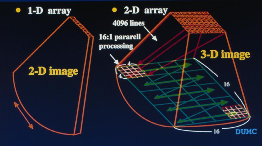
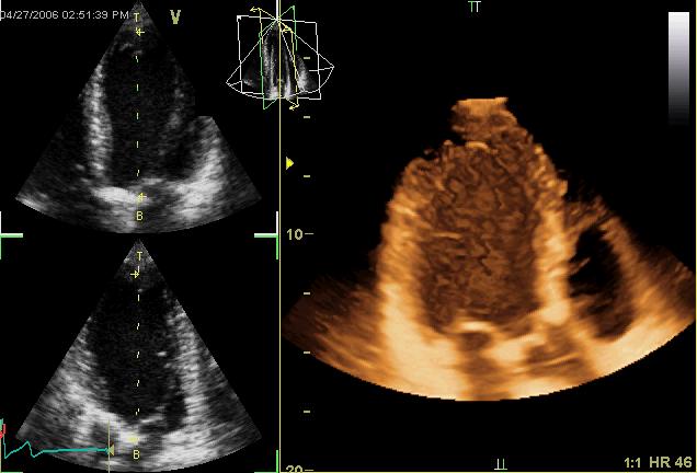
21 Real-time 3D echocardiography Matrix array transducer

22 Am J Cardiol Nov 1;100(9):


23 3D measurement of PISA Surface Area a b c J Am Soc Echocardiogr Apr;20(4):389-96
24 3D surface area of an ellipsoid is... a b c Not so simple
25 Am J Cardiol Oct 1;104(7):

26 Am J Cardiol Oct 1;104(7):
27 3D VCA Excellent Correlation with Cardiac MR

28 3D EROA (VCA) vs. 2D EROA 3D EROA > 2D EROA

29 3D Guided Measurement of vena contracta area in MR

30 Measuring the vena contracta area

31 3D Guided Vena Contracta Area
32 3D vena contracta method (from apical 4 window) Zeng X. et al. Circulation CVImaging 2011; Vol. 4
33 3D VCA vs. 2D ROA Central vs. Eccentric MR Jets A. Central MR jet B. Eccentric MR jet Zeng X. et al. Circulation CVImaging 2011; Vol. 4
34 3D VCA vs. 2D ROA Etiology of MR Degenerative Functional Zeng X. et al. Circulation CVImaging 2011; Vol. 4
35 Summary Increasing Validation of 3D color Doppler for MR Quantification Useful adjunct to 2D Integrative Method; especially in cases with mixed criteria 3D guided measurement of vena contracta area (effective regurgitant orifice area) is most validated Eccentric Jets Do not need Geometric Assumptions
36

37 The Good
38 EROA by 3D VCA vs 2D EROA PISA Zeng X. et al; Circ CVIM; October, 2011


39 How much MR? EROA = 2πR2 x Aliasing Velocity Velocity of MR
Disclosures Rebecca T. Hahn, MD, FASE
 The New ASE Guidelines for Native Valvular Regurgitation Mitral Regurgitation The New ASE Guidelines: Role of 2D/3D and CMR (With caveats and comments from R. Hahn) William A. Zoghbi MD, FASE, MACC Professor
PISA Evaluation of Mitral Regurgitation. Raymond Graber, MD Cardiac Anesthesia Group University Hospitals Case Medical Center 4/07/2011
 PISA Evaluation of Mitral Regurgitation Raymond Graber, MD Cardiac Anesthesia Group University Hospitals Case Medical Center 4/07/2011 Introduction Evaluation of MR. What is PISA? Physiologic basis Issues
Marti McCulloch, BS, MBA, RDCS, FASE Houston, Texas
 Marti McCulloch, BS, MBA, RDCS, FASE Houston, Texas Mitral Regurgitation What to Expect Review Specific Signs of Severity Supportive Signs of Severity Qualitative Parameters Structural Doppler Quantitative
Valvular Regurgitation: Can We Do Better Than Colour Doppler?
 Valvular Regurgitation: Can We Do Better Than Colour Doppler? A/Prof David Prior St Vincent s Hospital Melbourne Sports Cardiology Valvular Regurgitation Valve regurgitation volume loads the ventricles
Prof. JL Zamorano Hospital Universitario Ramón y Cajal
 Prof. JL Zamorano Hospital Universitario Ramón y Cajal Should we forget TR? Nath J et al. Impact of tricuspid regurgitation on long-term survival. J Am Coll Cardiol. 2004; 43:405-409 Why is it difficult
Professors Carpentier and McGoon Mechanism, resulting from the disease Severity of regurgitation, resulting from the mechanism Echo
 Professors Carpentier and McGoon Mechanism, resulting from the disease Severity of regurgitation, resulting from the mechanism Echo define the mechanism, quantify the regurgitation severity CP1293058-3
Quantifying Aortic Regurgitation
 Quantifying Aortic Regurgitation Linda D. Gillam, MD, MPH Morristown Medical Center Dorothy and Lloyd Huck Chair Cardiovascular Medicine Atlantic Health System No Disclosures 1 Valve Dysfunction Functional
CHAPTER 4 AN EFFICACIOUS APPROACH FOR THE QUANTIFICATION OF MITRAL REGURGITATION USING IMAGE PROCESSING AND PROXIMAL FLOW CONVERGENCE METHOD
 CHAPTER 4 AN EFFICACIOUS APPROACH FOR THE QUANTIFICATION OF MITRAL REGURGITATION USING IMAGE PROCESSING AND PROXIMAL FLOW CONVERGENCE METHOD 4.1. Introduction Mitral Regurgitation also called Mitral Insufficiency
Echocardiography. Guidelines for Valve and Chamber Quantification. In partnership with
 Echocardiography Guidelines for Valve and Chamber Quantification In partnership with Explanatory note & references These guidelines have been developed by the Education Committee of the British Society
How to assess ischaemic MR?
 ESC 2012 How to assess ischaemic MR? Luc A. Pierard, MD, PhD, FESC, FACC Professor of Medicine Head, Department of Cardiology University Hospital Sart Tilman, Liège ESC 2012 No conflict of interest Luc
Guidelines in perspective?
 EuroValve 2016 Challenges in the management Secondary MR Guidelines in perspective? Luc A. Pierard, MD, PhD Professor of Medicine Head of the Department of Cardiology, Liège, Belgium Heart Valve Clinic,
Quantification of MR
 Valvular Regurgitation: Putting the New Guidelines into Practice James D. Thomas, MD, FACC, FASE, FESC Director, Center for Heart Valve Disease Bluhm Cardiovascular Institute Professor of Medicine, Feinberg
Direct Planimetry of Mitral Valve Regurgitation Orifice Area by Real-time 3D Transesophageal Echocardiography
 Direct Planimetry of Mitral Valve Regurgitation Orifice Area by Real-time 3D Transesophageal Echocardiography Ertunc Altiok, Sandra Hamada, Silke van Hall, Mehtap Hanenberg, Eva Grabskaya, Michael Becker,
The Doppler Examination. Katie Twomley, MD Wake Forest Baptist Health - Lexington
 The Doppler Examination Katie Twomley, MD Wake Forest Baptist Health - Lexington OUTLINE Principles/Physics Use in valvular assessment Aortic stenosis (continuity equation) Aortic regurgitation (pressure
Echocardiography: Guidelines for Valve Quantification
 Echocardiography: Guidelines for Echocardiography: Guidelines for Chamber Quantification British Society of Echocardiography Education Committee Richard Steeds (Chair), Gill Wharton (Lead Author), Jane
Hemodynamic Assessment. Assessment of Systolic Function Doppler Hemodynamics
 Hemodynamic Assessment Matt M. Umland, RDCS, FASE Aurora Medical Group Milwaukee, WI Assessment of Systolic Function Doppler Hemodynamics Stroke Volume Cardiac Output Cardiac Index Tei Index/Index of myocardial
LUST trial. Echocardiography USER S MANUAL
 LUST trial Echocardiography USER S MANUAL Rosa Sicari, Luna Gargani Ins1tute of Clinical Physiology Na1onal Council of Research, Pisa, Italy Parameters required (1) Aortic root Measurement of aortic root
ASE Guidelines on Aortic Regurgitation What Do I Measure? Case Studies
 ASE Guidelines on Aortic Regurgitation What Do I Measure? Case Studies Mitral Regurgitation The New ASE Guidelines: Role of 2D/3D and CMR William A. Zoghbi MD, FASE, MACC Professor and Chairman, Department
AATS Annual Meeting Seattle, WA Irving Kron, M.D. Professor and Chairman Department of Surgery University of Virginia Hospital Charlottesville,
 AATS Annual Meeting Seattle, WA Irving Kron, M.D. Professor and Chairman Department of Surgery University of Virginia Hospital Charlottesville, Virginia AATS Ischemic MR Guideline Writing Group Roster
Titel Kardiologie-SG.ch hot topics in heart failure and mitral regurgitation
 Titel Kardiologie-SG.ch hot topics in heart failure and mitral regurgitation where and how to quantify mitral regurgitation: Echolab, Cathlab or MRI? Philipp K. Haager, St. Gallen Measuring mitral regurgitation?
Improvements in outcomes for heart valve surgery, in particular mitral valve repair, have dictated
 Imaging techniques DOPPLER ECHOCARDIOGRAPHIC ASSESSMENT OF VALVAR REGURGITATION c COLOUR Correspondence to: James D Thomas MD, Department of Cardiology, Desk F15, 9500 Euclid Avenue, The Cleveland Clinic
True morphology of mitral regurgitant flow assessed by three- dimensional transesophageal echocardiography
 DOI: 10.1111/echo.13395 ORIGINAL INVESTIGATION True morphology of mitral regurgitant flow assessed by three- dimensional transesophageal echocardiography Martin Lombardero M.D. Ruth Henquin D.L.S.H.T.M.,
What are the best diagnostic tools to quantify aortic regurgitation?
 What are the best diagnostic tools to quantify aortic regurgitation? Agnès Pasquet, MD, PhD Pôle de Recherche Cardiovasculaire Institut de Recherche Expérimentale et Clinique Université catholique de Louvain
Mitral regurgitation (MR) is a
 is a") Review Article Hellenic J Cardiol 2013; 54: 448-454 3D Vena Contracta Area to Quantify Severity of Mitral Regurgitation: A Practical New Tool? Dimitrios Maragiannis, Stephen H. Little The Methodist DeBakey
Review Article Hellenic J Cardiol 2013; 54: 448-454 3D Vena Contracta Area to Quantify Severity of Mitral Regurgitation: A Practical New Tool? Dimitrios Maragiannis, Stephen H. Little The Methodist DeBakey
Primary Mitral Regurgitation
 EURO VALVE Madrid News from Valves Guidelines 2012: What s new and Why? Primary Mitral Regurgitation Luc A. Pierard, MD, PhD Professor of Medicine Head of the Department of Cardiology Heart Valve Clinic,
ASCeXAM / ReASCE. Practice Board Exam Questions Monday Morning
 ASCeXAM / ReASCE Practice Board Exam Questions Monday Morning Ultrasound Physics Artifacts Doppler Physics Imaging, Knobology, and Artifacts Echocardiographic Evaluation of the RV Tricuspid and Pulmonary
EVALUATION OF CHRONIC MITRAL REGURGITATION: ASSESSING MECHANISMS AND QUANTIFYING SEVERITY 2018 STRUCTURAL HEART DISEASE CONFERENCE June 1, 2018
 1 EVALUATION OF CHRONIC MITRAL REGURGITATION: ASSESSING MECHANISMS AND QUANTIFYING SEVERITY 2018 STRUCTURAL HEART DISEASE CONFERENCE June 1, 2018 David A. Orsinelli, MD, FACC, FASE Professor, Internal
The difficult patient with mitral regurgitation
 Clinical pathways The difficult patient with mitral regurgitation Stress echo can be the best tool Challenging cases Maria João Andrade, Lisbon PT Management of Severe Chronic Organic MR Echo Exercise
Quantification of Aortic Regurgitation
 Quantification of Aortic Regurgitation ASE Review 2018 Boston Susan E Wiegers, MD, FASE, FACC Professor of Medicine And thanks to Dr. Roberto Lang Disclosure None related to this presentation 1 Objectives
Ioannis Alexanian, MD, PhD Department of Cardiology General Hospital of Chest Diseases Sotiria Athens
 MITRAL REGURGITATION IN PATIENT WITH SEVERE AORTIC VALVE STENOSIS Ioannis Alexanian, MD, PhD Department of Cardiology General Hospital of Chest Diseases Sotiria Athens I HAVE NOTHING TO DECLARE Management
Reproducibility of Proximal Isovelocity Surface Area, Vena Contracta, and Regurgitant Jet Area for Assessment of Mitral Regurgitation Severity
 JACC: CARDIOVASCULAR IMAGING VOL. 3, NO. 3, 2010 2010 BY THE AMERICAN COLLEGE OF CARDIOLOGY FOUNDATION ISSN 1936-878X/10/$36.00 PUBLISHED BY ELSEVIER INC. DOI:10.1016/j.jcmg.2009.09.029 Reproducibility
Quantification of Mitral Stenosis: Planimetry, pressure Half time, Continuity Common Errors
 Quantification of Mitral Stenosis: Planimetry, pressure Half time, Continuity Common Errors Christopher J Kramer RDCS Advanced Cardiovascular Services Aurora Health Care Milwaukee, WI No Disclosures Baumgartner,
MITRAL REGURGITATION ECHO PARAMETERS TOOL
 Comprehensive assessment of qualitative and quantitative parameters, along with the use of standardized nomenclature when reporting echocardiographic findings, helps to better define a patient s MR and
Doppler Basic & Hemodynamic Calculations
 Doppler Basic & Hemodynamic Calculations August 19, 2017 Smonporn Boonyaratavej MD Division of Cardiology, Department of Medicine Chulalongkorn University Cardiac Center, King Chulalongkorn Memorial Hospital
What Degree of MR Deserves Surgical or Transcatheter Intervention, and How Should It Be Assessed?
 What Degree of MR Deserves Surgical or Transcatheter Intervention, and How Should It Be Assessed? Robert J. Siegel, M.D., FACC Nov. 14-15, 2017, Beverly Hills Director, Cardiac Non-Invasive Laboratory
Diagnostic Value of Vena Contracta Area in the Quantification of Mitral Regurgitation Severity by Color Doppler 3D Echocardiography
 Diagnostic Value of Vena Contracta Area in the Quantification of Mitral Regurgitation Severity by Color Doppler 3D Echocardiography Xin Zeng, MD, PhD; Robert A. Levine, MD; Lanqi Hua, RDCS; Eleanor L.
Secondary MR joint with the mitral academy. What is new in our understanding of this disease? Luc Pierard University Hospital, Liège
 Secondary MR joint with the mitral academy What is new in our understanding of this disease? Luc Pierard University Hospital, Liège Faculty disclosure Luc Pierard I have no financial relationships to disclose.
Functional Mitral Regurgitation
 Club 35 - The best in heart valve disease - Functional Mitral Regurgitation Steven Droogmans, MD, PhD UZ Brussel, Jette, Belgium 08-12-2011 Euroecho & other Imaging Modalities 2011 No conflicts of interest
JOINT MEETING 2 Tricuspid club Chairpersons: G. Athanassopoulos, A. Avgeropoulou, M. Khoury, G. Stavridis
 JOINT MEETING 2 Tricuspid club Chairpersons: G. Athanassopoulos, A. Avgeropoulou, M. Khoury, G. Stavridis Similarities and differences in Tricuspid vs. Mitral Valve Anatomy and Imaging. Echo evaluation
Uncommon Doppler Echocardiographic Findings of Severe Pulmonic Insufficiency
 Uncommon Doppler Echocardiographic Findings of Severe Pulmonic Insufficiency Rahul R. Jhaveri, MD, Muhamed Saric, MD, PhD, FASE, and Itzhak Kronzon, MD, FASE, New York, New York Background: Two-dimensional
Who will Benefit from Percutaneous Management of Mitral Regurgitation? An Imaging Guide to Management
 Who will Benefit from Percutaneous Management of Mitral Regurgitation? An Imaging Guide to Management James D. Thomas, M.D., F.A.C.C. Department of Cardiovascular Medicine Heart and Vascular Institute
MR echo case. N.Koutsogiannis Department of Cardiology University Hospital Of Patras
 MR echo case N.Koutsogiannis Department of Cardiology University Hospital Of Patras Case A 35 years old male came to the echo lab for a third opinion for his valvulopathy. He reports a long standing MR
Clinical Outcome of Tricuspid Regurgitation. David Messika-Zeitoun
 Clinical Outcome of Tricuspid Regurgitation David Messika-Zeitoun I have financial relationships to disclose Consultant for: Edwards, Symetis and Valtech Tricuspid Regurgitation is a Common Finding Tricuspid
Tricuspid and Pulmonary Valve Disease
 Tricuspid and Pulmonary Valve Disease Lawrence Rudski MD FRCPC FACC FASE Professor of Medicine Director, Division of Cardiology Jewish General Hospital McGill University Question 1 All of the following
Tricuspid and Pulmonary Valve Disease
 Tricuspid and Pulmonary Valve Disease Lawrence Rudski MD FRCPC FACC FASE Professor of Medicine Director, Division of Cardiology Jewish General Hospital McGill University Right Sided Failure Edema Gut congestion
Diastolic Heart Function: Applying the New Guidelines Case Studies
 Diastolic Heart Function: Applying the New Guidelines Case Studies Mitral Regurgitation The New ASE William Guidelines: A. Zoghbi Role MD, of FASE, 2D/3D MACCand CMR Professor and Chairman, Department
3D Echo for Evaluation of Tricuspid Regurgitation Jong-Min Song, MD, PhD
 3D Echo for Evaluation of Tricuspid Regurgitation Jong-Min Song, MD, PhD Asan Medical Center University of Ulsan College of Medicine Seoul, Korea Causes of TR Primary causes (25%) Rheumatic Myxomatous
Back to Basics: Common Errors In Quantitation In Everyday Practice
 Back to Basics: Common Errors In Quantitation In Everyday Practice Deborah Agler, ACS, RDCS, FASE October 9, 2017 ASE: Echo Florida Rebecca T. Hahn, MD Director of Interventional Echocardiography Professor
Advanced Applica,on of Point- of- Care Echocardiography in Cri,cal Care. Dr. Mark Tutschka Dr. Rob ArnAield
 Advanced Applica,on of Point- of- Care Echocardiography in Cri,cal Care Dr. Mark Tutschka Dr. Rob ArnAield OBJECTIVES Provide an overview of common advanced echocardiographic techniques suitable for use
RIGHT VENTRICULAR SIZE AND FUNCTION
 RIGHT VENTRICULAR SIZE AND FUNCTION Edwin S. Tucay, MD, FPCC, FPCC, FPSE Philippine Society of Echocardiography Quezon City, Philippines Echo Mission, BRTTH, Legaspi City, July 1-2, 2016 NO DISCLOSURE
Revealing new insights. irotate electronic rotation and xplane adjustable biplane imaging. Ultrasound cardiology. irotate and xplane
 Ultrasound cardiology irotate and xplane Revealing new insights irotate electronic rotation and xplane adjustable biplane imaging Annemien van den Bosch and Jackie McGhie Department of Cardiology, Erasmus
aortic regurgitation, vena contracta area, vena contracta width, live three-dimensional echocardiography
 RESEARCH FROM THE UNIVERSITY OF ALABAMA AT BIRMINGHAM Assessment of Aortic Regurgitation by Live Three-Dimensional Transthoracic Echocardiographic Measurements of Vena Contracta Area: Usefulness and Validation
Imaging MV. Jeroen J. Bax Leiden University Medical Center The Netherlands Davos, feb 2015
 Imaging MV Jeroen J. Bax Leiden University Medical Center The Netherlands Davos, feb 2015 MV/MR: information needed on.. 1. MV anatomy 2. MR etiology - primary vs secondary 3. MR severity quantification
An understanding of the many factors involved in the
 Atrioventricular Valve Dysfunction: Evaluation by Doppler and Cross-Sectional Ultrasound Norman H. Silverman, MD, and Doff B. McElhinney, MD Division of Pediatric Cardiology, Department of Pediatrics,
NEW GUIDELINES. A Guideline Protocol for the Assessment of Aortic Regurgitation From the British Society of Echocardiography Education Committee
 NEW GUIDELINES A Guideline Protocol for the Assessment of Aortic Regurgitation From the British Society of Echocardiography Education Committee Gill Wharton, Prathap Kanagala (Lead Authors) Richard Steeds
Cover Page. The handle holds various files of this Leiden University dissertation.
 Cover Page The handle http://hdl.handle.net/1887/21650 holds various files of this Leiden University dissertation. Author: Shanks, Miriam Title: Evolving imaging techniques for the assessment of cardiac
Prosthetic valve dysfunction: stenosis or regurgitation
 Prosthetic valve dysfunction: stenosis or regurgitation Jean G. Dumesnil MD, FRCP(C), FACC, FASE(Hon) Quebec Heart and Lung Institute, Québec, Québec No disclosures Possible Causes of High Gradients in
Quantitative Assessment of Mitral Regurgitation
 JACC: CARDIOVASCULAR IMAGING VOL. 5, NO. 11, 2012 2012 BY THE AMERICAN COLLEGE OF CARDIOLOGY FOUNDATION ISSN 1936-878X/$36.00 PUBLISHED BY ELSEVIER INC. http://dx.doi.org/10.1016/j.jcmg.2012.07.013 STATE-OF-THE-ART
School, The University of Texas Health Science Center at Houston, TX.
 Chapter 4: Basics of echocardiography for transcatheter interventions Adrian DaSilva-DeAbreu, MD 1, Catalin Loghin, MD 2 1 Resident, Department of Internal Medicine, McGovern Medical School, The University
AIMI-HF PROCEDURE MANUAL TECHNICAL GUIDE FOR ECHOCARDIOGRAPHY. MHI Core Laboratory E. O Meara - J.C. Tardif J. Vincent, G. Grenier, C.
 AIMI-HF PROCEDURE MANUAL TECHNICAL GUIDE FOR ECHOCARDIOGRAPHY MHI Core Laboratory E. O Meara - J.C. Tardif J. Vincent, G. Grenier, C. Roy February 2016 Montreal Heart Institute HF Research Aude Turgeon,
The Normal Echocardiogram
 The Normal Echocardiogram Pravin V. Patil, MD FACC Lewis Katz School of Medicine at Temple University Acknowledgments Dr. Susan Wiegers Dr. Martin Keane Temple Cardiac Sonographers Disclosures No relevant
Congenital. Unicuspid Bicuspid Quadricuspid
 David Letterman s Top 10 Aortic Stenosis The victim can be anyone: Echo is the question and the answer!!!! Hilton Head Island Echocardiography Conference 2012 Timothy E. Paterick, MD, JD, MBA Christopher
Appendix II: ECHOCARDIOGRAPHY ANALYSIS
 Appendix II: ECHOCARDIOGRAPHY ANALYSIS Two-Dimensional (2D) imaging was performed using the Vivid 7 Advantage cardiovascular ultrasound system (GE Medical Systems, Milwaukee) with a frame rate of 400 frames
Concepts of Imaging and Knobology
 Concepts of Imaging and Knobology Pravin Patil, MD FACC FASE Associate Professor of Medicine Director, Cardiovascular Disease Training Program Lewis Katz School of Medicine at Temple University Disclosures
HEART VALVE DISEASES. Dr. James Kadouch Medical Officer Cardiologist Delphine Labojka Method & Process Manager
 NOVEMBER 2017 SCOR inform HEART VALVE DISEASES Dr. James Kadouch Medical Officer Cardiologist Delphine Labojka Method & Process Manager Introduction Cardiac cycle and heart valve diseases 03 The aortic
CHAPTER 5 EFFICIENT APPROACHES FOR QUANTIFICATION OF AORTIC REGURGITATION USING PROXIMAL ISOVELOCITY SURFACE AREA PROCESS
 CHAPTER 5 EFFICIENT APPROACHES FOR QUANTIFICATION OF AORTIC REGURGITATION USING PROXIMAL ISOVELOCITY SURFACE AREA PROCESS 5.1. Introduction Aortic Regurgitation is also known as Aortic Insufficiency (AI).
PART II ECHOCARDIOGRAPHY LABORATORY OPERATIONS ADULT TRANSTHORACIC ECHOCARDIOGRAPHY TESTING
 PART II ECHOCARDIOGRAPHY LABORATORY OPERATIONS ADULT TRANSTHORACIC ECHOCARDIOGRAPHY TESTING STANDARD - Primary Instrumentation 1.1 Cardiac Ultrasound Systems SECTION 1 Instrumentation Ultrasound instruments
Quantitative Assessment of Mitral Regurgitation
 Journal of the American College of Cardiology Vol. 60, No. 16, 2012 2012 by the American College of Cardiology Foundation ISSN 0735-1097/$36.00 Published by Elsevier Inc. http://dx.doi.org/10.1016/j.jacc.2012.05.048
Diagnostic approach to heart disease
 Diagnostic approach to heart disease Initial work up History Physical exam Chest radiographs ECG Special studies Echocardiography Cardiac catheterization Echocardiography principles Technique of producing
MITRAL VALVE PATHOLOGY WITH TRICUSPID REGURGITATION (AND PHT)
") UNIVERSITY OF PADUA, SCHOOL OF MEDICINE Department of Cardiac,Thoracic and Vascular Sciences Padua, Italy MITRAL VALVE PATHOLOGY WITH TRICUSPID REGURGITATION (AND PHT) Luigi P. Badano**, MD, PhD, FESC,
UNIVERSITY OF PADUA, SCHOOL OF MEDICINE Department of Cardiac,Thoracic and Vascular Sciences Padua, Italy MITRAL VALVE PATHOLOGY WITH TRICUSPID REGURGITATION (AND PHT) Luigi P. Badano**, MD, PhD, FESC,
A Practical Approach to Prosthetic Valves
 A Practical Approach to Prosthetic Valves Bonita Anderson DMU (Cardiac), MApplSc (Med Ultrasound), ACS, AMS, FASE https://doi.org/10.1161/circulationaha.108.778886 Disclosures None 1 Know the Product Know
Quantitative Doppler echocardiography
 BJA Education, 16 (2): 46 52 (2016) doi: 10.1093/bjaceaccp/mkv015 Advance Access Publication Date: 10 June 2015 Matrix reference 1A03, 2A04, 3G00 Quantitative Doppler echocardiography P Harris MBChB FRCA
Pulsed Wave Doppler and Color Flow Doppler Evaluation in Healthy Dogs and Dogs with Cardiac Disease
 Cloud Publications International Journal of Advanced Veterinary Science and Technology 2016, Volume 5, Issue 2, pp. 256-265, Article ID Sci-446 ISSN 2320-3595 Research Article Open Access Pulsed Wave Doppler
Echocardiographic evaluation of mitral stenosis
 Echocardiographic evaluation of mitral stenosis Euroecho 2011 Philippe Unger, MD, FESC Erasme Hospital, ULB, Brussels, Belgium I have nothing to declare EuroHeart Survey Etiology of single native left-sided
Quantitation of Aortic Regurgitation ASCeXAM / ReASCE Review Course
 Quantitation of Aortic Regurgitation 2017 ASCeXAM / ReASCE Review Course David Adams, ACS, RCS, RDCS, FASE Duke University Medical Center May 1, 2017 Disclosures None 1 Exam based on: Zoghbi et al. JASE
Adopted by Council March 2006, Revised March 2012, September 2015
 Guidelines, Policies and Statements E4 Education Protocol: Competences Required Of Cardiac Sonographers Who Practice Adult Transthoracic Cardiac Ultrasound Examinations Adopted by Council March 2006, Revised
M itral regurgitation (MR) is the most commonly
 is the most commonly") iv11 Assessment of mitral regurgitation T Irvine, X K Li, D J Sahn, A Kenny... M itral regurgitation (MR) is the most commonly encountered valve lesion in modern clinical practice. 1 The range of pathologies
iv11 Assessment of mitral regurgitation T Irvine, X K Li, D J Sahn, A Kenny... M itral regurgitation (MR) is the most commonly encountered valve lesion in modern clinical practice. 1 The range of pathologies
좌심실수축기능평가 Cardiac Function
 Basic Echo Review Course 좌심실수축기능평가 Cardiac Function Seonghoon Choi Cardiology Hallym university LV systolic function Systolic function 좌심실수축기능 - 심근의수축으로심실에서혈액을대동맥으로박출하는기능 실제임상에서 LV function 의의미 1Diagnosis
Introduction RECOMMENDATIONS
 European Journal of Echocardiography (2010) 11, 223 244 doi:10.1093/ejechocard/jeq030 RECOMMENDATIONS European Association of Echocardiography recommendations for the assessment of valvular regurgitation.
The diagnosis of mitral regurgitation (MR) caused
 caused") J Vet Intern Med 2014;28:1206 1213 Assessment of Mitral Regurgitation Severity by Doppler Color Flow Mapping of the Vena Contracta in Dogs M. Di Marcello, E. Terzo, C. Locatelli, V. Palermo, E. Sala, E.
J Vet Intern Med 2014;28:1206 1213 Assessment of Mitral Regurgitation Severity by Doppler Color Flow Mapping of the Vena Contracta in Dogs M. Di Marcello, E. Terzo, C. Locatelli, V. Palermo, E. Sala, E.
MITRAL STENOSIS. Joanne Cusack
 MITRAL STENOSIS Joanne Cusack BSE Breakdown Recognition of rheumatic mitral stenosis Qualitative description of valve and sub-valve calcification and fibrosis Measurement of orifice area by planimetry
Chapter 20. JACC Cardiovasc Imaging 2009;2:
 Chapter 20 Quantification of functional mitral regurgitation by real-time 3D echocardiography: comparison with 3D velocity-encoded cardiac magnetic resonance N Ajmone Marsan, J JM Westenberg, C Ypenburg,
10/7/2013. Systolic Function How to Measure, How Accurate is Echo, Role of Contrast. Thanks to our Course Director: Neil J.
 Systolic Function How to Measure, How Accurate is Echo, Role of Contrast Neil J. Weissman, MD MedStar Health Research Institute & Professor of Medicine Georgetown University Washington, D.C. No Disclosures
What echo measurements are key prior to MitraClip?
 APHP CHU Bichat - Claude Bernard What echo measurements are key prior to MitraClip? Eric Brochet,MD Cardiology Department Hopital Bichat Paris France No disclosure Conflict of interest Case 69 y.o man
Assessment of LV systolic function
 Tutorial 5 - Assessment of LV systolic function Assessment of LV systolic function A knowledge of the LV systolic function is crucial in the undertanding of and management of unstable hemodynamics or a
Goals of Reporting. Background of the STS/ACC TVT Registry. TVT Registry TAVR and MitraClip 10/11/ /11/2016 9:10 9:30 AM
 10/11/2016 9:10 9:30 AM Rebecca T. Hahn, MD Director of Interventional Echocardiography Columbia University Medical College Background of the STS/ACC TVT Registry The STS/ACC TVT Registry is a benchmarking
ECHOCARDIOGRAPHY DATA REPORT FORM
 Patient ID Patient Study ID AVM - - Date of form completion / / 20 Initials of person completing the form mm dd yyyy Study period Preoperative Postoperative Operative 6-month f/u 1-year f/u 2-year f/u
In the clinical management of patients with ventricular
 Quantitative Assessment of Severity of Ventricular Septal Defect by Three-Dimensional Reconstruction of Color Doppler Imaged Vena Contracta and Flow Convergence Region Masahiro Ishii, MD; Kanoko Hashino,
Normal TTE/TEE Examinations
 Normal TTE/TEE Examinations Geoffrey A. Rose, MD FACC FASE Sanger Heart & Vascular Institute Before you begin imaging... Obtain the patient s Height Weight BP PLAX View PLAX View Is apex @ 9-10 o clock?
The Key Questions in Mitral Valve Interventions. Where Are We in 2018?
 The Key Questions in Mitral Valve Interventions Where Are We in 2018? Gilles D. DREYFUS, MD, FRCS, FESC Professor of Cardiothoracic Surgery 30 GIORNATE CARDIOLOGICHE TORINESI - OCT 2018 Are guidelines
Usefulness of 3D-PISA as compared to guideline endorsed parameters for mitral regurgitation quantification
 DOI 10.1007/s10554-014-0496-7 ORIGINAL PAPER Usefulness of 3D-PISA as compared to guideline endorsed parameters for mitral regurgitation quantification Frank P. Schmidt Theresa Gniewosz Alexander Jabs
SURGICAL TREATMENT OF MODERATE ISCHEMIC MITRAL REGURGITATION: THE CARDIOTHORACIC SURGICAL TRIALS NETWORK
 SURGICAL TREATMENT OF MODERATE ISCHEMIC MITRAL REGURGITATION: THE CARDIOTHORACIC SURGICAL TRIALS NETWORK Robert E. Michler, M.D. For the CTSN Investigators AHA Late Breaking Clinical Trials November 18,
Dobutamine Stress testing In Low Flow, Low EF, Low Gradient Aortic Stenosis Case Studies
 Dobutamine Stress testing In Low Flow, Low EF, Low Gradient Aortic Stenosis Case Studies Mitral Regurgitation The New ASE Guidelines: Role of 2D/3D and CMR William A. Zoghbi MD, FASE, MACC Professor and
Aortic Stenosis: Spectrum of Disease, Low Flow/Low Gradient and Variants
 Aortic Stenosis: Spectrum of Disease, Low Flow/Low Gradient and Variants Martin G. Keane, MD, FASE Professor of Medicine Lewis Katz School of Medicine at Temple University Basic root structure Parasternal
When Does 3D Echo Make A Difference?
 When Does 3D Echo Make A Difference? Wendy Tsang, MD, SM Assistant Professor, University of Toronto Toronto General Hospital, University Health Network 1 Practical Applications of 3D Echocardiography Recommended
Ανεπάρκεια Τριγλώχινας Επιλογή Επεμβατικής Θεραπείας. Μ. Χρυσοχέρης Τμήμα Διαδερμικών Βαλβίδων ΔΘΚΑ ΥΓΕΙΑ
 Ανεπάρκεια Τριγλώχινας Επιλογή Επεμβατικής Θεραπείας Μ. Χρυσοχέρης Τμήμα Διαδερμικών Βαλβίδων ΔΘΚΑ ΥΓΕΙΑ Disclosures - Proctoring activities for Abbott Vascular, Edwards Lifesciences I and the HYGEIA Hospital
Quantitative Assessment of Pulmonary Regurgitation by Echocardiography in Patients After Repaired TOF
 Quantitative Assessment of Pulmonary Regurgitation by Echocardiography in Patients After Repaired TOF 2013. 4. 20. 서울대학교어린이병원소아청소년과 권보상 W. B. TOF (large VSD, infundibular stenosis) 19 mo, 8.5 kg Indication
Exercise-Induced Changes in Degenerative Mitral Regurgitation
 Journal of the American College of Cardiology Vol. 56, No. 4, 2010 2010 by the American College of Cardiology Foundation ISSN 0735-1097/$36.00 Published by Elsevier Inc. doi:10.1016/j.jacc.2009.12.073
American Society of Echocardiography
 Page 1 American Society of Echocardiography Recommendations for Quantification of Doppler Echocardiography A Report from the Doppler Quantification Task Force of the Nomenclature and Standards Committee