Circulatory system. Lecture #2
|
|
|
- Byron Knight
- 6 years ago
- Views:
Transcription

1 Circulatory system Lecture #2
2 The essential components of the human cardiovascular system: Heart Blood Blood vessels
 Umbilical arteries conduct venous blood from fetus to")
3 Arteries - blood vessels that conduct arterial blood from heart ventricle to organs and tissues Exceptions: 1) Pulmonary arteries conduct venous blood from right ventricle to lungs 2) Umbilical arteries conduct venous blood from fetus to placenta
 Umbilical veins conduct arterial blood from placenta to")
4 Veins - blood vessels that conduct venous blood from organs and tissues to heart atrium Exceptions: 1) Pulmonary veins conduct arterial blood from lungs to left atrium 2) Umbilical veins conduct arterial blood from placenta to fetus

5 Heart (Cor, Cardia)
6 Heart layers

7 Pericardium Serous pericardium layers: - Parietal - Visceral (=epicardium)

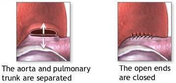
8 1. Transverse pericardial sinus - a space posterior to the ascending aorta and pulmonary trunk and anterior to the superior vena cava and pulmonary veins - it separates the great arteries from the great veins. The transverse sinus is useful in cardiac surgery to allow isolation of the aorta and pulmonary trunk. 2. Oblique pericardial sinus - the blind, inverted, U-shaped space posterior to the heart and bounded by reflection of serous pericardium around the 4 pulmonary veins and the inferior vena cava as they enter the heart.

9 Heart chambers

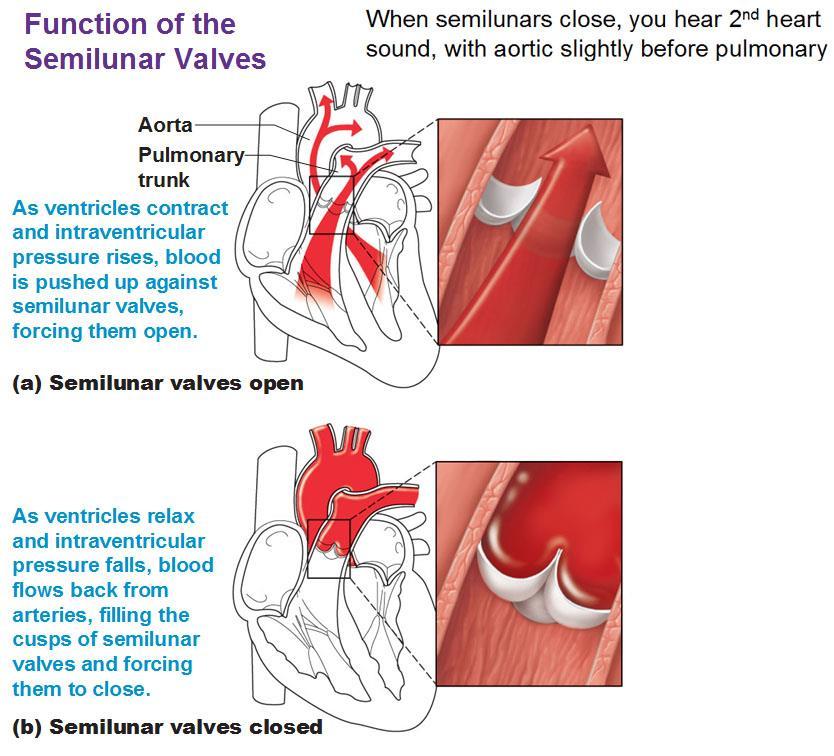
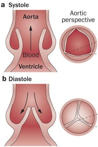
10 Heart valves Atrioventricular valves: - Between left atrium and left ventricle bicuspid (mitral) valve - Between right atrium and right ventricle tricuspid valve Semilunar valves: - Between left ventricle and aorta aortic valve - Between right ventricle and pulmonary arteries pulmonary valve


11 Mitral valve
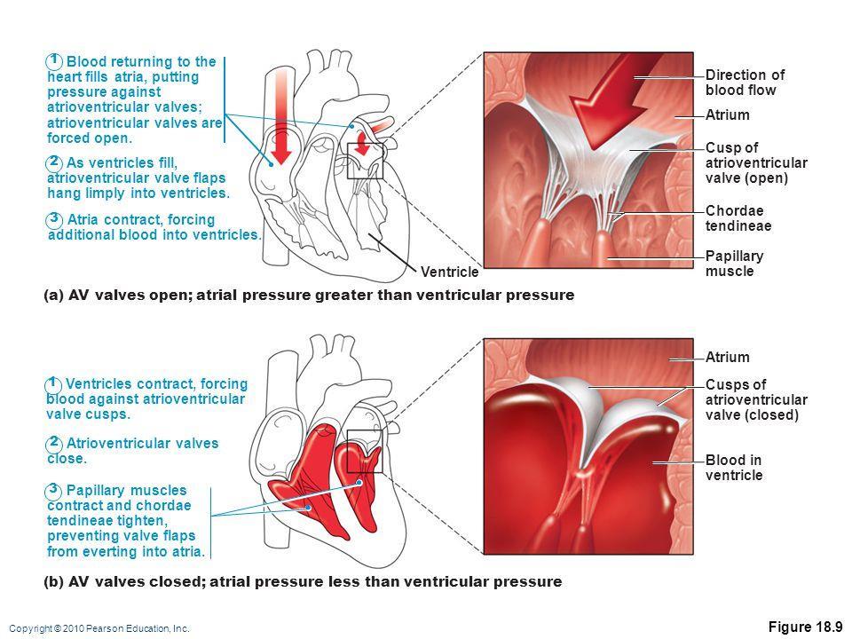
12

13 Bicuspid valve (mitral) between left atrium and left ventricle Tricuspid valve between right atrium and right ventricle

14 Semilunar cusps Aortic valve between left ventricle and aorta Pulmonary valve between right ventricle and pulmonary artery
15
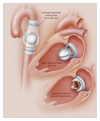

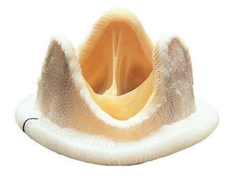
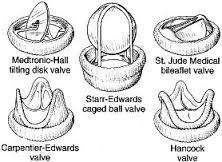
16 Cusp prosthetics
17

18 Coronary arteries Left coronary artery: - Anterior interventricular artery - Circumflex artery Right coronary artery: - Posterior interventricular artery - Marginal artery Widow-maker artery

 The great, middle and small cardiac veins 2) The")
19 Venous drainage of the heart: - Small veins (venae cordis minimae) directly into cardiac cavity - Anterior cardiac vein directly into right atrium - Coronary veins (accompany coronary arteries) that form coronary sinus opened into right atrium: 1) The great, middle and small cardiac veins 2) The oblique vein


20 The heart receives blood supply during diastole! The way coronary arteries pass between cardiomyocytes


21 - 25% of the heart nutrition diffuse through trabeculae carneae - present of evolution
22


23 Coronary artery bypass grafting (CABG)

24 Conducting system of the heart
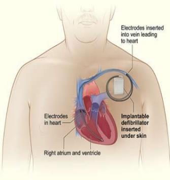
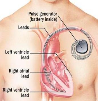
25 Artificial pacemaker (pulse generator)


26 Development of the heart 3 rd week - from splanchnic mesoderm within the cardiogenic area of the cranial end of the embryo - the cardiogenic cells condense to form a pair of primordial heart tubes
 2 endocardial bulbs 3 endocardial tubes 4 tubular")
27 Development of the heart 1 - epimyocardial plates (epicard and myocard) 2 endocardial bulbs 3 endocardial tubes 4 tubular heart

28 These structures can be distinguished in the tube Arterial conus Ventriculus Venous sinus

29 Development of the heart Venous sinus moves up and back Ventriculus goes front and to the left Arterial conus moves front and down
 end of the tube grows more rapidly and folds downward and ventrally and to the right.")
30 Blood flow The heart tube undergoes dextral looping (bends to the right) and rotation. The upper truncus arteriosus (ventricular) end of the tube grows more rapidly and folds downward and ventrally and to the right. The atria and sinus venosus lower part of the tube fold upward and dorsally and to the left. The 4 th week first contraction of the myocardium!

31 Development of the heart Cells of the endocardial tube Visceral mesoderm surrounding the primitive heart tube Endocardium Myocardium
 Somatic")

32 Development of the pericardium Intraembryonic body cavity(coelom) Somatic mesoderm Splanchnic mesoderm Pericardial cavity Parietal pericardium Visceral pericardium (epicardium)


33 Atrioventricular partition formation of the AV valves (5 th week)
34 Partition of the heart formation of the left (arterial) and right (venous) parts 1. Development of atrial septum 2. Development of ventricular septum 3. Development of arterial truncus septum
35 Partition of the left and right atrium
36 Foramen ovale

37 Circulatory changes at birth

38 Remnant of oval foramen fossa ovalis

")
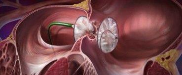
39 Patent foramen ovale (PFO)

40 Partition of the left and right ventricles
 perimembranous")
41 Ventricular septum defect (VSD) perimembranous muscular

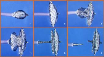
42 VSD Closure device

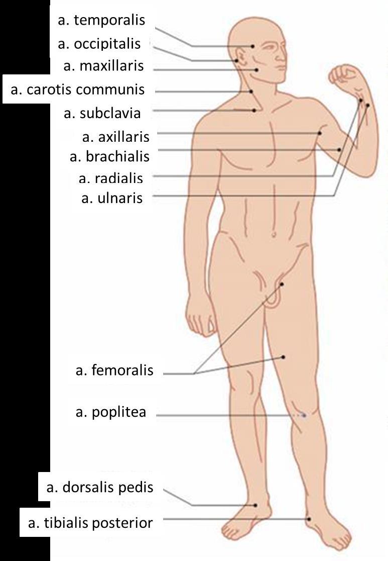
43 Partition of truncus arteriosus Longitudinal partition partition of aorta and pulmonary arteries Transverse partition formation of semilunar valves


44 Ductus arteriosus Ductus arteriosus Ligamentum arteriosum

45 Patent ductus arteriosus
 aortic arches (6 pairs) dorsal")
46 Development of the arteries Ventral aorta (paired) Blood of the embryo flows from heart into: aortic sac ventral aorta (paired) aortic arches (6 pairs) dorsal aorta

47 Development of the arteries dorsal end of ventral aorta external carotid artery I and II aortic arches reduce III aortic arch internal carotid artery right IV aortic arch right subclavian artery left IV aortic arch arch of aorta V aortic arch reduce VI aortic arch - pulmonary arteries
48


49 Coarctation of aorta - Congenital pathology of aorta development - Disproportional development of parts of the body
50 Arteries Arteries of pulmonary circulation Arteries of systemic circulation Arteries of the heart
51 Arteries Types of arteries (wall anatomy): - Elastic - Muscular - Combined (mixed)

52 Arteries Elastic arteries- large thick walled, aorta and its branches - has elastin in all tunics, especially tunica media - has pressure smoothing effect
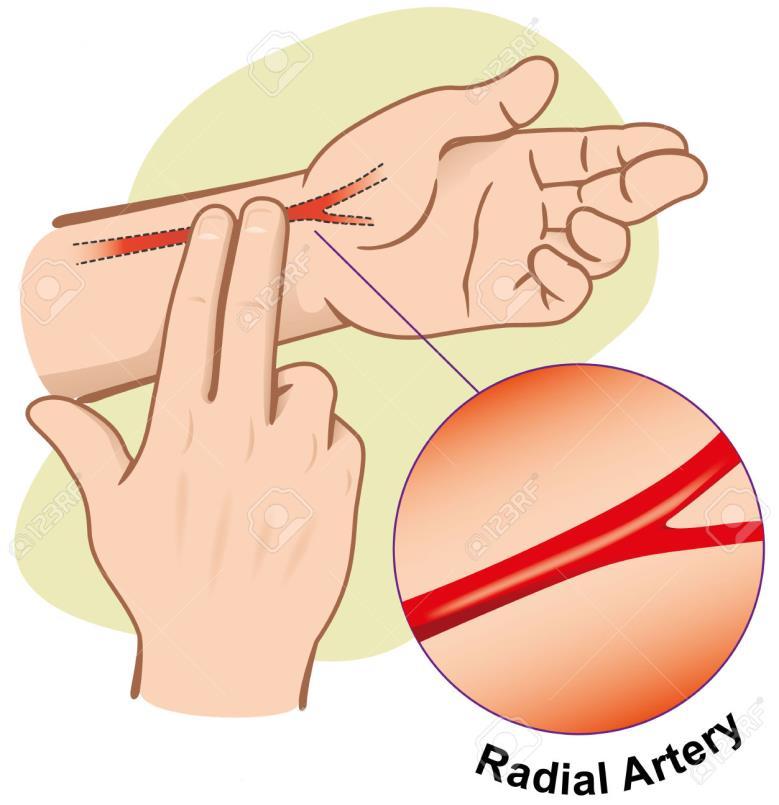
53 Arteries Muscular arteriesmost of the arteries - thickest media - more smooth muscle - active in vasoconstriction ( peripheral hearts )

54 3mm-10µm diameter of lumen blood flow to capillaries is determined by diameter of arterioles Arterioles


55 CAPILLARIES smallest 1mm long x 8-10µm diameter tunica intima only - endothelium exchange of gases, nutrients with interstitial fluid
 Fenestrated have windows very permeable to fluids and solutes Sinusoidal - modified, very")
56 Types of Capillaries Continuous - one endothelial cell wraps all around ends joined by tight junctions (brain) Fenestrated have windows very permeable to fluids and solutes Sinusoidal - modified, very leaky


57 Blood pressure 120/80? 120 cardiac (force of the heart) 80 tension of the vessels ( kidney )

58 Arteries - trunk parietal and visceral branches of aorta - paired and unpaired branches - arteries reaches organ along the shortest ways, usually together with nerves - every region has its own original main artery (head and neck carotid artery, abdomen abdominal part of aorta, etc.) - with or without anastomoses (connections)
 2) ventral visceral arteries (organs) 3) segmental")
59 Dorsal aorta descending aorta (thoracic and abdominal parts of aorta) branches: 1) dorsal parietal arteries (walls) 2) ventral visceral arteries (organs) 3) segmental arteries
60 Arterial blood supply of the limbs - mostly on the flexor side of the limbs - around joints form arterial network - on the palm and foot form arterial arches
 Inside system of one")
61 Anastomoses 1) Between different systems of arteries: - external and internal carotid arteries - external and internal iliac arteries - vessels of the right and left parts of the body 2) Inside system of one artery
 a.")
62 Anastomoses on the head a. dorsalis nasi (branch of ophthalmic artery internal carotid artery) a. angularis (branch of facial artery external carotid artery)

63 Arterial blood supply of the brain circle of Willis
")
64 Arterial anastomoses of the neck - between left and right external carotid arteries (arterial network of the organ)

65 Arterial anastomoses of the anterior abdominal wall
 leads to strong bleeding, which is very hard to")
66 Corona mortis crown of death % of people - wound of this anastomosis (during hernial sac operation) leads to strong bleeding, which is very hard to stop

67 Arteries clamping points


68 Tissues without vessels - transparent tissues of the eye (cornea, corpus vitreum, lens) - cartilage - epithelium - endothelium - dentin and enamel They are tolerant and can be transplanted without risk of rejection
Arteries. Lecture #2
 Arteries Lecture #2 The essential components of the human cardiovascular system: Heart Blood Blood vessels Arteries - blood vessels that conduct arterial blood from heart ventricle to organs and tissues
DEVELOPMENT OF THE CIRCULATORY SYSTEM L E C T U R E 5
 DEVELOPMENT OF THE CIRCULATORY SYSTEM L E C T U R E 5 REVIEW OF CARDIAC ANATOMY Heart 4 chambers Base and apex Valves Pericardial sac 3 layers: epi, myo, endo cardium Major blood vessels Aorta and its
Chapter 14. The Cardiovascular System
 Chapter 14 The Cardiovascular System Introduction Cardiovascular system - heart, blood and blood vessels Cardiac muscle makes up bulk of heart provides force to pump blood Function - transports blood 2
THE CARDIOVASCULAR SYSTEM. Part 1
 THE CARDIOVASCULAR SYSTEM Part 1 CARDIOVASCULAR SYSTEM Blood Heart Blood vessels What is the function of this system? What other systems does it affect? CARDIOVASCULAR SYSTEM Functions Transport gases,
Figure ) The specific chamber of the heart that is indicated by letter A is called the. Diff: 1 Page Ref: 364
 The specific chamber of the heart that is indicated by letter A is called the. Diff: 1 Page Ref: 364") Essentials of Anatomy and Physiology, 9e (Marieb) Chapter 11 The Cardiovascular System Short Answer Figure 11.1 Using Figure 11.1, identify the following: 1) The Purkinje fibers are indicated by label.
Essentials of Anatomy and Physiology, 9e (Marieb) Chapter 11 The Cardiovascular System Short Answer Figure 11.1 Using Figure 11.1, identify the following: 1) The Purkinje fibers are indicated by label.
Chapter 20 (1) The Heart
 The Heart") Chapter 20 (1) The Heart Learning Objectives Describe the location and structure of the heart Describe the path of a drop of blood from the superior vena cava or inferior vena cava through the heart out
Chapter 20 (1) The Heart Learning Objectives Describe the location and structure of the heart Describe the path of a drop of blood from the superior vena cava or inferior vena cava through the heart out
Blood Vessels. Types of Blood Vessels Arteries carry blood away from the heart Capillaries smallest blood vessels. Veins carry blood toward the heart
 C H A P T E R Blood Vessels 20 Types of Blood Vessels Arteries carry blood away from the heart Capillaries smallest blood vessels The site of exchange of molecules between blood and tissue fluid Veins
LECTURE 5. Anatomy of the heart
 LECTURE 5. Anatomy of the heart Main components of the CVS: Heart Blood circulatory system arterial compartment haemomicrocirculatory (=microvascular) compartment venous compartment Lymphatic circulatory
the Cardiovascular System I
 the Cardiovascular System I By: Dr. Nabil A Khouri MD, MsC, Ph.D MEDIASTINUM 1. Superior Mediastinum 2. inferior Mediastinum Anterior mediastinum. Middle mediastinum. Posterior mediastinum Anatomy of
Cardiovascular Anatomy Dr. Gary Mumaugh
 Cardiovascular Anatomy Dr. Gary Mumaugh Location of Heart Approximately the size of your fist Location o Superior surface of diaphragm o Left of the midline in mediastinum o Anterior to the vertebral column,
2. capillaries - allow exchange of materials between blood and tissue fluid
 Chapter 19 - Vascular System A. categories and general functions: 1. arteries - carry blood away from heart 2. capillaries - allow exchange of materials between blood and tissue fluid 3. veins - return
This lab activity is aligned with Visible Body s A&P app. Learn more at visiblebody.com/professors
 1 This lab activity is aligned with Visible Body s A&P app. Learn more at visiblebody.com/professors 2 PRE-LAB EXERCISES: A. Watch the video 29.1 Heart Overview and make the following observations: 1.
Embryology of the Heart
 *Page 1A: Embryology of the Heart Human embryonic disc is divided into three layers: ectoderm, intraembryonic mesoderm, and endoderm. The embryonic disc lies between the amniotic cavity and the primary
Figure 10.1A Transparency Master 79
 Brain Carotid arteries Jugular vein Right front leg Lungs (inflated) Cranial Right atrium To left front leg Left subclavian Bronchus capillaries Brachiocephalic vein Left atrium Dorsal aorta Right ventricle
CV Anatomy Quiz. Dr Ella Kim Dr Pip Green
 CV Anatomy Quiz Dr Ella Kim Dr Pip Green Q1 The location of the heart is correctly described as A) lateral to the lungs. B) medial to the sternum. C) superior to the diaphragm. D) posterior to the spinal
Anatomy of the Heart. Figure 20 2c
 Anatomy of the Heart Figure 20 2c Pericardium & Myocardium Remember, the heart sits in it s own cavity, known as the mediastinum. The heart is surrounded by the Pericardium, a double lining of the pericardial
The Heart. The Heart A muscular double pump. The Pulmonary and Systemic Circuits
 C H A P T E R 19 The Heart The Heart A muscular double pump circuit takes blood to and from the lungs Systemic circuit vessels transport blood to and from body tissues Atria receive blood from the pulmonary
2. right heart = pulmonary pump takes blood to lungs to pick up oxygen and get rid of carbon dioxide
 A. location in thorax, in inferior mediastinum posterior to sternum medial to lungs superior to diaphragm anterior to vertebrae orientation - oblique apex points down and to the left 2/3 of mass on left
Approximately the size of your fist Location Superior surface of diaphragm Left of the midline in mediastinum Anterior to the vertebral column,
 Dr. Gary Mumaugh Approximately the size of your fist Location Superior surface of diaphragm Left of the midline in mediastinum Anterior to the vertebral column, posterior to the sternum Posteriorly the
Major Function of the Cardiovascular System. Transportation. Structures of the Cardiovascular System. Heart - muscular pump
 Structures of the Cardiovascular System Heart - muscular pump Blood vessels - network of tubes Blood - liquid transport vehicle brachiocephalic trunk superior vena cava right pulmonary arteries right pulmonary
MODULE 2: CARDIOVASCULAR SYSTEM ANTOMY An Introduction to the Anatomy of the Heart and Blood vessels
 MODULE 2: CARDIOVASCULAR SYSTEM ANTOMY An Introduction to the Anatomy of the Heart and Blood vessels The cardiovascular system includes a pump (the heart) and the vessels that carry blood from the heart
Cardiovascular System
 Cardiovascular System Purpose Transport oxygen and nutrients Take waste products away from tissues & organs Things we learned Blood pressure: the force of blood pushing against the walls of blood vessels
THE HEART. A. The Pericardium - a double sac of serous membrane surrounding the heart
 THE HEART I. Size and Location: A. Fist-size weighing less than a pound (250 to 350 grams). B. Located in the mediastinum between the 2 nd rib and the 5 th intercostal space. 1. Tipped to the left, resting
Unit 11 - The Cardiovascular System 1
 Unit 11 - The Cardiovascular System 1 I. Unit 11: The Cardiovascular System A. The Cardiovascular System 1. A closed system of the heart and blood vessels a) The heart pumps blood b) Blood vessels allow
Lab Activity 23. Cardiac Anatomy. Portland Community College BI 232
 Lab Activity 23 Cardiac Anatomy Portland Community College BI 232 Cardiac Muscle Histology Branching cells Intercalated disc: contains many gap junctions connecting the adjacent cell cytoplasm, creates
Cardiovascular System Blood Vessels
 Cardiovascular System Blood Vessels Structure of Blood Vessels The three layers (tunics) Tunica intima composed of simple squamous epithelium Tunica media sheets of smooth muscle Contraction vasoconstriction
human anatomy 2016 lecture thirteen Dr meethak ali ahmed neurosurgeon
 Heart The heart is a hollow muscular organ that is somewhat pyramid shaped and lies within the pericardium in the mediastinum. It is connected at its base to the great blood vessels but otherwise lies
Cardiovascular System
 Cardiovascular System I. Structure of the Heart A. Average adult heart is 14 cm long and 9 cm wide. B. Lies in the mediastinum. C. Enclosed in the pericardium. 1. Fibrous pericardium- Outer, tough connective
Read Me. covering the Heart Anatomy. Labs. textbook. use. car: you
 Heart Anatomy Lab Pre-Lab Exercises Read Me These exercises should be done before coming to lab, after watching the videos covering the Heart Anatomy Labs. Answer the questions in this guide using the
Cardiovascular System. Heart Anatomy
 Cardiovascular System Heart Anatomy 1 The Heart Location & general description: Atria vs. ventricles Pulmonary vs. systemic circulation Coverings Walls The heart is found in the mediastinum, the medial
The cardiovascular system
 The cardiovascular system Components of the Cardiovascular system Heart Vessels: Arteries Capillaries Veins Functions of CVS: Transportation system where blood is the transporting vehicle Carries oxygen,
HUMAN HEART. Learn the following structures on the heart models.
 HUMAN HEART Learn the following structures on the heart models. The human heart has four chambers that consist of the right atrium, left atrium, right ventricle, and left ventricle. The atria are smaller
Unit 11: The Cardiovascular System
 Unit 11: The Cardiovascular System I. The Cardiovascular System A. A closed system of the heart and blood vessels 1. The heart pumps blood 2. Blood vessels allow blood to circulate to all parts of the
Middle mediastinum---- heart & pericardium. Dep. of Human Anatomy Zhou Hongying
 Middle mediastinum---- heart & pericardium Dep. of Human Anatomy Zhou Hongying eaglezhyxzy@163.com Subdivisions of the mediastinum Contents of Middle mediastinum Heart Pericardium: a serous sac enclosing
Large Arteries of Heart
 Cardiovascular System (Part A-2) Module 5 -Chapter 8 Overview Arteries Capillaries Veins Heart Anatomy Conduction System Blood pressure Fetal circulation Susie Turner, M.D. 1/5/13 Large Arteries of Heart
Practical Histology. Cardiovascular System. Dr Narmeen S. Ahmad
 Practical Histology Cardiovascular System Dr Narmeen S. Ahmad The Cardiovascular System A closed system of the heart and blood vessels Functions of cardiovascular system: Transport nutrients, hormones
Spleen. Vertebrate hearts Pericardial cavity division in coelum. Vessel walls. Endocardium = endothelium of blood vessels. Artery elastic tissue
 Spleen White pulp macrophages, monocyte storage Red pulp - (RBC) storage, and prod n (in nonmammals) Vertebrate hearts Pericardial cavity division in coelum Endocardium = endothelium of blood vessels Fig.
Heart Anatomy. 7/5/02 Stephen G Davenport 1
 Heart Anatomy Copyright 1999, Stephen G. Davenport, No part of this publication may be reproduced, stored in a retrieval system, or transmitted, in any form without prior written permission. 7/5/02 Stephen
AN ATOMY OF THE CARDIOVASCULAR SYSTEM
 Student Name CHAPTER 18 AN ATOMY OF THE CARDIOVASCULAR SYSTEM T he heart is actually two pumps one moves blood to the lungs, the other pushes it out into the body. These two functions seem rather elementary
Cardiovascular System. I. Structures of the heart A. : Pericardium sack that surrounds the heart
 Cardiovascular System I. Structures of the heart A. : Pericardium sack that surrounds the heart 1. : Pericardial Cavity serous fluid filled space between the heart and the pericardium B. Heart Wall 1.
Heart & vascular system I. Dawei Dong
 Heart & vascular system I Dawei Dong Lecture goal Learn the basics of heart and vascular development. Development of Heart, Blood, and Blood Vessels LEARNING GOALS: 1. explain the early development of
Chapter 20: Cardiovascular System: The Heart
 Chapter 20: Cardiovascular System: The Heart I. Functions of the Heart A. List and describe the four functions of the heart: 1. 2. 3. 4. II. Size, Shape, and Location of the Heart A. Size and Shape 1.
When you see this diagram, remember that you are looking at the embryo from above, through the amniotic cavity, where the epiblast appears as an oval
 When you see this diagram, remember that you are looking at the embryo from above, through the amniotic cavity, where the epiblast appears as an oval disc 2 Why the embryo needs the vascular system? When
The Cardiovascular System
 PowerPoint Lecture Slide Presentation by Patty Bostwick-Taylor, Florence-Darlington Technical College The Cardiovascular System 11PART A The Cardiovascular System A closed system of the heart and blood
Read Chapters 21 & 22, McKinley et al
 ACTIVITY 9: BLOOD AND HEART OBJECTIVES: 1) How to get ready: Read Chapters 21 & 22, McKinley et al., Human Anatomy, 5e. All text references are for this textbook. Read dissection instructions BEFORE YOU
THE CIRCULATORY SYSTEM
 Biology 30S THE CIRCULATORY SYSTEM Name: This module adapted from bblearn.merlin.mb.ca 1 Introduction to Circulation The first organ to form, and the last organ to die. The heart is the pump of life. The
10. Thick deposits of lipids on the walls of blood vessels, called, can lead to serious circulatory issues. A. aneurysm B. atherosclerosis C.
 Heart Student: 1. carry blood away from the heart. A. Arteries B. Veins C. Capillaries 2. What is the leading cause of heart attack and stroke in North America? A. alcohol B. smoking C. arteriosclerosis
Human Anatomy, First Edition
 Human Anatomy, First Edition McKinley & O'Loughlin Chapter 22 : Heart 1 Functions of the Heart Center of the cardiovascular system, the heart. Connects to blood vessels that transport blood between the
The Cardiovascular System
 C H A P T E R 1 4 The Cardiovascular System OBJECTIVES After studying this chapter, you should be able to: 1. Describe how the heart is positioned in the thoracic cavity. 2. List and describe the layers
The sinus venosus represent the venous end of the heart It receives 3 veins: 1- Common cardinal vein body wall 2- Umbilical vein from placenta 3-
 1 2 The sinus venosus represent the venous end of the heart It receives 3 veins: 1- Common cardinal vein body wall 2- Umbilical vein from placenta 3- Vitelline vein from yolk sac 3 However!!!!! The left
Sample Exam Biology 2050 Circulatory and Lymphatic Systems
 Sample Exam Biology 2050 Circulatory and Lymphatic Systems Note: Not all of the lymphatic system will be on the actual exam next Monday so disregard any questions that deal with something that wasn t covered
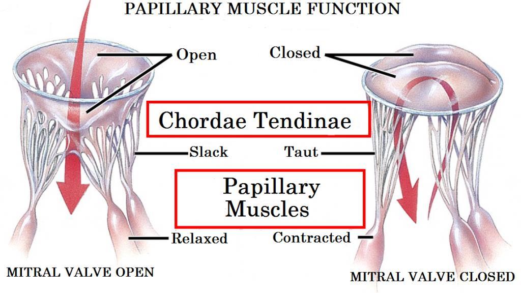
Anatomy lab -1- Imp note: papillary muscle Trabeculae Carneae chordae tendineae
 Anatomy lab -1- Imp note: the arrangement of this sheet is different than the lab recording, it has been arranged in a certain way to make it easier to study. When you open the left ventricle you can see
Development and teratology of cardiovascular and lymphatic systems. Repetition: Muscle tissue
 Development and teratology of cardiovascular and lymphatic systems Repetition: Muscle tissue Beginning of the cardiovascular system development the 3rd week: Hemangiogenesis (day 15 16) blood islets (insulae
Chapter 4: The thoracic cavity and heart. The Heart
 Chapter 4: The thoracic cavity and heart The thoracic cavity is divided into right and left pleural cavities by a central partition, the mediastinum. The mediastinum is bounded behind by the vertebral
CARDIOVASCULAR SYSTEM
 CARDIOVASCULAR SYSTEM Overview Heart and Vessels 2 Major Divisions Pulmonary Circuit Systemic Circuit Closed and Continuous Loop Location Aorta Superior vena cava Right lung Pulmonary trunk Base of heart
LAB 12-1 HEART DISSECTION GROSS ANATOMY OF THE HEART
 LAB 12-1 HEART DISSECTION GROSS ANATOMY OF THE HEART Because mammals are warm-blooded and generally very active animals, they require high metabolic rates. One major requirement of a high metabolism is
The Cardiovascular System
 PowerPoint Lecture Slide Presentation by Patty Bostwick-Taylor, Florence-Darlington Technical College The Cardiovascular System 11PART B The Heart: Cardiac Output Cardiac output (CO) Amount of blood pumped
Lab Photo Review Sheet
 9 8 0. Posterior Median Sulcus. Central Canal. Dorsal (Posterior) Horn. Ventral (Anterior) Horn. Grey Matter. White Matter. Anterior Median Fissure 8. Ventral (Anterior) Root (ramus) 9. Dorsal (Posterior)
ACTIVITY 9: BLOOD AND HEART BLOOD
 ACTIVITY 9: BLOOD AND HEART OBJECTIVES: 1) How to get ready: Read Chapters 21 & 22, McKinley et al., Human Anatomy, 4e. All text references are for this textbook. Read dissection instructions BEFORE YOU
The Cardiovascular System. Chapter 15. Cardiovascular System FYI. Cardiology Closed systemof the heart & blood vessels. Functions
 Chapter 15 Cardiovascular System FYI The heart pumps 7,000 liters (4000 gallons) of blood through the body each day The heart contracts 2.5 billion times in an avg. lifetime The heart & all blood vessels
KEY - Sample Exam Biology 2050 Circulatory and Lymphatic Systems - KEY
 KEY - Sample Exam Biology 2050 Circulatory and Lymphatic Systems - KEY Note: Not all of the lymphatic system will be on the actual exam next Monday so disregard any questions that deal with something that
Ch 19: Cardiovascular System - The Heart -
 Ch 19: Cardiovascular System - The Heart - Give a detailed description of the superficial and internal anatomy of the heart, including the pericardium, the myocardium, and the cardiac muscle. Trace the
STRUCTURES OF THE CARDIOVASCULAR SYSTEM
 STRUCTURES OF THE CARDIOVASCULAR SYSTEM CARDIOVASCULAR SYSTEM Also called the circulatory system Consists of the heart, arteries, veins, and capillaries Main function is to pump/circulate oxygenated blood
THE HEART OBJECTIVES: LOCATION OF THE HEART IN THE THORACIC CAVITY CARDIOVASCULAR SYSTEM
 BIOLOGY II CARDIOVASCULAR SYSTEM ACTIVITY #3 NAME DATE HOUR THE HEART OBJECTIVES: Describe the anatomy of the heart and identify and give the functions of all parts. (pp. 356 363) Trace the flow of blood
Cardiovascular System Module 3: Heart Anatomy *
 OpenStax-CNX module: m49683 1 Cardiovascular System Module 3: Heart Anatomy * Donna Browne Based on Heart Anatomy by OpenStax This work is produced by OpenStax-CNX and licensed under the Creative Commons
Chapter 14. Circulatory System Images. VT-122 Anatomy & Physiology II
 Chapter 14 Circulatory System Images VT-122 Anatomy & Physiology II The mediastinum Dog heart Dog heart Cat heart Dog heart ultrasound Can see pericardium as distinct bright line Pericardial effusion Fluid
THE VESSELS OF THE HEART
 1 THE VESSELS OF THE HEART The vessels of the heart include the coronary arteries, which supply the heart and the veins and lymph vessels, which drain the heart. THE CORONARY ARTERIES These are the blood
Function: Transportation of. Oxygen Nutrients Waste Hormones gases
 Function: Transportation of Oxygen Nutrients Waste Hormones gases Pericardium: double sac of serous membrane filled with fluid (pericardial fluid to be exact) that surrounds the heart. Parietal pericardium:
Part 1. Copyright 2011 Pearson Education, Inc. Figure Copyright 2011 Pearson Education, Inc.
 PowerPoint Lecture Slides prepared by Leslie Hendon University of Alabama, Birmingham C H A P T E R The Heart 19 Part 1 The Heart A muscular double pump circuit vessels transport blood to and from the
The Cardiovascular System (Part I) 黃敏銓 解剖學暨細胞生物學研究所
 黃敏銓 解剖學暨細胞生物學研究所") The Cardiovascular System (Part I) 黃敏銓 解剖學暨細胞生物學研究所 1 Congenital heart defects (CHDs) 台灣兒童心臟學會 Sinus venarum Membranous septum Conus arteiosus (infundibulum) Aortic vestibule The Cardiovascular System
The Cardiovascular System (Part I) 黃敏銓 解剖學暨細胞生物學研究所 1 Congenital heart defects (CHDs) 台灣兒童心臟學會 Sinus venarum Membranous septum Conus arteiosus (infundibulum) Aortic vestibule The Cardiovascular System
CIRCULATORY SYSTEM BLOOD VESSELS
 Name: Block: CIRCULATORY SYSTEM Multicellular organisms (above the level of roundworms) rely on a circulatory system to bring nutrients to, and take wastes away from, cells. In higher organisms such as
Anatomy and Physiology, Spring 2015 Exam II: Form A April 9, Name Student Number
 Anatomy and Physiology, Spring 2015 Exam II: Form A April 9, 2015 Name Student Number For Questions 1 2 refer to the following table. 1 Ventricular pressure is greater than aortic 6 AV valve is open 2
Chapter 12 Lecture Outline
 Chapter 12 Lecture Outline See separate PowerPoint slides for all figures and tables preinserted into PowerPoint without notes. Copyright The McGraw-Hill Companies, Inc. Permission required for reproduction
Anatomy of the Heart
 Biology 212: Anatomy and Physiology II Anatomy of the Heart References: Saladin, KS: Anatomy and Physiology, The Unity of Form and Function 8 th (2018). Required reading before beginning this lab: Chapter
11/10/2014. Muscular pump Two atria Two ventricles. In mediastinum of thoracic cavity 2/3 of heart's mass lies left of midline of sternum
 It beats over 100,000 times a day to pump over 1,800 gallons of blood per day through over 60,000 miles of blood vessels. During the average lifetime, the heart pumps nearly 3 billion times, delivering
The Heart. Happy Friday! #takeoutyournotes #testnotgradedyet
 The Heart Happy Friday! #takeoutyournotes #testnotgradedyet Introduction Cardiovascular system distributes blood Pump (heart) Distribution areas (capillaries) Heart has 4 compartments 2 receive blood (atria)
Development of the Heart *
 OpenStax-CNX module: m46673 1 Development of the Heart * OpenStax This work is produced by OpenStax-CNX and licensed under the Creative Commons Attribution License 3.0 By the end of this section, you will
The Cardiovascular and Lymphatic Systems Cardiovascular System Blood Vessels Blood Vessels Arteries Arteries Arteries
 CH 12 The Cardiovascular and s The Cardiovascular and s OUTLINE: Cardiovascular System Blood Vessels Blood Pressure Cardiovascular System The cardiovascular system is composed of Blood vessels This system
W.S. O The University of Hong Kong
 W.S. O The University of Hong Kong Objectives: Describe early angiogenesis. Describe the heart tube formation. Describe the partitioning into a 4- chambered heart. List the formation of heart valves and
6. Development of circulatory system II. Cardiac looping. Septation of atria and ventricles. Common heart malformations.
 6. Development of circulatory system II. Cardiac looping. Septation of atria and ventricles. Common heart malformations. Formation of heart tube paired endothelial-lined heart tube is formed from blood
The Cardiovascular System
 11 The Cardiovascular System Yong Jeong, MD, PhD Department of Bio and Brain Engineering The Cardiovascular System A closed system of the heart and blood vessels The heart pumps blood Blood vessels allow
Cardiovascular system:
 Cardiovascular system: Mediastinum: The mediastinum: lies between the right and left pleura and lungs. It extends from the sternum in front to the vertebral column behind, and from the root of the neck
CJ Shuster A&P2 Lab Addenum Beef Heart Dissection 1. Heart Dissection. (taken from Johnson, Weipz and Savage Lab Book)
") CJ Shuster A&P2 Lab Addenum Beef Heart Dissection 1 Heart Dissection. (taken from Johnson, Weipz and Savage Lab Book) Introduction When you have finished examining the model, you are ready to begin your
CJ Shuster A&P2 Lab Addenum Beef Heart Dissection 1 Heart Dissection. (taken from Johnson, Weipz and Savage Lab Book) Introduction When you have finished examining the model, you are ready to begin your
The Cardiovascular System
 11 PART A The Cardiovascular System PowerPoint Lecture Slide Presentation by Jerry L. Cook, Sam Houston University ESSENTIALS OF HUMAN ANATOMY & PHYSIOLOGY EIGHTH EDITION ELAINE N. MARIEB The Cardiovascular
The Cardiovascular System. The Structure of Blood Vessels. The Structure of Blood Vessels. The Blood Vessels. Blood Vessel Review
 The Cardiovascular System The Blood Vessels The Structure of Blood Vessels Blood Vessel Review Arteries carry blood away from the heart Pulmonary trunk to lungs Aorta to everything else Microcirculation
- what other structures, besides the heart, does the mediastinum contain?
 Basic A & P II Dr. L. Bacha Chapter Outline (Martini & Nath 2010) An Introduction to the Cardiovascular System - read the paragraphs under this heading on page 580 The Heart is a Four Chambered Organ describe
Circulation. Circulation = is a process used for the transport of oxygen, carbon! dioxide, nutrients and wastes through-out the body
 Circulation Circulation = is a process used for the transport of oxygen, carbon! dioxide, nutrients and wastes through-out the body Heart = muscular organ about the size of your fist which pumps blood.
Chapter 18 - Heart. I. Heart Anatomy: size of your fist; located in mediastinum (medial cavity)
") Chapter 18 - Heart I. Heart Anatomy: size of your fist; located in mediastinum (medial cavity) A. Coverings: heart enclosed in double walled sac called the pericardium 1. Fibrous pericardium: dense connective
Chapter 18 - Heart I. Heart Anatomy: size of your fist; located in mediastinum (medial cavity) A. Coverings: heart enclosed in double walled sac called the pericardium 1. Fibrous pericardium: dense connective
Cardiovascular System Notes: Physiology of the Heart
 Cardiovascular System Notes: Physiology of the Heart Interesting Heart Fact Capillaries are so small it takes ten of them to equal the thickness of a human hair. Review What are the 3 parts of the cardiovascular
The Cardiovascular System
 The Cardiovascular System The Cardiovascular System A closed system of the heart and blood vessels The heart pumps blood Blood vessels allow blood to circulate to all parts of the body The function of
This is not a required assignment but it is recommended.
 SU 12 Name: This is not a required assignment but it is recommended. BIO 116 - Anatomy & Physiology II Practice Assignment 2 - The Respiratory and Cardiovascular Systems 1. The exchange of oxygen and carbon
Heart Development and Congenital Heart Disease
 Heart Development and Congenital Heart Disease Sally Dunwoodie s.dunwoodie@victorchang.edu.au Developmental and Stem Cell Biology Division Victor Chang Cardiac Research Institute for the heart of Australia...
Ch.15 Cardiovascular System Pgs {15-12} {15-13}
 Ch.15 Cardiovascular System Pgs {15-12} {15-13} E. Skeleton of the Heart 1. The skeleton of the heart is composed of rings of dense connective tissue and other masses of connective tissue in the interventricular
Notes: 1)Membranous part contribute in the formation of small portion in the septal cusp.
Membranous part contribute in the formation of small portion in the septal cusp.") Embryology 9 : Slide 16 : There is a sulcus between primitive ventricular and bulbis cordis that will disappear gradually and lead to the formation of one chamber which is called bulboventricular chamber.
Embryology 9 : Slide 16 : There is a sulcus between primitive ventricular and bulbis cordis that will disappear gradually and lead to the formation of one chamber which is called bulboventricular chamber.
ANATDMY. lecture # : Date : Lecturer : Maher Hadidi
 ANATDMY 27 lecture # : Date : Lecturer : Maher Hadidi Pericardium A double-walled fibroserous conical-shaped sac, within middle mediastinum. Enclose the heart and roots of its large vessels. Vagus nerves
The Cardiovascular System. anatom.ua 1
 The Cardiovascular System anatom.ua 1 The Closed Circulatory System Humans have a closed circulatory system, typical of all vertebrates, in which blood is confined to vessels and is distinct from the interstitial
The Cardiovascular System. Preview of Heart Action. The CV system provides oxygen & nutrients to tissues-removes wastes.
 The Cardiovascular System BIO 250 Human Anatomy & Physiology Preview of Heart Action http://www.youtube.com/watch?v=d3zdj gfddk0&nr=1 The CV system provides oxygen & nutrients to tissues-removes wastes.
The blood returns from the body and enters right atrium using the vena cava. It passes through the tricuspid valve to the right ventricle.
 The blood returns from the body and enters right atrium using the vena cava. It passes through the tricuspid valve to the right ventricle. From this camber, it passes through the pulmonary semilunar valve
1. Distinguish among the types of blood vessels on the basis of their structure and function.
 Blood Vessels and Circulation Objectives This chapter describes the structure and functions of the blood vessels Additional subjects contained in Chapter 13 include cardiovascular physiology, regulation,
Identify the lines used in anatomical surface descriptions of the thorax. median line mid-axillary line mid-clavicular line
 L 14 A B O R A T O R Y Thorax THORACIC WALL Identify the lines used in anatomical surface descriptions of the thorax. median line mid-axillary line mid-clavicular line Identify the surface landmarks of