for Heart-Health Scanning
|
|
|
- Christopher O’Brien’
- 6 years ago
- Views:
Transcription
1 Magnetocardiography for Heart-Health Scanning CardioMag Imaging, Inc. 1
.")
2 Basic Principles of Magnetocardiography (MCG) The cardiac electric activity that produces a voltage difference on the body surface (resulting in the ECG trace) produces simultaneously a magnetic field that extends outside the body (resulting in an MCG trace). MCG signals, unlike ECG signals, are not attenuated by surrounding anatomical structures, tissue, and body fluids, thereby providing more accurate information Vortex currents in the myocardium produce magnetic signatures that are not visible on the ECG, thereby MCG provides more information. The MCG is a non-invasive electrophysiological mapping technique that provides unprecedented insight into the generation, localization, and dynamic behavior of electric current in the heart. Unique Advantages of Magnetocardiography Radiation is not used. Injections are not needed. Exercise stress and breath holds are not required. An MCG exam takes less than 10 minutes. Results are both qualitative and quantitative. The frequency of MCG exams is limitless, at the discretion of the treating physician. MCG results provide new, functional information complimentary to that obtained using CT, MRI, ECHO, and SPECT. MCG is also being used for Non-invasive localization of sources of arrhythmia, Diagnosis of fetal cardiac disorders, Screening patients for ischemic heart disease. Cost-Benefit Advantages: Higher confidence diagnoses More rapid turnaround of emergency department beds No biohazard waste Higher throughput scanning Effective, safe monitoring post-cabg, PCI and PTCA 2

 MCG traces and one (1) ECG trace [Lead I] are recorded continuously and displayed on a virtual oscilloscope (as shown above).")

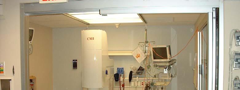
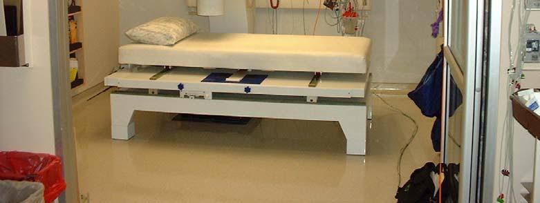
3 MCG Measurement Sequence The patient lies comfortably on the table; there is no need to remove clothing. Then the operator lowers the column containing the sensitive magnetic field probes so that it is close to but not touching the patient s chest. 20 cm The relative positions of the nine (9) magnetic field probes inside the column are shown as the white circles in the figure above. The outline of the column is in black. During a measurement, which lasts typically 90 seconds, nine (9) MCG traces and one (1) ECG trace [Lead I] are recorded continuously and displayed on a virtual oscilloscope (as shown above). The ECG trace is used as a time reference to average the 36 traces of MCG data acquired from four (4) consecutive scans covering a 20cm x 20cm area above the torso. A representative time-averaged MCG trace at each of the 36 positions is displayed in the 6 x 6 grid to the left. Interpolation among the 36 points of MCG data (small dots in the color map to the right) and assignment of a color to the strength and direction of the magnetic field yield magnetic field maps which display the electric activity of the heart during the entire cardiac cycle as seen from outside the body. 3

")
4 Magnetic Field Maps Since MCG data are acquired at a frequency of 1 khz, there are 1000 maps for every cardiac cycle. The sequence of 18 maps below shows the dynamics of interventricular septal depolarization, starting with the map at the upper left and proceeding left to right on every line from top to bottom. Consecutive maps are evenly spaced by a time interval of 2 msec, so that the process shown in the example to the right lasts 34 msec. Since effective cardiac electric current flows along the light blue line separating the red positive field from the purple negative field, one can see how the net cardiac current (white arrows above) starts flowing in the interventricular septum from the patient s left to right, and how it changes as expected, with a final net flow of depolarization current in the direction of the cardiac apex. 4
 Likewise, the complex magnetic field distribution of the MCG map can be represented")
 is perpendicular to this vector.")

.")
5 Localization and Effective Magnetic Dipoles The complex magnetic field generated by a simple bar magnet (figure to the far left) is readily visualized by iron filings (as seen in 3D in the figure to the immediate left) Likewise, the complex magnetic field distribution of the MCG map can be represented by a virtual bar magnet, or what is more appropriately named an effective magnetic vector, EMV (shown as a black arrow in the figure to the right). Note that the direction of the corresponding net electric current flow (as described on the opposite page) is perpendicular to this vector. One of the most important problems of biomagnetic investigations is the determination of the coordinates and the magnetic moments for the field sources based on the measured magnetic field. CMI MCG software solves this inverse problem and allows the user to track the dipole in space and time over any specified interval in the cardiac cycle. The figure above shows a series of EMV s representing electrical activity during ventricular repolarization (ascending T-wave). Plotting the EMV path is a unique, useful and much more convenient way to represent the electrical activity over any interval in the cardiac cycle. 5


 If any of the seven parameters lies in the shaded")

6 Diagnostic Algorithm for Ischemia The presence of ischemia is directly related to the direction and dynamic motion of the effective magnetic vector during ventricular repolarization (ascending and descending T-wave) as described by the seven (7) parameters shown in the table below. 90 ± Frontal Plane Angle Trajectory (black line) If any of the seven parameters lies in the shaded ischemic range, then the patient is considered ischemic. If none of the parameters lies in the ischemic range, the patient is considered not ischemic. Angular Deviation (blue line) 6


7 Diagnosis of Ischemia Using MCG It is estimated that as many as 8 million Americans present to the emergency department with chest pain or symptoms suggestive of acute cardiac ischemia. Because as many as one-half to two-thirds of those admitted do not have a cardiac cause for their symptoms, the opportunity for improved diagnostics is large. MCG, even in a resting test, provides a rapid, quantitative diagnosis with balanced diagnostic accuracy. MCG traces reinforce conventional methods based on ST segment analysis. Magnetic field maps show the degree of electrical homogeneity and reveal clues about the location of ischemia. Magnetic vector trajectories provide unique insight into abnormal repolarization. Demonstrated Clinical Value Resting magnetocardiographic imaging provides immediate results with very high positive predictive value for obstructive CAD that exceeds or meets the performance of SPECT but without the use of stress provocation, radiation, or injection of nuclear tracer. Reference: Comparison of Resting Magnetocardiography with Stress Single Photon Emission Computed Tomography in Patients with Stable and Unstable Angina, Tolstrup K. et al., presented at the American College of Cardiology 7
: 9 cardiac channels, 3 background environment reference channels Data")

 Spectral noise analysis Communication: fiber optic links between subsystems,")


8 Comprehensive Database Based on Windows System Specifications Sensors: SQUID (Superconducting Quantum Interference Device): 9 cardiac channels, 3 background environment reference channels Data Acquisition: 14 channels (12 SQUID, 1 cardiac trigger, 1 accessory), 24-bit ADC per channel, 1000 Hz sampling rate with a Hz range Frequency Response: 0 to 500 Hz, nominal Signal Processing: analog and digital filtering, low pass and high pass; signal averaging Analog and digital gain settings (programmable) Spectral noise analysis Communication: fiber optic links between subsystems, Power Supply: VAC input at 50/60 Hz Cryostat volume: 13.5 liters Liquid He consumption: < 2.5 liters/day Physical specifications Floor area: approx. 4 x 5 m with 2.4 m floor to ceiling clearance (computer workstation not shown) Weight: 280 kg CardioMag Imaging, Inc., 450 Duane Avenue, Schenectady, NY USA Tel.: Fax: cardiomag@cardiomag.com Web: 8
Multiple Gated Acquisition (MUGA) Scanning
 Scanning") Multiple Gated Acquisition (MUGA) Scanning Dmitry Beyder MPA, CNMT Nuclear Medicine, Radiology Barnes-Jewish Hospital / Washington University St. Louis, MO Disclaimers/Relationships Standard of care research
Multiple Gated Acquisition (MUGA) Scanning Dmitry Beyder MPA, CNMT Nuclear Medicine, Radiology Barnes-Jewish Hospital / Washington University St. Louis, MO Disclaimers/Relationships Standard of care research
Non-invasive diagnosis of coronary artery disease by exercise magnetocardiography
 Non-invasive diagnosis of coronary artery disease by exercise magnetocardiography Jai-Wun Park, MD Klinik für Kardiologie Campus Benjamin Franklin Charité - Universitätsmedizin Berlin JCR Busan, Korea
Magnetocardiogram Recordings in a Nonshielded Environment Reproducibility and Ischemia Detection
 Magnetocardiogram Recordings in a Nonshielded Environment Reproducibility and Ischemia Detection Benjamin A. Steinberg, B.A., Ariel Roguin, M.D., Ph.D., Stanley P. Watkins III, M.D., Peter Hill, M.D.,
Impaired Regional Myocardial Function Detection Using the Standard Inter-Segmental Integration SINE Wave Curve On Magnetic Resonance Imaging
 Original Article Impaired Regional Myocardial Function Detection Using the Standard Inter-Segmental Integration Ngam-Maung B, RT email : chaothawee@yahoo.com Busakol Ngam-Maung, RT 1 Lertlak Chaothawee,
Automated Heart Analysis.
 Automated Heart Analysis Yes, 5 minutes or less! Kardi is a revolutionizing non-invasive diagnostic tool used for testing ischaemic heart disease It s technology generates high quality 3D visuals projected
11/18/13 ECG SIGNAL ACQUISITION HARDWARE DESIGN. Origin of Bioelectric Signals
 ECG SIGNAL ACQUISITION HARDWARE DESIGN Origin of Bioelectric Signals 1 Cell membrane, channel proteins Electrical and chemical gradients at the semi-permeable cell membrane As a result, we get a membrane
Measurement of Respiratory and Cardiac Motion Using a Multi Antenna Continuous Wave Radar Operating in the Near Field
 Measurement of Respiratory and Cardiac Motion Using a Multi Antenna Continuous Wave Radar Operating in the Near Field Florian Pfanner 1,2, Thomas Allmendinger 2, Thomas Flohr 2, and Marc Kachelrieß 1,3
Introduction. Cardiac Imaging Modalities MRI. Overview. MRI (Continued) MRI (Continued) Arnaud Bistoquet 12/19/03
 MRI (Continued) Arnaud Bistoquet 12/19/03") Introduction Cardiac Imaging Modalities Arnaud Bistoquet 12/19/03 Coronary heart disease: the vessels that supply oxygen-carrying blood to the heart, become narrowed and unable to carry a normal amount
Introduction Cardiac Imaging Modalities Arnaud Bistoquet 12/19/03 Coronary heart disease: the vessels that supply oxygen-carrying blood to the heart, become narrowed and unable to carry a normal amount
ECG and Cardiac Electrophysiology
 ECG and Cardiac Electrophysiology Simon Some very basic electrophysiology Intracellular fluid: 10 mm Na, 140 mm K, etc. K Na-K ATPase Extracellular fluid: 140mM Na, 4mM K, etc. Na Ion gradient plus selective
CASE 10. What would the ST segment of this ECG look like? On which leads would you see this ST segment change? What does the T wave represent?
 CASE 10 A 57-year-old man presents to the emergency center with complaints of chest pain with radiation to the left arm and jaw. He reports feeling anxious, diaphoretic, and short of breath. His past history
MULTILEAD SIGNAL PREPROCESSING BY LINEAR TRANSFORMATION
 MULTILEAD SIGNAL PREPROCESSING BY LINEAR TRANSFORMATION TO DERIVE AN ECG LEAD WHERE THE ATYPICAL BEATS ARE ENHANCED Chavdar Lev Levkov Signa Cor Laboratory, Sofia, Bulgaria, info@signacor.com ECG signal
Signal Processing of Stress Test ECG Using MATLAB
 Signal Processing of Stress Test ECG Using MATLAB Omer Mukhtar Wani M. Tech ECE Geeta Engineering College, Panipat Abstract -Electrocardiography is used to record the electrical activity of the heart over
An electrocardiogram (ECG) is a recording of the electricity of the heart. Analysis of ECG
 is a recording of the electricity of the heart. Analysis of ECG") Introduction An electrocardiogram (ECG) is a recording of the electricity of the heart. Analysis of ECG data can give important information about the health of the heart and can help physicians to diagnose
Introduction An electrocardiogram (ECG) is a recording of the electricity of the heart. Analysis of ECG data can give important information about the health of the heart and can help physicians to diagnose
University of Groningen. Quantitative CT myocardial perfusion Pelgrim, Gert
 University of Groningen Quantitative CT myocardial perfusion Pelgrim, Gert IMPORTANT NOTE: You are advised to consult the publisher's version (publisher's PDF) if you wish to cite from it. Please check
Systolic and Diastolic Currents of Injury
 Systolic and Diastolic Currents of Injury Figure 1 Action Potentials of Normal and Ischemic Tissue In Figure 1 above, the action potential of normal myocardium is represented by the solid lines and the
Preface: Wang s Viewpoints
 AHA/ACCF/HRS Recommendations for the Standardization and Interpretation of the Electrocardiogram: Part IV, Ischemia and Infarction Presented by: WANG, TZONG LUEN, MD, PhD, JM, FACC, FESC, FCAPSC Professor,
CT Imaging at the Point-of-Care
 ENGLISH True Dedication The new Planmed Verity Extremity CT Scanner revolutionizes extremity CT imaging. The compact unit brings 3D imaging at emergency departments, orthopedic clinics or trauma centers
Identification of Ischemic Lesions Based on Difference Integral Maps, Comparison of Several ECG Intervals
 1.2478/v148-9-21-7 MEASUREMENT SCIENCE REVIEW, Volume 9, No. 5, 29 Identification of Ischemic Lesions Based on Difference Integral Maps, Comparison of Several ECG Intervals J. Švehlíková, M. Kania 1, M.
HST-582J/6.555J/16.456J-Biomedical Signal and Image Processing-Spring Laboratory Project 1 The Electrocardiogram
 HST-582J/6.555J/16.456J-Biomedical Signal and Image Processing-Spring 2007 DUE: 3/8/07 Laboratory Project 1 The Electrocardiogram 1 Introduction The electrocardiogram (ECG) is a recording of body surface
The Fundamentals of 12 Lead EKG. ECG Recording. J Point. Reviewing the Cardiac Conductive System. Dr. E. Joe Sasin, MD Rusty Powers, NRP
 The Fundamentals of 12 Lead EKG Dr. E. Joe Sasin, MD Rusty Powers, NRP SA Node Intranodal Pathways AV Junction AV Fibers Bundle of His Septum Bundle Branches Purkinje System Reviewing the Cardiac Conductive
Non-Invasive Techniques
 Non-Invasive Techniques Key: Does not hurt the organism Psychology 372 Physiological Psychology Steven E. Meier, Ph.D. Listen to the audio lecture while viewing these slides or view the video presentation
Non-Invasive Techniques
 Many Procedures Non-Invasive Techniques Key: Does not hurt the organism Psychology 372 Physiological Psychology Steven E. Meier, Ph.D. Listen to the audio lecture while viewing these slides or view the
Physiological and Physical Basis of Functional Brain Imaging 6. EEG/MEG. Kâmil Uludağ, 20. November 2007
 Physiological and Physical Basis of Functional Brain Imaging 6. EEG/MEG Kâmil Uludağ, 20. November 2007 Course schedule 1. Overview 2. fmri (Spin dynamics, Image formation) 3. fmri (physiology) 4. fmri
MR Advance Techniques. Cardiac Imaging. Class IV
 MR Advance Techniques Cardiac Imaging Class IV Heart The heart is a muscular organ responsible for pumping blood through the blood vessels by repeated, rhythmic contractions. Layers of the heart Endocardium
- why the T wave is deflected upwards although it's a repolarization wave?
 Cardiac Electrograph: - why the T wave is deflected upwards although it's a repolarization wave? After depolarization the ventricle contracts but since the heart is a volume conductor (3D not 2D), when
Detecting Acute Myocardial Ischemia by Evaluation of QRS Angles
 International Journal of Bioelectromagnetism Vol. 15, No. 1, pp. 77-82, 2013 www.ijbem.org Detecting Acute Myocardial Ischemia by Evaluation of QRS Angles Daniel Romero a,b, Pablo Laguna a,b, Esther Pueyo
Assessment of Reliability of Hamilton-Tompkins Algorithm to ECG Parameter Detection
 Proceedings of the 2012 International Conference on Industrial Engineering and Operations Management Istanbul, Turkey, July 3 6, 2012 Assessment of Reliability of Hamilton-Tompkins Algorithm to ECG Parameter
Outline. Electrical Activity of the Human Heart. What is the Heart? The Heart as a Pump. Anatomy of the Heart. The Hard Work
 Electrical Activity of the Human Heart Oguz Poroy, PhD Assistant Professor Department of Biomedical Engineering The University of Iowa Outline Basic Facts about the Heart Heart Chambers and Heart s The
Use of Nuclear Cardiology in Myocardial Viability Assessment and Introduction to PET and PET/CT for Advanced Users
 Use of Nuclear Cardiology in Myocardial Viability Assessment and Introduction to PET and PET/CT for Advanced Users February 1 5, 2011 University of Santo Tomas Hospital Angelo King A-V Auditorium Manila,
Fetal Rhythm and Blues
 Fetal Rhythm and Blues John Cotton, MD Professor of Pediatrics Division of Pediatric Cardiology Director, Fetal Cardiology Program UNC Chapel Hill, School of Medicine Objectives To review methods used
The Central Nervous System
 The Central Nervous System Cellular Basis. Neural Communication. Major Structures. Principles & Methods. Principles of Neural Organization Big Question #1: Representation. How is the external world coded
CARDIAC MRI. Cardiovascular Disease. Cardiovascular Disease. Cardiovascular Disease. Overview
 CARDIAC MRI Dr Yang Faridah A. Aziz Department of Biomedical Imaging University of Malaya Medical Centre Cardiovascular Disease Diseases of the circulatory system, also called cardiovascular disease (CVD),
The Value of Stress MRI in Evaluation of Myocardial Ischemia
 The Value of Stress MRI in Evaluation of Myocardial Ischemia Dr. Saeed Al Sayari, MBBS, EBCR, MBA Department of Radiology and Nuclear Medicine Mafraq Hospital, Abu Dhabi United Arab Emirates Introduction
Speed - Accuracy - Exploration. Pathfinder SL
 Speed - Accuracy - Exploration Pathfinder SL 98000 Speed. Accuracy. Exploration. Pathfinder SL represents the evolution of over 40 years of technology, design, algorithm development and experience in the
Electrocardiography I Laboratory
 Introduction The body relies on the heart to circulate blood throughout the body. The heart is responsible for pumping oxygenated blood from the lungs out to the body through the arteries and also circulating
12 Lead ECG Interpretation: Color Coding for MI s
 12 Lead ECG Interpretation: Color Coding for MI s Anna E. Story, RN, MS Director, Continuing Professional Education Critical Care Nurse Online Instructional Designer 2004 Anna Story 1 Objectives review
EKG. Danil Hammoudi.MD
 EKG Danil Hammoudi.MD What is an EKG? The electrocardiogram (EKG) is a representation of the electrical events of the cardiac cycle. Each event has a distinctive waveform, the study of which can lead to
Lect.6 Electrical axis and cardiac vector Cardiac vector: net result Vector that occurs during depolarization of the ventricles Figure:
 Lect.6 Electrical axis and cardiac vector Objectives: 1. State the relationship between the direction of cardiac vector with the direction (-ve, +ve) and amplitude of an ECG waves. 2. Draw diagram indicting
This presentation is the intellectual property of the author. Contact them for permission to reprint and/or distribute.
 Modified Combinatorial Nomenclature Montage, Review, and Analysis of High Density EEG Terrence D. Lagerlund, M.D., Ph.D. CP1208045-16 Disclosure Relevant financial relationships None Off-label/investigational
Magnetic Resonance Imaging on Soft Tissue. Jiten K. Mistry Calvin Gan
 Magnetic Resonance Imaging on Soft Tissue 1 Jiten K. Mistry Calvin Gan Outline Background of Medical Imaging Introduction to MRI How MRI works MRI of Soft Tissue Benefits & Risks Recent Advances 2 The
By the end of this lecture, you will be able to: Understand the 12 lead ECG in relation to the coronary circulation and myocardium Perform an ECG
 By the end of this lecture, you will be able to: Understand the 12 lead ECG in relation to the coronary circulation and myocardium Perform an ECG recording Identify the ECG changes that occur in the presence
INTRODUCTION TO ECG. Dr. Tamara Alqudah
 INTRODUCTION TO ECG Dr. Tamara Alqudah Excitatory & conductive system of the heart + - The ECG The electrocardiogram, or ECG, is a simple & noninvasive diagnostic test which records the electrical
Simulation of T-Wave Alternans and its Relation to the Duration of Ventricular Action Potentials Disturbance
 The Open Pacing, Electrophysiology & Therapy Journal, 2010, 3, 21-27 21 Open Access Simulation of T-Wave Alternans and its Relation to the Duration of Ventricular Action Potentials Disturbance Dariusz
Ways to Study Brain Structures and Functioning. Can physically trace connections. Ablation. Is the most primitive Can be done with any structures
 Ways to Study Brain Structures and Functioning Can physically trace connections Is the most primitive Can be done with any structures Ablation Can remove a piece of the brain and see what happens If the
Biopac Student Lab Lesson 6 ELECTROCARDIOGRAPHY (ECG) II Analysis Procedure. Rev
 II Analysis Procedure. Rev") 42 Aero Camino, Goleta, CA 93117 www.biopac.com Biopac Student Lab Lesson 6 ELECTROCARDIOGRAPHY (ECG) II Analysis Procedure Rev. 12292017 Richard Pflanzer, Ph.D. Associate Professor Emeritus Indiana University
42 Aero Camino, Goleta, CA 93117 www.biopac.com Biopac Student Lab Lesson 6 ELECTROCARDIOGRAPHY (ECG) II Analysis Procedure Rev. 12292017 Richard Pflanzer, Ph.D. Associate Professor Emeritus Indiana University
2017 Qualified Clinical Data Registry (QCDR) Performance Measures
 Performance Measures") 2017 Qualified Clinical Data Registry (QCDR) Performance Measures Description: This document contains the 15 performance measures that were approved by CMS for use in ASC's 2017 Qualified Clinical Data
2017 Qualified Clinical Data Registry (QCDR) Performance Measures Description: This document contains the 15 performance measures that were approved by CMS for use in ASC's 2017 Qualified Clinical Data
DETECTION OF HEART ABNORMALITIES USING LABVIEW
 IASET: International Journal of Electronics and Communication Engineering (IJECE) ISSN (P): 2278-9901; ISSN (E): 2278-991X Vol. 5, Issue 4, Jun Jul 2016; 15-22 IASET DETECTION OF HEART ABNORMALITIES USING
Imaging congestive heart failure: role of coronary computed tomography angiography (CCTA)
") Imaging congestive heart failure: role of coronary computed tomography angiography (CCTA) Gianluca Pontone, MD, PhD, FESC, FSCCT Director of MR Unit Deputy Director of Cardiovascul CT Unit Clinical Cardiology
Imaging congestive heart failure: role of coronary computed tomography angiography (CCTA) Gianluca Pontone, MD, PhD, FESC, FSCCT Director of MR Unit Deputy Director of Cardiovascul CT Unit Clinical Cardiology
Your surgeon will order pre-operative testing before you have surgery.
 Tests You May Need Prior to Surgery Your surgeon will order pre-operative testing before you have surgery. These tests give your surgeon valuable information regarding your current health condition. Below
PART I. Disorders of the Heart Rhythm: Basic Principles
 PART I Disorders of the Heart Rhythm: Basic Principles FET01.indd 1 1/11/06 9:53:05 AM FET01.indd 2 1/11/06 9:53:06 AM CHAPTER 1 The Cardiac Electrical System The heart spontaneously generates electrical
BME 365 Website. Project Directions
 Lecture 17 EKG BME 365 Website Project Directions Heart rate Factors Affecting CO Parasympathetic activity decreases HR Sympathetic activity increases HR Stroke volume Depends on force generated by cardiac
This presentation will deal with the basics of ECG description as well as the physiological basics of
 Snímka 1 Electrocardiography basics This presentation will deal with the basics of ECG description as well as the physiological basics of Snímka 2 Lecture overview 1. Cardiac conduction system functional
ECGSIM; AN INTERACTIVE TOOL FOR STUDYING THE GENESIS OF QRST WAVEFORMS
 ECGSIM; AN INTERACTIVE TOOL FOR STUDYING THE GENESIS OF QRST WAVEFORMS A. van Oosterom, PhD. Professor of Medical Physics Department of Medical Physics; University of Nijmegen; The Netherlands T.F. Oostendorp,
Cigna - Prior Authorization Procedure List: Radiology & Cardiology
 Cigna - Prior Authorization Procedure List: Radiology & Cardiology Product Category CPT Code CPT Code Description Radiology MR 70336 MRI Temporomandibular Joint(s), (TMJ) Radiology CT 70450 CT Head or
2018 OPTIONS FOR INDIVIDUAL MEASURES: REGISTRY ONLY. MEASURE TYPE: Efficiency
 Quality ID #323: Cardiac Stress Imaging t Meeting Appropriate Use Criteria: Routine Testing After Percutaneous Coronary Intervention (PCI) National Quality Strategy Domain: Efficiency and Cost Reduction
Heart Abnormality Detection Technique using PPG Signal
 Heart Abnormality Detection Technique using PPG Signal L.F. Umadi, S.N.A.M. Azam and K.A. Sidek Department of Electrical and Computer Engineering, Faculty of Engineering, International Islamic University
Entered: / / 20 Initials: Verified: / / 20 Initials: For office use only.
 Entered: / / 20 Initials: Verified: / / 20 Initials: For office use only. Research Coordinator Assessment Baseline (RCAB) Version: 06/15/2006 Patient ID - - Form Completion Date / / 20 mm dd yy Certification
Practice Exercises for the Cardiovascular System
 Practice Exercises for the Cardiovascular System On the diagram below, color the oxygen-rich blood red and the oxygen-poor blood blue. Label the parts: Continued on the next page... Label the parts on
Theta sequences are essential for internally generated hippocampal firing fields.
 Theta sequences are essential for internally generated hippocampal firing fields. Yingxue Wang, Sandro Romani, Brian Lustig, Anthony Leonardo, Eva Pastalkova Supplementary Materials Supplementary Modeling
Discrete Wavelet Transform-based Baseline Wandering Removal for High Resolution Electrocardiogram
 26 C. Bunluechokchai and T. Leeudomwong: Discrete Wavelet Transform-based Baseline... (26-31) Discrete Wavelet Transform-based Baseline Wandering Removal for High Resolution Electrocardiogram Chissanuthat
ECG MACHINE KEC- Series
 KIZLON ECG MACHINE KEC- Series 404 Crescent Royale, Off New Link Road Andheri West, Mumbai 40053, Phones: 022-6708747 Email: info@kizlon.com Website: www.kizlon.com ECG Machine KEC-A100 This ECG machine
Tissue Doppler Imaging in Congenital Heart Disease
 Tissue Doppler Imaging in Congenital Heart Disease L. Youngmin Eun, M.D. Department of Pediatrics, Division of Pediatric Cardiology, Kwandong University College of Medicine The potential advantage of ultrasound
PINPOINTING RADIATION THERAPY WITH THE PRECISION OF MR.
 GE Healthcare PINPOINTING RADIATION THERAPY WITH THE PRECISION OF MR. MR Radiation Oncology Suite MAXIMIZE YOUR PRECISION. HELP MINIMIZE PATIENT COMPLICATIONS. Our goal in MR radiation oncology is to
MYOCARDIALINFARCTION. By: Kendra Fischer
 MYOCARDIALINFARCTION By: Kendra Fischer Outline Definition Epidemiology Clinical Aspects Treatment Effects of Exercise Exercise Testing Exercise Rx Summary and Conclusions References Break it down MYOCARDIAL
Photon Attenuation Correction in Misregistered Cardiac PET/CT
 Photon Attenuation Correction in Misregistered Cardiac PET/CT A. Martinez-Möller 1,2, N. Navab 2, M. Schwaiger 1, S. G. Nekolla 1 1 Nuklearmedizinische Klinik der TU München 2 Computer Assisted Medical
Testing the Accuracy of ECG Captured by Cronovo through Comparison of ECG Recording to a Standard 12-Lead ECG Recording Device
 Testing the Accuracy of ECG Captured by through Comparison of ECG Recording to a Standard 12-Lead ECG Recording Device Data Analysis a) R-wave Comparison: The mean and standard deviation of R-wave amplitudes
Noninvasive Blood Glucose Analysis using Near Infrared Absorption Spectroscopy. Abstract
 Progress Report No. 2-3, March 31, 1999 The Home Automation and Healthcare Consortium Noninvasive Blood Glucose Analysis using Near Infrared Absorption Spectroscopy Prof. Kamal Youcef-Toumi Principal Investigator
2016 PQRS OPTIONS FOR INDIVIDUAL MEASURES: REGISTRY ONLY
 Measure #323: Cardiac Stress Imaging Not Meeting Appropriate Use Criteria: Routine Testing After Percutaneous Coronary Intervention (PCI) National Quality Strategy Domain: Efficiency and Cost Reduction
Cardiovascular Nursing Practice: A Comprehensive Resource Manual and Study Guide for Clinical Nurses 2 nd Edition
 Cardiovascular Nursing Practice: A Comprehensive Resource Manual and Study Guide for Clinical Nurses 2 nd Edition Table of Contents Volume 1 Chapter 1: Cardiovascular Anatomy and Physiology Basic Cardiac
Spatiotemporal Cardiac Activation Sites Localization Using ECG Precordial Leads
 Spatiotemporal Cardiac Activation Sites Localization Using ECG Precordial Leads Jaime R. De La Cruz BSEE a, Joseph H.Pierluissi PhD a, Ubaldo Robles BSEE a, Zainul Abedin MD b a Electrical and Computer
Testing with currently available computerized 2-lead resting ECG analysis devices consists of four steps:
 Medical Policy Manual Topic: Computerized 2-lead Resting Electrocardiogram Analysis for the Diagnosis of Coronary Artery Disease Date of Origin: December 2011 Section: Medicine Last Reviewed Date: September
Introduction to ECG Gary Martin, M.D.
 Brief review of basic concepts Introduction to ECG Gary Martin, M.D. The electrical activity of the heart is caused by a sequence of rapid ionic movements across cell membranes resulting first in depolarization
WELSH INFORMATION STANDARDS BOARD
 WELSH INFORMATION STANDARDS BOARD DSC Notice: DSCN / 01 Date of Issue: 22 nd March Ministerial / Official Letter: AC/AS/14/080/A9552379 Subject: Diagnostic and Therapy Services Waiting Times Return Sponsor:
AUTOMATIC ANALYSIS AND VISUALIZATION OF MULTILEAD LONG-TERM ECG RECORDINGS
 AUTOMATIC ANALYSIS AND VISUALIZATION OF MULTILEAD LONG-TERM ECG RECORDINGS Vessela Tzvetanova Krasteva 1, Ivo Tsvetanov Iliev 2 1 Centre of Biomedical Engineering Prof. Ivan Daskalov - Bulgarian Academy
A Snapshot on Nuclear Cardiac Imaging
 Editorial A Snapshot on Nuclear Cardiac Imaging Khalil, M. Department of Physics, Faculty of Science, Helwan University. There is no doubt that nuclear medicine scanning devices are essential tool in the
I have no financial disclosures
 Manpreet Singh MD I have no financial disclosures Exercise Treadmill Bicycle Functional capacity assessment Well validated prognostic value Ischemic assessment ECG changes ST segments Arrhythmias Hemodynamic
Cardiovascular nuclear imaging employs non-invasive techniques to assess alterations in coronary artery flow, and ventricular function.
 National Imaging Associates, Inc. Clinical guidelines CARDIOVASCULAR NUCLEAR MEDICINE -MYOCARDIAL PERFUSION IMAGING -MUGA Original Date: October 2015 Page 1 of 9 FOR CMS (MEDICARE) MEMBERS ONLY CPT4 Codes:
Previous MI with no intervention
 Previous MI with no intervention F. Mut, M. Beretta Nuclear Medicine Service, Asociacion Española Montevideo, Uruguay Clinical history Woman 68 y.o. Recent acute MI (3 weeks) with no intervention. Discharged
Electrocardiography. Hilal Al Saffar College of Medicine,Baghdad University
 Electrocardiography Hilal Al Saffar College of Medicine,Baghdad University Which of the following is True 1. PR interval, represent the time taken for the impulse to travel from SA node to AV nose. 2.
ECG CONVENTIONS AND INTERVALS
 1 ECG Waveforms and Intervals ECG waveforms labeled alphabetically P wave== represents atrial depolarization QRS complex=ventricular depolarization ST-T-U complex (ST segment, T wave, and U wave)== V repolarization.
BIADC. Computer Controlled Biomedical Auditory and Diagnostic Teaching Unit, with SCADA. Key features:
 Engineering and Technical Teaching Equipment Computer Controlled Biomedical Auditory and Diagnostic Teaching Unit, with SCADA BIADC Teaching Technique used EDIBON SCADA System 2 Control Interface Box 3
4/14/15. The Electrocardiogram. In jeopardy more than a century after its introduction by Willem Einthoven? Time for a revival. by Hein J.
 The Electrocardiogram. In jeopardy more than a century after its introduction by Willem Einthoven? Time for a revival. by Hein J. Wellens MD 1 Einthoven, 1905 The ECG! Everywhere available! Easy and rapid
Circulatory System. Functions and Components of the Circulatory System. Chapter 13 Outline. Chapter 13
 Circulatory System Chapter 13 Chapter 13 Outline Functions and Components of the Circulatory System Composition of Blood Structure of the Heart Cardiac Cycle and Heart Sounds Electrical Activity of the
Angular Measurements with BIOPAC Goniometers & Torsiometers
 APPLICATION NOTES 42 Aero Camino, Goleta, CA 93117 Tel (805) 685-0066 Fax (805) 685-0067 info@biopac.com support@.biopac.com Application Note 140 Angular Measurements with BIOPAC Goniometers & Torsiometers
Northeast Center for Special Care Grant Avenue Lake Katrine, NY
 300 Grant Avenue Lake Katrine, NY 12449 845-336-3500 Information Bulletin What is Brain Mapping? By Victor Zelek, Ph.D., Director of Neuropsychological Services Diplomate, National Registry of Neurofeedback
iworx Sample Lab Experiment HH-4: The Six-Lead Electrocardiogram
 Experiment HH-4: The Six-Lead Electrocardiogram Background The cardiac cycle involves a sequential contraction of the atria and the ventricles. These contractions are triggered by the coordinated electrical
Eavesdropping on the Mind. COGS 17 - Winter 2019 Andrew Shibata
 Eavesdropping on the Mind COGS 17 - Winter 2019 Andrew Shibata Announcements - Midterm I is next Tuesday! - Exam is worth 25% of your grade - Homework 1 is due at the exam (worth 2.5% of grade) - Review
PCA Enhanced Kalman Filter for ECG Denoising
 IOSR Journal of Electronics & Communication Engineering (IOSR-JECE) ISSN(e) : 2278-1684 ISSN(p) : 2320-334X, PP 06-13 www.iosrjournals.org PCA Enhanced Kalman Filter for ECG Denoising Febina Ikbal 1, Prof.M.Mathurakani
Nature Methods: doi: /nmeth Supplementary Figure 1. Activity in turtle dorsal cortex is sparse.
 Supplementary Figure 1 Activity in turtle dorsal cortex is sparse. a. Probability distribution of firing rates across the population (notice log scale) in our data. The range of firing rates is wide but
Preview of Presentation
 Preview of Presentation Discuss Healthcare Environment Clinical Implementation of Technical Innovations SNMMI/ASNC Joint Statement on Rb-82 Cardiac PET Imaging Protocol Principles of Rb-82 Cardiac Imaging
EEG, ECG, EMG. Mitesh Shrestha
 EEG, ECG, EMG Mitesh Shrestha What is Signal? A signal is defined as a fluctuating quantity or impulse whose variations represent information. The amplitude or frequency of voltage, current, electric field
Falls clinic tests explained
 Falls clinic tests explained Day Hospital RDaSH Doncaster Community Integrated Services Have you had one or more falls recently? It happens to more people than you think. It can be a common problem and
On the feasibility of speckle reduction in echocardiography using strain compounding
 Title On the feasibility of speckle reduction in echocardiography using strain compounding Author(s) Guo, Y; Lee, W Citation The 2014 IEEE International Ultrasonics Symposium (IUS 2014), Chicago, IL.,
Stress Testing:Which Study is Indicated for My Patient?
 Stress Testing:Which Study is Indicated for My Patient? Cardiology-Primary Care Conference 7/14/17 Peter Casterella, MD Co-Executive Director Swedish Heart and Vascular Institute 1 Stress Testing Options
Division of Nuclear Medicine Procedure / Protocol
 Division of Nuclear Medicine Procedure / Protocol MYOCARDIAL PLANAR STRESS &/OR REST CPT CODE: 78460-61, 78464-65, 78478, 78480 FOR USE WITH THALLIUM 201 AGENTS ONLY UPDATED: MARCH 2012 Indications: Diagnosis
Prior Authorization for Non-emergency Cardiac Imaging Procedures
 Attention: All Providers Prior Authorization for Non-emergency Cardiac Imaging Procedures The N.C. Medicaid Program is considering implementation of a prior authorization (PA) program for non-emergency
OTHER NON-CARDIAC USES OF Tc-99m CARDIAC AGENTS Tc-99m Sestamibi for parathyroid imaging, breast tumor imaging, and imaging of other malignant tumors.
 DEFINITION OF CARDIAC RADIOPHARMACEUTICAL: A radioactive drug which, when administered for purpose of diagnosis of heart disease, typically elicits no physiological response from the patient. Even though
The Electrocardiogram part II. Dr. Adelina Vlad, MD PhD
 The Electrocardiogram part II Dr. Adelina Vlad, MD PhD Basic Interpretation of the ECG 1) Evaluate calibration 2) Calculate rate 3) Determine rhythm 4) Determine QRS axis 5) Measure intervals 6) Analyze
1. LV function and remodeling. 2. Contribution of myocardial ischemia due to CAD, and
 1 The clinical syndrome of heart failure in adults is commonly associated with the etiologies of ischemic and non-ischemic dilated cardiomyopathy, hypertrophic cardiomyopathy, hypertensive heart disease,
An Effective Technique for Enhancing an Intrauterine Catheter Fetal Electrocardiogram
 EURASIP Journal on Applied Signal Processing 2003:3, 287 311 c 2003 Hindawi Publishing Corporation An Effective Technique for Enhancing an Intrauterine Catheter Fetal Electrocardiogram Steven L. Horner