Optimising your Doppler settings for an accurate PI. Alison McGuinness Mid Yorks Hospitals
|
|
|
- Polly Harper
- 6 years ago
- Views:
Transcription
1 Optimising your Doppler settings for an accurate PI Alison McGuinness Mid Yorks Hospitals
2 Applications Both maternal uterine and fetal circulations can be studied with doppler sonography Uterine arteries Umbilical artery Fetal Middle cerebral artery Fetal internal carotid artery Fetal thoracic aorta Fetal renal arteries Ductus venosus
3 Umbilical artery Doppler
4 Umbilical artery doppler is a marker or uteroplacental insufficiency and consequent intrauterine growth restriction (IUGR) or suspected pre-eclampsia It has been shown in multiple studies to identifying patients with abnormal UA reduces perinatal mortality and morbidity Not useful in the low risk population Used in 3 rd trimester where there is increased risk of fetal growth restriction or poor perinatal outcome
5 Parameters Commonly used parameters are: Umbilical arterial S/D ratio(sdr) systolic velocity/diastolic velocity Pulsatility index (PI) PSV - EDV/temporal average frequency over 1 cardiac cycle Resistance index (RI) PSV - EDV/PSV Nationally we have adopted PI and RI but which charts?
6 Investigations and management of Smallfor-Gestational-Age Fetus RCOG green top guideline 31 (2013) In high risk population, uterine artery doppler at weeks has a moderate prediction value for a severely SGA baby. Women who have a major risk factor should be referred for serial ultrasound measurements of fetal size and assessment of wellbeing with umbilical artery doppler from weeks.
7 Investigations and management of Small-for-Gestational-Age Fetus RCOG greentop guideline 31 (2013) When umbilical artery Doppler flow indices are abnormal (pulsatility or resistance index > +2 SDs above mean for gestational age) and delivery is not indicated repeat surveillance twice weekly in fetuses with end diastolic velocities present and daily in fetuses with absent/reversed end diastolic frequencies.
8 Consequences of inaccurate indices This means that if the PI or RI are falsely positive sonographer will cause: Parental anxiety Unnecessary scans safety issues Increased workload radiology and maternity Increased stress radiology and maternity More importantly if falsely negative we may miss a potential at risk fetus
9 Optimising the image for an accurate PI
10 THE DOPPLER PRINCIPLE The Doppler principle is based on doppler shift. When red blood cells move relative to an U/S probe this will cause a frequency shift in the returning U/S signal. This frequency shift is proportional to the velocity at which the structure moves.
11 THE DOPPLER PRINCIPLE The Doppler shift is affected by the relative angle between the direction of the U/S beam and the direction of the red blood cell. In summary, the frequency shift increases as the structure moves towards the probe and decreases as the structure moves away from the probe.
12 Colour flow vs Pulsed wave Doppler Color flow Doppler Overall view of flow in a region Limited flow information Poor temporal resolution/flow dynamics (frame rate can be low when scanning deep) Color flow map (different color maps) Direction information Velocity information (high velocity & low velocity) Turbulent flows Pulsed wave Doppler Examines flow at one site Detailed analysis of distribution of flow Good temporal resolution can examine flow waveform Allows calculations of velocity and indices
13 Triplex mode When these modes are used simultaneously, the performance of each is decreased. When transducer elements are employed in three modes (B-mode, color flow and pulsed wave Doppler) the frame rate is decreased the color flow box is reduced in size the available pulse repetition frequency is reduced leading to increased susceptibility to aliasing. Top Tip - always freeze B-mode and colour flow before measuring PI
14 COLOUR DOPPLER KNOBOLOGY COLOUR GAIN - used to amplify the overall strength of the echoes displayed in the colour. To correctly set the colour gain, turn the gain up until there is colour bleeding, then reduce the gain until the noise reduces. COLOUR MAPS - situated on the touch screen and scrolled through in the same way as Gray maps. Top Tip - Personal preference some colours give better contrast resolution
15
16 Colour Doppler Knobology COLOUR SCAN AREA - The size can be adjusted by Reducing the width and maximum depth of the color flow area under investigation will usually improve frame rate and may allow a higher color scan line density with improved spatial resolution COLOUR FOCUS - Always move the focus to the region of interest COLOUR FREQUENCY - Adjusted to improve colour penetration/resolution. Only available on some equipment/transducers. Top Tip - High frequencies give better sensitivity to low flow and have better spatial resolution
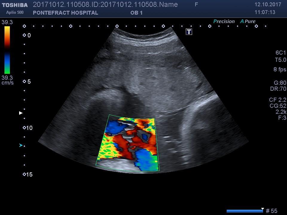
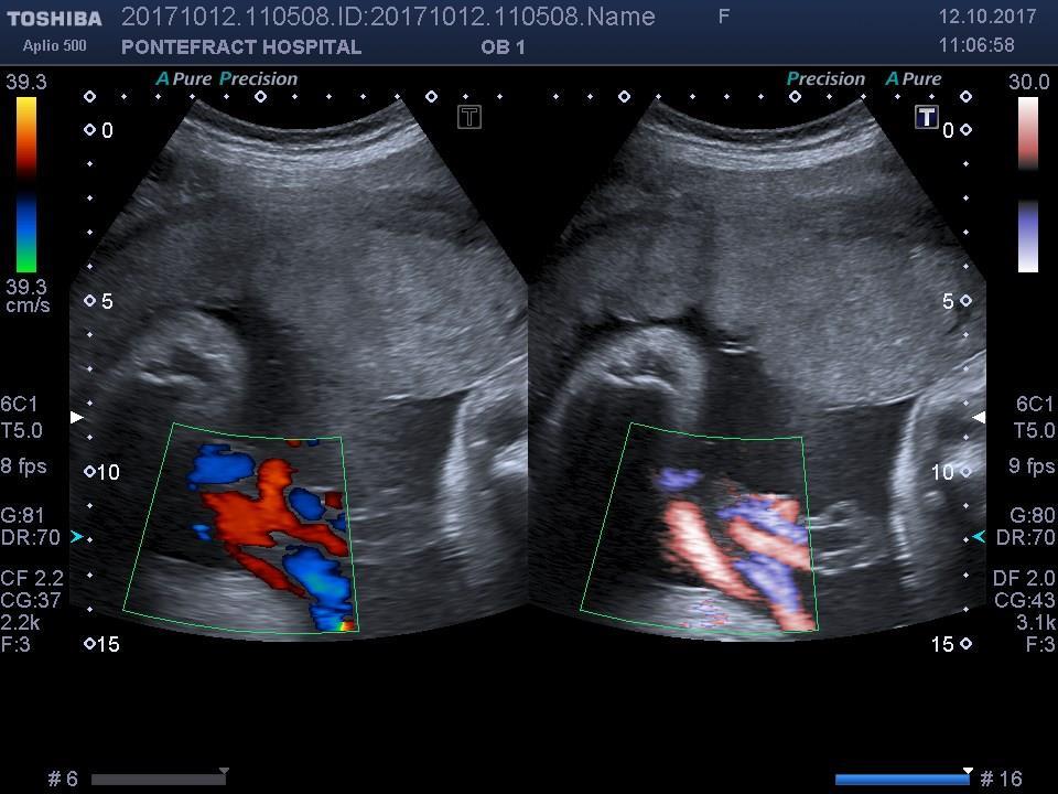
17 Colour Doppler Knobology Flow towards the probe is coloured RED Flow away from the probe is coloured BLUE. Turbulent flow shows a mosaic pattern of colours. Aliasing is demonstrated as colours wrapped into each other. TOP TIP -eliminate aliasing by increasing colour PRF or lowering the baseline
18 Colour Doppler Knobology Top Tips: TO INCREASE COLOUR SENSITIVITY Increase colour gain & decrease PRF. Decrease wall filter. Decrease size of colour box. Decrease / increase frequency or change transducer.
19
20
21 B-FLOW and Advanced dynamic flow (ADF) B-Flow maps the B-mode reflections from the blood components. B-Flow is not doppler it will not alias, is not angle dependant and does not bleed over vessel walls. ADF uses high resolution doppler techniques to accurately resolve small vessels Top Tip: Both the above will help distinguish the arteries and vein in the umbilical cord (different names on different manufacturers)
22
23 Pulsed wave Doppler
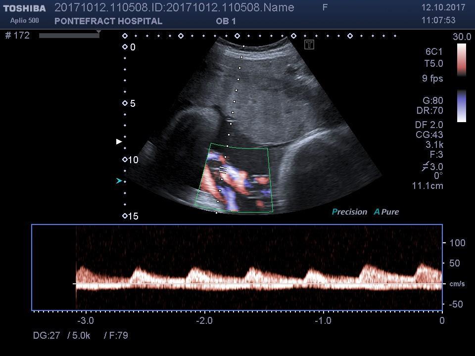
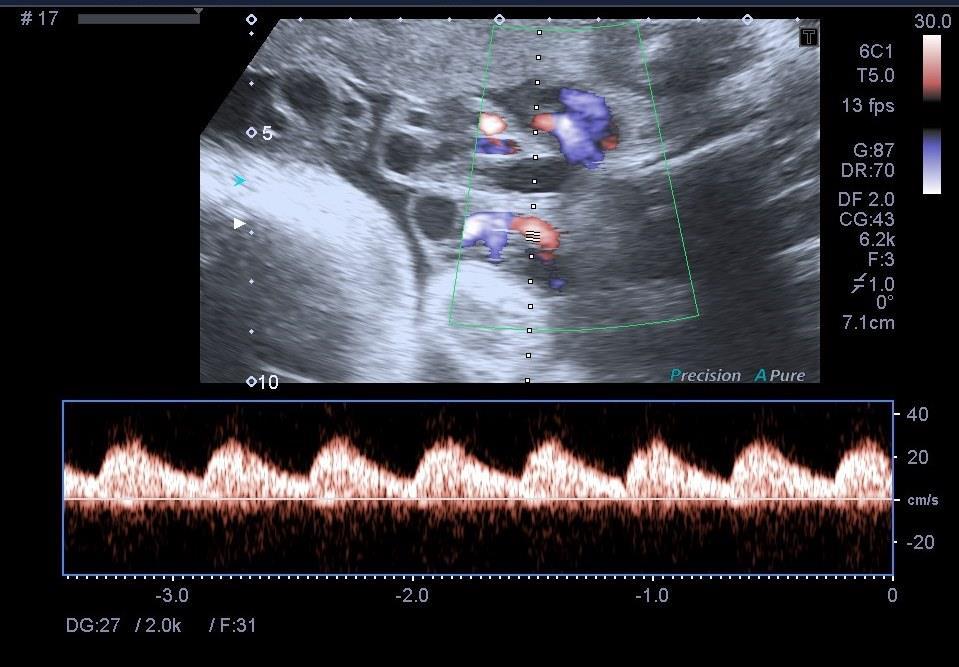
24 Pulsed doppler display spectral trace khz Peak Systole End Diastole M/s Provides a measure of the changing velocity throughout the cardiac cycle and the distribution of velocities in the sample volume (or gate) PEAK SYSTOLE-The peak velocity of the cardiac cycle. END DIASTOLE-The end point of the cardiac cycle.
25 DOPPLER ANGLE Doppler Angle 60 Degrees 0 If an accurate angle correction is made, then absolute velocities can be measured. The perfect doppler angle is 0. In practice the doppler angle should be maintained as close to 60 as possible to achieve reproducible results.
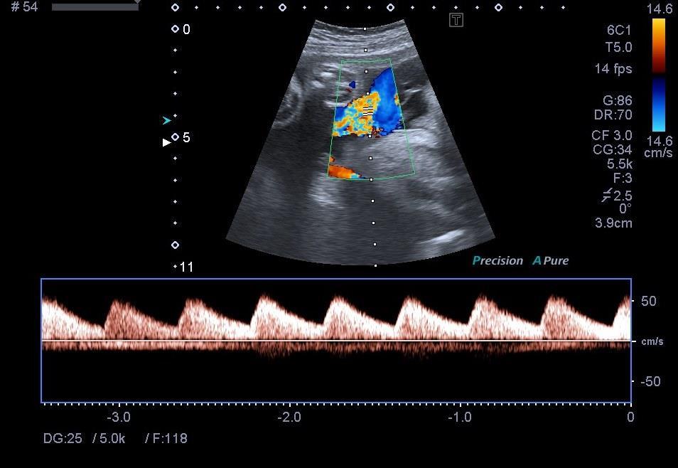
26 2 1
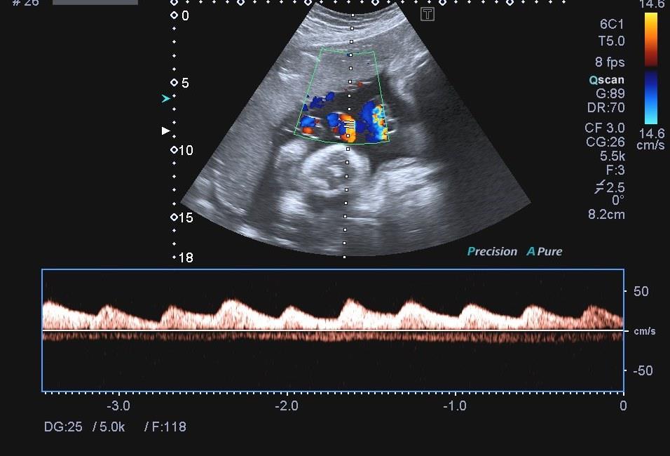
27 No 1
28 No 2
29 Pulsed wave Doppler Knobology BASELINE- changes the spectrum baseline or the colour bar to accommodate higher velocity flow. ANGLE CORRECT can t change the angle with a curvi-linear probe, must change probe position PULSE WAVE GAIN can be adjusted to increase amplitude of returning echoes Top Tip too high will affect PI.
30
31 Pulsed wave Doppler Knobology SAMPLE GATE SIZE - If flow measurements are being attempted, the whole vessel should be insonated. A large gate may include signals from adjacent vessels TRACE DIRECTION - the cursors can be altered to trace above or below the baseline. TIMELINE - if selected will display only the doppler and not the B-mode image Top Tip - not recommended because you can t see the angle used
32
33 Pulsed wave Doppler Knobology DOPPLER FREQUENCY - Altering the transmit doppler frequency can improve penetration/resolution. DISPLAY/TIMELINE - will display only the spectral trace and not the B-mode image - Top Tip - not recommended because you can t see the angle used SWEEP SPEED - should be fast enough to separate successive waveforms. Top Tip Ideal is a display of four to six (but no more than eight to 10) complete cardiac cycles.
34 Pulsed wave Doppler Knobology PULSE REPETITION FREQUENCY -The doppler is pulsed thousands of times a second (Pulse Repetition Frequency (PRF)) through a small sample box. If the speed of the blood is faster than half of the PRF rate an artefact called aliasing is produced.

35 PRF Aliasing is displayed by wrapping the waveform around the baseline and the peaks are situated below the baseline. Top tip- reduce the PRF or lower the baseline. PI cannot be measured if aliasing is present
36 PRF When there is low flow a lower PRF is required, when there is high velocity flow a higher PRF is required PRF is the first factor that needs to be altered if aliasing or low flow is present. On most machines PRF button is usually called SCALE Top Tip -Reduce scale to see end diastolic flow better
37 Abnormal spectral trace PI should not be calculated when there is: Fetal breathing movements Fetal movements Fetal arrhythmia Hiccoughs Top Tip: wait or walk
38
39 PI values The end diastolic flow should become thicker with increased gestational age. Therefore the ratio of PSV to EDV will reduce therefore PI will decrease with gestational age TOP TIP if the PI is above the 95 th centile but there is thick EDV your technique may be wrong false positive
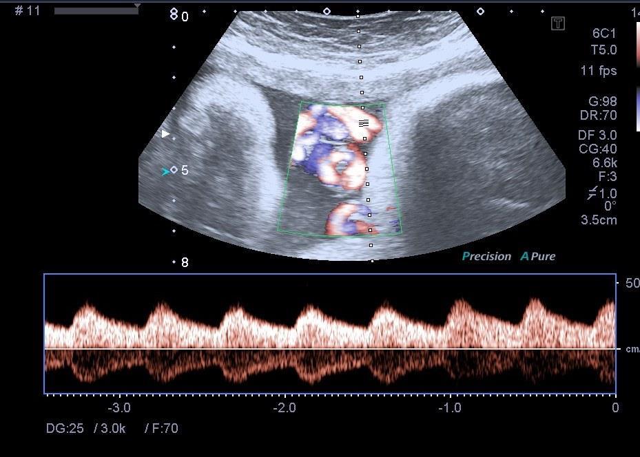
40 Calculations - Automatic trace Manufacturers assure us that auto trace is accurate If more than one peak is measured then average is displayed However, if one of the peaks has some irregularity the average PI will be lower No guidance on how many peaks to choose Top Tip don t choose highest or lowest CHOOSE THE BEST choose the best peak 1 peak may be too little 8 peaks may be too many
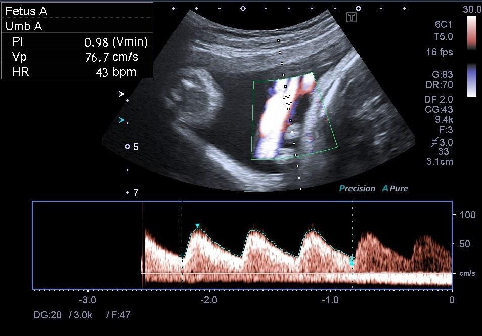
41 PI Values
42 PI values 5 peaks
43 PI values 2 peaks
44 Which Chart?

45 Which Chart
46 Raised PI
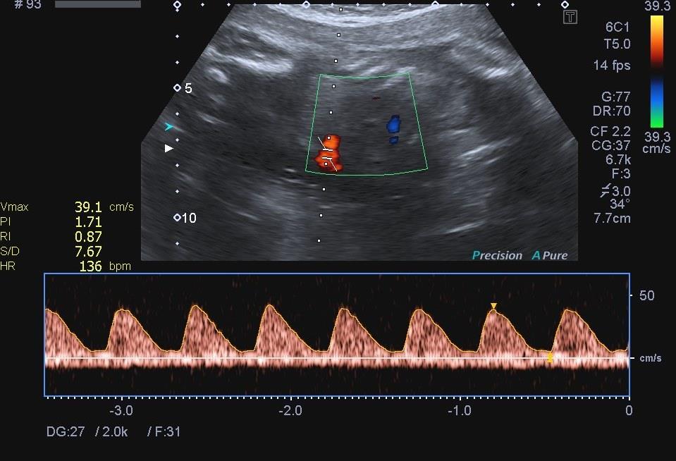
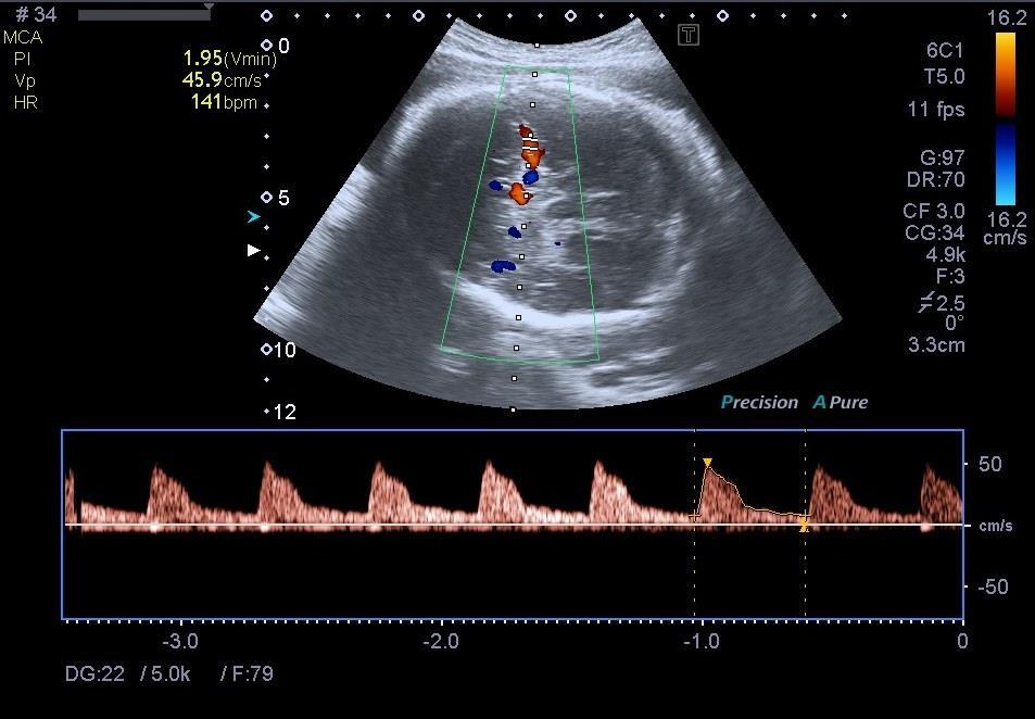
47 Artefacts forward and reverse flow If the line of pulse doppler crosses a vessel that is folded or 2 umbilical arteries running in opposite directions forward and reverse flow will be seen on the spectral trace
48
49 Artefacts umbilical vein If the the pulsed doppler line crosses the artery and the vein the spectral trace of the vein will be seen below the baseline- this can obscure possible reversed flow If caught at an angle it can mimic reversed flow

50 Absent EDV vein below

51 Reported as reversed EDV - vein below

52 NO EDV reduced scale, reduced sweep speed, correct


53 Wall filters remove the low frequency signal created by wall thump. Setting the wall filter too high may erase low velocity flow. TOP TIP-reduce the wall filter in low flow situations e.g. No end diastolic flow Can t measure PI if wall filter too high PW WALL FILTER

54 Fetal middle cerebral artery Head in the transverse plane Axial section of the brain including them thalami and sphenoid wing bones Find vessels on colour/power doppler overlying the anterior wing near the base of the skull
55 Fetal middle cerebral artery Normal high resistance flow In pathology low resistance flow as a result of head sparing theory (redistribution) Reading obtained close to the internal carotid artery

56 Fetal middle cerebral artery
57

58 Real reversed flow?
59 Conclusion Operator dependent Optimise B mode, colour and oulse wave setting before measuring PI know your equipment Use all criteria to decide which PI value to use PI value is only part of the story Don t measure if trace irregular
4/19/2018. St. Cloud Hospital Perinatology Kristin Olson, RDMS, RVT
 St. Cloud Hospital Perinatology Kristin Olson, RDMS, RVT Review Fetal Circulation Provide Indications for Umbilical Artery, Middle Cerebral Artery, and Ductus Venosus Doppler studies. Demonstrate normal
What effects will proximal or distal disease have on a waveform?
 Spectral Doppler Interpretation Director of Ultrasound Education & Quality Assurance Baylor College of Medicine Division of Maternal-Fetal Medicine Maternal Fetal Center Imaging Manager Texas Children
Diploma of Medical Ultrasonography (DMU) Physical Principles of Ultrasound and Instrumentation Syllabus
 Physical Principles of Ultrasound and Instrumentation Syllabus") Diploma of Medical Ultrasonography (DMU) Physical Principles of Ultrasound and Instrumentation Syllabus Page 1 of 7 11/18 Candidates are expected to cover all of the content of this syllabus when preparing
Diploma of Medical Ultrasonography (DMU) Physical Principles of Ultrasound and Instrumentation Syllabus Page 1 of 7 11/18 Candidates are expected to cover all of the content of this syllabus when preparing
The Fetus: Five Top Do Not Miss Diagnoses. Doppler Ultrasound
 The Fetus: Five Top Do Not Miss Diagnoses Doppler Ultrasound Giancarlo Mari, MD, MBA Professor and Chair Department of Obstetrics and Gynecology University of Tennessee Health Science Center Memphis, TN
Vascular Sonography Examination
 Vascular Sonography Examination The purpose of The American Registry of Radiologic Technologists (ARRT ) Vascular Sonography Examination is to assess the knowledge and cognitive skills underlying the intelligent
Circulatory System MARE HEIFER. aorta cvc. iia eia ipa. aorta. dca. (uov) oa. uboa. uma. ubva va. bua. iia. uma. ubva. eia. ua bua. cvc.
 oa. uboa. uma. ubva va. bua. iia. uma. ubva. eia. ua bua. cvc.") Circulatory System 13 MARE aorta cvc ov (uov) oa dca uboa iia eia ipa ua uma bua ubva va HEIFER iia aorta cvc eia oa uma ua bua ubva va ov (uov) uboa 18 Chapter 1 Hemodynamics Plug flow Natural disturbed
Circulatory System 13 MARE aorta cvc ov (uov) oa dca uboa iia eia ipa ua uma bua ubva va HEIFER iia aorta cvc eia oa uma ua bua ubva va ov (uov) uboa 18 Chapter 1 Hemodynamics Plug flow Natural disturbed
STRUCTURED EDUCATION REQUIREMENTS IMPLEMENTATION DATE: JULY 1, 2016
 STRUCTURED EDUCATION REQUIREMENTS Vascular Sonography The purpose of structured education is to provide the opportunity for individuals to develop mastery of discipline-specific knowledge that, when coupled
Image Formation (10) 2 Evaluation and Selection of Representative Images (10)
 2 Evaluation and Selection of Representative Images (10)") STRUCTURED SELF ASSESSMENT CONTENT SPECIFICATIONS SSA LAUNCH DATE: JANUARY 1, 2018 Vascular Sonography The purpose of continuing qualifications requirements (CQR) is to assist registered technologists
STRUCTURED SELF ASSESSMENT CONTENT SPECIFICATIONS SSA LAUNCH DATE: JANUARY 1, 2018 Vascular Sonography The purpose of continuing qualifications requirements (CQR) is to assist registered technologists
What effects will proximal or distal disease have on an waveform?
 Spectral Doppler Interpretation Director Director of of Ultrasound Ultrasound Education Education & & Quality Quality Assurance Assurance Baylor Baylor College College of of Medicine Medicine Division
Lesson 07: Ultrasound Transducers. This lesson contains 73 slides plus 16 multiple-choice questions.
 Lesson 07: Ultrasound Transducers This lesson contains 73 slides plus 16 multiple-choice questions. This lesson was derived from pages 33 through 42 in the textbook: Ultrasound Transducers Ultrasound Transducers
Doppler Basic & Hemodynamic Calculations
 Doppler Basic & Hemodynamic Calculations August 19, 2017 Smonporn Boonyaratavej MD Division of Cardiology, Department of Medicine Chulalongkorn University Cardiac Center, King Chulalongkorn Memorial Hospital
Ultrasound Physics & Terminology
 Ultrasound Physics & Terminology This module includes the following: Basic physics terms Basic principles of ultrasound Ultrasound terminology and terms Common artifacts seen Doppler principles Terms for
8/20/18. The Doppler Effect. Objectives. What is the Doppler Effect. Doppler principles. Spectral Waveform. Image recognition. Vascular Ultrasound
 Vascular Ultrasound: Physics and Haemodynamics Objectives Doppler principles Spectral Waveform Key factors Haemodynamics: Stenosis Waveforms Image recognition Vascular Ultrasound: A flawed paradigm What
Basic Training Programme. 16 Februrary 2018, ROTTERDAM. Pre and Post-Course Test Answers
 Basic Training Programme 16 Februrary 2018, ROTTERDAM Pre and Post-Course Test Answers Your details: Name: Conference registration number/ BT delegate number: Email address: Are you already performing
Carotid Abnormalities Coils, Kinks and Tortuosity David Lorelli M.D., RVT, FACS Michigan Vascular Association Conference Saturday, October 20, 2012
 Carotid Abnormalities Coils, Kinks and Tortuosity David Lorelli M.D., RVT, FACS Michigan Vascular Association Conference Saturday, October 20, 2012 Page 1 Table of Contents Carotid Anatomy Carotid Duplex
Guide to Small Animal Vascular Imaging using the Vevo 770 Micro-Ultrasound System
 Guide to Small Animal Vascular Imaging using the Vevo 770 Micro-Ultrasound System January 2007 Objectives: After completion of this module, the participant will be able to accomplish the following: Understand
Transducer Selection. Renal Artery Duplex Exam. Renal Scan. Renal Scan Echogenicity. How to Perform an Optimal Renal Artery Doppler Examination
 How to Perform an Optimal Renal Artery Doppler Examination Director of Ultrasound Education & Quality Assurance Baylor College of Medicine Division of Maternal-Fetal Medicine Maternal Fetal Center Imaging
Basic Ultrasound Physics Board Review Questions
 Basic Ultrasound Physics Board Review Questions Sidney K. Edelman, PhD ESP Ultrasound The Woodlands, TX Question 1 What is the wavelength of 2 MHz sound in soft tissue? 1. 1.54 mm 2. 0.75 mm 3. 0.75 cm
39 th Annual Perinatal Conference Vanderbilt University December 6, 2013 IUGR. Diagnosis and Management
 39 th Annual Perinatal Conference Vanderbilt University December 6, 2013 IUGR Diagnosis and Management Giancarlo Mari, M.D., M.B.A. Professor and Chair Department of Obstetrics and Gynecology University
ULTRASOUND IMAGING EE 472 F2018. Prof. Yasser Mostafa Kadah
 ULTRASOUND IMAGING EE 472 F2018 Prof. Yasser Mostafa Kadah www.k-space.org Recommended Textbook Diagnostic Ultrasound: Physics and Equipment, 2nd ed., by Peter R. Hoskins (Editor), Kevin Martin (Editor),
The 2 nd Cambridge Advanced Emergency Ultrasound Course
 The 2 nd Cambridge Advanced Emergency Ultrasound Course Addenbrooke s Hospital Cambridge Sept 2008 1 2 Faculty! UK! USA! Australia! Toshiba! Emergency Medicine! Radiology 3 Programme! Day 1 Introduction
NCVH. Ultrasongraphy: State of the Art Vein Forum 2015 A Multidisciplinary Approach to Otptimizing Venous Circulation From Wounds to WOW
 Ultrasongraphy: State of the Art 2015 NCVH New Cardiovascular Horizons Vein Forum 2015 A Multidisciplinary Approach to Otptimizing Venous Circulation From Wounds to WOW Anil K. Chagarlamudi, M.D. Cardiovascular
From Head to Toe Use of Advanced Dynamic Flow in prenatal ultrasound
 From Head to Toe Use of Advanced Dynamic Flow in prenatal ultrasound Without doubt, the B- Schwerdtfeger, R. tant diagnostic instrument. Furthermore, we use colour in feto- mode imaging is the most important
Policies and Statements D16. Intracranial Cerebrovascular Ultrasound
 Policies and Statements D16 Intracranial Cerebrovascular Ultrasound SECTION 1: INSTRUMENTATION Policies and Statements D16 Intracranial Cerebrovascular Ultrasound May 2006 (Reaffirmed July 2007) Essential
Diagnostic Ultrasound. Sutiporn Khampunnip, M.D.
 Diagnostic Ultrasound Sutiporn Khampunnip, M.D. Definition of Ultrasound Ultrasound is simply sound waves, like audible sound. High-frequency sound and refers to mechanical vibrations above 20 khz. Human
Diagnostic approach to heart disease
 Diagnostic approach to heart disease Initial work up History Physical exam Chest radiographs ECG Special studies Echocardiography Cardiac catheterization Echocardiography principles Technique of producing
Concepts of Imaging and Knobology
 Concepts of Imaging and Knobology Pravin Patil, MD FACC FASE Associate Professor of Medicine Director, Cardiovascular Disease Training Program Lewis Katz School of Medicine at Temple University Disclosures
DC-6. Diagnostic Ultrasound System
 DC-6 Diagnostic Ultrasound System MINDRAY is proud to introduce DC-6, a color Doppler ultrasound system for general applications. DC-6 incorporates the latest digital ultrasound image processing technology
Ultrasound Applied Physics
 Ultrasound Applied Physics University of Toronto Department of Medical Imaging Applied Physics Mini-Course #3 2016 Ultrasound Laboratory Manual and Examination Booklet 1/21/2016 Ultrasound Applied Physics
Volume Flow. Volume Flow
 Volume Flow Jonathan M. Rubin, M.D., Ph.D. Department of Radiology Volume Flow Technique initially described by Hottenger and Meindl in 1974 Describes method for measuring the total flux across a flow
VFI Technology to Change the Way You Work
 Analogic Ultrasound VFI Technology to Change the Way You Work Vascular Ultrasound Made Easier Vector Flow Imaging VFI VFI is a ground-breaking technology that can revolutionize the workflow for many Doppler
PIAF study: Placental insufficiency and aortic isthmus flow Jean-Claude Fouron, MD
 Dear colleagues, I would like to thank you very sincerely for agreeing to participate in our multicentre study on the clinical significance of recording fetal aortic isthmus flow during placental circulatory
Introduction to Biomedical Imaging
 Alejandro Frangi, PhD Computational Imaging Lab Department of Information & Communication Technology Pompeu Fabra University www.cilab.upf.edu Basic principles. Comparison to X-rays Ultrasound > 20kHz
Image optimization for critical care US
 Image optimization for critical care US 1 Although we assume you are already familiar with focused US in the ED, it might not hurt to revise the basics: Machines & transducers US appearance of normal tissues
Diagnosis of Middle Cerebral Artery Occlusion with Transcranial Color-Coded Real-Time Sonography
 Diagnosis of Middle Cerebral Artery Occlusion with Transcranial Color-Coded Real-Time Sonography Kazumi Kimura, Yoichiro Hashimoto, Teruyuki Hirano, Makoto Uchino, and Masayuki Ando PURPOSE: To determine
Ultrasound Physics and Knobology Alan Macfarlane. Consultant Anaesthetist Glasgow Royal Infirmary
 Ultrasound Physics and Knobology Alan Macfarlane Consultant Anaesthetist Glasgow Royal Infirmary RAPM 2009; 34: 40-46 Ultrasound Proficiency Understanding US image generation and device operation Image
Fetal cardiovascular parameters for the prediction of postnatal cardiovascular risk in intrauterine growth-restriction?
 17 th International Conference on Prenatal Diagnosis and Therapy Lisbon, June 2013 Fetal cardiovascular parameters for the prediction of postnatal cardiovascular risk in intrauterine growth-restriction?
Diagnosis and Management of the Early Growth Restricted Fetus
 11 th Congress of Maternal Fetal Medicine and Perinatology Society of Turkey Diagnosis and Management of the Early Growth Restricted Fetus Giancarlo Mari, MD, MBA, FACOG, FAIUM Professor and Chair Department
ULTRASOUND. OB/Gyn (Core) Ultrasound PIEZOELECTRIC EFFECT. Principles of Ultrasound Physics and Instrumentation. Nathan Pinkney, BS, CDOS
 Ultrasound PIEZOELECTRIC EFFECT. Principles of Ultrasound Physics and Instrumentation. Nathan Pinkney, BS, CDOS") 1 OB/Gyn (Core) Ultrasound Principles of Ultrasound Physics and Instrumentation Nathan Pinkney, BS, CDOS Philadelphia College of Osteopathic Medicine 2016 ULTRASOUND CATEGORIES OF SOUND INFRASOUND = below
1 OB/Gyn (Core) Ultrasound Principles of Ultrasound Physics and Instrumentation Nathan Pinkney, BS, CDOS Philadelphia College of Osteopathic Medicine 2016 ULTRASOUND CATEGORIES OF SOUND INFRASOUND = below
Goals. Access flow and renal artery stenosis evaluation by Doppler ultrasound. Reimbursement. WHY use of Doppler Ultrasound
 Access flow and renal artery stenosis evaluation by Doppler ultrasound Adina Voiculescu, MD Interventional Nephrology Brigham and Women s Hospital Boston Instructor at Harvard Medical School Understand
GUNDERSEN/LUTHERAN ULTRASOUND DEPARTMENT POLICY AND PROCEDURE MANUAL
 GUNDERSEN/LUTHERAN ULTRASOUND DEPARTMENT POLICY AND PROCEDURE MANUAL SUBJECT: Carotid Duplex Ultrasound SECTION: Vascular Ultrasound ORIGINATOR: Deborah L. Richert, BSVT, RDMS, RVT DATE: October 15, 2015
Vikram Dogra, M.D. Professor of Radiology, Urology & BME Department of Imaging Sciences University Of Rochester Medical Center
 Ultrasound of the Scrotum Vikram Dogra, M.D. Professor of Radiology, Urology & BME Department of Imaging Sciences University Of Rochester Medical Center Etiologies of Acute Scrotal Pain Epididymitis/Orchitis
Introduction. Cardiac Imaging Modalities MRI. Overview. MRI (Continued) MRI (Continued) Arnaud Bistoquet 12/19/03
 MRI (Continued) Arnaud Bistoquet 12/19/03") Introduction Cardiac Imaging Modalities Arnaud Bistoquet 12/19/03 Coronary heart disease: the vessels that supply oxygen-carrying blood to the heart, become narrowed and unable to carry a normal amount
Introduction Cardiac Imaging Modalities Arnaud Bistoquet 12/19/03 Coronary heart disease: the vessels that supply oxygen-carrying blood to the heart, become narrowed and unable to carry a normal amount
Doppler assessment of fetal aortic isthmus blood flow in two different sonographic planes during the second half of gestation
 Ultrasound Obstet Gynecol 2005; 26: 170 174 Published online in Wiley InterScience (www.interscience.wiley.com). DOI: 10.1002/uog.1955 Doppler assessment of fetal aortic isthmus blood flow in two different
Management of IUGR Prof. Dr. Acar KOÇ
 Management of IUGR Prof. Dr. Acar KOÇ Ankara University School of Medicine Department of OB&GYN Department of Perinatology Definition and Diagnosis: SGA IUGR EFW: < 10th percentile EFW: < 10th percentile
Disclosure Statement:
 Marsha M. Neumyer, BS, RVT, FSVU, FSDMS, FAIUM International Director Vascular Diagnostic Educational Services Vascular Resource Associates Harrisburg, PA Disclosure Statement: CME Calendar QR Code Marsha
Imaging of the Basal Cerebral Arteries and Measurement of Blood Velocity in Adults by Using Transcranial Real-Time Color Flow Doppler Sonography
 497 Imaging of the Basal Cerebral Arteries and Measurement of Blood Velocity in Adults by Using Transcranial Real-Time Color Flow Doppler Sonography Takashi Tsuchiya 1 Masahiro Yasaka Takenori Yamaguchi
Basic Physics of Ultrasound and Knobology
 WELCOME TO UTMB Basic Physics of Ultrasound and Knobology By Daneshvari Solanki, FRCA Laura B. McDaniel Distinguished Professor Anesthesiology and Pain Medicine University of Texas Medical Branch Galveston,
Tissue Doppler Imaging
 Cronicon OPEN ACCESS Hesham Rashid* Tissue Doppler Imaging CARDIOLOGY Editorial Department of Cardiology, Benha University, Egypt *Corresponding Author: Hesham Rashid, Department of Cardiology, Benha University,
Basic Doppler Assessment of Fetal Distress
 Basic Doppler Assessment of Fetal William J. Polzin, M.D. Co-Director, Fetal Care Center of Cincinnati Director, Division of Maternal-Fetal Medicine Good Samaritan Hospital Cincinnati, OH No Relevant Disclosures
GLOBAL INNOVATION BY DESIGN
 GLOBAL INNOVATION BY DESIGN For over 130 years Toshiba s research and development has improved the health and welfare of people around the world. Today, Toshiba Medical Systems offers a full range of diagnostic
2015 ARDMS Sonography Principles & Instrumentation Job Task Analysis Summary Report
 P a g e 1 2015 ARDMS Sonography Principles & Instrumentation Job Task Analysis Summary Report American Registry for Diagnostic Medical Sonography (ARDMS) P a g e 2 Table of Contents ABOUT THE REPORT...
Ultrasound 10/1/2014. Basic Echocardiography for the Internist. Mechanical (sector) transducer Piezoelectric crystal moved through a sector sweep
 transducer Piezoelectric crystal moved through a sector sweep") Ultrasound Basic Echocardiography for the Internist Carol Gruver, MD, FACC UT Erlanger Cardiology Mechanical wave of compression and rarefaction Requires a medium for transmission Ultrasound frequency
Ultrasound Basic Echocardiography for the Internist Carol Gruver, MD, FACC UT Erlanger Cardiology Mechanical wave of compression and rarefaction Requires a medium for transmission Ultrasound frequency
GE Healthcare. Logiq P5 Premium
 GE Healthcare Logiq P5 Premium LOGIQ P5 PREMIUM The GE Logiq P5 premium is an economical shared service ultrasound machine that is unique in it s price range by offering deep support for all applications
Standardising echocardiography and images. Version 2, 13/04/15
 Standardising echocardiography and images 1. Review of ECHO eligibility criteria - trial entry - rescue treatment 2. Assessments - personnel - timing 3. Technical aspects of ECHO examination 1. Trial entry
Duplex Ultrasound of the Renal Arteries. Duplex Ultrasound. In the Beginning
 Duplex Ultrasound of the Renal Arteries DIMENSIONS IN HEART AND VASCULAR CARE 2013 PENN STATE HEART AND VASCULAR INSTITUTE ROBERT G. ATNIP MD PROFESSOR OF SURGERY AND RADIOLOGY Duplex Ultrasound Developed
Basic of Ultrasound Physics E FAST & Renal Examination. Dr Muhammad Umer Ihsan MBBS,MD, DCH CCPU,DDU1,FACEM
 Basic of Ultrasound Physics E FAST & Renal Examination Dr Muhammad Umer Ihsan MBBS,MD, DCH CCPU,DDU1,FACEM What is Sound? Sound is Mechanical pressure waves What is Ultrasound? Ultrasounds are sound waves
Guidelines, Policies and Statements D20 Statement on Peripheral Venous Ultrasound
 Guidelines, Policies and Statements D20 Statement on Peripheral Venous Ultrasound Disclaimer and Copyright The ASUM Standards of Practice Board have made every effort to ensure that this Guideline/Policy/Statement
Background & Indications Probe Selection
 Teresa S. Wu, MD, FACEP Director, EM Ultrasound Program & Fellowship Co-Director, Simulation Based Training Program & Fellowship Associate Program Director, EM Residency Program Maricopa Medical Center
Physical Principles of Ultrasound
 Physical Principles of Ultrasound Grateful appreciation to Richard A. Lopchinsky, MD, FACS and Nancy H. Van Name, RDMS, RTR, and MarleneKattaron, RDMS 2000 UIC All Rights Reserved. Course Objectives Identify
Abdominal Ultrasound
 Abdominal Ultrasound What is Ultrasound Imaging of the Abdomen? What are some common uses of the procedure? How should I prepare? What does the equipment look like? How does the procedure work? How is
Ultrasound in Medicine
 Ultrasound in Medicine Experimental Equipment for Medical Education Universities Colleges Medical Schools Medical and Med-Technical Training Education can befun! WELCOME TO GAMPT Devices and accessories
Ultrasound Principles cycle Frequency Wavelength Period Velocity
 ! Teresa S. Wu, MD, FACEP Director, EM Ultrasound Program & Fellowship Co-Director, Simulation Based Training Program & Fellowship Associate Program Director, EM Residency Program Maricopa Medical Center
A (quasi)evidence-based approach to the management of early-onset IUGR
evidence-based approach to the management of early-onset IUGR") A (quasi)evidence-based approach to the management of early-onset IUGR Eduard Gratacós Barcelona Center for Maternal-Fetal and Neonatal Medicine Hospital Clínic and Hospital Sant Joan de Deu, University
A (quasi)evidence-based approach to the management of early-onset IUGR Eduard Gratacós Barcelona Center for Maternal-Fetal and Neonatal Medicine Hospital Clínic and Hospital Sant Joan de Deu, University
Confident Diagnosis, Confident Decisions
 Confident Diagnosis, Confident Decisions ALPINION MEDICAL SYSTEMS We are Ultrasound Professionals The E-CUBE Value Creation Optimal Imaging Suite Image Quality Enhancements Optimal Imaging Suite, ALPINION
Accuracy Versatility Mobility
 M6 Hand-Carried Ultrasound System Accuracy Versatility Mobility People you know, Service you can trust. Bring high performance ultrasound to the bedside It has been a long, challenging path for clinicians
Normal TTE/TEE Examinations
 Normal TTE/TEE Examinations Geoffrey A. Rose, MD FACC FASE Sanger Heart & Vascular Institute Before you begin imaging... Obtain the patient s Height Weight BP PLAX View PLAX View Is apex @ 9-10 o clock?
Ultrasound. Principles of Medical Imaging. Contents. Prof. Dr. Philippe Cattin. MIAC, University of Basel. Oct 17th, 2016
 Ultrasound Principles of Medical Imaging Prof. Dr. Philippe Cattin MIAC, University of Basel Contents Abstract 1 Image Generation Echography A-Mode B-Mode M-Mode 2.5D Ultrasound 3D Ultrasound 4D Ultrasound
MESENTERIC ISCHEMIA. Phillip J Bendick, PhD
 MESENTERIC ISCHEMIA Phillip J Bendick, PhD Arterial Celiac - Hepatic - Splenic Superior Mesenteric Artery Inferior Mesenteric Artery Venous Mesenteric system Porto - hepatic system Inferior Vena Cava Acute
ACUSON P500. Ultrasound Anytime, Anywhere. ACUSON P500. siemens.com/acusonp500. siemens.com/acusonp500 1
 Ultrasound Anytime, Anywhere. ACUSON P500 1 Enabling ultrasound imaging anytime, anywhere. 2 Ultrasound Anytime, Anywhere Siemens Healthineers engineered the compact and powerful ACUSON P500, a portable
Ultrasound Physics & Doppler
 Ultrasound Physics & Doppler Endocrine University 2018 Mark Lupo, MD, FACE, ECNU Objectives Review the essential components of ultrasound physics in neck sonography Demonstrate the importance of ultrasound
Appendix II: ECHOCARDIOGRAPHY ANALYSIS
 Appendix II: ECHOCARDIOGRAPHY ANALYSIS Two-Dimensional (2D) imaging was performed using the Vivid 7 Advantage cardiovascular ultrasound system (GE Medical Systems, Milwaukee) with a frame rate of 400 frames
No financial or commercial relationships to disclose
 Deanna New, RVT No financial or commercial relationships to disclose IAC REQUIREMENTS: The main duty of a sonographer is to make the physician or radiologists job easier by capturing images and doing
Performance in style. Ultrasound system H60 SAMSUNG MEDISON CO., LTD. Scan code or visit to learn more
 founded in 1985. With a mission to bring health and well-being to people's lives, the company manufactures diagnostic ultrasound systems around the world across in 2001 and since being part of Samsung
5 Working With Measurements
 5 Working With Measurements Measurement Overview Measurements accompanying ultrasound images supplement other clinical procedures available to the attending physician. Accuracy of the measurements is determined
DOW-RAD, DOW DIAGNOSTIC COMPLEX, DUHS TRAINING PROGRAM HANDBOOK 2013
 DOW-RAD, DOW DIAGNOSTIC COMPLEX, DUHS TRAINING PROGRAM HANDBOOK 2013 CERTIFICATE COURSE INVASCULAR/DOPPLER ULTRASOUND: Introduction: Ultrasound is an evolving technology with wide spectrum application
PRACTICAL GUIDE TO FETAL ECHOCARDIOGRAPHY IC Huggon and LD Allan
 PRACTICAL GUIDE TO FETAL ECHOCARDIOGRAPHY IC Huggon and LD Allan Fetal Cardiology Unit, Harris Birthright Research Centre for Fetal Medicine, King's College Hospital, London, UK IMPORTANCE OF PRENATAL
BEDSIDE ULTRASOUND BEDSIDE ULTRASOUND. Deep Vein Thrombosis. Probe used
 BEDSIDE ULTRASOUND Part 2 Diagnosis of deep vein thrombosis Kishore Kumar Pichamuthu, Professor, Department of Critical Care, CMC, Vellore Summary: Deep vein thrombosis (DVT) is a problem encountered in
Introduction & Physics of ED Ultrasound. Objectives. What? - Limited Studies. Who? - ED Docs
 Introduction & Physics of ED Ultrasound Martine Sargent, MD Ultrasound Director, Assistant Professor UCSF Department of Emergency Medicine San Francisco General Hospital & Trauma Center Objectives Who?
COMPREHENSIVE EVALUATION OF FETAL HEART R. GOWDAMARAJAN MD
 COMPREHENSIVE EVALUATION OF FETAL HEART R. GOWDAMARAJAN MD Disclosure No Relevant Financial Relationships with Commercial Interests Fetal Echo: How to do it? Timing of Study -optimally between 22-24 weeks
Introduction to Fetal Doppler Echocardiography
 Chapter 32 Introduction to Fetal Doppler Echocardiography Dev Maulik Introduction Evaluation of the fetal heart constitutes one of the critical areas of prenatal diagnosis. Advances in diagnostic medical
PART II ECHOCARDIOGRAPHY LABORATORY OPERATIONS ADULT TRANSTHORACIC ECHOCARDIOGRAPHY TESTING
 PART II ECHOCARDIOGRAPHY LABORATORY OPERATIONS ADULT TRANSTHORACIC ECHOCARDIOGRAPHY TESTING STANDARD - Primary Instrumentation 1.1 Cardiac Ultrasound Systems SECTION 1 Instrumentation Ultrasound instruments
What is Ultrasound? Resolution Image production Attenuation Imaging modes Ultrasound artifacts... 7
 What is Ultrasound?... 1 Resolution... 3 Image production... 3 Attenuation... 4 Imaging modes... 5 Ultrasound artifacts... 7 0 What is Ultrasound? High frequency sound of frequencies 2-50 MHz is used in
Hitachi Aloka Medical manufactured one of the world s first diagnostic ultrasound platforms, and today this imaging modality has become the first
 Hitachi Aloka Medical manufactured one of the world s first diagnostic ultrasound platforms, and today this imaging modality has become the first choice examination for many disorders. If the subtlest
Routine Quality Assurance Cookbook
 This Cookbook is a companion guide to the AIUM Routine Quality Assurance (QA) for Diagnostic Ultrasound Equipment document, which outlines the basic QA requirements for AIUM-accredited practices. The Guide
Assessment of fetal heart function and rhythm
 Assessment of fetal heart function and rhythm The fetal myocardium Early Gestation Myofibrils 30% of myocytes Less sarcoplasmic reticula Late Gestation Myofibrils 60% of myocytes Increased force per unit
Carotid Artery Doppler
 Carotid Artery Doppler Patient Position supine or semisupine head slightly hyper extended rotated 45 away from the side being examined. Higher frequency linear transducers (7 MHz) Vessels should be imaged
What Do We Know? Disclosure Statement: 3/11/2015. Deep abdominal imaging
 Marsha M. Neumyer, BS, RVT, FSVU, FSDMS, FAIUM International Director Vascular Diagnostic Educational Services Vascular Resource Associates Harrisburg, PA Disclosure Statement: CME Calendar QR Code Marsha
Section II: Patient Interview Grade: 5
 Only written competency completed with this EXACT form will be accepted for grading. No modifications to the LAYOUT of the form will be accepted for a written competency. Failure to comply will result
Protokollanhang zur SPACE-2-Studie Neurology Quality Standards
 Protokollanhang zur SPACE-2-Studie Neurology Quality Standards 1. General remarks In contrast to SPACE-1, the neurological center participating in the SPACE-2 trial will also be involved in the treatment
Certificate in Clinician Performed Ultrasound (CCPU) Syllabus. Vascular Access (venous (peripheral and central) and arterial)
 Syllabus. Vascular Access (venous (peripheral and central) and arterial)") Certificate in Clinician Performed Ultrasound (CCPU) Syllabus Vascular Access (venous (peripheral and central) and arterial) Page 1 of 8 04/16 Vascular Access (venous (peripheral and central) and arterial)
Certificate in Clinician Performed Ultrasound (CCPU) Syllabus Vascular Access (venous (peripheral and central) and arterial) Page 1 of 8 04/16 Vascular Access (venous (peripheral and central) and arterial)
Transcranial Doppler (Basic Step) Dae-il Chang, M.D., Sung Sang Yoon, M.D. Department of Neurology, College of Medicine, Kyunghee university
 Dae-il Chang, M.D., Sung Sang Yoon, M.D. Department of Neurology, College of Medicine, Kyunghee university") Transcranial Doppler (Basic Step) Dae-il Chang, M.D., Sung Sang Yoon, M.D. Department of Neurology, College of Medicine, Kyunghee university Principles of Doppler Ultrasonography Major target Speed & direction
Transcranial Doppler (Basic Step) Dae-il Chang, M.D., Sung Sang Yoon, M.D. Department of Neurology, College of Medicine, Kyunghee university Principles of Doppler Ultrasonography Major target Speed & direction
Tissue Doppler Imaging in Congenital Heart Disease
 Tissue Doppler Imaging in Congenital Heart Disease L. Youngmin Eun, M.D. Department of Pediatrics, Division of Pediatric Cardiology, Kwandong University College of Medicine The potential advantage of ultrasound
Mobile excellence SAMSUNG. ULTRASOUND H60 Ultrasound system HM70A with Plus. Excellence on the Move SAMSUNG MEDISON CO., LTD.
 Samsung Medison, an affiliate of Samsung Electronics, is a global medical company founded in 1985. With a mission to bring health and well-being to people's lives, the company manufactures diagnostic ultrasound
Bits and Bobs secondary causes of heart problems. Dr Angela McBrien 9 th September 2017
 Bits and Bobs secondary causes of heart problems Dr Angela McBrien 9 th September 2017 Not the heart Dextroposition Heart in the right chest with the apex to the left Often caused by left sided chest mass
Normal pericardial fluid in the fetus: color and spectral Doppler analysis
 Ultrasound Obstet Gynecol 2001; 18: 248 252 Normal pericardial fluid in the fetus: color and spectral Blackwell ORIGINAL Science, PAPERLtd Doppler analysis S.-J. YOO*, J.-Y. MIN and Y.-H. LEE *Department
Doppler Color Flow Imaging #4
 Doppler Color Flow Imaging #4 Joseph A. Kisslo, MD David B. Adams, RDCS INTRODUCTION Doppler color flow imaging is a method for noninvasively imaging blood flow through the heart by displaying flow data
Quick practical guide to Cranial Ultrasound in the newborn
 Quick practical guide to Cranial Ultrasound in the newborn Introduction A standard set of views is taken to assist with consistent visualisation of structures and in the interpretation of possible abnormalities.
Principles of Ultrasound. Cara C. Prideaux, M.D. University of Utah PM&R Sports Medicine Fellow March 14, 2012
 Principles of Ultrasound Cara C. Prideaux, M.D. University of Utah PM&R Sports Medicine Fellow March 14, 2012 None Disclosures Outline Introduction Benefits and Limitations of US Ultrasound (US) Physics
Ultrasound Guidance Needle Techniques
 Ultrasound Guidance Needle Techniques Dr TANG Ho-ming AED/UCH USG Guidance Needle Techniques Commonly used in EM 1. Vessel cannulation-peripheral & central 2. Foreign body removal 3. Peripheral nerve/plexus