Background Focused Assessment with Sonography in Trauma. Johann Baptist Dormagen, MD, PhD
|
|
|
- Janice Spencer
- 6 years ago
- Views:
Transcription
1 Focused Assessment with Sonography in Trauma Johann Baptist Dormagen, MD, PhD Unit of Abdominal and Oncologic Radiology Department of Radiology and Nuclear Medicine Oslo University Hospital, Norway 8 th NORDIC TRAUMA RADIOLOGY COURSE Stockholm, Sweden May 19-22, 2014 Background Technique Pitfalls Controversies New trends Background Quick assessment of presence of free fluid Lack of uniform definition Pericard Pleura Extended FAST Free air? Parenchymal organs 1
 Urine bladder")
2 No diff. between blood and other fluid No assessment of diaphragm, retroperit. Operator dependent No reliable assessment of organ injury FAST No radiation, no complications Repeatable Non invasiv Cheoooap and fast Background DPL Complications with DPL(0,5-1%) Urine bladder Bowel Mesentery Ovarian Infections Time: From fasciotomia to correctly placed catheter: 11 minutter (3-40) From fasciotomia to backflow 27min Velmahos Innledning - DPL Saunders 1998 ATLS recommendations 4 point examination Pericard Morrissons pouch Perisplenic view ( left upper quadrant) Pelvic view, the retrouterine pouch (pouch of Douglas) or retrovesical pouch 2

3 After Korner, M. et al. Radiographics 2008;28: Axial/Coronal view Left shoulder 8 9 3
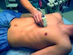
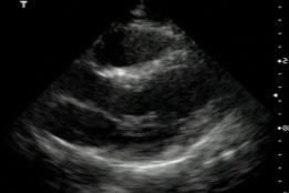
4 10 Longitudinal view 11 Parasternal view 12 4


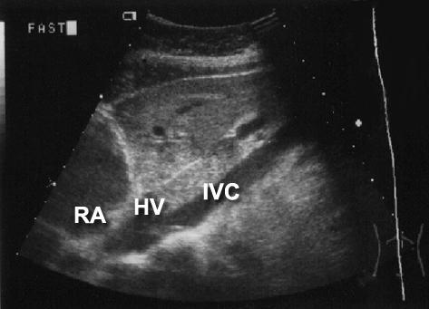
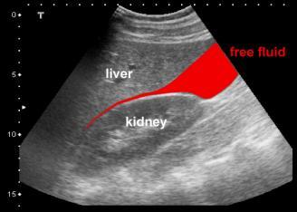
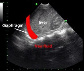
5 Intraperitoneal fluid Right side: One cine-loop from anterior to posterior starting at the anterior axilar line. Cranial portion of the liver and diaphragm One cine-loop from anterior to posterior aside the lower right liverlbe

6


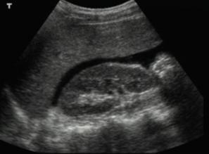
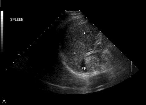
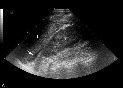
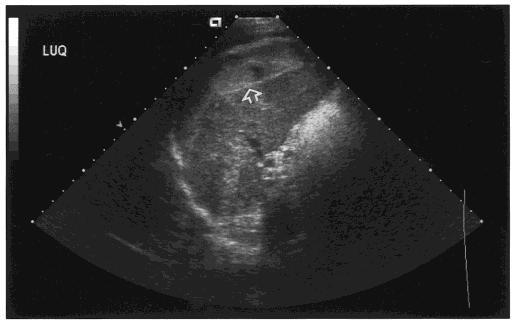
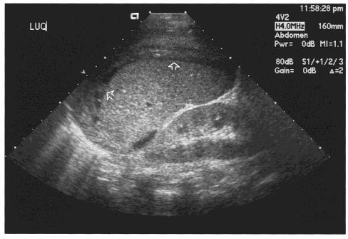
7 19 Left side: One intercostal view from posterior axial line to the anterior axial line. Cranial portion of the spleen and diaphragm One intercostal or subcostal view from posterior to anterior axial line. Caudal spleen and cranial portion of left kidney

8

9


10 Bladder One cine-loop longitudinal from left to right with good depth One cine-loop from cranial to caudal
11 Pitfalls False negative findings due to Overlying bowel gas Empty bladder Obesity Surgical emphysema in chest or abdominal wall Too early scanning Incorrect settings of the equipment FAST in unstable patients Author n Exam. Year Sens. Spec. Rozcyski 30 FAST % 100% Kuncir 62 FAST % 95% Gaarder 104 FAST % 95% Cha 70 FAST % Ikke angitt Kuncir 62 DPL % 89% Cha i.a. DPL % 100% Pitfalls False positive findings Preexisting ascites Ovarian cyst accidents, plevic inflammatory disease Physiological amount of free fluid in the retrouterine pouch in women of reproductive age 11


12 Recent publications ( 2012) Controversies No data mortality, hospital stay No data on cost reduction Usufulness in unstable patients is questionable Small, often hand-held scanners at the cheaper end of the market must be used with particular care, as the image quality may be inadequate for some examinations 12

13 New trends Paramedical examination before the patient reaches hospital e-fast Technique of Chest US Chest US as part of the e-fast examination Before CT and before placement of thoracostomy tube All patients hemodynamically unstable 7.5 Mhz linear probe 13

14 Four signs Lung sliding: Visceral pleura moving against the parietal pleura In pneumothax the pleural line appears motionless Highly negative predictive B-lines: Comet tail artifacts, arise from the pleural line and spread up to the edge of the screen. Synchroneous to the respiratory movements Presence of B-lines indicate absence of pneumothroax Lung point at the lateral inferior chest wall: Transition between pneumothorax and adherent lung Lung pulse: Vertical movement of the pleural line, synchroneous to the heart beat, indicating consolidated motionless lung through wich the heart beat is transmitted B-lines Diagnostic algorythm for diagnosis of pneumothorax Ianello,

15 Accuracy Ianello 2013 Prehospital FAST and efast Development of handheld, battery-powered, low-weight US machines has created the possibility of bringing US to the prehospital setting, thus gaining a potential for early diagnosis and treatment The data regarding the use of US in the prehospital setting is sparse, often of low quality and describing a broad variety of patients and clinical challenges 15
16 14 studies included Large heterogenity Lack of quality in all studies No radiologist participated 885 patients included Results Prehospital ultrasound is working and feasible Favours early diagnosis Potential for change of admittance Time delay 0-6 min Higly reliable in detedtion of haemoperitoneum and haemopericardium compared with low accuracy of physical examination and haemodynamic measurements Specificity and Sensitivity: 0.93, 0.99! It is currently not possible to conclude that prehospital-performed US improves treatment of the trauma patient. However, data indicate that prehospital management is altered by the use of US in respect to early and precise diagnosis, treatment and visitation. 16
17 Summary FAST requires high experience and a large turn-over of traumapatients for maintanance of practical skills More a rule-inn tool than rule-out tool Impact on treatment and outcome remains unclear Extensions of FAST seem useful in some clinical settings 17
Ultrasound. FAST Focused Assessment with Sonography in Trauma
 Ultrasound FAST Focused Assessment with Sonography in Trauma Rohit Patel, MD University of Florida Health Director, Critical Care Ultrasound Surgical ICU Center for Intensive Care Gainesville, Florida
FAST Focused Assessment with Sonography in Trauma
 FAST Focused Assessment with Sonography in Trauma Wilma Rodriguez Mojica,MD,FACR Professor of Radiology UPR School of Medicine Ultrasound Section - Radiological Sciences Department OBJECTIVES Understand
Extended FAST Exam. Goal of Trauma Care. Golden Hour of Trauma
 Extended FAST Exam Goal of Trauma Care Golden Hour of Trauma Best INITIAL screening modality in trauma efast 2014 LLSA Article (ACEP Policy Statement) Level B Recommendation: In hemodynamically unstable
EFAST. Extended Focussed Assessment with Sonography for Trauma. Ultrasound Logbook. Name
 EFAST Extended Focussed Assessment with Sonography for Trauma Ultrasound Logbook ame Contents EFAST Accreditation Requirements 25 Abdominal Aorta Report Forms 3 Formative Assessments 1 Summative Assessment
Certificate in Clinician Performed Ultrasound (CCPU) Syllabus. Extended Focussed Abdominal Scan for Trauma (E-FAST)
 Syllabus. Extended Focussed Abdominal Scan for Trauma (E-FAST)") Certificate in Clinician Performed Ultrasound (CCPU) Syllabus Extended Focussed Abdominal Scan for Trauma (E-FAST) Page 1 of 6 01/17 ACN 001 679 161 ABN 64 001 679 Extended Focussed Abdominal Scan for
Certificate in Clinician Performed Ultrasound (CCPU) Syllabus Extended Focussed Abdominal Scan for Trauma (E-FAST) Page 1 of 6 01/17 ACN 001 679 161 ABN 64 001 679 Extended Focussed Abdominal Scan for
Objectives. The Extended FAST Exam. Focused Assessment e With Sonography In. Trauma (FAST)
") Northern California Emergency Ultrasound Course Objectives The Extended FAST Exam Rimon Bengiamin, MD, RDMS UC SF Discuss the components of the EFAST exam Evaluate the utility of the EFAST Review how to
Northern California Emergency Ultrasound Course Objectives The Extended FAST Exam Rimon Bengiamin, MD, RDMS UC SF Discuss the components of the EFAST exam Evaluate the utility of the EFAST Review how to
Abdominal Ultrasonography
 Abdominal Ultrasonography David A. Masneri, DO, FACEP, FAAEM Assistant Professor of Emergency Medicine Assistant Director, Emergency Medicine Residency Medical Director, Operational Medicine Division Center
Basic of Ultrasound Physics E FAST & Renal Examination. Dr Muhammad Umer Ihsan MBBS,MD, DCH CCPU,DDU1,FACEM
 Basic of Ultrasound Physics E FAST & Renal Examination Dr Muhammad Umer Ihsan MBBS,MD, DCH CCPU,DDU1,FACEM What is Sound? Sound is Mechanical pressure waves What is Ultrasound? Ultrasounds are sound waves
Certificate in Clinician Performed Ultrasound (CCPU) Syllabus. Lung
 Syllabus. Lung") Certificate in Clinician Performed Ultrasound (CCPU) Syllabus Lung Page 1 of 8 01/17 Lung Syllabus Purpose: This unit is designed to cover the theoretical and practical curriculum for lung ultrasound in
Certificate in Clinician Performed Ultrasound (CCPU) Syllabus Lung Page 1 of 8 01/17 Lung Syllabus Purpose: This unit is designed to cover the theoretical and practical curriculum for lung ultrasound in
Advanced Imaging Practice CSB068
 Advanced Imaging Practice CSB068 Week 1 Peer Review - Evaluation of work by one or more people of similar competence to the producer - A form of self-regulation about improving quality and upholding standards
Lung sonography in the diagnosis of pneumothorax.
 Lung sonography in the diagnosis of pneumothorax. Poster No.: C-0526 Congress: ECR 2011 Type: Educational Exhibit Authors: K. Stefanidis, K. Vintzilaios, D. D. Cokkinos, E. Antypa, S. Dimopoulos, S. Nanas,
The Role of the FAST exam in the EDRU
 The Role of the FAST exam in the EDRU A. Robb McLean, MD, MHCM Vice Chair of Clinical Operations, Department of Emergency Medicine Joint Trauma Conference June 20, 2017 Disclosures Goals Describe the performance,
Focused Assessment Sonography of Trauma (FAST) Scanning Protocol
 Scanning Protocol") Focused Assessment Sonography of Trauma (FAST) Scanning Protocol Romolo Gaspari CHAPTER 3 GOAL OF THE FAST EXAM Demonstrate free fluid in abdomen, pleural space, or pericardial space. EMERGENCY ULTRASOUND
Focused Assessment Sonography of Trauma (FAST) Scanning Protocol Romolo Gaspari CHAPTER 3 GOAL OF THE FAST EXAM Demonstrate free fluid in abdomen, pleural space, or pericardial space. EMERGENCY ULTRASOUND
Point of Care Ultrasound (PoCUS)
") Point of Care Ultrasound (PoCUS) Competency Assessment Forms AORTA Competency A Focussed Assessment of the Aorta (AAA) Guidance Please follow this guidance as closely as possible to ensure consistency
Point of Care Ultrasound (PoCUS) Competency Assessment Forms AORTA Competency A Focussed Assessment of the Aorta (AAA) Guidance Please follow this guidance as closely as possible to ensure consistency
Pediatric Lung Ultrasound (PLUS) In Diagnosis of Community Acquired Pneumonia (CAP)
 In Diagnosis of Community Acquired Pneumonia (CAP)") Pediatric Lung Ultrasound (PLUS) In Diagnosis of Community Acquired Pneumonia (CAP) Dr Neetu Talwar Senior Consultant, Pediatric Pulmonology Fortis Memorial Research Institute, Gurugram Study To compare
Pediatric Lung Ultrasound (PLUS) In Diagnosis of Community Acquired Pneumonia (CAP) Dr Neetu Talwar Senior Consultant, Pediatric Pulmonology Fortis Memorial Research Institute, Gurugram Study To compare
Certificate in Clinician Performed Ultrasound (CCPU) Syllabus. Lung
 Syllabus. Lung") Certificate in Clinician Performed Ultrasound (CCPU) Syllabus Lung ASUM Quality CCPU Syllabi Released: 21 March 2013 Approved by: CEO Lung Purpose: This unit is designed to cover the theoretical and practical
Certificate in Clinician Performed Ultrasound (CCPU) Syllabus Lung ASUM Quality CCPU Syllabi Released: 21 March 2013 Approved by: CEO Lung Purpose: This unit is designed to cover the theoretical and practical
This appendix was part of the submitted manuscript and has been peer reviewed. It is posted as supplied by the authors.
 This appendix was part of the submitted manuscript and has been peer reviewed. It is posted as supplied by the authors. - Figure S1: The four quadrant approach lung ultrasound at the bedside. * The anterolateral
MISSED FINDINGS IN EMERGENCY RADIOLOGY: CASE BASE SESSION 5 th Nordic Trauma Radiology Course Oslo, Norway
 MISSED FINDINGS IN EMERGENCY RADIOLOGY: CASE BASE SESSION 5 th Nordic Trauma Radiology Course Oslo, Norway K.SHANMUGANATHAN M.D. EASILY MISSED FINDINGS IN EMERGENCY RADIOLOGY OBJECTIVES Commonly missed
Ultrasound in the ICU
 Ultrasound in the ICU Kristine E. W. Breyer, MD Assistant Professor Anesthesia & Critical Care Medicine UCSF DISCLOSURES: NONE Definition The Ultrasound Exam Types & Uses Training Clinical Examples Objectives
Chest Ultrasound: Pneumothorax
 WINFOCUS BASIC ECHO (WBE) Chest Ultrasound: Pneumothorax Mark Hamlin, MD, MS Associate Professor of Anesthesiology and Surgery University of Vermont College of Medicine Co-Director of Surgical Critical
Background & Indications
 Teresa S. Wu, MD, FACEP Director, EM Ultrasound Program & Fellowship Co-Director, Simulation Based Training Program & Fellowship Maricopa Medical Center Simulation Curriculum Director Associate Professor,
Certificate in Clinician Performed Ultrasound (CCPU) Syllabus. Lung
 Syllabus. Lung") Certificate in Clinician Performed Ultrasound (CCPU) Syllabus Lung Page 1 of 8 12/15 Lung Syllabus Purpose: This unit is designed to cover the theoretical and practical curriculum for lung ultrasound in
Certificate in Clinician Performed Ultrasound (CCPU) Syllabus Lung Page 1 of 8 12/15 Lung Syllabus Purpose: This unit is designed to cover the theoretical and practical curriculum for lung ultrasound in
SPECIAL DIAGNOSTIC STUDIES IN BLUNT TRAUMA OLEH : Prof.DR.Dr Abdul Rasyid SpRad (K),Ph.D Dr.Evo Elidar Sp.Rad
,Ph.D Dr.Evo Elidar Sp.Rad") SPECIAL DIAGNOSTIC STUDIES IN BLUNT TRAUMA OLEH : Prof.DR.Dr Abdul Rasyid SpRad (K),Ph.D Dr.Evo Elidar Sp.Rad Trauma Emergency Room layout Ideally the trauma emergency room is centrally located to provide
SPECIAL DIAGNOSTIC STUDIES IN BLUNT TRAUMA OLEH : Prof.DR.Dr Abdul Rasyid SpRad (K),Ph.D Dr.Evo Elidar Sp.Rad Trauma Emergency Room layout Ideally the trauma emergency room is centrally located to provide
Definitions and diagnostic implications of terms used in the chest radiograph and lung ultrasound diagnoses of pneumonia.
 Supplementary 1 Definitions and diagnostic implications of terms used in the chest radiograph and lung ultrasound diagnoses of pneumonia. Imaging finding Definition Implication CR Consolidation Interstitial
Focused Assessment with Sonography in Trauma (FAST) UC Irvine School of Medicine
 UC Irvine School of Medicine") Focused Assessment with Sonography in Trauma (FAST) UC Irvine School of Medicine Purpose of FAST exam Quickly evaluate patient s status in emergency situations Blunt or penetrating trauma Visualize fluid
Focused Assessment with Sonography in Trauma (FAST) UC Irvine School of Medicine Purpose of FAST exam Quickly evaluate patient s status in emergency situations Blunt or penetrating trauma Visualize fluid
A Practical Approach to Ultrasound Assessment of Respiratory Distress
 A Practical Approach to Ultrasound Assessment of Respiratory Distress Yanick Beaulieu, MD, FRCPC Director, Bedside Ultrasound Curriculum Division of Cardiology and Critical Care Hôpital du Sacré-Coeur
Lung ultrasound in the critically ill patient BASICS
 Lung ultrasound in the critically ill patient BASICS Rohit Patel, MD University of Florida Health Director, Critical Care Ultrasound Surgical ICU Center for Intensive Care Gainesville, Florida Introduction
The faculty will include physicians with international reputations as outstanding ultrasound educators.
 Ultrasound Courses Course Description Whether you re a beginner or a seasoned sonographer, this year s AAEM pre-conference ultrasound course will be worth your time. We will be offering a half day course
Guidelines, Policies and Statements D5 Statement on Abdominal Scanning
 Guidelines, Policies and Statements D5 Statement on Abdominal Scanning Disclaimer and Copyright The ASUM Standards of Practice Board have made every effort to ensure that this Guideline/Policy/Statement
Bedside Sonographic Diagnosis of Pneumothorax in Pediatric Patients: A Preliminary Report Chia-Wang Tang 1, Kai-Sheng Hsieh 1 1
 ORIGINAL ARTICLE Bedside Sonographic Diagnosis of in Pediatric Patients: A Preliminary Report Chia-Wang Tang 1, Kai-Sheng Hsieh 1 1 Division of Pediatric Pulmonology, Department of Pediatrics, Kaohsiung
The Human Body: An Overview of Anatomy. Anatomy. Physiology. Anatomy - Study of internal and external body structures
 C H A P T E R 1 The Human Body: An Orientation An Overview of Anatomy Anatomy The study of the structure of the human body Physiology The study of body function Anatomy - Study of internal and external
The Focused Assessment with Sonography for Trauma, (FAST) procedure.
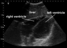
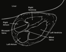
 procedure.") The Focused Assessment with Sonography for Trauma, (FAST) procedure. ROBERT H. WRIGLEY Professor Veterinary Diagnostic Imaging University of Sydney Veterinary Teaching Hospital Professor Emeritus Colorado
The Focused Assessment with Sonography for Trauma, (FAST) procedure. ROBERT H. WRIGLEY Professor Veterinary Diagnostic Imaging University of Sydney Veterinary Teaching Hospital Professor Emeritus Colorado
Bedside RUQ Ultrasound. Replace Formal ULS? Why Bedside ULS RUQ? RUQ Ultrasound. Bedside ULS is Limited, Goal-Directed
 Bedside RUQ Ultrasound RUQ Ultrasound Why do it How to do it Elizabeth Kwan UCSF Emergency Ultrasound Fellow Why Bedside ULS RUQ? Dx or Rule Out Acute Cholecystitis Cholelithiasis, Choledocolithiasis Earlier
Ex. 1 :Language of Anatomy
 Collin College BIOL 2401 : Human Anatomy & Physiology Ex. 1 :Language of Anatomy The Anatomical Position Used as a reference point when referring to specific areas of the human body Body erect Head and
L o o k L i s t e n F e e l S c a n. Your Pocus Cards For Your Every Day Scanning.
 L o o k L i s t e n F e e l S c a n Your Pocus Cards For Your Every Day Scanning E-FAST Extended Focused Assessment by Sonography in Trauma Subcostal Heart View Pleural Sliding on M-mode (Sea-shore sign)
Archiving in Qpath Defining Adequate
 General Archiving Information for QPath Users As you become familiar with Qpath and how to archive your clips you will want to be sure you are capturing good quality clips for review. The properly captured,
(FAST) Peter Logan FRCS(Ed) FFAEM FACEM David Lewis FRCS FFAEM. Focused Assessment with Sonography for Trauma
 Peter Logan FRCS(Ed) FFAEM FACEM David Lewis FRCS FFAEM. Focused Assessment with Sonography for Trauma") Focused Assessment with Sonography for Trauma (FAST) Peter Logan FRCS(Ed) FFAEM FACEM David Lewis FRCS FFAEM 1 (FAST) Introduction Physical examination of the abdomen in blunt trauma is subjective and
Focused Assessment with Sonography for Trauma (FAST) Peter Logan FRCS(Ed) FFAEM FACEM David Lewis FRCS FFAEM 1 (FAST) Introduction Physical examination of the abdomen in blunt trauma is subjective and
Ultrasound basics Part 1
 Ultrasound basics Part 1 'Ultrasound enhanced critical care medicine' Rohit Patel, MD University of Florida Health Director, Critical Care Ultrasound Surgical ICU Center for Intensive Care Gainesville,
Perioperative Ultrasonography Ehab Farag, MD, FRCA Hesham Elsharkawy David G. Anthony, M.D.
 Perioperative Ultrasonography Ehab Farag, MD, FRCA Hesham Elsharkawy David G. Anthony, M.D. Cleveland Clinic, Cleveland OH 1 Complications during central venous catheterization (CVC) occur 2% -15% of the
POCUS for the Internist: Lungs & Pericardial Effusions
 POCUS for the Internist: Lungs & Pericardial Effusions Jeremy S. Boyd, MD, FACEP Asst. Professor of Emergency Medicine Vanderbilt University Medical Illustrations courtesy of Robinson Ferre, MD, FACEP
Small animal point of care ultrasound techniques
 Small animal point of care ultrasound techniques The role of veterinary point of care ultrasound in determining the presence or absence of specific pathologies is examined by Jantina McMurray DVM; Søren
NON INVASIVE LIFE SAVERS. Ultrasound in PICU
 VOL 1 NO.1 Jan March 2014 54 Table 1. Selected Applications of Point-of-Care Ultrasonography, According to Medical Specialty. Specialty Ultrasound Applications Anesthesia Cardiology Guidance for vascular
Policies, Standards, and Guidelines. Guidelines for Abdominal Ultrasound Examination
 Policies, Standards, and Guidelines Guidelines for Abdominal Ultrasound Examination Approved by Council Feb 2018 Disclaimer and Copyright The ASUM Standards of Practice Board have made every effort to
Abdominal Ultrasound
 Abdominal Ultrasound Imaging Control Buttons Depth The organ imaged should take up 3/4 of the screen Frequency = Penetration Use high frequencies (harmonics) for fluid filled and superficial structures
Introduction. Chapter 1. Structure and Function. Introduction. Anatomy and Physiology Integrated. Anatomy and Physiology Integrated Anatomy
 Introduction Chapter 1 An Introduction to A&P Study strategies crucial for success Attend all lectures, labs, and study sessions Read your lecture and laboratory assignments before going to class or lab
Introduction to Anatomical Terms. Packet #3
 Introduction to Anatomical Terms Packet #3 Directional Terms Directional terms describe the positions of structures relative to other structures or locations in the body. Introduction Superior vs. Inferior
A Frame of Reference for Anatomical Study. Anatomy and Physiology Mr. Knowles Chapter 1 Liberty Senior High School
 A Frame of Reference for Anatomical Study Anatomy and Physiology Mr. Knowles Chapter 1 Liberty Senior High School Anatomical Terms of Direction and Position Created for communicating the direction and
Welcome to ANAT 10A! What is Anatomy? Different levels of Anatomy The Language of Anatomy Pearson Education, Inc.
 Welcome to ANAT 10A! What is Anatomy? Different levels of Anatomy The Language of Anatomy Introduction Anatomy means to dissect: (ANAT 10A) The study of internal & external body structures The study of
Anatomical Terminology
 Anatomical Terminology Dr. A. Ebneshahidi Anatomy Anatomy : is the study of structures or body parts and their relationships to on another. Anatomy : Gross anatomy - macroscopic. Histology - microscopic.
Brugge Mars 2009 P.R.E.P. Programme Rapide Échographie Polytraumatisé. Aalst - December 2009 C.F.F.E.
 Brugge Mars 2009 Programme Rapide Échographie Polytraumatisé www.ultrason.com Aalst - December 2009 C.F.F.E. PREVIOUSLY! About «formal ultrasonography»! Ultrasonography for the diagnosis! Performed by
Thoracic Ultrasound: Pictorial review of pneumothorax, the fastest and easiest method to diagnose.
 Thoracic Ultrasound: Pictorial review of pneumothorax, the fastest and easiest method to diagnose. Poster No.: C-1588 Congress: ECR 2014 Type: Educational Exhibit Authors: J. A. Guirola, V. Mayoral Campos,
Bedside ultrasound - Lung ultrasound in the Intensive Care Unit
 Bedside ultrasound - Lung ultrasound in the Intensive Care Unit Kishore K. Pichamuthu, Professor, Department of Critical Care, Christian Medical College, Vellore. Summary In an ICU setting, ultrasonographic
FHS Appendicitis US Protocol
 FHS Appendicitis US Protocol Reviewed By: Shireen Khan, MD; Sarah Farley, MD; Anna Ellermeier, MD Last Reviewed: May 2018 Contact: (866) 761-4200 **NOTE for all examinations: 1. If documenting possible
Caudal Edge of the Liver in the Right Upper Quadrant (RUQ) View Is the Most Sensitive Area for Free Fluid on the FAST Exam
 View Is the Most Sensitive Area for Free Fluid on the FAST Exam") Original Research Caudal Edge of the Liver in the Right Upper Quadrant (RUQ) View Is the Most Sensitive Area for Free Fluid on the FAST Exam Viveta Lobo, MD* Michelle Hunter-Behrend, MD* Erin Cullnan,
Original Research Caudal Edge of the Liver in the Right Upper Quadrant (RUQ) View Is the Most Sensitive Area for Free Fluid on the FAST Exam Viveta Lobo, MD* Michelle Hunter-Behrend, MD* Erin Cullnan,
Lung ultrasound in the critically ill patient Pleural Effusions
 Lung ultrasound in the critically ill patient Pleural Effusions Rohit Patel, MD University of Florida Health Director, Critical Care Ultrasound Surgical ICU Center for Intensive Care Gainesville, Florida
Abdominal ultrasound:
 Abdominal ultrasound: Non-traumatic acute abdomen Wittanee Na-ChiangMai, MD Department of Radiology ChiangMai University 26/04/2017 Contents Technique of examination Normal anatomy Emergency conditions
**Confirm accuracy of above with your instructor.** Revised 8/22/2017 1
 AP1 Lab 1 Cavities, Organs, Serous Membranes, Quadrants, Regions, and Directional Terms, Planes & Sections Project 1 Directional Terminology Step 1: Define/Describe what is known as the "ANATOMICAL POSITION."
The 2 nd Cambridge Advanced Emergency Ultrasound Course
 The 2 nd Cambridge Advanced Emergency Ultrasound Course Addenbrooke s Hospital Cambridge Sept 2008 1 2 Faculty! UK! USA! Australia! Toshiba! Emergency Medicine! Radiology 3 Programme! Day 1 Introduction
Radiological Investigations of Abdominal Trauma
 76 77 Investigations of Abdominal Trauma Introduction: Trauma to abdominal organs is a common cause of patient morbidity and mortality among trauma patients. Causes of abdominal trauma include blunt injuries,
2 Blunt Abdominal Trauma
 2 Blunt Abdominal Trauma Ricardo Ferrada, Diego Rivera, and Paula Ferrada Pearls and Pitfalls Patients suffering a high-energy trauma have solid viscera rupture in the abdomen and/or aortic rupture in
Ultrasound in abdominal trauma: an
 Archives of Emergency Medicine, 1988, 5, 26-33 Ultrasound in abdominal trauma: an alternative to peritoneal lavage J. A. CHAMBERS & W. J. PILBROW Accident Unit, Countess of Chester Hospital, Chester, England
ASSESSING THE PLAIN ABDOMINAL RADIOGRAPH M A A M E F O S U A A M P O F O
 ASSESSING THE PLAIN ABDOMINAL RADIOGRAPH M A A M E F O S U A A M P O F O Introduction The abdomen (less formally called the belly, stomach, is that part of the body between the thorax (chest) and pelvis,
RADIOLOGIC TECHNOLOGY (526)
") RADIOLOGIC TECHNOLOGY (526) 526-133 DMS General Procedures 2 Radiologic Technology (526) 1 526-130 Introduction to Diagnostic Medical Sonography This course introduces the student to the history of ultrasound
RADIOLOGIC TECHNOLOGY (526) 526-133 DMS General Procedures 2 Radiologic Technology (526) 1 526-130 Introduction to Diagnostic Medical Sonography This course introduces the student to the history of ultrasound
Children are not small adults Children are Not Small Adults Anatomic considerations Pliable bony & cartilagenous structures - Significant thoracic inj
 PEDIATRIC CHEST TRAUMA Children are not small adults Role of imaging Spectrum of injury Children are not small adults Children are Not Small Adults Anatomic considerations Pliable bony & cartilagenous
The FAST Exam! Dr. David Easton MD FRCPC Critical Care and Emergency Medicine University of Manitoba Canada
 The FAST Exam! Dr. David Easton MD FRCPC Critical Care and Emergency Medicine University of Manitoba Canada Dr. David Easton MD FRCPC Assistant Professor Section of Critical Care and Emergency Medicine
Case Conference. Discussion. Indications of Trauma Blue. Trauma Protocol In SKH. Trauma Blue VS. Trauma Red. Supervisor:VS 楊毓錚 Presenter:R1 周光緯
 Case Conference Supervisor:VS 楊毓錚 Presenter:R1 周光緯 Discussion 2010.7.14 2/81 Trauma Protocol In SKH Indications of Trauma Blue Trauma Blue VS. Trauma Red 3/81 Severe trauma mechanism : 1. Trauma to multiple
Role of Focused Assessment with Sonography for Trauma (FAST) and in abdominal trauma: Radiologist s perspective
 and in abdominal trauma: Radiologist s perspective") Original Research Article Role of Focused Assessment with Sonography for Trauma (FAST) and CT scan in abdominal trauma: Radiologist s perspective Nirav Patel 1*, Niket Domadia 2, Konark Sarvaiya 1, Anil
Original Research Article Role of Focused Assessment with Sonography for Trauma (FAST) and CT scan in abdominal trauma: Radiologist s perspective Nirav Patel 1*, Niket Domadia 2, Konark Sarvaiya 1, Anil
Contents& & & 1.! Ultrasound&basics& 1! 2.! Image&generation& 15!
 A l i n e press é % % % Contents& & & 1. Ultrasound&basics& 1 1.1. What,is,ultrasound?, 1 1.2. Ultrasound,probes,send,and,receive,ultrasound, 3 1.3. How,does,ultrasound,behave,travelling,through,tissue?,
Imaging of Thoracic Trauma: Tips and Traps. Arun C. Nachiappan, MD Associate Professor of Clinical Radiology University of Pennsylvania
 Imaging of Thoracic Trauma: Tips and Traps Arun C. Nachiappan, MD Associate Professor of Clinical Radiology University of Pennsylvania None Disclosures Objectives Describe blunt and penetrating traumatic
Chapter 1: The Human Organism
 Chapter 1: The Human Organism I. Anatomy and Physiology A. Anatomy - study of structure 1. Studying structural changes from conception to adulthood is called: 2. Embryology is the study of 3. The study
Free fluid accumulation following blunt abdominal trauma: potential for expansion of the FAST protocol
 Hong Kong Journal of Emergency Medicine Free fluid accumulation following blunt abdominal trauma: potential for expansion of the FAST protocol N Simpson, P Page, DM Taylor Objective: To determine sites
9. Which term refers to the back? A. Inferior B. Lateral C. Posterior D. Peripheral 10. The heart is to the lungs. A. dorsal B. superior C.
 1 Student: 1. Which term refers to the study of how an organ functions? A. Anatomy B. Physiology C. Ecology D. Homeostasis 2. Observing the parts of the brain would be part of the study of A. homeostasis.
Which Blunt Trauma Patients Should Be Studied by Abdominal CT?
 MDCT of Bowel and Mesenteric Injury: How Findings Influence Management 4 th Nordic Trauma Radiology Course 2006 4 th Nordic Trauma Radiology Course 2006 Stuart E. Mirvis, M.D., FACR Department of Radiology
Basic Training Programme. 16 Februrary 2018, ROTTERDAM. Pre and Post-Course Test Answers
 Basic Training Programme 16 Februrary 2018, ROTTERDAM Pre and Post-Course Test Answers Your details: Name: Conference registration number/ BT delegate number: Email address: Are you already performing
Pre Hospital Ultrasound: Direction for the future?
 Pre Hospital Ultrasound: Direction for the future? Craig Manifold, DO Medical Director San Antonio Fire Department San Antonio AirLIFE Historical Perspective Like most new technology, there is a risk
Semiotics in Radiology
 Adelino Santos Health Technology College Coimbra, Portugal Collaboration of António Agudo Student of Radiology College of Health Technology Coimbra, Portugal What are the most important points to evaluate
Introduction to The Human Body
 1 Introduction to The Human Body FOCUS: The human organism is often examined at seven structural levels: chemical, organelle, cell, tissue, organ, organ system, and the organism. Anatomy examines the structure
ULTRASOUND NOMENCLATURE
 Chapter 1: Ultrasound Nomenclature, Image Orientation, and Basic Instrumentation CYNTHIA SIKOWSKI Ultrasound waves are sound waves that have a frequency exceeding 20,000 Hz. When sound waves are transmitted
2. Blunt abdominal Trauma
 Abdominal Trauma 1. Evaluation and management depends on: a. Mechanism (Blunt versus Penetrating) b. Injury complex in addition to abdomen c. Haemodynamic stability assessment: i. Classically patient s
Children's (Pediatric) Ultrasound - Abdomen
 Ultrasound - Abdomen") Scan for mobile link. Children's (Pediatric) Ultrasound - Abdomen Children s (pediatric) ultrasound imaging of the abdomen is a safe, noninvasive test that uses sound waves to produce a clear picture of
Scan for mobile link. Children's (Pediatric) Ultrasound - Abdomen Children s (pediatric) ultrasound imaging of the abdomen is a safe, noninvasive test that uses sound waves to produce a clear picture of
Initially for cardiac echo Subsequent studies non-cardiac applications
 No disclosures But Heavy accent Initially for cardiac echo Subsequent studies non-cardiac applications 1973: Goldberg et al in JCUS 30 mediastinal masses in pts. age 1-84 yrs. 1977: Kangarloo et al in
Courtesy of Dr. Susan Maskel Western Connecticut State University
 Courtesy of Dr. Susan Maskel Western Connecticut State University 1 2 SECTION vs. PLANE SECTION = a cut PLANE: when the section is made through the body wall or through an organ, it is made along an imaginary
OVERVIEW. Need for USG. Weaning assessment. Mechanics of USG. Pneumonia / VAP. Principles of lung USG. Prone position ventilation assessment
 OVERVIEW Need for USG Mechanics of USG Principles of lung USG BLUE protocol Alveolar syndrome Interstitial syndrome Weaning assessment Pneumonia / VAP Prone position ventilation assessment ETT positioning
MDCT signs differentiating retroperitoneal and intraperitoneal lesions- diagnostic pearls
 MDCT signs differentiating retroperitoneal and intraperitoneal lesions- diagnostic pearls Poster No.: C-0987 Congress: ECR 2015 Type: Educational Exhibit Authors: D. V. Bhargavi, R. Avantsa, P. Kala; Bangalore/IN
performed to help sway the clinician in what the appropriate diagnosis is, which can substantially alter the treatment of management.
 Hello, I am Maura Polansky at the University of Texas MD Anderson Cancer Center. I am a Physician Assistant in the Department of Gastrointestinal Medical Oncology and the Program Director for Physician
Tubes and lines in neonatal chest radiograph
 Tubes and lines in neonatal chest radiograph Poster No.: C-1008 Congress: ECR 2014 Type: Educational Exhibit Authors: R. TUMMA, N. AHMED, V. Prasad ; Hyderabad/IN, 1 2 1 1 2 HYDERABAD, ANDHRA PRADESH/IN
MAKERERE UNIVERSITY DR PETER CLAVER MBIDDE MUSIITWA. MBchB (MUST)
") MAKERERE UNIVERSITY SONOGRAPHIC SCORING FOR OPERATING ROOM TRIAGE IN TRAUMA; ACCURACY FOR THERAPEUTIC LAPAROTOMY AMONG BLUNT ABDOMINAL TRAUMA PATIENTS IN MULAGO HOSPITAL. BY DR PETER CLAVER MBIDDE MUSIITWA
MAKERERE UNIVERSITY SONOGRAPHIC SCORING FOR OPERATING ROOM TRIAGE IN TRAUMA; ACCURACY FOR THERAPEUTIC LAPAROTOMY AMONG BLUNT ABDOMINAL TRAUMA PATIENTS IN MULAGO HOSPITAL. BY DR PETER CLAVER MBIDDE MUSIITWA
Dr.Susan Maskel. BIO 105 Western Connecticut State University A&P I BIO 211 Naugatuck Valley Community College A&P I.
 1 Please wait 20 seconds before starting slide show. Mouse click to advance. Arrow keys etc.also work. Dr.Susan Maskel BIO 105 Western Connecticut State University A&P I BIO 211 Naugatuck Valley Community
Urogenital Injuries The role of radiology
 Urogenital Injuries The role of radiology NORDTER 7 th Nordic Trauma Radiology Course Helsinki, Finland May 21-24, 2012 Johann Baptist Dormagen, MD, PhD Oslo University Hospital, Norway Kidney injuries
LAB 1: INTRODUCTION TO ANATOMY AND PHYSIOLOGY
 LAB 1: INTRODUCTION TO ANATOMY AND PHYSIOLOGY ANSWERS TO Pre- Lab Assignments Pre-Lab Activity 1: 1. b 2. a. 3 b. 7 c. 5 d. 6 e. 4 f. 1 g. 8 h. 2 i. 10 j. 9 3. a. frontal b. cervical c. antecubital d.
Pulmonary Embolism. Thoracic radiologist Helena Lauri
 Pulmonary Embolism Thoracic radiologist Helena Lauri 8.5.2017 Statistics 1-2 out of 1000 adults annually are diagnosed with deep vein thrombosis (DVT) and/or pulmonary embolism (PE) About half of patients
Scientific Exhibit Authors: V. Moustakas, E. Karallas, K. Koutsopoulos ; Rodos/GR, 2
 Diagnosis of Acute Appendicitis: the role of Color Doppler Ultrasound as first-line imaging method and evaluation of the higher diagnostic performances of CT against its disadvantages. Poster No.: C-0708
Interpreting thoracic x-ray of the supine immobile patient: Syllabus
 Interpreting thoracic x-ray of the supine immobile patient: Syllabus Johannes Godt Dep. of Radiology and Nuclear Medicine Oslo University Hospital Ullevål NORDTER 2017, Helsinki Content - Why bedside chest
My Patient Has Abdominal Pain PoCUS of the Biliary Tract and the Urinary Tract
 My Patient Has Abdominal Pain PoCUS of the Biliary Tract and the Urinary Tract Objectives PoCUS for Biliary Disease PoCUS for Renal Colic PoCUS for Urinary Retention Biliary Disease A patient presents
Positron Emission Tomography Computed Tomography (PET/CT)
") Positron Emission Tomography Computed Tomography (PET/CT) What is Positron Emission Tomography Computed Tomography (PET/CT) Scanning? What are some common uses of the procedure? How should I prepare for
Positron Emission Tomography Computed Tomography (PET/CT) What is Positron Emission Tomography Computed Tomography (PET/CT) Scanning? What are some common uses of the procedure? How should I prepare for
Ultrasound (US) evaluation of peritoneal thickness in children and young patients on peritoneal dialysis (PD): A single centre experience
 evaluation of peritoneal thickness in children and young patients on peritoneal dialysis (PD): A single centre experience") Ultrasound (US) evaluation of peritoneal thickness in children and young patients on peritoneal dialysis (PD): A single centre experience Poster No.: C-2812 Congress: ECR 2010 Type: Scientific Exhibit
Ultrasound (US) evaluation of peritoneal thickness in children and young patients on peritoneal dialysis (PD): A single centre experience Poster No.: C-2812 Congress: ECR 2010 Type: Scientific Exhibit
The Human Body: An Orientation
 The Human Body: An Orientation Body standing upright Anatomical Position feet slightly apart palms facing forward thumbs point away from body Directional Terms Superior and inferior toward and away from
Elastography in the. technically difficult patient. EPIQ ultrasound system. Ultrasound
 Ultrasound Elastography in the technically difficult patient EPIQ ultrasound system Chairman Department of Diagnostic Radiology Allegheny General Hospital Pittsburgh, PA, USA You can offer more information
An abdominal ultrasound produces a picture of the organs and other structures in the upper abdomen.
 Scan for mobile link. Ultrasound - Abdomen Ultrasound imaging of the abdomen uses sound waves to produce pictures of the structures within the upper abdomen. It is used to help diagnose pain or distention