Functional Chest MRI in Children Hyun Woo Goo
|
|
|
- Jane Horn
- 6 years ago
- Views:
Transcription
1 Functional Chest MRI in Children Hyun Woo Goo Department of Radiology and Research Institute of Radiology Asan Medical Center, University of Ulsan College of Medicine, Seoul, Korea
2 No ionizing radiation Excellent soft tissue contrast High temporal resolution Easier implementation of dynamic protocols
3 Low signal-to-noise ratio (SNR) due to the low proton density of the lung Motion artifacts from respiration & cardiac pulsation Long examination time Susceptibility artifacts from multiple air-tissue interfaces
4 Parallel imaging technique to accelerate scan speed Multi (e.g. 32)-channel body-array coil to increase SNR Better data acquisition (e.g. radial, spiral) & reconstruction schemes
5 Respiratory triggering using a pressure-sensing belt Respiratory gating using a navigator echo Saturation bands to suppress artifacts Signal averaging Motion-insensitive pulse sequences Vector ECG triggering
6 High SNR Greater susceptibility artifacts Much more motion sensitive Overall, no apparent benefits In the future,
7 Complex functions, including ventilation, perfusion, respiratory motion, gas exchange Anatomic chest MRI: difficult Functional chest MRI: much more challenging
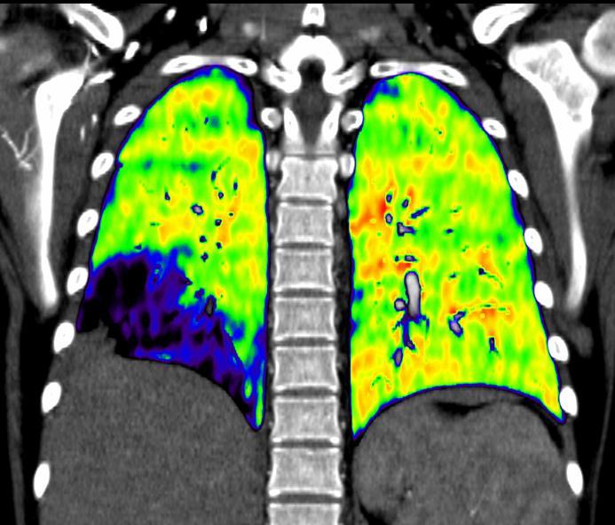
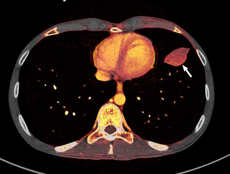
8 Lung perfusion Lung ventilation Mass evaluation Respiratory mechanics Lung stiffness


9 Contrast-enhanced time-resolved MRA Qualitative or quantitative assessment Correlation analysis: increased SNR Respiratory registration or motion correction


10 Arterial spin labeling (ASL) techniques No external contrast agent Animal experiments, volunteer studies Low SNR, further technical developments SVC LPA RPA IVC Goo HW (Unpublished data)


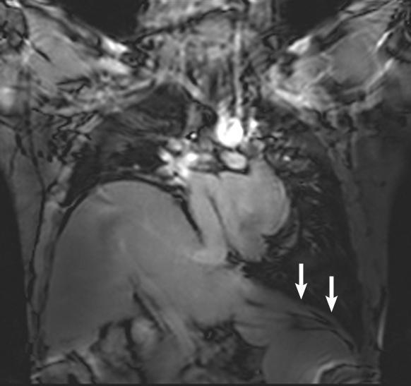
11 Non-contrast Lung Perfusion MRI Using ECG-triggered TSE Sequence A. Systole B. Diastole B - A


12 Phase contrast imaging Quantitative measurement of pulmonary blood flow Flow-curve pattern analysis No direct information on lung tissue perfusion Normal PA Pattern PA Hypertension Proximal PA Stenosis
13 Oxygen-enhanced MRI Low SNR Procedural difficulties in children Hyperpolarized gas MRI Very restricted availability Procedural difficulties in children


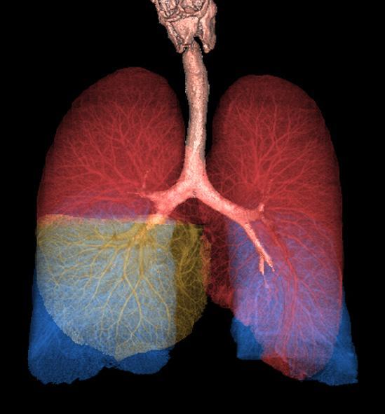
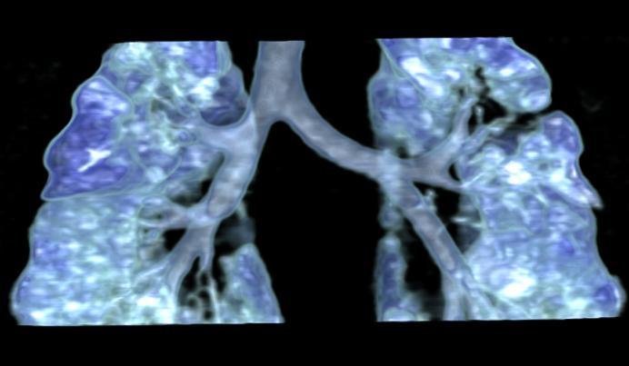
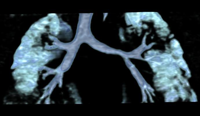
14 Easy & promising Free-breathing, untriggered acquisition of balanced SSFP for 4 min Perfusion-weighted Good agreement with ventilation & perfusion SPECT Ventilation-weighted
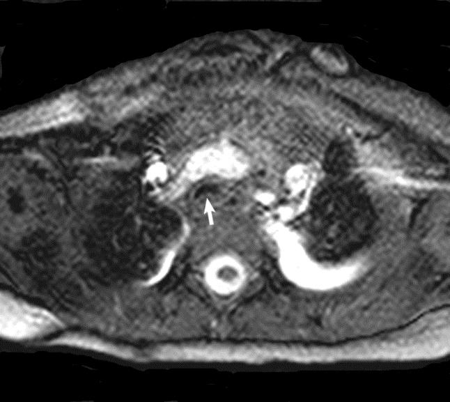
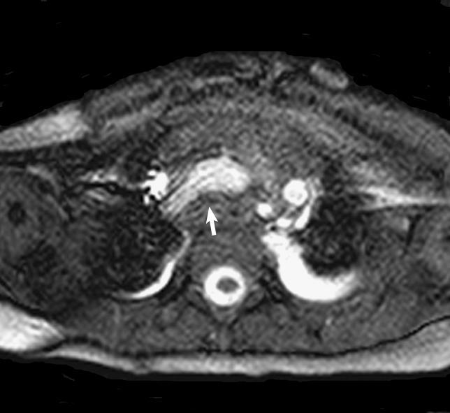
15 Dynamic contrast-enhanced (DCE) imaging Tumor vascular density, permeability (K trans ) Tumor grade, treatment responses Respiratory registration or motion correction Diffusion-weighted imaging Water diffusivity within tissues Tumor cellularity, treatment responses Degraded image quality by motion artifacts
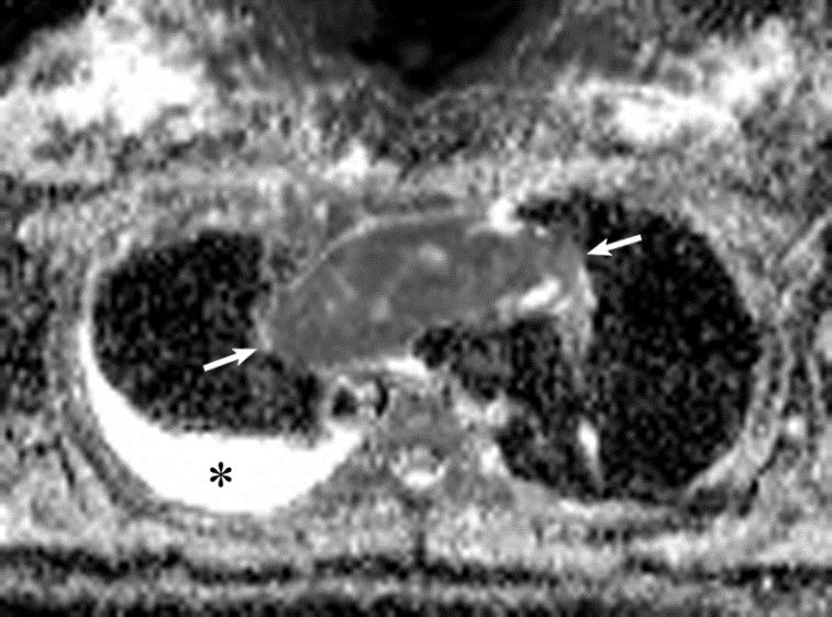
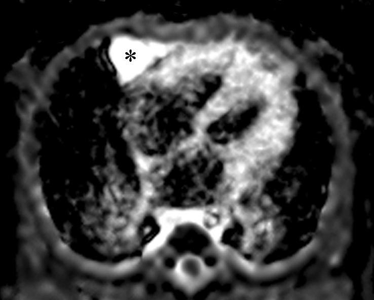


16 Qualitative Quantitative Lymphoma Benign cyst Mature cystic teratoma
17 Real-time dynamic MRI (B-SSFP) Motion of lung, airway, chest wall, and diaphragm Restricted motion in both tumor infiltration & benign adhesion Measured lung volume parameters: a good correlation with pulmonary function test Tracheomalacia, diaphragm palsy/paralysis


18 Tracheomalacia Right Diaphragm Palsy Inspiration Expiration

19 MR elastography Non-invasive measurement of shear stiffness of human lungs in 10 volunteers Mariappan et al. J Magn Reson Imaging 2011 Further technical refinement, particularly in children

20
21 Functional chest MR imaging has potential to detect early or mild change of lung diseases, to quantify altered lung function caused by lung diseases, to give insights into the pathophysiology of lung diseases, and to evaluate the effectiveness of established and new therapies. A majority of these imaging techniques can be applied to pediatric population.
22 Functional Chest MRI in Children Hyun Woo Goo Department of Radiology and Research Institute of Radiology Asan Medical Center, University of Ulsan College of Medicine, Seoul, Korea
Fulfilling the Promise
 Fulfilling the Promise of Cardiac MR Non-contrast, free-breathing technique generates comprehensive evaluation of the coronary arteries By Maggie Fung, MR Cardiovascular Clinical Development Manager; Wei
Non Contrast MRA. Mayil Krishnam. Director, Cardiovascular and Thoracic Imaging University of California, Irvine
 Non Contrast MRA Mayil Krishnam Director, Cardiovascular and Thoracic Imaging University of California, Irvine No disclosures Non contrast MRA-Why? Limitations of CTA Radiation exposure Iodinated contrast
MRI protocol for post-repaired TOF
 2012 NASCI MRI protocol for post-repaired TOF Taylor Chung, M.D. Associate Director, Body and Cardiovascular Imaging Department of Diagnostic Imaging Children s Hospital & Research Center Oakland Oakland,
DISCLOSURE OBJECTIVES PULMONARY VEIN STENOSIS DIAGNOSTIC TOOLS. Echo with Doppler Catheterization with angiography CT angiography MRI
 1 2 ND INTERNATIONAL CONFERENCE: NEONATAL AND CHILDHOOD PULMONARY VASCULAR DISEASE, MARCH 13-14, 2009, SAN FRANCISCO, USA PATHOPHYSIOLOGY OF PULMONARY VEIN FLOW: IMAGING NORMAL AND ABNORMAL PULMONARY VEIN
Advances in MRI for Radiation Therapy
 Advances in MRI for Radiation Therapy Jing Cai, PhD, DABR Associate Professor Department of Radiation Oncology Duke University Medical Center, Durham NC Advances in MRI Structural Imaging Fast Imaging
Fundamentals, Techniques, Pitfalls, and Limitations of MDCT Interpretation and Measurement
 Fundamentals, Techniques, Pitfalls, and Limitations of MDCT Interpretation and Measurement 3 rd Annual Imaging & Physiology Summit November 20-21, 21, 2009 Seoul, Korea Wm. Guy Weigold, MD, FACC Cardiovascular
Raja Muthupillai, PhD. Department of Diagnostic and Interventional Radiology St. Luke s Episcopal Hospital. Research Support: Philips Healthcare
 3D Cardiac Imaging Raja Muthupillai, PhD Department of Diagnostic and Interventional Radiology St. Luke s Episcopal Hospital Houston, TX Disclosures Research Support: Philips Healthcare This presentation
Abdominal applications of DWI
 Postgraduate course, SPR San Antonio (Texas), May 14-15, 2013 Abdominal applications of DWI Rutger A.J. Nievelstein Wilhelmina Children s s Hospital, Utrecht (NL) Outline What is DWI? How to perform? Challenges
1Pulse sequences for non CE MRA
 MRI: Principles and Applications, Friday, 8.30 9.20 am Pulse sequences for non CE MRA S. I. Gonçalves, PhD Radiology Department University Hospital Coimbra Autumn Semester, 2011 1 Magnetic resonance angiography
MR Advance Techniques. Cardiac Imaging. Class IV
 MR Advance Techniques Cardiac Imaging Class IV Heart The heart is a muscular organ responsible for pumping blood through the blood vessels by repeated, rhythmic contractions. Layers of the heart Endocardium
MR Advance Techniques. Vascular Imaging. Class II
 MR Advance Techniques Vascular Imaging Class II 1 Vascular Imaging There are several methods that can be used to evaluate the cardiovascular systems with the use of MRI. MRI will aloud to evaluate morphology
Introduction. Cardiac Imaging Modalities MRI. Overview. MRI (Continued) MRI (Continued) Arnaud Bistoquet 12/19/03
 MRI (Continued) Arnaud Bistoquet 12/19/03") Introduction Cardiac Imaging Modalities Arnaud Bistoquet 12/19/03 Coronary heart disease: the vessels that supply oxygen-carrying blood to the heart, become narrowed and unable to carry a normal amount
Introduction Cardiac Imaging Modalities Arnaud Bistoquet 12/19/03 Coronary heart disease: the vessels that supply oxygen-carrying blood to the heart, become narrowed and unable to carry a normal amount
General Cardiovascular Magnetic Resonance Imaging
 2 General Cardiovascular Magnetic Resonance Imaging 19 Peter G. Danias, Cardiovascular MRI: 150 Multiple-Choice Questions and Answers Humana Press 2008 20 Cardiovascular MRI: 150 Multiple-Choice Questions
Perfusion Physics. ICMRI2018 March 29-31, 2018 Grand Hilton Hotel, Seoul, Korea. Asian Forum Ⅱ: Perfusion MRI SY24-1.
 SY24-1 Perfusion Physics Hiroyuki Kabasawa MR Collaborations and Development, GE Healthcare, Tokyo, Japan Perfusion is referred as the blood supply to micro capillary in tissue. Perfusion parameter such
How I do it: Non Contrast-Enhanced MR Angiography (syngo NATIVE)
") Clinical How-I-do-it Cardiovascular How I do it: Non Contrast-Enhanced MR Angiography (syngo NATIVE) Manuela Rick, Nina Kaarmann, Peter Weale, Peter Schmitt Siemens Healthcare, Erlangen, Germany Introduction
Clinical How-I-do-it Cardiovascular How I do it: Non Contrast-Enhanced MR Angiography (syngo NATIVE) Manuela Rick, Nina Kaarmann, Peter Weale, Peter Schmitt Siemens Healthcare, Erlangen, Germany Introduction
Non-Contrast MRA. How and When 1996! Why Non-Contrast MRA? Angiography: What are our goals? Inflow Techniques Differences in excitation hx
 A major teaching hospital of Harvard Medical School Angiography: What are our goals? Non-Contrast MRA: How and When Neil M. Rofsky, M.D. Professor of Radiology, Harvard Medical School Director of MRI &
ASL BASICS II. Learning Objectives. Outline. Acquisition. M. A. Fernández-Seara, Ph. D. Arterial spin labeled perfusion MRI: basic theory
 Acquisition ASL BASICS II M. A. Fernández-Seara, Ph. D. Neuroimaging Laboratory Center for Applied Medical Research University of Navarra Pamplona, Spain Outline Arterial spin labeled perfusion MRI: basic
Can SCMR CMR protocol recommendations
 Can SCMR CMR protocol recommendations V1.3 - April 2009 CanSCMR CMR Protocol and SOP Recommendation 2009 (15 minutes) 2 Planning of LV fct. real time multiple axes Realtime 3 cine long axis 6 long axes
Imaging Emphysema 3-Helium MR Imaging
 Imaging Emphysema 3-Helium MR Imaging Edwin J.R. van Beek MD PhD MEd FRCR Professor of Radiology and Medicine Carver College of Medicine, University of Iowa, USA. Permanent Visiting Professor of Radiology,
Master Thesis in Radiation Physics 09/10 Annie Olsson. Supervisors: Frank Risse Lars E. Olsson. Imaging centre AstraZeneca R&D Mölndal
 Determination of a preclinical protocol for quantitative measurements of perfusion and permeability in the rat lung using dynamic contrast enhanced-mri Master Thesis in Radiation Physics 9/1 Annie Olsson
Cardiovascular Imaging
 Cardiovascular Imaging Cardiovascular Imaging Cardio and Vascular Imaging Vascularization / Angiogenesis Cardiovascular Imaging metabolic imaging of the heart myocardial perfusion imaging Cardiac CT Vascularization
T2, T2*, ute. Yeo Ju Kim. Radiology, Inha University Hospital, Incheon, Korea
 SY28-1 T2, T2*, ute Yeo Ju Kim Radiology, Inha University Hospital, Incheon, Korea T2 relaxation times relate to the rate of transverse magnetization decay, caused by the loss of phase coherence induced
Initial Clinical Experience of TOSHIBA 3T MRI
 The 21st Conference of the Japanese Society of Cardiovascular Imaging & Dynamics Sponsored Seminar The Leading Edge of CT/MRI Diagnosis for the Cardiovascular System Initial Clinical Experience of TOSHIBA
Disclosures. GETTING TO THE HEART OF THE MATTER WITH MULTIMODALITY CARDIAC IMAGING Organ Review Meeting 25 September. Overview
 GETTING TO THE HEART OF THE MATTER WITH MULTIMODALITY CARDIAC IMAGING Organ Review Meeting 25 September Disclosures None relevant to this presentation Mini Pakkal Assistant Professor of Radiology University
Publication for the Philips MRI Community Issue 39 December 2009
 FieldStrength Publication for the Philips MRI Community Issue 39 December 2009 32-channel coil boosts 3.0T neuro imaging at Kennedy Krieger Kennedy Krieger Institute sees significantly better fmri, DTI,
Anatomical and Functional MRI of the Pancreas
 Anatomical and Functional MRI of the Pancreas MA Bali, MD, T Metens, PhD Erasme Hospital Free University of Brussels Belgium mbali@ulb.ac.be Introduction The use of MRI to investigate the pancreas has
CARDIAC MRI. Cardiovascular Disease. Cardiovascular Disease. Cardiovascular Disease. Overview
 CARDIAC MRI Dr Yang Faridah A. Aziz Department of Biomedical Imaging University of Malaya Medical Centre Cardiovascular Disease Diseases of the circulatory system, also called cardiovascular disease (CVD),
Clinical Applications
 C H A P T E R 16 Clinical Applications In selecting pulse sequences and measurement parameters for a specific application, MRI allows the user tremendous flexibility to produce variations in contrast between
MR Advance Techniques. Cardiac Imaging. Class III
 MR Advance Techniques Cardiac Imaging Class III Black Blood Imaging & IR Blue= O2 poor blood Red=O2 rich blood Inversion pulses can produce black blood imaging in GRE pulse sequences. Specially on the
MSc. eng. Artur Wujcicki. Ventilation-perfusion lung imaging by filtering MR image sequence
 TECHNICAL UNIVERSITY OF LODZ FACULTY OF ELECTRICAL, ELECTRONIC, COMPUTER AND CONTROL ENGINEERING INSTITUTE OF ELECTRONICS MSc. eng. Artur Wujcicki Abstract of Ph.D. Thesis Ventilation-perfusion lung imaging
4D-MRI for Radiotherapy of moving Tumours: Latest developments, comparison to 4D-CT. J. Biederer Vancouver, August 2, 2011
 4D-MRI for Radiotherapy of moving Tumours: Latest developments, comparison to 4D-CT Vancouver, August 2, 2011 Indications for Radiotherapy of Lung Cancer primary radiotherapy in NCSLC - stage III Oertel
Qualitative and Quantitative Assessment of Perfusion
 APCDE 2011 Qualitative and Quantitative Assessment of Perfusion Hyun Ju Yoon Chonnam National University Hospital Gwangju, Korea ISCHEMIC CASCADE Blood flow mismatch Perfusion defects on nuclear imaging,
Cardiac Imaging Tests
 Cardiac Imaging Tests http://www.medpagetoday.com/upload/2010/11/15/23347.jpg Standard imaging tests include echocardiography, chest x-ray, CT, MRI, and various radionuclide techniques. Standard CT and
Measurement of Respiratory and Cardiac Motion Using a Multi Antenna Continuous Wave Radar Operating in the Near Field
 Measurement of Respiratory and Cardiac Motion Using a Multi Antenna Continuous Wave Radar Operating in the Near Field Florian Pfanner 1,2, Thomas Allmendinger 2, Thomas Flohr 2, and Marc Kachelrieß 1,3
PHYSICS OF MRI ACQUISITION. Alternatives to BOLD for fmri
 PHYSICS OF MRI ACQUISITION Quick Review for fmri HST-583, Fall 2002 HST.583: Functional Magnetic Resonance Imaging: Data Acquisition and Analysis Harvard-MIT Division of Health Sciences and Technology
Speed, Comfort and Quality with NeuroDrive
 Speed, Comfort and Quality with NeuroDrive Echelon Oval provides a broad range of capabilities supporting fast, accurate diagnosis of brain conditions and injuries. From anatomical depiction to vascular
Magnetic Resonance Angiography
 Magnetic Resonance Angiography 1 Magnetic Resonance Angiography exploits flow enhancement of GR sequences saturation of venous flow allows arterial visualization saturation of arterial flow allows venous
MR Assessment of Myocardial Viability
 MR Assessment of Myocardial Viability Definition of Viability Clinical Metabolism: Presence of glucose uptake Perfusion / Perfusion reserve Morphology: Wall thickness, wall thickening Contractility: Recovery
iu22 Liver Shear Wave ElastPQ
 iu22 Liver Shear Wave ElastPQ Clinical Case Study Lucy Wang Clinical Application Specialist ASEAN Case Study: History: 58-year-old male patient, hepatitis B virus (HBV) carrier, with non clinical symptoms
Use of Cardiac Computed Tomography for Ventricular Volumetry in Late Postoperative Patients with Tetralogy of Fallot
 Korean J Thorac Cardiovasc Surg 2017;50:71-77 ISSN: 2233-601X (Print) ISSN: 2093-6516 (Online) CLINICAL RESEARCH https://doi.org/10.5090/kjtcs.2017.50.2.71 Use of Cardiac Computed Tomography for Ventricular
Abdominal MRI Techniques in Pediatric Oncology
 Abdominal MRI Techniques in Pediatric Oncology Jonathan R. Dillman, M.D. Assistant Professor Departments of Radiology & Urology Section of Pediatric Radiology C.S. Mott Children s Hospital Disclosures
MRI Assessment of the Right Ventricle and Pulmonary Blood Flow, Perfusion and Ventilation
 MRI Assessment of the Right Ventricle and Pulmonary Blood Flow, Perfusion and Ventilation Dr. Richard Thompson Department of Biomedical Engineering University of Alberta Heart and Lung Imaging Many Constantly
8/4/2016. Optimizing Pediatric Cardiovascular MRI. Disclosure. Outline. Jie Deng, PhD, DABMP, Cynthia Rigsby, MD
 Optimizing Pediatric Cardiovascular MRI Jie Deng, PhD, DABMP, Cynthia Rigsby, MD Department of Medical Imaging Radiology, Feinberg School of Medicine, Northwestern University Aug 4 th, 2016 Disclosure
Cardiac MRI at 7T Syllabus contribution: Matthew Robson
 Cardiac MRI at 7T Syllabus contribution: Matthew Robson Field strength escalation has occurred over the last 10 years. Whilst 1.5T remains the most prevalent field strength the loss of 1T and the popularity
MR coronary artery imaging with 3D motion adapted gating (MAG) in comparison to a standard prospective navigator technique
 in comparison to a standard prospective navigator technique") Journal of Cardiovascular Magnetic Resonance (2005) 7, 793 797 Copyright D 2005 Taylor & Francis Inc. ISSN: 1097-6647 print / 1532-429X online DOI: 10.1080/10976640500287547 ANGIOGRAPHY MR coronary artery
Journal of Cardiovascular Magnetic Resonance (2005) 7, 793 797 Copyright D 2005 Taylor & Francis Inc. ISSN: 1097-6647 print / 1532-429X online DOI: 10.1080/10976640500287547 ANGIOGRAPHY MR coronary artery
High Field MR of the Spine
 Department of Radiology University of California San Diego 3T for MR Applications Advantages High Field MR of the Spine Increased signal-to-noise Better fat suppression Increased enhancement with gadolinium
Pulmonary Embolism. Thoracic radiologist Helena Lauri
 Pulmonary Embolism Thoracic radiologist Helena Lauri 8.5.2017 Statistics 1-2 out of 1000 adults annually are diagnosed with deep vein thrombosis (DVT) and/or pulmonary embolism (PE) About half of patients
ASL Perfusion Imaging: Concepts and Applications
 ASL Perfusion Imaging: Concepts and Applications David C. Alsop, Ph.D. Beth Israel Deaconess Medical Center and Harvard Medical School, Boston USA INTRODUCTION Arterial Spin Labeling (ASL) perfusion imaging
PINPOINTING RADIATION THERAPY WITH THE PRECISION OF MR.
 GE Healthcare PINPOINTING RADIATION THERAPY WITH THE PRECISION OF MR. MR Radiation Oncology Suite MAXIMIZE YOUR PRECISION. HELP MINIMIZE PATIENT COMPLICATIONS. Our goal in MR radiation oncology is to
MRI/MRS Biomarkers. Robert E. Lenkinski, Ph.D.
 MRI/MRS Biomarkers Robert E. Lenkinski, Ph.D. Disclosure GE Healthcare-Research Grant Aspect MR-Scientific Advisor Aposense-Scientific Advisor Brainwatch-Scientific Advisor I will be discussing off-label
On the feasibility of speckle reduction in echocardiography using strain compounding
 Title On the feasibility of speckle reduction in echocardiography using strain compounding Author(s) Guo, Y; Lee, W Citation The 2014 IEEE International Ultrasonics Symposium (IUS 2014), Chicago, IL.,
Objectives. CMR Volumetric Analysis 8/25/11. CMR Volumetric Analysis Technique. Cardiac imaging plane acquisition. CMR Volumetric Analysis
 Objectives Cynthia K. Rigsby Children s Memorial Hospital Chicago, IL CMR volumetric analysis Techniques Normalized data Sources of error CMR phase contrast flow analysis Techniques What we can do with
Objectives 8/17/2011. Challenges in Cardiac Imaging. Challenges in Cardiac Imaging. Basic Cardiac MRI Sequences
 8/17/2011 Traditional Protocol Model for Tomographic Imaging Cardiac MRI Sequences and Protocols Frandics Chan, M.D., Ph.D. Stanford University Medical Center Interpretation Lucile Packard Children s Hospital
6/23/2009. Inversion Recovery (IR) Techniques and Applications. Variations of IR Technique. STIR, FLAIR, TI and TI Null. Applications of IR
 Techniques and Applications. Variations of IR Technique. STIR, FLAIR, TI and TI Null. Applications of IR") The Anatomy of Basic R Pulse Sequences Inversion Recovery () Techniques and Applications Chen Lin, PhD Indiana University School of edicine & Clarian Health Partners agnetization Preparation Section Chemical
The Anatomy of Basic R Pulse Sequences Inversion Recovery () Techniques and Applications Chen Lin, PhD Indiana University School of edicine & Clarian Health Partners agnetization Preparation Section Chemical
Cor pulmonale. Dr hamid reza javadi
 1 Cor pulmonale Dr hamid reza javadi 2 Definition Cor pulmonale ;pulmonary heart disease; is defined as dilation and hypertrophy of the right ventricle (RV) in response to diseases of the pulmonary vasculature
RECENT ADVANCES IN CLINICAL MR OF ARTICULAR CARTILAGE
 In Practice RECENT ADVANCES IN CLINICAL MR OF ARTICULAR CARTILAGE By Atsuya Watanabe, MD, PhD, Director, Advanced Diagnostic Imaging Center and Associate Professor, Department of Orthopedic Surgery, Teikyo
Multiple Gated Acquisition (MUGA) Scanning
 Scanning") Multiple Gated Acquisition (MUGA) Scanning Dmitry Beyder MPA, CNMT Nuclear Medicine, Radiology Barnes-Jewish Hospital / Washington University St. Louis, MO Disclaimers/Relationships Standard of care research
Multiple Gated Acquisition (MUGA) Scanning Dmitry Beyder MPA, CNMT Nuclear Medicine, Radiology Barnes-Jewish Hospital / Washington University St. Louis, MO Disclaimers/Relationships Standard of care research
Perfusion MRI. Youngkyoo Jung, PhD Associate Professor Radiology, Biomedical Engineering, and Clinical & Translational Science Institute
 Perfusion MRI Youngkyoo Jung, PhD Associate Professor Radiology, Biomedical Engineering, and Clinical & Translational Science Institute Perfusion The delivery of blood to a capillary bed in tissue Perfusion
Outline. Why Image Animals?
 Small Animal Magnetic Resonance Imaging: Current Trends, Challenges and Perspectives for Pathological Imaging C. Chad Quarles Vanderbilt University Institute of Imaging Science Outline Why image animals?
Go With The Flow: Role of 4D Flow Imaging
 4D Flow Go With The Flow: Role of 4D Flow Imaging Niti R. Aggarwal, MD Associate Director of Cardiac MRI Assistant Professor of Medicine & Radiology University of Wisconsin Madison Disclosures GE Healthcare
What effects will proximal or distal disease have on a waveform?
 Spectral Doppler Interpretation Director of Ultrasound Education & Quality Assurance Baylor College of Medicine Division of Maternal-Fetal Medicine Maternal Fetal Center Imaging Manager Texas Children
Isolated congenital coronary anomalies: Evaluation by multislice-ct or MRI
 Isolated congenital coronary anomalies: Evaluation by multislice-ct or MRI B.K. Velthuis, Dept. of Radiology UMC Utrecht, the Netherlands ESC 2010 Coronary artery anomalies CAA Uncommon 0.3-5% normal population
Tips and Tricks of State of the art MRA
 Tips and Tricks of State of the art MRA Mayil Krishnam, MD,MBA, MRCP,FRCR(UK) Professor of Radiology Director, Cardiovascular and Thoracic Imaging University of California, Irvine Objectives Technical
Why Cardiac MRI? Presented by:
 Why Cardiac MRI? Presented by: Lisa G. Carkner, MD, FACC 1 Disclosures I have no financial disclosures Objectives Review basic principles of Cardiac MRI. What patient characteristics do I need to consider
Cardiac magnetic resonance (CMR) imaging is widely considered
 imaging is widely considered") ORIGINAL ARTICLE Motion-Corrected Real-Time Cine Magnetic Resonance Imaging of the Heart Initial Clinical Experience Amir Ali Rahsepar, MD,* Haris Saybasili, PhD, Ahmadreza Ghasemiesfe, MD,* Ryan S. Dolan,
ORIGINAL ARTICLE Motion-Corrected Real-Time Cine Magnetic Resonance Imaging of the Heart Initial Clinical Experience Amir Ali Rahsepar, MD,* Haris Saybasili, PhD, Ahmadreza Ghasemiesfe, MD,* Ryan S. Dolan,
Arterial Spin Labeling in Body MR
 Arterial Spin Labeling in Body MR Neil M. Rofsky, MD FACR, FISMRM, FSCBTMR Department of Radiology and Advanced Imaging Research Center None Disclosures Acknowledgements Ananth J. Madhuranthakam, Ph.D.
Newborn Hypoxic Ischemic Brain Injury. Hisham Dahmoush, MBBCh FRCR Lucile Packard Children s Hospital at Stanford
 Newborn Hypoxic Ischemic Brain Injury Hisham Dahmoush, MBBCh FRCR Lucile Packard Children s Hospital at Stanford NO DISCLOSURES INTRODUCTION Neonatal hypoxic-ischemic encephalopathy (HIE) is a major cause
Time-Of-Flight MRA. Faculty Disclosures Vincent B. Ho, M.D. Presentation Objectives. MRA Techniques. Pros and Cons of MRA
 Faculty Disclosures Vincent B. Ho, M.D. MR Angiography Techniques and Pitfalls Financial Disclosure Grant/Research Support General Electric Medical Systems Off-Label/Investigational Drug Use Dr. Ho will
Cardiac MRI in ACHD What We. ACHD Patients
 Cardiac MRI in ACHD What We Have Learned to Apply to ACHD Patients Faris Al Mousily, MBChB, FAAC, FACC Consultant, Pediatric Cardiology, KFSH&RC/Jeddah Adjunct Faculty, Division of Pediatric Cardiology
Handzettel 1. Multiparametric Functional Imaging in Radiation Therapy. Functional and Quantitative Imaging with MR
 Multiparametric Functional Imaging in Radiation Therapy Himanshu Bhat, Ph.D. Siemens Healthcare MR in RT Adding valuable information on tissue properties CT provides: Geometric accuracy Delineation of
Essentials of Clinical MR, 2 nd edition. 99. MRA Principles and Carotid MRA
 99. MRA Principles and Carotid MRA As described in Chapter 12, time of flight (TOF) magnetic resonance angiography (MRA) is commonly utilized in the evaluation of the circle of Willis. TOF MRA allows depiction
PET-MRI in Cardiac Imaging: Initial Experience
 PET-MRI in Cardiac Imaging: Initial Experience Mallinckrodt Institute of Radiology Washington University School of Medicine Pamela K. Woodard, M.D. Professor of Radiology and Biomedical Engineering Head,
In vivo diffusion tensor imaging (DTI) of articular cartilage as a biomarker for osteoarthritis
 of articular cartilage as a biomarker for osteoarthritis") In vivo diffusion tensor imaging (DTI) of articular cartilage as a biomarker for osteoarthritis Jose G. Raya 1, Annie Horng 2, Olaf Dietrich 2, Svetlana Krasnokutsky 3, Luis S. Beltran 1, Maximilian F.
In vivo diffusion tensor imaging (DTI) of articular cartilage as a biomarker for osteoarthritis Jose G. Raya 1, Annie Horng 2, Olaf Dietrich 2, Svetlana Krasnokutsky 3, Luis S. Beltran 1, Maximilian F.
MRI qbold Based Evaluation. Renal Oxidative Metabolism. Department of Radiology and Hernando Gomez, MD Critical Care Medicine
 MRI qbold Based Evaluation of Renal Oxidative Metabolism Xiang He, PhD Department of Radiology and Hernando Gomez, MD Critical Care Medicine Background High oxygen-demand and lower medullary blood flow
Ultrasound. Computed tomography. Case studies. Utility of IQon Spectral CT in. cardiac imaging
 Ultrasound Computed tomography Case studies Utility of IQon Spectral CT in cardiac imaging Cardiac imaging is a challenging procedure where it is necessary to image a motion-free heart. This requires a
Index. cardiology.theclinics.com. Note: Page numbers of article titles are in boldface type.
 Index Note: Page numbers of article titles are in boldface type. A ABI. See Ankle-brachial index (ABI). Afterload, deconstructing of, in ventricular vascular interaction in heart failure, 449 Air plethysmography
Cardiac CT Lowering the Dose Dramatically
 Cardiac CT Lowering the Dose Dramatically U. Joseph Schoepf, MD, FAHA, FSCBT MR, FSCCT Professor of Radiology, Medicine, and Pediatrics Director of Cardiovascular Imaging Disclosures Consultant for / research
Matthias Stuber, PhD Associate Professor Division of MRI Research Johns Hopkins University Baltimore, MD
 Coronary Magnetic Resonance Imaging Matthias Stuber, PhD Associate Professor Division of MRI Research Johns Hopkins University Baltimore, MD The Need for MRI Background X-ray coronary angiograpy (gold
Jeffrey C. Weinreb, MD, FACR Yale School of Medicine Yale-New Haven Hospital
 Jeffrey C. Weinreb, MD, FACR Yale School of Medicine Yale-New Haven Hospital jeffrey.weinreb@yale.edu 1991 1997 Whole body MRI: multistation approach x z Isocenter: Table Move: Multiple Steps Whole body
Previous talks. Clinical applications for spiral flow imaging. Clinical applications. Clinical applications. Coronary flow: Motivation
 for spiral flow imaging Joao L. A. Carvalho Previous talks Non-Cartesian reconstruction (2005) Spiral FVE (Spring 2006) Aortic flow Carotid flow Accelerated spiral FVE (Fall 2006) 2007? Department of Electrical
CT Perfusion. U. Joseph Schoepf, MD, FAHA, FSCBT MR, FSCCT Professor of Radiology, Medicine, and Pediatrics Director of Cardiovascular Imaging
 CT Perfusion U. Joseph Schoepf, MD, FAHA, FSCBT MR, FSCCT Professor of Radiology, Medicine, and Pediatrics Director of Cardiovascular Imaging Disclosures Consultant for / research support from Bayer Bracco
High-Resolution MR Angiography: Results in Diseased Arteries
 IAGS Proceedings NEW IMAGING FOR NEW AND OLD DISEASES High-Resolution MR Angiography: Results in Diseased Arteries Peter Gonschior, M D, Ingo Pragst, M D, Gregor Valassis, M D, Claudia Vo g e l - Wiens,
Fellows on this rotation are expected to attend nuclear conferences and multimodality imaging conference.
 Rotation: Imaging 1 Imaging 1 provides COCATS Level 1 experience for nuclear cardiology (including SPECT and PET) and cardiac CT. Fellows will administer, process, and read cardiac nuclear studies with
Rotation: Imaging 2. Nuclear Cardiology (in Imaging 1 and 2)
") Rotation: Imaging 2 Imaging 2 provides addition nuclear cardiology experience and COCATS Level 1 cardiac MRI experience. Fellows administer, process, and read VHVI cardiac nuclear studies with cardiology
Rotation: Imaging 2 Imaging 2 provides addition nuclear cardiology experience and COCATS Level 1 cardiac MRI experience. Fellows administer, process, and read VHVI cardiac nuclear studies with cardiology
Tissue Doppler Imaging in Congenital Heart Disease
 Tissue Doppler Imaging in Congenital Heart Disease L. Youngmin Eun, M.D. Department of Pediatrics, Division of Pediatric Cardiology, Kwandong University College of Medicine The potential advantage of ultrasound
Using Radial k-space Sampling and Steady-State Free Precession Imaging
 MRI of Coronary Vessel Walls Cardiac Imaging Original Research A C D E M N E U T R Y L I A M C A I G O F I N G Marcus Katoh 1 Elmar Spuentrup 1 Arno Buecker 1 Tobias Schaeffter 2 Matthias Stuber 3 Rolf
FieldStrength. Tips for abdomen/pelvis oncology imaging. SPECIAL ISSUE MR in oncology. Publication for the Philips MRI Community
 FieldStrength Publication for the Philips MRI Community Issue 41 September 2010 Tips for abdomen/pelvis oncology imaging SPECIAL ISSUE MR in oncology This article is part of Field Strength issue 41, September
Coronary Artery Anomalies from Birth to Adulthood; the Role of CT Coronary Angiography in Sudden Cardiac Death Screening
 Coronary Artery Anomalies from Birth to Adulthood; the Role of CT Coronary Angiography in Sudden Cardiac Death Screening E O Dwyer 1, C O Brien 1, B Loo 1, A Snow Hogan 1, O Buckley1 2, B 1. Department
MRI Abdomen Protocol Pancreas/MRCP with Contrast
 MRI Abdomen Protocol Pancreas/MRCP with Contrast Reviewed By: Brett Mollard, MD; Anna Ellermeier, MD Last Reviewed: July 2018 Contact: (866) 761-4200 Standard uses: 1. Characterization of cystic and solid
The first MR with IQ. Philips Intera 1.5T With SmartExam
 The first MR with IQ Philips Intera 1.5T With SmartExam Smart Exam reproducibility 2 consistency, and efficiency Philips Intera 1.5T gives you the image quality, patient comfort and range of clinical applications
Low Dose Era in Cardiac CT
 Low Dose Era in Cardiac CT DIANA E. LITMANOVICH, MD Department of Radiology Beth Israel Deaconess Medical Center Harvard Medical School Disclosures Neither I nor my immediate family members have a financial
CT Versus MR for the Runoff
 CT Versus MR for the Runoff Robert R. Edelman, M.D. Dept. of Radiology NorthShore University HealthSystem Feinberg School of Medicine, Northwestern University Magnetic Resonance Computed Tomography Radio
FOR CMS (MEDICARE) MEMBERS ONLY NATIONAL COVERAGE DETERMINATION (NCD) FOR MAGNETIC RESONANCE IMAGING:
 MEMBERS ONLY NATIONAL COVERAGE DETERMINATION (NCD) FOR MAGNETIC RESONANCE IMAGING:") National Imaging Associates, Inc. Clinical guidelines BONE MARROW MRI Original Date: July 2008 Page 1 of 5 CPT Codes: 77084 Last Review Date: September 2014 NCD 220.2 MRI Last Effective Date: July 2011
National Imaging Associates, Inc. Clinical guidelines BONE MARROW MRI Original Date: July 2008 Page 1 of 5 CPT Codes: 77084 Last Review Date: September 2014 NCD 220.2 MRI Last Effective Date: July 2011
Review Article Cardiovascular Imaging
 Review Article Cardiovascular Imaging eissn 2005-8330 Korean J Radiol 2019;20(2):190-204 User-Friendly Vendor-Specific Guideline for Pediatric Cardiothoracic Computed Tomography Provided by the Asian Society
Cardiovascular magnetic resonance artefacts
 Ferreira et al. Journal of Cardiovascular Magnetic Resonance 2013, 15:41 REVIEW Open Access Cardiovascular magnetic resonance artefacts Pedro F Ferreira 1,2*, Peter D Gatehouse 1,2, Raad H Mohiaddin 1,2
Improvement of Image Quality with ß-Blocker Premedication on ECG-Gated 16-MDCT Coronary Angiography
 16-MDCT Coronary Angiography Shim et al. 16-MDCT Coronary Angiography Sung Shine Shim 1 Yookyung Kim Soo Mee Lim Received December 1, 2003; accepted after revision June 1, 2004. 1 All authors: Department
4D PET: promises and limitations
 4D PET: promises and limitations Tinsu Pan, Ph.D. M.D. Anderson Cancer Center The University of Texas Background Outlines Gating techniques: Deep inspiration breath hold 4D PET/CT Non-gating techniques
Case Reports: Tumor Detection by Diffusion-Weighted MRI and ADC-Mapping with Correlation to PET/CT Results
 Case Reports: Tumor Detection by Diffusion-Weighted MRI and ADC-Mapping with Correlation to PET/CT Results Matthias Philipp Lichy, M.D.; Philip Aschoff, M.D.; Christina Pfannenberg, M.D.; Schlemmer Heinz-Peter,
Liver Fat Quantification
 Liver Fat Quantification Jie Deng, PhD, DABMP Department of Medical Imaging May 18 th, 2017 Disclosure Research agreement with Siemens Medical Solutions 2 Background Non-alcoholic fatty liver diseases
ACR MRI Accreditation: Medical Physicist Role in the Application Process
 ACR MRI Accreditation: Medical Physicist Role in the Application Process Donna M. Reeve, MS, DABR, DABMP Department of Imaging Physics University of Texas M.D. Anderson Cancer Center Educational Objectives