Vascular Imaging in the Pediatric Abdomen. Jonathan Swanson, MD
|
|
|
- Delphia Spencer
- 6 years ago
- Views:
Transcription


1 Vascular Imaging in the Pediatric Abdomen Jonathan Swanson, MD
2 Goals and Objectives To understand the imaging approach, appearance, and clinical manifestations of the common pediatric abdominal vascular diseases.
3 Outline Hepatic Vascular Lesions Portal Hypertension Complications Liver Transplant Vascular Complications Renal Transplant Vascular Complications
4 Case #1: 2-day-old female with abnormal echocardiogram
5 Question #1: What is the most likely diagnosis given these radiographic findings? A. Ebstein s anomaly B. Tricuspid atresia C. Hemangioendothelioma D. Metastatic Neuroblastoma

6 2-day-old female with abnormal echocardiogram Findings: Cardiomegaly Left upper quadrant soft tissue density with rightward displacement of the stomach and downward displacement of the bowel Diagnosis: Hemangioendothelioma
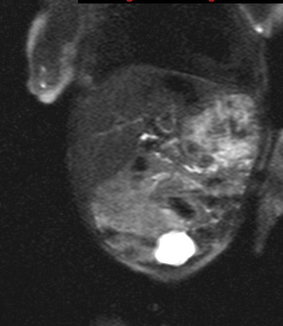
7 Hemangioendothelioma Cross-sectional imaging Coronal T2

8 Hemangioendothelioma: Cross-sectional Imaging Dynamic MRA in Arterial Phase, Coronal Plane
9 Hemangioendothelioma: Background Definition: Benign endothelial-lined vascular mass in liver Demographics 85% in the first 6 months of life 2:1 female-to-male ratio Clinical Presentation: 50% have cutaneous hemangiomas Can present in high-output cardiac failure, consumptive coagulopathy
10 Hemangioendothelioma Imaging Characteristics Hypervascular and heterogeneous Most often large (1 20cm) Solitary or multiple Aorta caliber change below the level of the celiac artery Larger lesions often have central core of necrosis


11 DDX: Infantile hemagioma Axial T2 Haste


12 Infantile Hemangioma Dynamic MRA, arterial phase, coronal Dynamic MRA, arterial phase, coronal
13 Hemangioendothelioma Imaging Recommendations Ultrasound for initial imaging, confirms vascular hepatic lesion(s). MRI provides most flexible approach for multiphase imaging. CT can avoid need for sedation, but multi-phase CT not recommended in the neonate population.


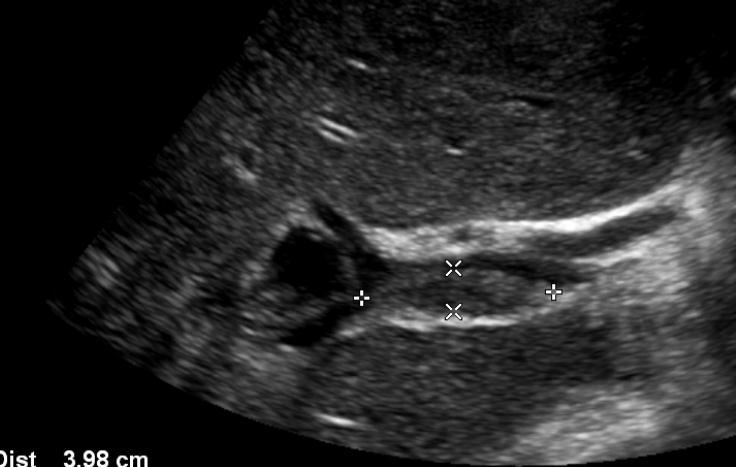
14 Case #2: 16 year old boy with long standing portal hypertension Contrast-enhanced CT abdomen, venous phase. Ultrasound of splenic hilum, greyscale and doppler.

15 Question #2: What additional sequence could be used to determine the nature of the central vascular prominence? A. Arterial Phase CECT B. Delayed Phase CECT C. Upper GI D. Celiac Angiogram

16 Additional Imaging in patient with portal hypertension Arterial Phase 3-D Reconstruction of Arterial Phase CECT Venous Phase

17 Splenic Artery Aneurysm: Background Epidemiology: Occur in 1.6% of the population Clinical Presentation: Often asymptomatic Risk Factors: Portal hypertension Pregnancy Vasculititis Increasing age
18 Splenic Artery Aneurysm: Imaging Findings: 75% are found in the distal third of the splenic artery Can have rim calcification Modality: Ultrasound diagnostic challenge in setting of venous collaterals CECT Single phase or mixed arterial/venous phase CT can obscure the splenic artery Dual-phase CT may be necessary to diagnose SAA
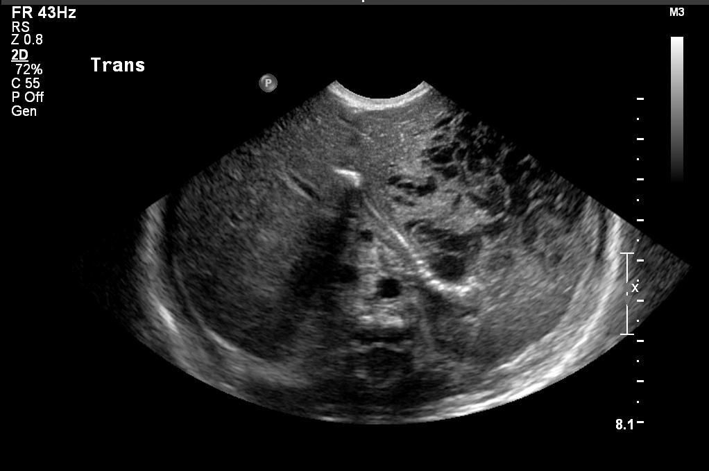
19 Case #3: 11-year-old girl status post liver transplant
20 Question #3: What is the most likely diagnosis? A. Hepatic artery thrombosis B. Hepatic artery stenosis C. Normal arterial waveforms in immediate postoperative setting D. Hepatic vein thrombosis E. Expected findings in prolonged cold ischemia time of donor liver
21 Pediatric Liver Transplant: Background The number of cadaveric donor livers is not sufficient. Three options to meet the demand Segmental liver transplantation with living donor Reduced-size cadaveric Split cadaveric allografts Left lateral to small child Right extended liver to large child or small adult
22 Pediatric Liver Transplant: Anastomosis Hepatic artery Bile duct Standard vs. Biliary Atresia Portal vein Hepatic veins and IVC
23 Pediatric Liver Transplant: Imaging Imaging studies are critical for early diagnosis Clinical signs are nonspecific with wide variation Imaging Options US CT MR Angiography
24 Pediatric Liver Transplant: Ultrasound Protocol Intraoperative Within 12 hours Every 24 hours while in ICU

25 Pediatric Liver Transplant: Normal hepatic arteries Doppler Normal hepatic arteries waveforms: Rapid upstroke with continuous diastolic flow acceleration time from end diastole to first systolic peak less than 80 ms resistive indices peak systolic velocity >30 cm/s and < 250 cm\s


26 Pediatric Liver Transplant: Immediate Postoperative Doppler Absent diastolic flow (RI =1.0) to postoperative edema causing increased peripheral resistance. Differential would also include hepatic arterial thrombosis. Follow-up confirms resolving edema with lowered RI s secondary to improved diastolic flow.
27 Pediatric Liver Transplant: Hepatic Artery Stenosis Findings: HA flow present but with tardus parvus pattern Low resistive indices


28 Pediatric Liver Transplant: Hepatic Artery Stenosis
29 Pediatric Liver Transplant: Imaging hepatic artery stenosis Hepatic artery stenosis occurs in up to 14% of pediatric liver transplants Doppler US imaging modality of choice Sensitivity = 80-90% Diagnosis usually made on Doppler US finding distal to narrowing Tardus Parvus Tardus parvus can be normal in first 72 hours due to edema at anastomotic site Confirm with CT/MR angiography, treat with IR

30 Pediatric Liver Transplant: Hepatic Artery Thrombosis Immediate post-op 24 hrs. post-op Syndrome of Impending Thrombosis 48 hrs. post-op
31 Pediatric Liver Transplant: Imaging hepatic artery thrombosis Prevalence is 5-7%, More common in split or living donor transplantations. Doppler US imaging modality of choice Absence of proper and intrahepatic arterial flow Syndrome of impending thrombosis Both MR and CT can be used to confirm

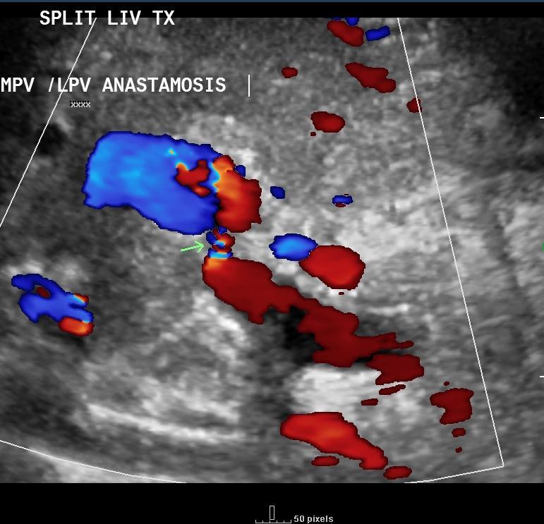
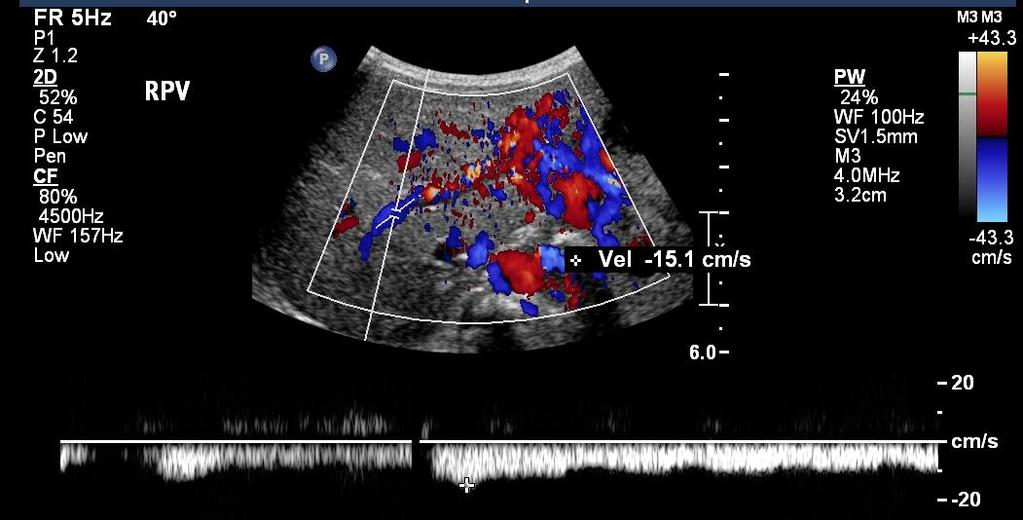
32 Pediatric Liver Transplant: Imaging Portal Vein Stenosis
33 Pediatric Liver Transplant: Imaging Portal Vein Stenosis Uncommon complication occurring at anastomotic site. Potential to lead to thrombosis More likely in reduced-size liver transplantation Doppler US demonstrates >= 50% reduction or 2.5mm or less vessel caliber Aliasing at vascular anastomosis Stenosis vs. Pseudostenosis MR and IR can confirm portal vein stenosis

34 Case #4: 15-year-old female post-op renal transplant
35 Question #4: What is the diagnosis? A. Renal vein thrombosis B. Renal artery thrombosis C. Expected post-operative waveform secondary to edema D. Renal artery stenosis
36 Pediatric Renal Transplantation Renal vein thrombosis Occurs in <5% of patients. Presents in first postoperative week Clinical Presentation: Anuria Tenderness over graft Etiology Hypovolemia, venous compression, slow flow secondary to rejection

37 Pediatric Renal Transplantation Imaging renal vein thrombosis Gray-scale ultrasound Enlarged kidney Doppler Absent venous flow Increased resistance on the arterial side Reversed diastolic flow Upside down m sign


38 Pediatric Renal Transplantation Imaging Renal Artery Stenosis Ultrasound Color Doppler demonstrates stenotic segments Velocities greater than 2 m/sec Marked distal disturbance MR angiography MRA can confirm, but limited in patients with low GFR


39 Pediatric Renal Transplantation Biopsy Induced Pseudoaneurysm Result of arterial laceration secondary to biopsy Gray-scale hypo-echoic mass mimicking simple or complex renal cyst Intracystic color Doppler flow with classic swirling pattern
40 Conclusions MR angiography excellent tool for infantile hepatic vascular masses Multi-phase imaging improves detection of splenic artery aneurysms in the setting of portal hypertension Repetitive liver Doppler ultrasound is effective in following postoperative liver transplant patients Doppler ultrasound provides an effective method to evaluate for the immediate and delayed vascular complications of renal transplant
What effects will proximal or distal disease have on an waveform?
 Spectral Doppler Interpretation Director Director of of Ultrasound Ultrasound Education Education & & Quality Quality Assurance Assurance Baylor Baylor College College of of Medicine Medicine Division
Case 8038 Renal allograft complicated with renal artery stenosis
 Case 8038 Renal allograft complicated with renal artery stenosis Santiago I, Canelas A, Pinto AP Section: Cardiovascular Published: 2009, Nov. 30 Patient: 61 year(s), male Clinical History A 61-year-old
What effects will proximal or distal disease have on a waveform?
 Spectral Doppler Interpretation Director of Ultrasound Education & Quality Assurance Baylor College of Medicine Division of Maternal-Fetal Medicine Maternal Fetal Center Imaging Manager Texas Children
Treatment of choice for end stage renal disease Imaging to establish baseline and diagnosis of potential complications Review common surgical
 Treatment of choice for end stage renal disease Imaging to establish baseline and diagnosis of potential complications Review common surgical techniques Review normal appearance Discuss US diagnosis of
DISCLOSURE TEST YOUR WAVEFORM IQ. Partial volume artifact. 86 yo female with right arm swelling, picc line. AVF on left? Dx?
 Deborah Rubens University of Rochester Rochester, NY DISCLOSURE Neither I nor my immediate family have a financial relationship with a commercial organization that may have a direct or indirect interest
The role for contrast-enhanced ultrasonography outside of focal liver lesions
 The role for contrast-enhanced ultrasonography outside of focal liver lesions Paul S. Sidhu King s College Hospital, London, UK Introduction Contrast-enhanced ultrasonography (US) of focal liver lesions
US of Renovascular Hypertension. Jonathan R. Dillman, MD, MSc Associate Professor Director, Thoracoabdominal Imaging
 US of Renovascular Hypertension Jonathan R. Dillman, MD, MSc Associate Professor Director, Thoracoabdominal Imaging Disclosures Nothing Relevant Unrelated grant funding Siemens US Toshiba US Objectives
Visceral Vascular Ultrasound. Joel Thompson, MD, MPH Borg & Ide Imaging
 Visceral Vascular Ultrasound Joel Thompson, MD, MPH Borg & Ide Imaging Objectives: Review major abdominal vascular structures Identify normal peak systolic velocity (PSV) for major abdominal arteries.
Abdominal Doppler Mastering the next level of vascular anatomy in the belly. Cindy A. Owen, RDMS, RVT
 Abdominal Doppler Mastering the next level of vascular anatomy in the belly Cindy A. Owen, RDMS, RVT Introduction Abdominal Doppler is a tough exam Success is dependent on: Patient body habitus Patient
Job Task Analysis for ARDMS Abdomen Data Collected: June 30, 2011
 Job Task Analysis for ARDMS Abdomen Data Collected: June 30, 2011 Reported: Analysis Summary for: Abdomen Examination Survey Dates 06/13/2011-06/26/2011 Invited Respondents 6,000 Surveys with Demographics
Vascular Technology Examination Content Outline
 Vascular Technology Examination Content Outline (Outline Summary) # Domain Subdomain Percentage 1 Normal Anatomy, Perfusion, and Function Evaluate normal anatomy, perfusion, function 2 Pathology, Perfusion,
Renal Transplant Surgery
 Renal Transplant Surgery Mr Somaiah Aroori MS MD EBS in HPB FRCS Consultant HPB & Renal Transplant Surgeon SWTC, Derriford Hospital, Plymouth Over next few minutes Aim to cover Details of Transplant procedure
Overall Goals and Objectives for Transplant Hepatology EPAs:
 Overall Goals and Objectives for Transplant Hepatology EPAs: 1. DIAGNOSTIC LIST During the one-year Advanced Pediatric Transplant Hepatology Program, fellows are expected to develop comprehensive skills
Abdomen Sonography Examination Content Outline
 Abdomen Sonography Examination Content Outline (Outline Summary) # Domain Subdomain Percentage 1 2 3 Anatomy, Perfusion, and Function Pathology, Vascular Abnormalities, Trauma, and Postoperative Anatomy
STRUCTURED EDUCATION REQUIREMENTS IMPLEMENTATION DATE: JULY 1, 2016
 STRUCTURED EDUCATION REQUIREMENTS Vascular Sonography The purpose of structured education is to provide the opportunity for individuals to develop mastery of discipline-specific knowledge that, when coupled
Looking Outside the Box: Incidental Extracardiac Finding in Echo
 Looking Outside the Box: Incidental Extracardiac Finding in Echo Dr. Aijaz Shah Head of Division, Adult Echocardiography Laboratory Prince Sultan Cardiac Centre Riyadh Case 1 17 year old boy presented
Vascular Sonography Examination
 Vascular Sonography Examination The purpose of The American Registry of Radiologic Technologists (ARRT ) Vascular Sonography Examination is to assess the knowledge and cognitive skills underlying the intelligent
Liver Transplantation
 1 Liver Transplantation Department of Surgery Yonsei University Wonju College of Medicine Kim Myoung Soo M.D. ysms91@wonju.yonsei.ac.kr http://gs.yonsei.ac.kr History Development of Liver transplantation
Image Formation (10) 2 Evaluation and Selection of Representative Images (10)
 2 Evaluation and Selection of Representative Images (10)") STRUCTURED SELF ASSESSMENT CONTENT SPECIFICATIONS SSA LAUNCH DATE: JANUARY 1, 2018 Vascular Sonography The purpose of continuing qualifications requirements (CQR) is to assist registered technologists
STRUCTURED SELF ASSESSMENT CONTENT SPECIFICATIONS SSA LAUNCH DATE: JANUARY 1, 2018 Vascular Sonography The purpose of continuing qualifications requirements (CQR) is to assist registered technologists
MINIMALLY INVASIVE MANAGEMENT OF RENOVASCULAR COMPLICATIONS AFTER RENAL GRAFT TRANSPLANTATION
 MINIMALLY INVASIVE MANAGEMENT OF RENOVASCULAR COMPLICATIONS AFTER RENAL GRAFT TRANSPLANTATION Gortes, Francisco Javier B.S; Salsamendi, Jason Thomas M.D LEARNING OBJECTIVES Educate physicians on the prompt
Erin Kane, HMS III Dr. Gillian Lieberman BIDMC Radiology Core Clerkship March 2009
 Erin Kane, HMS III Dr. Gillian Lieberman BIDMC Radiology Core Clerkship March 2009 Agenda Patient OH: Initial presentation Kidney transplantation: Menu of tests Routine imaging for kidney donors Selected
Ultrasound in Liver Trasplantation
 Ultrasound in Liver Trasplantation Poster No.: C-1892 Congress: ECR 2011 Type: Educational Exhibit Authors: B. Molinares, A. Marquez, M. Ochoa, S. Alvarez; CO Keywords: Ultrasound-Spectral Doppler, Ultrasound-Colour
Doppler Ultrasound Findings in the Hepatic Artery Shortly After Liver Transplantation
 Gastrointestinal Imaging Pictorial Essay García-riado et al. Hepatic rtery fter Liver Transplantation Gastrointestinal Imaging Pictorial Essay FOUS ON: Ángeles García-riado 1 Rosa Gilabert nnalisa erzigotti
Duplex Ultrasound of the Renal Arteries. Duplex Ultrasound. In the Beginning
 Duplex Ultrasound of the Renal Arteries DIMENSIONS IN HEART AND VASCULAR CARE 2013 PENN STATE HEART AND VASCULAR INSTITUTE ROBERT G. ATNIP MD PROFESSOR OF SURGERY AND RADIOLOGY Duplex Ultrasound Developed
Ultrasound of the Renal Arteries
 Ultrasound of the Renal Arteries Greg Curry Vascular Ultrasound Workshop Aug 2017 The Examination Technique Pathophysiology Role of US then and now Background Live Scanning Ultrasound Population: 20% Hypertensive
Portal Venous Thrombosis: Tumor VS Bland Thrombus
 June 2015 Portal Venous Thrombosis: Tumor VS Bland Thrombus SERGIO ALFARO, HARVARD MEDICAL SCHOOL YEAR III GILLIAN LIEBERMAN, MD Overview 2 Index Patient History Portal Venous Thrombosis (PVT) Imaging
Imaging abdominal vascular emergencies. V.Stoynova
 Imaging abdominal vascular emergencies V.Stoynova Abdominal vessels V. Stoynova 2 Acute liver bleeding trauma anticoagulant therapy liver disease : HCC, adenoma, meta, FNH, Hemangioma Diagnosis :CT angiography
Transducer Selection. Renal Artery Duplex Exam. Renal Scan. Renal Scan Echogenicity. How to Perform an Optimal Renal Artery Doppler Examination
 How to Perform an Optimal Renal Artery Doppler Examination Director of Ultrasound Education & Quality Assurance Baylor College of Medicine Division of Maternal-Fetal Medicine Maternal Fetal Center Imaging
Radiology of hepatobiliary diseases
 GI cycle - Lecture 14 436 Teams Radiology of hepatobiliary diseases Objectives 1. To Interpret plan x-ray radiograph of abdomen with common pathologies. 2. To know the common pathologies presentation.
CT 101 :Pancreas and Spleen
 CT 101 :Pancreas and Spleen Shikha Khullar,, MD, MPH Division of Radiology University of South Alabama The Pancreas Normal Pancreas 3 Phase Pancreatic CT Non contrast Arterial phase : 30-35 35 second
Alice Fung, MD Oregon Health and Science University
 Alice Fung, MD Oregon Health and Science University Disclosure Comments The speaker Alice Fung, MD Has relevant financial relationships to disclose. Received honorarium from (Guerbet). This individual
Vascular complications after adult liver transplantation: Evaluation with Doppler ultrasound
 Vascular complications after adult liver transplantation: Evaluation with Doppler ultrasound Poster No.: C-2588 Congress: ECR 2012 Type: Educational Exhibit Authors: M. S. C. Sousa, M. C. Cordeiro, R.
HEPATO-BILIARY IMAGING
 HEPATO-BILIARY IMAGING BY MAMDOUH MAHFOUZ MD PROF.OF RADIOLOGY CAIRO UNIVERSITY mamdouh.m5@gmail.com www.ssregypt.com CT ABDOMEN Indications Patient preparation Patient position Scanogram Fasting 4-6 hours
What Do We Know? Disclosure Statement: 3/11/2015. Deep abdominal imaging
 Marsha M. Neumyer, BS, RVT, FSVU, FSDMS, FAIUM International Director Vascular Diagnostic Educational Services Vascular Resource Associates Harrisburg, PA Disclosure Statement: CME Calendar QR Code Marsha
Diagnosis of Renal Artery Stenosis (RAS)
") May 2001 Diagnosis of Renal Artery Stenosis (RAS) Kurt Fink, Harvard Medical School, Year III Epidemiology Hypertension -Affects 60 million Americans Essential HTN >95% of cases Secondary HTN 1-5% of cases
May 2001 Diagnosis of Renal Artery Stenosis (RAS) Kurt Fink, Harvard Medical School, Year III Epidemiology Hypertension -Affects 60 million Americans Essential HTN >95% of cases Secondary HTN 1-5% of cases
General Imaging. Imaging modalities. Incremental CT. Multislice CT Multislice CT [ MDCT ]
![General Imaging. Imaging modalities. Incremental CT. Multislice CT Multislice CT [ MDCT ]](/thumbs/76/74079340.jpg "General Imaging. Imaging modalities. Incremental CT. Multislice CT Multislice CT [ MDCT ]") General Imaging Imaging modalities Conventional X-rays Ultrasonography [ US ] Computed tomography [ CT ] Radionuclide imaging Magnetic resonance imaging [ MRI ] Angiography conventional, CT,MRI Interventional
General Imaging Imaging modalities Conventional X-rays Ultrasonography [ US ] Computed tomography [ CT ] Radionuclide imaging Magnetic resonance imaging [ MRI ] Angiography conventional, CT,MRI Interventional
Journal of Medical Imaging and Radiation Oncology
 Journal of Medical Imaging and Radiation Oncology 62 (2018) 504 511 MEDICAL IMAGING PICTORIAL ESSAY Imaging in pancreas transplantation complications: Temporal classification Paula Gallego Ferrero and
Goals. Access flow and renal artery stenosis evaluation by Doppler ultrasound. Reimbursement. WHY use of Doppler Ultrasound
 Access flow and renal artery stenosis evaluation by Doppler ultrasound Adina Voiculescu, MD Interventional Nephrology Brigham and Women s Hospital Boston Instructor at Harvard Medical School Understand
Sonography of soft-tissue vascular lesions
 Sonography of soft-tissue vascular lesions Oscar M. Navarro Associate Professor, University of Toronto Dept. of Diagnostic Imaging, The Hospital for Sick Children Toronto, Canada Declaration of Disclosure
CTA/MRA of Pediatric Hepatic Masses Radiology-Pathology Correlation
 Acta Radiológica Portuguesa, Vol.XVIII, nº70, pág. 41-50, Abr.-Jun., 2006 CTA/MRA of Pediatric Hepatic Masses Radiology-Pathology Correlation Marilyn J. Siegel Mallinckrodt Institute of Radiology, Washington
Zoltan Harkanyi M.D., Ph.D. Department of Radiology, Heim Pal Children s Hospital, Budapest, Hungary
 Zoltan Harkanyi M.D., Ph.D. Department of Radiology, Heim Pal Children s Hospital, Budapest, Hungary CEUS expereince 10 years Department of Radiology, Heim Pal Children s Hospital, Budapest US N o 1 study
Renal Artery Stenosis With Severe Hypertension: A Case Report
 CASE REPORT Renal Artery Stenosis With Severe Hypertension: A Case Report Suwaid MA ABSTRACT Background: Renal artery stenosis (RAS) is found in 77% of hypertensive patients and is responsible for 1-2%
Contrast Enhanced Ultrasound of Parenchymal Masses in Children
 Contrast Enhanced Ultrasound of Parenchymal Masses in Children Sue C Kaste, DO On behalf of Beth McCarville, MD St. Jude Children s Research Hospital Memphis, TN Overview Share St. Jude experience with
11 TH ANNUAL VASCULAR NONINVASIVE TESTING SYMPOSIUM NOVEMBER 10, 2018
 11 TH ANNUAL VASCULAR NONINVASIVE TESTING SYMPOSIUM NOVEMBER 10, 2018 RENAL ARTERY DISEASE AND RENOVASCULAR HYPERTENSION 1 WHAT IS RENOVASCULAR HYPERTENSION? https://my.clevelandclinic.org/health/diseases/16459-renovascular-hypertension
Financial Disclosure
 Benign Liver Masses Adil Abdalla, MBBS Creighton University-CHI Health August 25, 2018 Financial Disclosure Nothing to disclose Financial Disclosure 1 Objectives To assess patients with benign liver tumors
Appendix 5. EFSUMB Newsletter. Gastroenterological Ultrasound
 EFSUMB Newsletter 87 Examinations should encompass the full range of pathological conditions listed below A log book listing the types of examinations undertaken should be kept Training should usually
No financial or commercial relationships to disclose
 Deanna New, RVT No financial or commercial relationships to disclose IAC REQUIREMENTS: The main duty of a sonographer is to make the physician or radiologists job easier by capturing images and doing
AMR in Liver Transplantation: Incidence
 AMR in Liver Transplantation: Incidence Primary AMR 1/3 to 1/2 of ABO-incompatible transplants Uncommon with ABO-compatible transplant Secondary AMR Unknown incidence: rarely tested Why is AMR uncommon
Liver Transplantation in Children: Techniques and What the Surgeon Wants to Know from Imaging
 Liver Transplantation in Children: Techniques and What the Surgeon Wants to Know from Imaging Jaimie D. Nathan, MD Associate Professor of Surgery and Pediatrics Associate Surgical Director, Liver Transplant
Bits and Bobs secondary causes of heart problems. Dr Angela McBrien 9 th September 2017
 Bits and Bobs secondary causes of heart problems Dr Angela McBrien 9 th September 2017 Not the heart Dextroposition Heart in the right chest with the apex to the left Often caused by left sided chest mass
Lung sequestration and Scimitar syndrome
 Lung sequestration and Scimitar syndrome Imaging approaches M. Mearadji International Foundation for Pediatric Imaging Aid Rotterdam, The Netherlands Pulmonary sequestration Pulmonary sequestration (PS)
Doppler Ultrasound Velocities and Resistive Indexes Immediately After Pediatric Liver Transplantation: Normal Ranges and Predictors of Failure
 Pediatric Imaging Original Research Jamieson et al. Doppler Ultrasound After Pediatric Liver Transplantation Pediatric Imaging Original Research Lucy H. Jamieson 1 Bo Arys 2 Gavin Low 1 Ravi Bhargava 1
Liver transplant for biliary atresia
 Jean de Ville de Goyet ISMETT Director of the Department for the Treatment and Study of Pediatric Abdominal Diseases and Abdominal Transplantation The first human liver transplant was performed on a pediatric
Visceral aneurysm. Diagnosis and Interventions M.NEDEVSKA
 Visceral aneurysm Diagnosis and Interventions M.NEDEVSKA History 1953 De Bakeyand Cooley Visceral aneurysm VAAs rare, reported incidence of 0.01 to 0.2% on routine autopsies. Clinically important Potentially
Uroradiology For Medical Students
 Uroradiology For Medical Students Lesson 8 Computerized Tomography 2 American Urological Association Objectives In this lesson you will: Gain more experience reading CT images Learn how computer generated
IT 의료융합 1 차임상세미나 복부질환초음파 이재영
 IT 의료융합 1 차임상세미나 2013-4-3 복부질환초음파 이재영 나는오늘누구를위하여 종을울리나? 전통적의료 의사 공학설계자 의사 최첨단진단장비들 USG, CT, MRI 환자 환자 현대의료 사용자중심의사고 US in the Abdomen Detection DDx Look Behavior Response by external stimuli Guiding Tool
Adult Echocardiography Examination Content Outline
 Adult Echocardiography Examination Content Outline (Outline Summary) # Domain Subdomain Percentage 1 2 3 4 5 Anatomy and Physiology Pathology Clinical Care and Safety Measurement Techniques, Maneuvers,
Indications: following: embolization. artery that has diseases 5. The evaluation. of suspected. such entities. a cold hand. biopsy
 Peripheral Arterial Ultrasound Protocol Using Color and Spectral Doppler Reviewed by: Mark Yuhasz, MD Last Review Date: January 2015 Contact: (866) 761 4200, Option 1 Indications: The indications for peripheral
Case 9799 Stanford type A aortic dissection: US and CT findings
 Case 9799 Stanford type A aortic dissection: US and CT findings Accogli S, Aringhieri G, Scalise P, Angelini G, Pancrazi F, Bemi P, Bartolozzi C Department of Diagnostic and Interventional Radiology, University
Nasogastric tube. Stomach. Pylorus. Duodenum 1. Duodenum 2. Duodenum 3. Duodenum 4
 Esophagus Barium Swallow Stomach and Duodenum 4 year old Upper GI Nasogastric tube Stomach and Duodenum 4 year old Upper GI Nasogastric tube Stomach Pylorus Duodenum 1 Duodenum 2 Duodenum 3 Duodenum 4
Cardiac MRI in ACHD What We. ACHD Patients
 Cardiac MRI in ACHD What We Have Learned to Apply to ACHD Patients Faris Al Mousily, MBChB, FAAC, FACC Consultant, Pediatric Cardiology, KFSH&RC/Jeddah Adjunct Faculty, Division of Pediatric Cardiology
Imaging of liver and pancreas
 Imaging of liver and pancreas.. Disease of the liver Focal liver disease Diffusion liver disease Focal liver disease Benign Cyst Abscess Hemangioma FNH Hepatic adenoma HCC Malignant Fibrolamellar carcinoma
The role of abdominal CT and MRI in detection of complications after transplantations of liver, kidney and pancreas.
 The role of abdominal CT and MRI in detection of complications after transplantations of liver, kidney and pancreas. Poster No.: C-1319 Congress: ECR 2015 Type: Educational Exhibit Authors: R. Muslimov,
CT abdomen and pelvis
 CT abdomen and pelvis General indications: Assessment of vague abdominal symptoms (pain, colics,distenstion,...) Varifecation of a lesion discovered by other diagnostic modalities as US, barium,ivp, Staging
1. Long images of aorta (prox, mid, and dist) with AP measurements. 2. Trans images of aorta (prox, mid, and dist) with R/L measurements.
 with AP measurements. 2. Trans images of aorta (prox, mid, and dist) with R/L measurements.") Aorta 1. Long images of aorta (prox, mid, and dist) with AP measurements. 2. Trans images of aorta (prox, mid, and dist) with R/L measurements. 3. Long images of R/L common iliac arteries with AP measurements.
Aorta 1. Long images of aorta (prox, mid, and dist) with AP measurements. 2. Trans images of aorta (prox, mid, and dist) with R/L measurements. 3. Long images of R/L common iliac arteries with AP measurements.
NYU School of Medicine Department of Radiology Rotation-Specific House Staff Evaluation
 Vascular & Interventional Radiology Rotation 1 Core competency in vascular and interventional radiology during the first resident rotation consists of clinical objectives, technical objectives and image
Post-operative complications following hepatobiliary surgery: imaging findings and current radiological treatment options
 Post-operative complications following hepatobiliary surgery: imaging findings and current radiological treatment options Poster No.: C-1501 Congress: ECR 2015 Type: Educational Exhibit Authors: A. Hadjivassiliou,
Pediatric Hepatobiliary, Pancreatic & Splenic US
 Pediatric Hepatobiliary, Pancreatic & Splenic US Susan J. Back, MD Department of Radiology, The Children s Hospital of Philadelphia No Disclosures Objectives Normal Abnormal: cases and US advances Objectives
HD Scanning: Velocities and Volume Flow
 HD Scanning: Velocities and Volume Flow Non-Invasive Lab Symposium West Orange, NJ April 27, 2018 Volume Flow Cindy Sturt, MD, FACS, RVT 500,000 Americans on dialysis 20-25% annual mortality 65% 5 year
Liver Tumors. Prof. Dr. Ahmed El - Samongy
 Liver Tumors Prof. Dr. Ahmed El - Samongy Objective 1. Identify the most important features of common benign liver tumors 2. Know the risk factors, diagnosis, and management of hepatocellular carcinoma
Imaging for Peripheral Vascular Disease
 Imaging for Peripheral Vascular Disease James G. Jollis, MD Director, Rex Hospital Cardiovascular Imaging Imaging for Peripheral Vascular Disease 54 year old male with exertional calf pain in his right
Masateru Yamamoto 1, Toshiyuki Itamoto 1,2*, Akihiko Oshita 1,2 and Yasuhiro Matsugu 1
 Yamamoto et al. Journal of Medical Case Reports (2018) 12:92 https://doi.org/10.1186/s13256-018-1614-2 CASE REPORT Open Access Celiac axis stenosis due to median arcuate ligament compression in a patient
Guidelines, Policies and Statements D5 Statement on Abdominal Scanning
 Guidelines, Policies and Statements D5 Statement on Abdominal Scanning Disclaimer and Copyright The ASUM Standards of Practice Board have made every effort to ensure that this Guideline/Policy/Statement
Chief Complain. Liver lesion found in routine health check 41 days ago
 Chief Complain Liver lesion found in routine health check 41 days ago Present Illness On 2005-7-26 at 台北署立醫院 he underwent a health check for the first time. Abdominal US showed suspicious of a 6*5 cm hepatoma,
Pancreatic Adenocarcinoma: Everything You Need to Know From Cross-Sectional Imaging to Treatment
 Pancreatic Adenocarcinoma: Everything You Need to Know From Cross-Sectional Imaging to Treatment Andrew W. Bowman, MD PhD Assistant Professor of Radiology Mayo Clinic Florida SCBT-MR Annual Meeting Nashville,
Tranjugular Intrahepatic Portosystemic Shunt
 Tranjugular Intrahepatic Portosystemic Shunt Christopher Selhorst July 25, 2005 BIDMC Radiology Overview Portal Hypertension Indications, Contraindications The Procedure Case Review Complications Outcomes
PERPHERAL ARTERY ANEURYSM. By Pooja Sharma and Susanna Sebastianpillai
 PERPHERAL ARTERY ANEURYSM By Pooja Sharma and Susanna Sebastianpillai Defintions True Aneurysm Involves all three layers of the vessel. Have two basic shapes; Fusiform = symmetric widening of the vessels
Doppler Ultrasonography of the Liver: What Every General Radiologist Should Know
 Doppler Ultrasonography of the Liver: What Every General Radiologist Should Know Poster No.: C-1658 Congress: ECR 2014 Type: Authors: Keywords: DOI: Educational Exhibit T. González de la Huebra Labrador,
Recommendations for Follow-up After Vascular Surgery Arterial Procedures SVS Practice Guidelines
 Recommendations for Follow-up After Vascular Surgery Arterial Procedures 2018 SVS Practice Guidelines vsweb.org/svsguidelines About the guidelines Published in the July 2018 issue of Journal of Vascular
Personal data. Age : 63 Gender : male
 Personal data Age : 63 Gender : male Chief complain No specific symptom or discomfort A hepatic mass, found by abdominal sonography of routine health exam on 88-12-08 Past history 1984-3-3 Old CVA with
Torsion of a Wandering Spleen Presenting as a Painful Pelvic Mass Post Pregnancy: Imaging Diagnosis
 CASE REPORT Torsion of a Wandering Spleen Presenting as a Painful Pelvic Mass Post Pregnancy: Imaging Diagnosis Abbey P 1, Aarushi A 1, Andley M 2, Anand R 1 1 Department of Radio-Diagnosis, 2 Department
Diagnostic Imaging
 www.fisiokinesiterapia.biz Diagnostic Imaging Diagnostic Imaging is no longer limited to radiography. Major technological advancements have lead to the use of new and improved imaging technologies. The
Ultrasound imaging of vascular anomalies: pearls and pitfalls
 Ultrasound imaging of vascular anomalies: pearls and pitfalls Oscar Navarro, MD Dept. of Medical Imaging, University of Toronto Dept. of Diagnostic Imaging, The Hospital for Sick Children Declaration of
Variations in Surgical Anatomy of the Portal Vein in Living Donor Liver Transplantation
 Kasr El Aini Journal of Surgery VOL., 9, NO 3 January 2008 19 Variations in Surgical Anatomy of the Portal Vein in Living Donor Liver Transplantation A. Ayad ; W. Tobar; M.Hassan; A.Hosny; M.El Shazly;
Hematologic Malignancies of the Liver : Spectrum of Disease. Zhou Jian
 Hematologic Malignancies of the Liver : Spectrum of Disease Zhou Jian 2015-7-8 Hematologic malignancies include a wide spectrum of lymphoproliferative and myeloproliferative disorders with nodal and extranodal
Applications of magnetic resonance imaging and magnetic resonance angiography to evaluate the hepatic vasculature in the pediatric patient
 Pediatr Radiol (1999) 29: 238±243 Ó Springer-Verlag 1999 Eu-Leong H. J. Teo Peter J.Strouse Martin R. Prince Applications of magnetic resonance imaging and magnetic resonance angiography to evaluate the
ACUTE PANCREATITIS: NEW CLASSIFICATION OF AN OLD FOE. T Barrow, A Nasrullah, S Liong, V Rudralingam, S A Sukumar
 ACUTE PANCREATITIS: NEW CLASSIFICATION OF AN OLD FOE T Barrow, A Nasrullah, S Liong, V Rudralingam, S A Sukumar LEARNING OBJECTIVES q Through a series of cases illustrate the updated Atlanta symposium
Carotid Artery Doppler
 Carotid Artery Doppler Patient Position supine or semisupine head slightly hyper extended rotated 45 away from the side being examined. Higher frequency linear transducers (7 MHz) Vessels should be imaged
Index. Crit Care Clin 19 (2003)
") Crit Care Clin 19 (2003) 331 335 Index A ACVECC. See American College of Veterinary Emergency and Critical Care (ACVECC). Aging. See also Elderly; Geriatric critical care. respiratory function effects
Crit Care Clin 19 (2003) 331 335 Index A ACVECC. See American College of Veterinary Emergency and Critical Care (ACVECC). Aging. See also Elderly; Geriatric critical care. respiratory function effects
Heart and Lungs. LUNG Coronal section demonstrates relationship of pulmonary parenchyma to heart and chest wall.
 Heart and Lungs Normal Sonographic Anatomy THORAX Axial and coronal sections demonstrate integrity of thorax, fetal breathing movements, and overall size and shape. LUNG Coronal section demonstrates relationship
Ultrasound of soft-tissue vascular anomalies
 Ultrasound of soft-tissue vascular anomalies Oscar M. Navarro Associate Professor, University of Toronto Dept. of Diagnostic Imaging, The Hospital for Sick Children Toronto, Canada Declaration of Disclosure
Lab Monitor Images Dissection of the Abdominal Vasculature + Lower Digestive System
 Lab Monitor Images Dissection of the Abdominal Vasculature + Lower Digestive System Stomach & Duodenum Frontal (AP) View Nasogastric tube 2 1 3 4 Stomach Pylorus Duodenum 1 Duodenum 2 Duodenum 3 Duodenum
MRI (AND CT) FOR REPAIRED TETRALOGY OF FALLOT
 FOR REPAIRED TETRALOGY OF FALLOT") MRI (AND CT) FOR REPAIRED TETRALOGY OF FALLOT Linda B Haramati MD, MS Departments of Radiology and Medicine Bronx, New York OUTLINE Pathogenesis Variants Initial surgical treatments Basic MR protocols
MRI (AND CT) FOR REPAIRED TETRALOGY OF FALLOT Linda B Haramati MD, MS Departments of Radiology and Medicine Bronx, New York OUTLINE Pathogenesis Variants Initial surgical treatments Basic MR protocols
Mesenteric/Splanchnic Artery Duplex Imaging
 VASCULAR TECHNOLOGY PROFESSIONAL PERFORMANCE GUIDELINES Mesenteric/Splanchnic Artery Duplex Imaging This Guideline was prepared by members of the Society for Vascular Ultrasound (SVU) as a template to
Traumatic Renocaval Fistula With Pseudoaneurysm Leading To Renal Atrophy
 ISPUB.COM The Internet Journal of Radiology Volume 6 Number 2 Traumatic Renocaval Fistula With Pseudoaneurysm Leading To Renal Atrophy M Kukkady, A Deena, S Raj, Ramachandra Citation M Kukkady, A Deena,
Abdominal Doppler. Cases of Where, Why, and How
 Abdominal Doppler Cases of Where, Why, and How Jill D. Trotter, BS, RT(R), RDMS, RVT Director, Diagnostic Medical Sonography Program Vanderbilt University/Vanderbilt Medical Center Nashville, Tennessee
Supplementary Online Content
 Supplementary Online Content Smith D, Chudgar A, Daly B, Cooper M. Evaluation of potential renal transplant recipients with computed tomography angiography. Arch Intern Med. doi: 10.1001/archsurg.2012.1466.
Physician s Vascular Interpretation Examination Content Outline
 Physician s Vascular Interpretation Examination Content Outline (Outline Summary) # Domain Subdomain Percentage 1 2 3 4 5 6 Cerebrovascular Abdominal Peripheral Arterial - Duplex Imaging Peripheral Arterial
Scanning Mesenteric and Hypogastric Artery Aneurysms
 Scanning Mesenteric and Hypogastric Artery Aneurysms Marsha M. Neumyer, BS, RVT, FSVU, FSDMS, FAIUM International Director Vascular Diagnostic Education Services Vascular Resource Associates Harrisburg,
Likes ML, Johnston TA. Gastric pseudoaneurysm in the setting of Loey s Dietz Syndrome. Images Paediatr Cardiol. 2012;14(3):1-5
:1-5") IMAGES in PAEDIATRIC CARDIOLOGY Likes ML, Johnston TA. Gastric pseudoaneurysm in the setting of Loey s Dietz Syndrome. Images Paediatr Cardiol. 2012;14(3):1-5 University of Washington, Pediatrics, Seattle
IMAGES in PAEDIATRIC CARDIOLOGY Likes ML, Johnston TA. Gastric pseudoaneurysm in the setting of Loey s Dietz Syndrome. Images Paediatr Cardiol. 2012;14(3):1-5 University of Washington, Pediatrics, Seattle