12 Lead Electrocardiogram (ECG) PFN: SOMACL17. Terminal Learning Objective. References
|
|
|
- Catherine Johnson
- 6 years ago
- Views:
Transcription
1 12 Lead Electrocardiogram (ECG) PFN: SOMACL17 Slide 1 Terminal Learning Objective Action: Communicate knowledge of 12 Lead Electrocardiogram (ECG) Condition: Given a lecture in a classroom environment Standard: Received a minimum score of 84% on the written exam IAW course AHA standards Slide 2 References Advanced Cardiovascular Life Support, Provider Manual 2010 The 12 lead ECG in Acute Myocardial Infarction 1996 The 12 lead ECG for Acute and Critical Care Providers 2005 Slide 3 1
2 Reason Ischemic heart disease causes the greatest number of deaths in the United States. 50% die before arriving at a hospital. If treatment is not received within 2 hours of onset of symptoms, chance of survival is 25%. By treating a heart attack within one hour of onset of symptoms, the chance of survival is 49%! Slide 4 Reason The keys to quick management of an acute myocardial infarction (AMI) patient are: Prompt recognition of the symptoms Rapid performance and interpretation of a 12 Lead ECG Provide pre arrival notification to the receiving facility Slide 5 Agenda Recall the pathophysiology of acute myocardial infarction (AMI) Identify the electrode placement for a 12 Lead ECG Identify the 12 Lead ECG physiology to assess an AMI Identify an AMI using a 12 Lead ECG Slide 6 2
 Slide 8 Pathophysiology of AMI A heart attack occurs when a thrombus or blood clot forms in a coronary artery, cutting off the blood supply to a segment of the heart muscle.")
3 Agenda Identify the limitations and complications of recognizing an AMI using a 12 Lead ECG Participate in a 12 Lead ECG interpretation practical exercise Slide 7 Pathophysiology of Acute Myocardial Infarction (AMI) Slide 8 Pathophysiology of AMI A heart attack occurs when a thrombus or blood clot forms in a coronary artery, cutting off the blood supply to a segment of the heart muscle. Prompt restoration of blood flow can stop and minimize (or even prevent) the heart damage. Slide 9 3
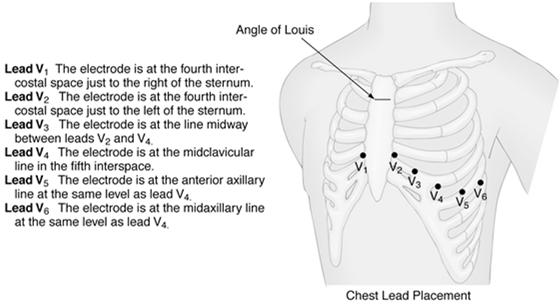
4 Pathophysiology of AMI The blood flow can be restored by: Percutaneous coronary intervention (PCI) Coronary artery bypass graft (CABG) Administration of a clot dissolving (thrombolytic) drug Slide 10 Pathophysiology of AMI ACS symptom review: Crushing chest pain lasting more than 15 minutes Chest Pain spreading to the shoulders, neck, arms or jaw Chest discomfort with lightheadedness, fainting, sweating or nausea Shortness of breath with or without chest discomfort Slide 11 Electrode Placement for a 12 Lead ECG Slide 12 4


5 ECG Leads Bipolar Leads I, II, and III Unipolar Leads avr, avl, and avf Precordial V1, V2, V3, V4, V5, and V6 Slide Lead ECG Preparation Slide 14 Bipolar and Unipolar Leads Slide 15 5
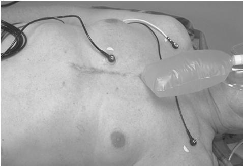
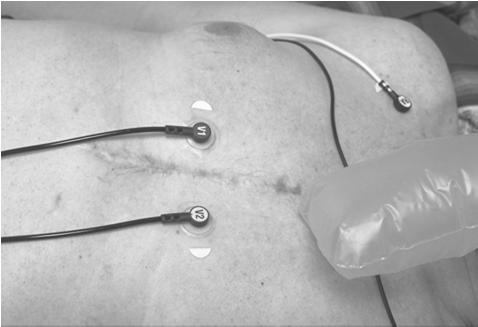
6 Precordial Leads Slide 16 V1 Slide 17 V2 Slide 18 6


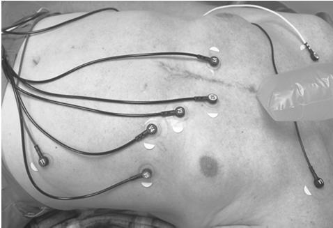
7 V4 Slide 19 V3 Slide 20 V5 Slide 21 7


8 V6 Slide Lead ECG Electrode Placement Slide Lead ECG Physiology to Assess an AMI Slide 24 8
9 12 Lead ECG Relationship to Coronary Artery Anatomy Let s look at the heart Page 28 in the ACLS Handbook The right coronary artery (RCA) supplies oxygen to the inferior (bottom) surface of the heart Slide Lead ECG Relationship to Coronary Artery Anatomy The left coronary artery (LCA) splits in two: The left anterior descending coronary artery (LAD) The left circumflex coronary artery (LCX or Cx) Slide Lead ECG Relationship to Coronary Artery Anatomy (cont) Slide 27 9

10 12 Lead ECG Relationship to Coronary Artery Anatomy The left anterior descending coronary artery (LAD) courses down the anterior (front) surface of the heart and supplies oxygen to the septal, anterior, and lateral sides of the heart. Slide Lead ECG Relationship to Coronary Artery Anatomy (cont) Slide Lead ECG Relationship to Coronary Artery Anatomy The left circumflex coronary artery (Cx) supplies oxygen to the left lateral (side) surface of the heart. Slide 30 10


11 12 Lead ECG Relationship to Coronary Artery Anatomy (cont) Slide Lead ECG Relationship to Coronary Artery Anatomy Slide Lead ECG Relationship to Coronary Artery Anatomy (cont) I lateral avr V 1 septal V 4 anterior II inferior avl lateral V 2 septal V 5 lateral III inferior avf inferior V 3 anterior V 6 lateral Slide 33 11


12 12 Lead ECG Relationship to Coronary Artery Anatomy Slide 34 Identify an AMI Using a 12 Lead ECG Slide 35 Analyze the 12 Lead ECG Slide 36 12
, ST segment may be depressed (angina)")
13 Normal 12 Lead ECG Slide Lead Versus 3 Lead ECG Differences 2.5 second views instead of a 6 second view Legend Views mimic anatomy of the heart Slide 38 Identify an AMI Baseline Ischemia tall or inverted T wave (infarct), ST segment may be depressed (angina) Injury elevated ST segment, T wave may invert Infarction (Acute) abnormal Q wave, ST segment may be elevated and T wave may be inverted Infarction (Age Unknown) abnormal Q wave, ST segment and T wave returned to normal Slide 39 13

 Acute Anterior Wall Infarct")
14 ECG Recording Slide 40 Identify an AMI Know what to look for: Measure from J point ST elevation or depression >1 mm 2 anatomical leads Pathologic Q wave greater than 40 ms or greater than 1/3 height of R wave J point plus 0.04 second PR baseline ST segment deviation = 4.5 mm Slide 41 ST Segment Elevation Myocardial Infarctions (STEMIs) Acute Anterior Wall Infarct Anterolateral Infarct Acute Inferior Wall Infarct Slide 42 14


15 Acute Anterior Wall Infarct Slide 43 Anterolateral Infarct Slide 44 Acute Inferior Wall Infarct Slide 45 15
16 Limitations and Complications of Recognizing an AMI Using a 12 Lead ECG Slide 46 Limitations of 12 Lead ECG 12 Lead ECG: Does not look at the right ventricle of the heart Acute Right Ventricular Infarct Does not look at the posterior (rear) of the heart Posterior Wall Infarct Patient can still be having an AMI even if ST elevation is not seen on a 12 lead ECG Slide 47 Right Ventricular Infarct Slide 48 16

17 Right Ventricular Infarct Looks just like an inferior infarct Take lead V4 and place it 5th ICS MCL on the right side of the chest RVI suspected if ST elevation is present in that lead Usually this patient is in cardiogenic shock due to a heavy reliance on preload Don t administer Nitro or Morphine Slide 49 Posterior Wall Infarct Slide 50 Posterior Wall Infarct ST segment depression is reciprocal of ST segment elevation Taller R wave is reciprocal of pathological Q wave Usually noticed in leads V1, V2, and V3 Slide 51 17


18 Complications of 12 Lead ECG AMI can affect the conduction system of the heart New Left Bundle Branch Block Lethal Dysrhythmias Examples: VF or Pulseless VT Cardiogenic Shock Slide 52 Left Bundle Branch Block Slide 53 Left Bundle Branch Block Slide 54 18

19 Left Bundle Branch Block Look at the QS complex in V1 QRS > 0.12 seconds Pointing down indicates LBBB Slide Lead ECG Interpretation Practical Exercise Slide 56 Practical Exercise Slide 57 19
20 Questions? Slide 58 Terminal Learning Objective Action: Communicate knowledge of 12 Lead Electrocardiogram (ECG) Condition: Given a lecture in a classroom environment Standard: Received a minimum score of 84% on the written exam IAW course AHA standards Slide 59 Agenda Recall the pathophysiology of acute myocardial infarction (AMI) Identify the electrode placement for a 12 Lead ECG Identify the 12 Lead ECG physiology to assess an AMI Identify an AMI using a 12 Lead ECG Slide 60 20
21 Agenda Identify the limitations and complications of recognizing an AMI using a 12 Lead ECG Participate in a 12 Lead ECG interpretation practical exercise Slide 61 Reason Ischemic heart disease causes the greatest number of deaths in the United States. 50% die before arriving at a hospital. If treatment is not received within 2 hours of onset of symptoms, chance of survival is 25%. By treating a heart attack within one hour of onset of symptoms, the chance of survival is 49%! Slide 62 Reason The keys to quick management of an acute myocardial infarction (AMI) patient are: Prompt recognition of the symptoms Rapid performance and interpretation of a 12 Lead ECG Provide pre arrival notification to the receiving facility Slide 63 21
12 Lead ECGs: Ischemia, Injury & Infarction. Kevin Handke NRP, FP-C, CCP, CMTE STEMI Coordinator Flight Paramedic
 12 Lead ECGs: Ischemia, Injury & Infarction Kevin Handke NRP, FP-C, CCP, CMTE STEMI Coordinator Flight Paramedic None Disclosures Objectives Upon completion of this program the learner will be able to
The Fundamentals of 12 Lead EKG. ECG Recording. J Point. Reviewing the Cardiac Conductive System. Dr. E. Joe Sasin, MD Rusty Powers, NRP
 The Fundamentals of 12 Lead EKG Dr. E. Joe Sasin, MD Rusty Powers, NRP SA Node Intranodal Pathways AV Junction AV Fibers Bundle of His Septum Bundle Branches Purkinje System Reviewing the Cardiac Conductive
Electrocardiography for Healthcare Professionals. Chapter 14 Basic 12-Lead ECG Interpretation
 Electrocardiography for Healthcare Professionals Chapter 14 Basic 12-Lead ECG Interpretation 2012 The Companies, Inc. All rights reserved. Learning Outcomes 14.1 Discuss the anatomic views seen on a 12-lead
All About STEMIs. Presented By: Brittney Urvand, RN, BSN, CCCC. Essentia Health Fargo Cardiovascular Program Manager.
 All About STEMIs Presented By: Brittney Urvand, RN, BSN, CCCC Essentia Health Fargo Cardiovascular Program Manager Updated 10/2/2018 None Disclosures Objectives Identify signs and symptoms of a heart attack
Electrocardiography. Hilal Al Saffar College of Medicine,Baghdad University
 Electrocardiography Hilal Al Saffar College of Medicine,Baghdad University Which of the following is True 1. PR interval, represent the time taken for the impulse to travel from SA node to AV nose. 2.
12 Lead ECG Interpretation: Color Coding for MI s
 12 Lead ECG Interpretation: Color Coding for MI s Anna E. Story, RN, MS Director, Continuing Professional Education Critical Care Nurse Online Instructional Designer 2004 Anna Story 1 Objectives review
Section V. Objectives
 Section V Landscape of an MI Objectives At the conclusion of this presentation the participant will be able to Outline a systematic approach to 12 lead ECG interpretation Demonstrate the process for determining
12 Lead EKG. The Basics
 12 Lead EKG The Basics Objectives Demonstrate proper 12 EKG lead placement Determine electrical axis Identify ST and T wave changes as they relate to myocardial ischemia Describe possible complications
By the end of this lecture, you will be able to: Understand the 12 lead ECG in relation to the coronary circulation and myocardium Perform an ECG
 By the end of this lecture, you will be able to: Understand the 12 lead ECG in relation to the coronary circulation and myocardium Perform an ECG recording Identify the ECG changes that occur in the presence
Relax and Learn At the Farm 2012
 Relax and Learn At the Farm 2012 Session 2: 12 Lead ECG Fundamentals 101 Cynthia Webner DNP, RN, CCNS, CCRN-CMC, CHFN Though for Today Mastery is not something that strikes in an instant, like a thunderbolt,
12 Lead ECG Interpretation
 12 Lead ECG Interpretation Julie Zimmerman, MSN, RN, CNS, CCRN Significant increase in mortality for every 15 minutes of delay! N Engl J Med 2007;357:1631-1638 Who should get a 12-lead ECG? Also include
Preface: Wang s Viewpoints
 AHA/ACCF/HRS Recommendations for the Standardization and Interpretation of the Electrocardiogram: Part IV, Ischemia and Infarction Presented by: WANG, TZONG LUEN, MD, PhD, JM, FACC, FESC, FCAPSC Professor,
STAT 12 Lead ECG Workshop: Basics & ACS
 STAT 12 Lead ECG Workshop: Basics & ACS Part 2: Acute Coronary Syndrome WAYNE W RUPPERT, CVT, CCCC, NREMT-P Cardiovascular Coordinator Bayfront Health Seven Rivers Crystal River, Florida Interventional
12 Lead ECG. Presented by Rebecca Sevigny BSN, RN Professional Practice & Development Dept.
 12 Lead ECG Presented by Rebecca Sevigny BSN, RN Professional Practice & Development Dept. Two Main Coronary Arteries RCA LCA which branches into Left Anterior Descending Circumflex Artery Two Main Coronary
12 Lead Interpretation
 12 Lead Interpretation Objectives Ischemia, injury and infarction ECG complex review J point ST segment STEMI recognition Ischemia to Infarct Infarction is an evolving process As the infarct evolves ECG
ACUTE CORONARY SYNDROME
 12 LEAD ECG INTERPRETATION in ACUTE CORONARY SYNDROME WAYNE W RUPPERT, CVT, CCCC, NREMT-P Cardiovascular Clinical Coordinator Bayfront Health Seven Rivers Crystal River, FL Education Specialist St. Joseph
ECG Basics Sonia Samtani 7/2017 UCI Resident Lecture Series
 ECG Basics Sonia Samtani 7/2017 UCI Resident Lecture Series Agenda I. Introduction II.The Conduction System III.ECG Basics IV.Cardiac Emergencies V.Summary The Conduction System Lead Placement avf Precordial
Acute Coronary Syndromes. Disclosures
 Acute Coronary Syndromes Disclosures I work for Virginia Garcia Memorial Health Center, Beaverton, OR. Jon Tardiff, BS, PA-C OHSU Clinical Assistant Professor And I am a medical editor for Jones & Bartlett
Understanding the 12-lead ECG, part II
 Bundle-branch blocks Understanding the 12-lead ECG, part II Most common electrocardiogram (ECG) abnormality Appears as a wider than normal S complex Occurs when one of the two bundle branches can t conduct
ECG pre-reading manual. Created for the North West Regional EMET training program
 ECG pre-reading manual Created for the North West Regional EMET training program Author:- Dr Juan Carlos Ascencio-Lane juan.ascencio-lane@ths.tas.gov.au 1 Disclaimer This handbook has been created for
Acute Coronary Syndromes Unstable Angina Non ST segment Elevation MI (NSTEMI) ST segment Elevation MI (STEMI)
 ST segment Elevation MI (STEMI)") Leanna R. Miller, RN, MN, CCRN-CSC, PCCN-CMC, CEN, CNRN, CMSRN, NP Education Specialist LRM Consulting Nashville, TN Objectives Evaluate common abnormalities that mimic myocardial infarction. Identify
Leanna R. Miller, RN, MN, CCRN-CSC, PCCN-CMC, CEN, CNRN, CMSRN, NP Education Specialist LRM Consulting Nashville, TN Objectives Evaluate common abnormalities that mimic myocardial infarction. Identify
Myocardial Infarction. Reading Assignment (p66-78 in Outline )
") Myocardial Infarction Reading Assignment (p66-78 in Outline ) Objectives 1. Why do ST segments go up or down in ischemia? 2. STEMI locations and culprit vessels 3. Why 15-lead ECGs? 4. What s up with avr?
Myocardial Infarction Reading Assignment (p66-78 in Outline ) Objectives 1. Why do ST segments go up or down in ischemia? 2. STEMI locations and culprit vessels 3. Why 15-lead ECGs? 4. What s up with avr?
Chapter 3 for 12 Lead Training -Precourse-
 ONTARIO BASE HOSPITAL GROUP Chapter 3 for 12 Lead Training -Precourse- Ontario Base Hospital Group Education Subcommittee 2008 TIME IS MUSCLE ONTARIO BASE HOSPITAL GROUP Introduction and Purpose Introduction
A Review of Cardiac Pathophysiology and EKG. Jamie Dyson PT, DPT Kathy Swanick PT, DPT, OCS
 A Review of Cardiac Pathophysiology and EKG Jamie Dyson PT, DPT Kathy Swanick PT, DPT, OCS Cardiac Pathophysiology Coronary Artery Disease Congestive Heart Failure Valvular Heart Disease Athletic Heart
12 Lead ECG Skills: Building Confidence for Clinical Practice. Presented By: Cynthia Webner, BSN, RN, CCRN-CMC. Karen Marzlin, BSN, RN,CCRN-CMC
 12 Lead ECG Skills: Building Confidence for Clinical Practice NTI 2009 Preconference Session 803 Presented By: Karen Marzlin, BSN, RN,CCRN-CMC 1 12 Lead ECG Fundamentals: The Starting Place for Linking
Acute Myocardial Infarction. Willis E. Godin D.O., FACC
 Acute Myocardial Infarction Willis E. Godin D.O., FACC Acute Myocardial Infarction Definition: Decreased delivery of oxygen and nutrients to the myocardium Myocardial tissue necrosis causing irreparable
12 Lead Acquisition and Interpretation APRIL 23 11:00 AM
 12 Lead Acquisition and Interpretation APRIL 23 11:00 AM Presented by : Jennifer Robson, Prehospital Care Specialist Dr. Don Eby, Local Medical Director Objectives Upon completion of this webinar, you
12 LEAD EKG BASICS. By: Steven Jones, NREMT P CLEMC
 12 LEAD EKG BASICS By: Steven Jones, NREMT P CLEMC ECG Review Waves and Intervals P wave: the sequential activation (depolarization) of the right and left atria QRS complex: right and left ventricular
Part One Objectives. Don t Worry About It. All done for you Paper Speed 25 mm/sec Calibration 1 mv charge over 20 ms = 10 mm tall Lincoln Hat
 12-lead and ACS Review North Lyon Refresher Part One Objectives 12 lead ECG Basics Anatomy and Physiology STEMI Diagnosis Types of MI ACS Review STEMI System and Interventional Cardiology Review The Value
A few new tools for better detection and understanding of STEMIs in the field.
 A few new tools for better detection and understanding of STEMIs in the field. Let s talk, prep and placement. Try to shoot for quality, consistency and no artifact! (looking sometimes for 1 or 2 mm changes)
Foundations EKG I - Unit 1 Summary
 Foundations EKG I - Unit 1 Summary The accurate diagnosis of ST elevation myocardial infarction (STEMI) is one of the most time critical duties in the practice of EM. Diagnosis is not always easy so guidelines
Basic electrocardiography reading. R3 lee wei-chieh
 Basic electrocardiography reading R3 lee wei-chieh The Normal Conduction System Lead Placement avf Limb Leads Precordial Leads Interpretation Rate Rhythm Interval Axis Chamber abnormality QRST change What
Electrocardiography Normal 5. Faisal I. Mohammed, MD, PhD
 Electrocardiography Normal 5 Faisal I. Mohammed, MD, PhD 1 Objectives 2 1. Describe the different waves in a normal electrocardiogram. 2. Recall the normal P-R and Q-T interval time of the QRS wave. 3.
Chapter 76 Acute Coronary Syndromes Part A
 Chapter 76 Acute Coronary Syndromes Part A Episode Overview: 1. Define Stable Angina, UA, AMI 2. Describe the pathophysiology of AMI 3. What are the components of prehospital management of AMI 4. List
also aid the clinician in recognizing both the obvious and subtle abnormalities that may help guide therapy.
 Karen Lieberman, MS, CRNP f the many diagnostic tools used to screen for and evaluate cardiac abnormalities, the 12-lead electrocardiogram (ECG) is among the most basic. This inexpensive and noninvasive
CORONARY ARTERIES HEART
 CARDIAC/ECG MODULE THE HEART CORONARY ARTERIES FIBRILLATING HEART CORONARY ARTERIES HEART PRACTICE RHYTHMS PRACTICE RHYTHMS ELECTRICAL CONDUCTION SA Node (60 100) Primary pacemaker AV Node (40 60) ***Creates
Acute Myocardial Infarction
 Acute Myocardial Infarction Hafeza Shaikh, DO, FACC, RPVI Lourdes Cardiology Services Asst.Program Director, Cardiology Fellowship Associate Professor, ROWAN-SOM Acute Myocardial Infarction Definition:
Hot Topics in Cardiac Arrest. Should the patient go To the Cath Lab?
 Hot Topics in Cardiac Arrest Should the patient go To the Cath Lab? Tim Russert 1950-2008 Host of NBC s Meet the Press Sudden Cardiac Arrest : Autopsy showed plaque rupture in his LAD ( per LA Times,
Preface: Wang s Viewpoints
 AHA/ACCF/HRS Recommendations for the Standardization and Interpretation of the Electrocardiogram: Ischemia and Infarction 103.10.07 Presented by: WANG, TZONG LUEN, MD, PhD, JM, FACC, FESC, FCAPSC Professor,
Cardiac Ischemia ECG Workshop
 Cardiac Ischemia ECG Workshop Classic, Confusing, and Confounding Patterns Amal Mattu, MD, NE Professor and Vice Chair Department of Emergency Medicine University of Maryland School of Medicine amalmattu@comcast.net
UPDATE ON THE MANAGEMENTACUTE CORONARY SYNDROME. DR JULES KABAHIZI, Psc (Rwa) Lt Col CHIEF CONSULTANT RMH/KFH 28 JUNE18
 Lt Col CHIEF CONSULTANT RMH/KFH 28 JUNE18") UPDATE ON THE MANAGEMENTACUTE CORONARY SYNDROME DR JULES KABAHIZI, Psc (Rwa) Lt Col CHIEF CONSULTANT RMH/KFH 28 JUNE18 INTRODUCTION The clinical entities that comprise acute coronary syndromes (ACS)-ST-segment
UPDATE ON THE MANAGEMENTACUTE CORONARY SYNDROME DR JULES KABAHIZI, Psc (Rwa) Lt Col CHIEF CONSULTANT RMH/KFH 28 JUNE18 INTRODUCTION The clinical entities that comprise acute coronary syndromes (ACS)-ST-segment
ECGs: Everything a finalist needs to know. Dr Amy Coulden As part of the Simply Finals series
 ECGs: Everything a finalist needs to know Dr Amy Coulden As part of the Simply Finals series Aims and objectives To be able to interpret basic ECG abnormalities To be able to recognise commonly tested
INTERPRETAZIONE ECG NEL PAZIENTE CON SOSPETTO STEMI
 INTERPRETAZIONE ECG NEL PAZIENTE CON SOSPETTO STEMI Giacomo Veronese Scuola di Specializzazione Medicina d Emergenza e Urgenza Università Milano-Bicocca Siete d accordo se vi propongo per una relazione..
BTL CardioPoint ST Maps. ST maps. Graphic tool for displaying the spatial orientation of ST deviations
 TL CardioPoint ST Maps ST maps Graphic tool for displaying the spatial orientation of ST deviations TL CardioPoint ST Maps 2 Introduction Since the ST-segment analysis gives highly valuable information
ST SEGMENT IN LEAD A VR IN ACUTE INFERIOR MYOCARDIAL INFARCTION
 ST SEGMENT IN LEAD A VR IN ACUTE INFERIOR MYOCARDIAL INFARCTION Pages with reference to book, From 365 To 366 Shehbaz A. Kureshi, Yoshiharu Yonekura, Yutaka Konishi, Kanji Torizuka ( Kyoto University School
REtrive. REpeat. RElearn Design by. Test-Enhanced Learning based ECG practice E-book
 Test-Enhanced Learning Test-Enhanced Learning Test-Enhanced Learning Test-Enhanced Learning based ECG practice E-book REtrive REpeat RElearn Design by S I T T I N U N T H A N G J U I P E E R I Y A W A
12 Lead EKG Chapter 4 Worksheet
 Match the following using the word bank. 1. A form of arteriosclerosis in which the thickening and hardening of the vessels walls are caused by an accumulation of fatty deposits in the innermost lining
Acute chest pain and ECG need for immediate coronary angiography?
 Acute chest pain and ECG need for immediate coronary angiography? Kjell Nikus, MD, PhD Heart Center, Tampere University Hospital, Finland and Samuel Sclarovsky, MD, PhD Tel Aviv University, Israel There
Abnormal, Autoquant Adenosine Myocardial Perfusion Heart Imaging. ID: GOLD Date: Age: 46 Sex: M John Doe Phone (310)
") Background: Reason: preoperative assessment of CAD, Shortness of Breath Symptom: atypical chest pain Risk factors: hypertension Under influence: a beta blocker Medications: digoxin Height: 66 in. Weight:
Background: Reason: preoperative assessment of CAD, Shortness of Breath Symptom: atypical chest pain Risk factors: hypertension Under influence: a beta blocker Medications: digoxin Height: 66 in. Weight:
This presentation will deal with the basics of ECG description as well as the physiological basics of
 Snímka 1 Electrocardiography basics This presentation will deal with the basics of ECG description as well as the physiological basics of Snímka 2 Lecture overview 1. Cardiac conduction system functional
ECG INTERPRETATION MANUAL
 Lancashire & South Cumbria Cardiac Network ECG INTERPRETATION MANUAL THE NORMAL ECG Lancashire And South Cumbria Cardiac Physiologist Training Manual THE NORMAL ECG E.C.G CHECKLIST 1) Name, Paper Speed,
Medical Management of Acute Coronary Syndrome: The roles of a noncardiologist. Norbert Lingling D. Uy, MD Professor of Medicine UERMMMCI
 Medical Management of Acute Coronary Syndrome: The roles of a noncardiologist physician Norbert Lingling D. Uy, MD Professor of Medicine UERMMMCI Outcome objectives of the discussion: At the end of the
ECG in coronary artery disease. By Sura Boonrat Central Chest Institute
 ECG in coronary artery disease By Sura Boonrat Central Chest Institute EKG P wave = Atrium activation PR interval QRS = Ventricle activation T wave= repolarization J-point EKG QT interval Abnormal repolarization
12-Lead ECG Interpretation. Kathy Kuznar, RN, ANP
 12-Lead ECG Interpretation Kathy Kuznar, RN, ANP The 12-Lead ECG Objectives Identify the normal morphology and features of the 12- lead ECG. Perform systematic analysis of the 12-lead ECG. Recognize abnormalities
Other 12-Lead ECG Findings
 Other 12-Lead ECG Findings Left Atrial Enlargement Left atrial enlargement is illustrated by increased P wave duration in lead II, top ECG, and by the prominent negative P terminal force in lead V1, bottom
ECG Workshop. Nezar Amir
 ECG Workshop Nezar Amir Myocardial Ischemia ECG Infarct ECG in STEMI is dynamic & evolving Common causes of ST shift Infarct Localisation Left main artery occlusion: o diffuse ST-depression with ST elevation
Atherosclerotic Heart Disease: Coronary Vessels, EKG Localization of STEMI and Complications/Derivatives for USMLE Step One
 Atherosclerotic Heart Disease: Coronary Vessels, EKG Localization of STEMI and Complications/Derivatives for USMLE Step One Howard J. Sachs, MD Associate Professor of Medicine University of Massachusetts
December 2018 Tracings
 Tracings Tracing 1 Tracing 4 Tracing 1 Answer Tracing 4 Answer Tracing 2 Tracing 5 Tracing 2 Answer Tracing 5 Answer Tracing 3 Tracing 6 Tracing 3 Answer Tracing 6 Answer Questions? Contact Dr. Nelson
Coronary Heart Disease. Raja Nursing Instructor RN, DCHN, Post RN. BSc.N
 Coronary Heart Disease Raja Nursing Instructor RN, DCHN, Post RN. BSc.N 31/03/2016 Objectives Define coronary heart disease (CHD). Identify the causes and risk factors of CHD Discuss the pathophysiological
FUNDAMENTAL CARDIAC WORKBOOK
 FUNDAMENTAL CARDIAC WORKBOOK SELF DIRECTED TESTS A MARK OF 100% MUST BE ACHIEVED IN ALL TESTS IN ORDER TO RECEIVE A CERTIFICATE AND PROGRESS TO THE ADVANCED CARDIAC STUDY DAY COMPREHENSION EXERCISES MODULE
Marcin Dada, MD December 03, 2013
 STEMI Imposters Marcin Dada, MD December 03, 2013 Marcin Dada, MD Associate Director, Chest Pain Center Hartford Hospital, Hartford, CT Member, AHA Mission Lifeline Steering Committee Outline of Topics
10 ECGs No Practitioner Can Afford to Miss. Objectives
 10 ECGs No Practitioner Can Afford to Miss Mary L. Dohrmann, MD Professor of Clinical Medicine Division of Cardiovascular Medicine University of Missouri School of Medicine No disclosures Objectives 1.
12 Lead ECGs: Ischemia, Injury, Infarction
 12 Lead ECGs: Ischemia, Injury, Infarction This course has been awarded four (4) contact hours. This course expires on March 31, 2019. Copyright 2015 by RN.com. All Rights Reserved. Reproduction and distribution
A walk through a STEMI
 A walk through a STEMI M.M. s Story Kim Robison Ashley Corcoran Situation M.M. is an 82 year old male brought in by private vehicle on 10/22/17 to the Emergency Department Pt. c/o left arm numbness, pain
ECG. Prepared by: Dr.Fatima Daoud Reference: Guyton and Hall Textbook of Medical Physiology,12 th edition Chapters: 11,12,13
 ECG Prepared by: Dr.Fatima Daoud Reference: Guyton and Hall Textbook of Medical Physiology,12 th edition Chapters: 11,12,13 The Concept When the cardiac impulse passes through the heart, electrical current
Acute Coronary Syndrome. Emergency Department Updated Jan. 2017
 Acute Coronary Syndrome Emergency Department Updated Jan. 2017 Goals and Objectives To reduce mortality and morbidity for people who have cardiovascular disease, with a focus on those who experience an
Matters of the Heart: Comprehensive Cardiology SARAH BEANLANDS RN BSCN MSC
 Matters of the Heart: Comprehensive Cardiology SARAH BEANLANDS RN BSCN MSC Who am I? Class Outline Gross anatomy of the heart Trip around the heart Micro anatomy: cellular and tissue level Introduction
Arrhythmic Complications of MI. Teferi Mitiku, MD Assistant Clinical Professor of Medicine University of California Irvine
 Arrhythmic Complications of MI Teferi Mitiku, MD Assistant Clinical Professor of Medicine University of California Irvine Objectives Brief overview -Pathophysiology of Arrhythmia ECG review of typical
Family Medicine for English language students of Medical University of Lodz ECG. Jakub Dorożyński
 Family Medicine for English language students of Medical University of Lodz ECG Jakub Dorożyński Parts of an ECG The standard ECG has 12 leads: six of them are considered limb leads because they are placed
Results of Ischemic Heart Disease
 Ischemic Heart Disease: Angina and Myocardial Infarction Ischemic heart disease; syndromes causing an imbalance between myocardial oxygen demand and supply (inadequate myocardial blood flow) related to
Can be felt where an artery passes near the skin surface and over a
 1 Chapter 14 Cardiovascular Emergencies 2 Cardiovascular Emergencies Cardiovascular disease has been leading killer of Americans since. Accounts for 1 of every 2.8 deaths Cardiovascular disease (CVD) claimed
Huseng Vefali MD St. Luke s University Health Network Department of Cardiology
 Huseng Vefali MD St. Luke s University Health Network Department of Cardiology Learning Objectives Establish Consistent Approach to Interpreting ECGs Review Essential Cases for Paramedics and first responders
Cardiovascular Disorders Lecture 3 Coronar Artery Diseases
 Cardiovascular Disorders Lecture 3 Coronar Artery Diseases By Prof. El Sayed Abdel Fattah Eid Lecturer of Internal Medicine Delta University Coronary Heart Diseases It is the leading cause of death in
ECG ABNORMALITIES D R. T AM A R A AL Q U D AH
 ECG ABNORMALITIES D R. T AM A R A AL Q U D AH When we interpret an ECG we compare it instantaneously with the normal ECG and normal variants stored in our memory; these memories are stored visually in
Goals: Widen Your Understanding of the Wide QRS!
 Goals: Widen Your Understanding of the Wide QRS! 1. Describe an approach to diagnosis of LBBB 2. Describe the predictive value of New LBBB 3. Describe the ST segment changes that are diagnostic of AMI
Ekg pra pr c a tice D.HAMMOUDI.MD
 Ekg practice D.HAMMOUDI.MD Anatomy Revisited RCA (Right Coronary Artery) Right ventricle Inferior wall of LV Posterior wall of LV (75%) SA Node (60%) AV Node (>80%) LCA (Left Coronary Artery) Septal wall
UNDERSTANDING TREATMENT OPTIONS FOR HEART DISEASE. Visit
 Visit www.absorbstent.com UNDERSTANDING TREATMENT OPTIONS FOR HEART DISEASE UNDERSTANDING YOUR BLOCKED ARTERIES CORONARY ARTERY DISEASE MAY BE AFFECTING YOU OR SOMEONE YOU LOVE. Your heart needs a constant
CASE 10. What would the ST segment of this ECG look like? On which leads would you see this ST segment change? What does the T wave represent?
 CASE 10 A 57-year-old man presents to the emergency center with complaints of chest pain with radiation to the left arm and jaw. He reports feeling anxious, diaphoretic, and short of breath. His past history
Introduction to ECG Gary Martin, M.D.
 Brief review of basic concepts Introduction to ECG Gary Martin, M.D. The electrical activity of the heart is caused by a sequence of rapid ionic movements across cell membranes resulting first in depolarization
Pennsylvania Academy of Family Physicians Foundation & UPMC 43rd Refresher Course in Family Medicine CME Conference March 10-13, 2016
 Pennsylvania Academy of Family Physicians Foundation & UPMC 43rd Refresher Course in Family Medicine CME Conference March 10-13, 2016 Disclosures: EKG Workshop Louis Mancano, MD Speaker has no disclosures
Electrocardiographic Diagnosis of ST-elevation Myocardial Infarction
 Cardiol Clin 24 (2006) 343 365 Electrocardiographic Diagnosis of ST-elevation Myocardial Infarction Shaul Atar, MD, Alejandro Barbagelata, MD, Yochai Birnbaum, MD* Division of Cardiology, University of
HeartRescue Global Policy Brief STEMI: ST-segment elevation myocardial infarction. HeartRescue Global MEDTRONIC FOUNDATION
 HeartRescue Global Policy Brief STEMI: ST-segment elevation myocardial infarction HeartRescue Global MEDTRONIC FOUNDATION STEMI, or ST Segment Elevation Myocardial Infarction, is one form of cardiovascular
Module 1: 12-Lead ECG Interpretation
 Module 1: 12-Lead ECG Interpretation York Region Base Hospital Program Markham Stouffville Hospital April 2003 Acknowledgements Prepared by: Edited by: John Anderson, Program Coordinator Warren Beckett,
Chapter 14 Cardiovascular Emergencies Cardiovascular Emergencies Cardiovascular disease has been leading killer of Americans since.
 1 2 3 4 5 Chapter 14 Cardiovascular Emergencies Cardiovascular Emergencies Cardiovascular disease has been leading killer of Americans since. Accounts for 1 of every 2.8 deaths Cardiovascular disease (CVD)
myocardial infarction
 202 22Accid Emerg Med 1999;16:202-207 Department of Emergency Medicine, University of Virginia School of Medicine, Charlottesville, Virginia, USA W J Brady Department of Accident and Emergency Medicine,
DR QAZI IMTIAZ RASOOL OBJECTIVES
 PRACTICAL ELECTROCARDIOGRAPHY DR QAZI IMTIAZ RASOOL OBJECTIVES Recording of electrical events in heart Established electrode pattern results in specific tracing pattern Health of heart i. e. Anatomical
SIMPLY ECGs. Dr William Dooley
 SIMPLY ECGs Dr William Dooley Content Basic ECG interpretation pattern Some common (examined) abnormalities Presenting ECGs in context Setting up an ECG Setting up an ECG 1 V1-4 th Right intercostal space
Chapter 14. Cardiovascular Emergencies
 Chapter 14 Cardiovascular Emergencies Introduction (1 of 2) Cardiovascular disease has been leading killer of Americans since 1900. Accounts for 1 of every 2.8 deaths Introduction (2 of 2) EMS can help
Case 1. Case 2. Case 3
 Case 1 The correct answer is D. Occasionally, the Brugada syndrome can present similar morphologies to A and also change depending on the lead position but in the Brugada pattern the r is wider and ST
APPENDIX F: CASE REPORT FORM
 APPENDIX F: CASE REPORT FORM Instruction: Complete this form to notify all ACS admissions at your centre to National Cardiovascular Disease Registry. Where check boxes are provided, check ( ) one or more
CORONARY ARTERY DISEASE OVERVIEW
 CORONARY ARTERY DISEASE OVERVIEW Your heart is a strong muscular pump that is responsible for moving about 3,000 gallons of blood through your body every day. Like other muscles, your heart requires a
Cardiovascular Emergencies. Chapter 12
 Cardiovascular Emergencies Chapter 12 Cardiovascular Emergencies Cardiovascular disease (CVD) claimed 931,108 lives in the US during 2001. 2,551 per day Almost two people per minute! CVD accounts for 38.5%
Axis. B.G. Petty, Basic Electrocardiography, DOI / _2, Springer Science+Business Media New York 2016
 Axis 2 The electrical axis of any electrocardiogram (EKG) waveform is the average direction of electrical activity. It is not a vector, because by definition a vector has both direction and amplitude,
2017 Cardiology Survival Guide
 2017 Cardiology Survival Guide Chapter 4: Cardiac Catheterization/Percutaneous Coronary Intervention A cardiac catheterization involves a physician inserting a thin plastic tube (catheter) into an artery
Common Codes for ICD-10
 Common Codes for ICD-10 Specialty: Cardiology *Always utilize more specific codes first. ABNORMALITIES OF HEART RHYTHM ICD-9-CM Codes: 427.81, 427.89, 785.0, 785.1, 785.3 R00.0 Tachycardia, unspecified
 100 100 100 100 100 200 200 200 200 200 300 300 300 300 300 400 400 400 400 400 500 500 500 500 500 Interpret this ECG Include rhythm, STEMI-location, Axis deviation, BBB, hypertrophy. What interventions
June 2009 CE. Site code # E-1209
 1 June 2009 CE Site code # 107200E-1209 Region X SOP Review ACS, Asystole/PEA, Bradycardia, VF/Pulseless VT, Conscious Sedation, Region X Field Triage Criteria Prepared by: Sharon Hopkins, RN, BSN, EMT-P
ECG CONVENTIONS AND INTERVALS
 1 ECG Waveforms and Intervals ECG waveforms labeled alphabetically P wave== represents atrial depolarization QRS complex=ventricular depolarization ST-T-U complex (ST segment, T wave, and U wave)== V repolarization.
Left posterior hemiblock (LPH)/
/") ECG OF THE MONTH Left Postero-inferior Depolarization Delay Keywords Electrocardiography Intraventricular conduction delay, Inferoposterior hemiblock, Left posterior fascicular block, Left posterior hemiblock
ECG OF THE MONTH Left Postero-inferior Depolarization Delay Keywords Electrocardiography Intraventricular conduction delay, Inferoposterior hemiblock, Left posterior fascicular block, Left posterior hemiblock
PARA210 SUMMARY Hyperglycaemia (DKA & HHS) Brain & Nervous System Anatomy & Physiology Degenerative Neurological Disorders
 Brain & Nervous System Anatomy & Physiology Degenerative Neurological Disorders") PARA210 SUMMARY Page Topic 01-03 Diabetes Mellitus 04-05 Hyperglycaemia (DKA & HHS) 06-13 Toxicology 14-18 12 Lead ECG 19-21 Brain & Nervous System Anatomy & Physiology 22-24 Degenerative Neurological
PARA210 SUMMARY Page Topic 01-03 Diabetes Mellitus 04-05 Hyperglycaemia (DKA & HHS) 06-13 Toxicology 14-18 12 Lead ECG 19-21 Brain & Nervous System Anatomy & Physiology 22-24 Degenerative Neurological