CORONARY ARTERIES. LAD Anterior wall of the left vent Lateral wall of left vent Anterior 2/3 of interventricluar septum R & L bundle branches
|
|
|
- Wesley Rose
- 6 years ago
- Views:
Transcription

1 CORONARY ARTERIES RCA Right atrium Right ventricle SA node 55% AV node 90% Posterior wall of left ventricle in 90% Posterior third of interventricular septum 90% LAD Anterior wall of the left vent Lateral wall of left vent Anterior 2/3 of interventricluar septum R & L bundle branches Left Circumflex SA node 45%, AV node 10% Lateral wall of left ventricle Posterior wall of left ventricle & 1/3 of interventricular septum 10% 1
 Located at the base of the right atrium.")
2 SA Node (Sinoatrial Node) Main pacemaker of the heart in a healthy heart the SA node is in charge. The impulse starts here and has an intrinsic rate of beats per minute. Internodal tracts and Bachmann s Bundle- impulse travels through the right and left atrium causing depolarization AV Node (Atrioventricular Node) Located at the base of the right atrium. Impulse from the atria enters the AV node. As the impulse travels through the node there is a natural delay in conduction to the ventricles, thus allowing for the blood in the atria to empty into the ventricles. Bundle of His Located just below the AV node, this is the normal connector for the impulse from the atria into the ventricles. The Bundle of His contains pacemaker cells that are capable of discharging at a rate of beats per minute. The bundle then divides into the right and left bundle branches to spread the impulse through the venticules. Purkinje Fibers A special network of fibers at the ends of the bundle branches that rapidly spread the impulse throughout the muscle of the ventricles to cause depolarization. There are pacemaker cells here as well. They can create an impulse, but the rate is usually only beats per minute. 2

3 THE CARDIAC CYCLE During the cardiac cycle, the pressures in each chamber of the heart rise in systole and fall in diastole. Valves assist in blood flow by directing the blood in the right direction. The two phases are defined as follows: Systole is the period during which the chamber is contracting and blood is being ejected from the chamber. Diastole is the period of relaxation when the chamber is allowed to fill. The heart s conduction system provides the necessary timing for these events to occur 3
4 Waveforms and ECG Paper When using EKG paper remember 5 X.20 = 1 second 30 X.20 = 6 seconds 4

5 Ventricular Refractory Periods Absolute Refractory period no impulse no matter how strong can cause depolarization to begin at this point. Relative Refractory period beginning a the peak of the T wave, the cardiac cells have repolarized enough to respond to a strong stimulus. This time can also be called the vulnerable period and can initiate an undesirable rhythm such as Ventricular Tachycardia. 5
6 Lead-placement for Telemetry Monitoring Be sure to explain to the patient what you are doing. Preparing the patient s skin: Be sure that the patient s skin is clean and dry. Wash the area with soap and water and dry it completely. If the patient has a large amount of chest hair, you can clip the area with scissors. Leads are to be placed as shown RA-White LA Black RL-Green LL-Red V 1 -Brown The telemetry cables should be attached to the electrodes before placing them on the patient whenever possible. Leads should be changed every 48 hours or when they become loose or do not provide a good signal. You should observe the area where the lead has been placed, if redness/irritation is present, do not replace the lead exactly where the previous lead was placed. It is ok to shift placement enough to avoid the area of redness. BE SURE ALL LEADS ARE REMOVED FROM THE PATIENT BEFORE DISCHARGE. 6
7 Rate Chart (To be used with regular rhythms only) Number of Squares Heart Rate Number of Squares Heart Rate Number of Squares Heart Rate For irregular rhythms Identify a 6 second strip Count the p waves for the atrial rate or the QRS for the ventricular rate and multiply by 10 7


8 SINUS ARRHYTHMIAS SINUS RHYTHM 1. Atrial & Ventricular rate: /minute 2. P-P interval is regular 3. R-R Interval is regular 4. PR interval: seconds 5. QRS is 0.1 or less SINUS TACHYCARDIA 1. Atrial & Ventricular rate /minute 2. P-P interval is regular 3. R-R Interval is regular 4. PR interval: seconds 5. QRS is 0.1 or less SINUS BRADYCARDIA 1. Atrial & Ventricular rate: less than 60/minute 2. P-P interval is regular 3. R-R Interval is regular 4. PR interval is seconds 5. QRS is 0.1 or less 8

 1.")
9 SINUS ARRHYTHMIA 1. P-P interval: irregular in a pattern 2. Difference between the longest P-P and the shortest P-P interval is 0.12 seconds or greater 3. PR interval: seconds 4. QRS is 0.1 or less SINUS BLOCK/SINUS ARREST (within a rhythm) 1. Complete absence of P-QRS-T complex creating a pause in that rhythm; if marches = block, if irregular = arrest 9


 5. QRS is 0.")
10 ATRIAL ARRHYTHMIAS PREMATURE ATRIAL BEATS/COMPLEX (PAB) 1.Underlying rhythm may be regular, but will be altered by an early beat. 2.QRS complex can be present or absent 3. If QRS complex is present, PR interval is or greater 4. Early beat interrupts or resets the basic rhythm ATRIAL TACHYCARDIA 1. Atrial & Ventricular rate: /minute 2. P-P interval: regular 3. R-R interval: regular or irregular 4. PR interval: 0.12 seconds or greater (with 1:1 conduction) 5. QRS is 0.1 or less SUPRAVENTRICULAR TACHYCARDIA 1. Ventricular rate is /minute 2. R-R interval is regular 3. P wave: questionable 4. QRS is 0.1 or less 10


11 ATRIAL FLUTTER 1. Atrial rate: /minute 2. Ventricular rate: varies 3. P-P interval: regular 4. R-R interval: regular or irregular 5. Baseline: saw-toothed ATRIAL FIBRILLATION 1. Ventricular rate: varies 2. P waves: unidentifiable 3. R-R interval: irregular 11
 in front of the QRS complex with a PR interval less than")
 PREMATURE JUNCTION BEAT/COMPLEX (PJB) 1.")
 3.")
 1. A late QRS complex 2.")
12 JUNCTIONAL ARRHYTHMIAS In all Junctional Rhythms: The P wave can be: (a) in front of the QRS complex with a PR interval less than 0.12 seconds (b) after the QRS complex (c) in the QRS complex (not seen) PREMATURE JUNCTION BEAT/COMPLEX (PJB) 1. Underlying rhythm may be regular, but will be altered by an early beat. 2. The P wave... (see above) 3. QRS complex of the early beat: is 0.1 or less JUNCTION ESCAPE BEAT (JEB) 1. A late QRS complex 2. The P wave... (see above) 3. QRS complex of the late beat is 0.1 or less 12
 in front of the QRS complex with a PR interval less than 0.")


13 JUNCTIONAL RHYTHM 1. Ventricular rate: 40-60/minute 2. The P wave can be: (a) in front of the QRS complex with a PR interval less than 0.12 seconds (b) after the QRS complex (c) in the QRS complex (not seen) 3. R-R interval: regular 4. QRS is 0.1 or less ACCELERATED JUNCTIONAL RHYTHM 1. Ventricular rate: /minute 2. The P wave. (see above) 3. R-R interval: regular 4. QRS is 0.1 or less JUNCTIONAL TACHYCARDIA 1. Ventricular rate: greater than 100/minute 2. The P wave(see above) 3. R-R interval: regular 4. QRS is 0.1 or less 13
: None of the impulses get through from the atria to the ventricles FIRST DEGREE BLOCK (within a Sinus rhythm) 1. PR interval: greater than 0.")
 (except in 2:1 conduction) 4.")
14 Heart Blocks First Degree: Every impulse gets through from the atria to the ventricles, but conduction in slowed Second Degree: Some impulse get through from the atria to the ventricles, but others are blocked Third Degree (Complete): None of the impulses get through from the atria to the ventricles FIRST DEGREE BLOCK (within a Sinus rhythm) 1. PR interval: greater than 0.20 seconds and usually constant 2. P-P interval: regular 3. R-R interval: regular 4. QRS is usually less than 0.10 seconds SECOND DEGREE BLOCK - MOBITZ TYPE I (WENCKEBACH) 1. Atrial rate higher than ventricular rate 2. P-P interval: regular 3. R-R interval: irregular (in groups) (except in 2:1 conduction) 4. PR interval: lengthens between consecutively conducted beats until a QRS complex is dropped (a P wave appears without a QRS complex) 5. QRS is usually less than 0.10 seconds 14


15 SECOND DEGREE BLOCK - MOBITZ TYPE II 1. Atrial rate higher than ventricular rate 2. P-P interval: regular 3. R-R interval: regular or irregular 4. PR interval: constant 5. Every P wave is not followed by a QRS complex 6. QRS is usually greater than 0.10 seconds THIRD DEGREE BLOCK (COMPLETE HEART BLOCK) 1. P-P interval: regular 2. R-R interval: regular 3. PR interval: varies: There is no real PR Interval due To the atria and ventricles working independently of one another 15


16 VENTRICULAR ARRHYTHMIAS PREMATURE VENTRICULAR BEAT/COMPLEX (PVB) 1. Underlying rhythm may be regular, but will be altered by an early beat. 2.QRS complex of the early beat: wide and bizarre, greater than 0.12 seconds 3. No associated P wave IDIOVENTRICULAR RHYTHM 1. Ventricular rate: less than 40/minute 2. QRS complexes: wide and bizarre, greater than 0.12 seconds 3. No associated P waves 4. R-R interval: usually regular ACCELERATED VENTRICULAR RHYTHM 1. Ventricular rate: /minute 2. QRS complexes: wide and bizarre greater than 0.12 seconds 3. No associated P waves 4. R-R interval: regular 16


17 VENTRICULAR TACHYCARDIA 1. Ventricular rate: greater than 100/minute 2. QRS complexes: wide and bizarre greater than 0.12 seconds 3. No associated P waves 4. R-R interval: usually regular VENTRICULAR FIBRILLATION 1. Baseline: wavy, undulating 2. P waves: absent 3. QRS complexes: absent ASYSTOLE 1. No PQRST 2. Baseline straight, or slightly wavy. (Should be confirmed in two leads) 17

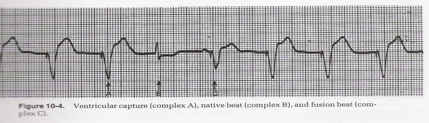
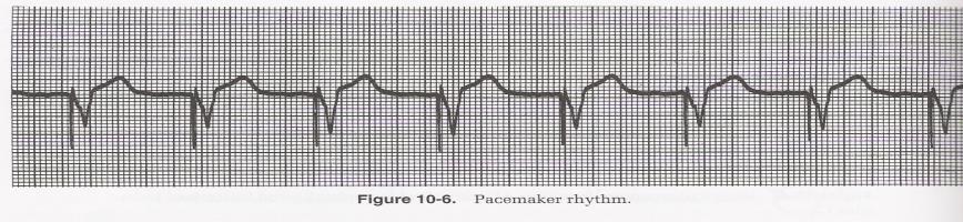
18 Pacemaker Rhythms Ventricular Capture 18


19 Marquette Electronics Copyright 1996 Marquette Electronics Copyright
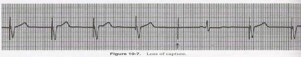
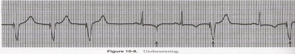
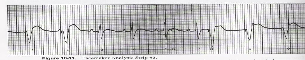
20 Pacemaker Troubleshooting Loss of Capture Under-sensing=Overpacing 20
21 Basic Rhythm Criteria Rhythm Criteria Treatment Normal Sinus Rhythm Sinus Brady Sinus Tach Sinus Arrhythmia PAC SVT (if no identifiable P waves) Atrial Tach (identifiable P waves) Atrial Flutter P wave uniform and upright for each QRS PRI= , QRS=.10 or less. Every p-wave is followed by a QRS, P to P is regular, R to R is regular Rate Same as above except rate is <60 Same as above except rate is P to P irregular in a pattern (must be 3 or more blocks off), Normal PR interval and QRS width, every P wave followed by a QRS. Beat is early and P-wave normally followed by QRS (except with nonconducted PAC). P- wave may be slightly different in appearance from the sinus p-wave. Regular rhythm, rate , p-wave may be difficult to see at higher rates Baseline Sawtooth pattern (f-waves) The f-waves are regular, R to R regular or irregular, usually at a rate of Monitor/Observe O2, IV, If symptomatic- Atropine, Pacemaker O2, IV, treat underlying cause (fever, pain, anxiety) Usually none required Usually none required. Monitor for increased frequency. Eliminate cause (caffeine, tobacco, stress) O2, IV, Vagal Maneuvers, Adenosine, Cardizem, synchronized cardioversion O2, IV. If pt. symptomatic: synchronized cardioversion, anticoagulation (if onset >24 hours), Cardizem, Amiodarone Asymptomatic: Cardizem, Amiodarone Anticoagulate 21
22 Atrial Fibrillation Irregular QRS, baseline is wavy with no discernable p-waves associated with QRS complexes. R to R irregular. O2, IV. If pt. symptomatic: synchronized cardioversion, anticoagulation (onset >24 hours), Cardizem, Amiodarone Asymptomatic: Cardizem, Amiodarone, Anticoagulate Junctional Rhythm P-wave is inverted in front of, buried within, or immediately behind the QRS, R to R regular, QRS normal. Rate Accelerated Junctional P-wave is inverted in front of, buried within, or immediately behind the QRS, R to R regular, QRS normal. Rate Junctional Tachycardia P-wave is inverted in front of, buried within, or immediately behind the QRS, R to R regular, QRS normal, rate >100 PJC Underlying rate reg but altered by early beat. P- wave is inverted in front of, buried within, or immediately behind the QRS 1 st Degree Block Meets criteria for a Sinus rhythm except PR interval is prolonged > nd Degree Block Mobitz Type I/Wenckebach 2 nd Degree Block Mobitz Type II PR interval progressively lengthens until a QRS is dropped. P to P regular. More P s than QRS s. QRS normal. P to P regular, PRI constant but with a dropped QRS. More P s than QRS s. QRS may be >.10 O2, IV, monitor pt If symptomatic: Pacemaker, Atropine Treat cause of rhythm (medications, MI) O2, IV, monitor pt. Reverse underlying cause (digoxin toxicity, Inf MI, rheumatic fever). Usually well tolerated. O2, IV, monitor pt. Treat underlying cause (digoxin toxicity, MI, CHF,) can use Cardizem. Monitor for frequency O2, IV, monitor pt, check cause (Beta Blocker, Calcium Channel Blocker, digoxin toxicity) O2, IV, monitor pt, Treat underlying cause (drugs, MI, Hyperkalemia) If symptomatic: Atropine. Usually a well tolerated rhythm by the pt. O2, IV, Pacemaker ASAP Dopamine or Epi gtt for BP support 22
23 Complete Heart Block 3 rd Degree Block Idioventricular PVC Atrial and Ventricular beats are regular but independent of each other with no relationship between the two. Ventricular rate P can fall into QRS. More P s than QRS s. QRS >.12 Rate < 40, regular rhythm, no p-wave, no PRI, QRS>0.12 (wide) QRS is early, wide, and has bizarre configuration: it is not preceded by a p- wave O2, IV, Pacemaker ASAP Dopamine or Epi gtt for BP support O2, IV, monitor pt, Pacemaker, Atropine, O2, IV monitor frequency, Lidocaine, Amiodarone, Reverse cause (stress, alcohol, caffeine, ischemia) Ventricular Tachycardia Regular rhythm, no p- wave, no PRI, QRS >0.12, rate >100, or group of 3 or more PVCs in a row Ventricular Fibrillation Irregular baseline, no discernable ECG waveform, no P waves, no QRS, no pulse O2, IV, If pulse present: synchronized cardioversion, if NO pulse defibrillate, CPR. ACLS protocol. Amiodarone, Epi, Magnesium for Torsades. O2, IV, defibrillate, CPR ACLS protocol Epinephrine, Amiodarone, Asystole No PQRST, baseline straight or slightly wavy. Confirm in 2 leads ACLS protocol. O2, IV, Epinephrine,Treat cause (H s and T s) 23
TEST BANK FOR ECGS MADE EASY 5TH EDITION BY AEHLERT
 Link download full: http://testbankair.com/download/test-bank-for-ecgs-made-easy-5thedition-by-aehlert/ TEST BANK FOR ECGS MADE EASY 5TH EDITION BY AEHLERT Chapter 5 TRUE/FALSE 1. The AV junction consists
EKG Rhythm Interpretation Exam
 as EKG Rhythm Interpretation Exam Name: Date: ID# Unit Assume each strip is a 6 second strip. Passing is 80%. 1. Identify the following rhythm: a. Asystole b. Ventricular fibrillation c. Atrial fibrillation
Course Objectives. Proper Lead Placements. Review the ECG print paper. Review the mechanics of the Myocardium. Review basics of ECG Rhythms
 ECG Interpretations Course Objectives Proper Lead Placements Review the ECG print paper Review the mechanics of the Myocardium Review basics of ECG Rhythms How Leads Work The ECG Leads we use are Bipolar
-RHYTHM PRACTICE- By Dr.moanes Msc.cardiology Assistant Lecturer of Cardiology Al Azhar University. OBHG Education Subcommittee
 -RHYTHM PRACTICE- By Dr.moanes Msc.cardiology Assistant Lecturer of Cardiology Al Azhar University The Normal Conduction System Sinus Node Normal Sinus Rhythm (NSR) Sinus Bradycardia Sinus Tachycardia
Rhythm ECG Characteristics Example. Normal Sinus Rhythm (NSR)
") Normal Sinus Rhythm (NSR) Rate: 60-100 per minute Rhythm: R- R = P waves: Upright, similar P-R: 0.12-0.20 second & consistent P:qRs: 1P:1qRs Sinus Tachycardia Exercise Hypovolemia Medications Fever Substances
Normal Sinus Rhythm (NSR) Rate: 60-100 per minute Rhythm: R- R = P waves: Upright, similar P-R: 0.12-0.20 second & consistent P:qRs: 1P:1qRs Sinus Tachycardia Exercise Hypovolemia Medications Fever Substances
Review Packet EKG Competency This packet is a review of the information you will need to know for the proctored EKG competency test.
 Review Packet EKG Competency 2015 This packet is a review of the information you will need to know for the proctored EKG competency test. Normal Sinus Rhythm Rhythm: Regular Ventricular Rate: 60-100 bpm
UNDERSTANDING YOUR ECG: A REVIEW
 UNDERSTANDING YOUR ECG: A REVIEW Health professionals use the electrocardiograph (ECG) rhythm strip to systematically analyse the cardiac rhythm. Before the systematic process of ECG analysis is described
Arrhythmia Study Guide 3 Junctional and Ventricular Rhythms
 Arrhythmia Study Guide 3 Junctional and Ventricular Rhythms JUNCTIONAL RHYTHMS The AV Junction (Bundle of His and surrounding cells) only acts as pacemaker of the heart when the SA Node is not firing normally
The ABCs of EKGs/ECGs for HCPs. Al Heuer, PhD, MBA, RRT, RPFT Professor, Rutgers School of Health Related Professions
 The ABCs of EKGs/ECGs for HCPs Al Heuer, PhD, MBA, RRT, RPFT Professor, Rutgers School of Health Related Professions Learning Objectives Review the basic anatomy of the heart Describe the cardiac conducting
Basic ECG Interpretation Module Notebook
 Basic ECG Interpretation Module Notebook ECG_Notebook_04.27.05 Page 1 of 142 Basic ECG Interpretation Table of Contents Module Objectives... 3 Module Outline... 6 Lesson I... 6 Lesson II... 8 Lesson III...
Arrhythmic Complications of MI. Teferi Mitiku, MD Assistant Clinical Professor of Medicine University of California Irvine
 Arrhythmic Complications of MI Teferi Mitiku, MD Assistant Clinical Professor of Medicine University of California Irvine Objectives Brief overview -Pathophysiology of Arrhythmia ECG review of typical
2017 BDKA Review. Regularity Rate P waves PRI QRS Interpretation. Regularity Rate P waves PRI QRS Interpretation 1/1/2017
 1. 2017 BDKA Review 2. 3. 4. Interpretation 5. QT 6. 7. 8. 9. 10. QT 11. 12. 13. 14. 15. 16. 17. 18. QT 19. 20. QT 21. 22. QT 23. 24. Where are pacer spikes? Before the P wave or before the QRS complex?
ABCs of ECGs. Shelby L. Durler
 ABCs of ECGs Shelby L. Durler Objectives Review the A&P of the cardiac conduction system Placement and obtaining 4-lead and 12-lead ECGs Overview of the basics of ECG rhythm interpretation Intrinsic
EKG Competency for Agency
 EKG Competency for Agency Name: Date: Agency: 1. The upper chambers of the heart are known as the: a. Atria b. Ventricles c. Mitral Valve d. Aortic Valve 2. The lower chambers of the heart are known as
Cardiac Telemetry Self Study: Part One Cardiovascular Review 2017 THINGS TO REMEMBER
 Please review the above anatomy of the heart. THINGS TO REMEMBER There are 3 electrolytes that affect cardiac function o Sodium, Potassium, and Calcium When any of these electrolytes are out of the normal
ACLS Study Guide for Precourse Self-Assessment
 20 rhythm strips on Precourse Self-Assessment with the following matching choices: Agonal Rhythm/Asystole Atrial Fibrillation Flutter Ventricular Fibrillation Monomorphic Ventricular Normal Sinus Rhythm
PATIENT WITH ARRHYTHMIA IN DENTIST S OFFICE. Małgorzata Kurpesa, MD., PhD. Chair&Department of Cardiology
 PATIENT WITH ARRHYTHMIA IN DENTIST S OFFICE Małgorzata Kurpesa, MD., PhD. Chair&Department of Cardiology Medical University of Łódź The heart is made up of four chambers Left Atrium Right Atrium Left Ventricle
Rate: The atrial and ventricular rates are equal; heart rate is greater than 100 bpm (usually between bpm).
.") Sinus Bradycardia Regularity: The R-R intervals are constant; the rhythm is regular. Rate: The atrial and ventricular rates are equal; heart rate is less than 60 bpm. P wave: There is a uniform P wave
Sinus Bradycardia Regularity: The R-R intervals are constant; the rhythm is regular. Rate: The atrial and ventricular rates are equal; heart rate is less than 60 bpm. P wave: There is a uniform P wave
Step by step approach to EKG rhythm interpretation:
 Sinus Rhythms Normal sinus arrhythmia Small, slow variation of the R-R interval i.e. variation of the normal sinus heart rate with respiration, etc. Sinus Tachycardia Defined as sinus rhythm with a rate
Basic Dysrhythmia Interpretation
 Basic Dysrhythmia Interpretation Objectives 2 To understand the Basic ECG To understand the meaning of Dysrhythmia To describe the normal heart conduction system. To describe the normal impulse pathways.
CRC 431 ECG Basics. Bill Pruitt, MBA, RRT, CPFT, AE-C
 CRC 431 ECG Basics Bill Pruitt, MBA, RRT, CPFT, AE-C Resources White s 5 th ed. Ch 6 Electrocardiography Einthoven s Triangle Chest leads and limb leads Egan s 10 th ed. Ch 17 Interpreting the Electrocardiogram
EKG Intermediate Tips, tricks, tools
 Birmingham Regional Emergency Medical Services System 2018 ALCTE Summer Conference EKG Intermediate Tips, tricks, tools Brian Gober, MAT, ATC, NRP, CSCS Education Services Manager ECC Training Center Coordinator
EKG Abnormalities. Adapted from:
 EKG Abnormalities Adapted from: http://www.bem.fi/book/19/19.htm Some key terms: Arrhythmia-an abnormal rhythm or sequence of events in the EKG Flutter-rapid depolarizations (and therefore contractions)
CORONARY ARTERIES HEART
 CARDIAC/ECG MODULE THE HEART CORONARY ARTERIES FIBRILLATING HEART CORONARY ARTERIES HEART PRACTICE RHYTHMS PRACTICE RHYTHMS ELECTRICAL CONDUCTION SA Node (60 100) Primary pacemaker AV Node (40 60) ***Creates
4/14/15 HTEC 91. Topics for Today. Guess That Rhythm. Premature Ventricular Contractions (PVCs) Ventricular Rhythms
 Ventricular Rhythms") 4/14/15 Topics for Today HTEC 91 Medical Office Diagnostic Tests Week 5 Ventricular Rhythms PVCs: Premature Ventricular Contractions VT: Ventricular Tachycardia VF: Ventricular Fibrillation Asystole Study
4/14/15 Topics for Today HTEC 91 Medical Office Diagnostic Tests Week 5 Ventricular Rhythms PVCs: Premature Ventricular Contractions VT: Ventricular Tachycardia VF: Ventricular Fibrillation Asystole Study
Northwest Community Healthcare Paramedic Education Program AV Conduction Defects/AV Blocks Connie J. Mattera, M.S., R.N., EMT-P
 Northwest Community Healthcare Paramedic Education Program AV Conduction Defects/ Connie J. Mattera, M.S., R.N., EMT-P Reading assignments: Bledsoe Vol. 3: pp. 88-93; 120-121 (atropine, norepinephrine,
Electrocardiography for Healthcare Professionals
 Electrocardiography for Healthcare Professionals Chapter 9: Ventricular Dysrhythmias 2012 The Companies, Inc. All rights reserved. Learning Outcomes 9.1 Describe the various ventricular dysrhythmias 9.2
Chapter 9. Learning Objectives. Learning Objectives 9/11/2012. Cardiac Arrhythmias. Define electrical therapy
 Chapter 9 Cardiac Arrhythmias Learning Objectives Define electrical therapy Explain why electrical therapy is preferred initial therapy over drug administration for cardiac arrest and some arrhythmias
INDEX. Kingston General Hospital May Introduction Page 2. Cardiac Conduction System Page 3. Introduction to Cardiac Monitoring Page 5
 Kingston General Hospital May 2008 INDEX Introduction Page 2 Cardiac Conduction System Page 3 Introduction to Cardiac Monitoring Page 5 Recording Electrical Events Page 9 Waves of the ECG Complex Page
Basic EKG Interpretation. Nirja Parikh, PT, DPT
 Basic EKG Interpretation Nirja Parikh, PT, DPT Electrocardiogram (EKG) Using surface electrodes record the electrical activity of the heart electrical activity (usually) correlates to mechanical function
Lake EMS Basic EKG Review: Atrial Rhythms. The Lake EMS Quality Development Team
 Lake EMS Basic EKG Review: Atrial Rhythms The Lake EMS Quality Development Team This program is the Intellectual Property of Lake Emergency Medical Services Use of this program is limited to training and
The ECG Course. Boone County Fire Protection District EMS Education
 The ECG Course Level I G rated material AV Blocks What Causes AV Block? Long list of bad things that includes ischemia and.. Old age / disease Medications or drugs Electrolyte imbalances Physiologic Blocks
CSI Skills Lab #5: Arrhythmia Interpretation and Treatment
 CSI 202 - Skills Lab #5: Arrhythmia Interpretation and Treatment Origins of the ACLS Approach: CSI 202 - Skills Lab 5 Notes ACLS training originated in Nebraska in the early 1970 s. Its purpose was to
Cardiac Arrhythmia How to approach นพ.พ น จ แกวส วรรณะ หน วยโรคห วใจและหลอดเล อด
 Cardiac Arrhythmia How to approach นพ.พ น จ แกวส วรรณะ หน วยโรคห วใจและหลอดเล อด EKG paper is a grid where time is measured along the horizontal axis. Each small square is 1 mm in length and represents
I have no conflicts of interest relative to this lecture.
 PATHWAYS AROUND THE PITFALLS OF EKG RHYTHM INTERPRETATION Donald D. Brown, MD April 4, 2019 I have no conflicts of interest relative to this lecture. OBJECTIVES FOR PATHWAYS AROUND PITFALLS OF EKG RHYTHM
COURSE DESCRIPTION. Rev 2.0 7/2013. Page 1 of 26
 COURSE DESCRIPTION Ventricular dysrhythmias (arrhythmias) are unique and potentially dangerous cardiac rhythms. They are often associated with Code Blue calls, and life and death situations. The only two
ECG Interpretation and Clinical Significance
 Confirming Pages ECG Interpretation and Clinical Significance Chapter Outline 5.1 5.2 5.3 5.4 5.5 5.6 5.7 5.8 5.9 5.10 Learning Outcomes Key Terms Introduction (p. 114) Identifying the Components of the
Electrocardiography for Healthcare Professionals
 Electrocardiography for Healthcare Professionals Chapter 7: Junctional Dysrhythmias 2012 The Companies, Inc. All rights reserved. Learning Outcomes 7.1 Describe the various junctional dysrhythmias 7.2
8/20/2012. Learning Outcomes (Cont d)
") 1 2 3 4 Electrocardiography for Healthcare Professionals Chapter 7: Junctional Dysrhythmias Learning Outcomes 7.1 Describe the various junctional dysrhythmias 7.2 Identify premature junctional complexes
1 2 3 4 Electrocardiography for Healthcare Professionals Chapter 7: Junctional Dysrhythmias Learning Outcomes 7.1 Describe the various junctional dysrhythmias 7.2 Identify premature junctional complexes
HTEC 91. Performing ECGs: Procedure. Normal Sinus Rhythm (NSR) Topic for Today: Sinus Rhythms. Characteristics of NSR. Conduction Pathway
 Topic for Today: Sinus Rhythms. Characteristics of NSR. Conduction Pathway") HTEC 91 Medical Office Diagnostic Tests Week 3 Performing ECGs: Procedure o ECG protocol: you may NOT do ECG if you have not signed up! If you are signed up and the room is occupied with people who did
HTEC 91 Medical Office Diagnostic Tests Week 3 Performing ECGs: Procedure o ECG protocol: you may NOT do ECG if you have not signed up! If you are signed up and the room is occupied with people who did
Full file at
 MULTIPLE CHOICE. Choose the one alternative that best completes the statement or answers the question. 1) What electrical event must occur for atrial kick to occur? 1) A) Atrial repolarization B) Ventricular
1 Cardiology Acute Care Day 22 April 2013 Arrhythmia Tutorial Course Material
 1 Cardiology Acute Care Day 22 April 2013 Arrhythmia Tutorial Course Material Arrhythmia recognition This tutorial builds on the ECG lecture and provides a framework for approaching any ECG to allow the
2) Heart Arrhythmias 2 - Dr. Abdullah Sharif
 Heart Arrhythmias 2 - Dr. Abdullah Sharif") 2) Heart Arrhythmias 2 - Dr. Abdullah Sharif Rhythms from the Sinus Node Sinus Tachycardia: HR > 100 b/m Causes: o Withdrawal of vagal tone & Sympathetic stimulation (exercise, fight or flight) o Fever
2) Heart Arrhythmias 2 - Dr. Abdullah Sharif Rhythms from the Sinus Node Sinus Tachycardia: HR > 100 b/m Causes: o Withdrawal of vagal tone & Sympathetic stimulation (exercise, fight or flight) o Fever
Chad Morsch B.S., ACSM CEP
 What Is Cardiac Stress Testing? Chad Morsch B.S., ACSM CEP A Cardiac Stress Test is a test used to measure the heart's ability to respond to external stress in a controlled clinical environment. Cardiac
Answer: The electrical current has difficulty traveling down the normal conduction pathway.
 1 Electrocardiography for Healthcare Professionals Chapter 8: Heart Block Dysrhythmias 2 3 4 5 6 Learning Outcomes 8.1 Describe the various heartblock dysrhythmias 8.2 Identify first degree heartblock
Lab Activity 24 EKG. Portland Community College BI 232
 Lab Activity 24 EKG Reference: Dubin, Dale. Rapid Interpretation of EKG s. 6 th edition. Tampa: Cover Publishing Company, 2000. Portland Community College BI 232 Graph Paper 1 second equals 25 little boxes
Dysrhythmias that every Learn how to recognize an abnormal cardiac rhythm and intervene appropriately. By AnneMarie Palatnik, RN, APN-BC, MSN
 Too fast, too slow, too ugly: Dysrhythmias that every Learn how to recognize an abnormal cardiac rhythm and intervene appropriately. By AnneMarie Palatnik, RN, APN-BC, MSN 2.5 ANCC CONTACT HOURS CONTINUOUS
ECG Interpretation Cat Williams, DVM DACVIM (Cardiology)
") ECG Interpretation Cat Williams, DVM DACVIM (Cardiology) Providing the best quality care and service for the patient, the client, and the referring veterinarian. GOAL: Reduce Anxiety about ECGs Back to
ECG Interpretation Cat Williams, DVM DACVIM (Cardiology) Providing the best quality care and service for the patient, the client, and the referring veterinarian. GOAL: Reduce Anxiety about ECGs Back to
BEDSIDE ECG INTERPRETATION
 BEDSIDE ECG INTERPRETATION Presented by: Ryan Dean, RN, MSN, CCRN, CCNS, CFRN Flight Nurse 2017 Based on presentations originally by Gennifer DePaoli, RN Objectives Hospital policies Electrical conduction
Understanding the 12-lead ECG, part II
 Bundle-branch blocks Understanding the 12-lead ECG, part II Most common electrocardiogram (ECG) abnormality Appears as a wider than normal S complex Occurs when one of the two bundle branches can t conduct
ECGs and Arrhythmias: Family Medicine Board Review 2009
 Rate Rhythm Intervals Hypertrophy ECGs and Arrhythmias: Family Medicine Board Review 2009 Axis Jess (Fogler) Waldura, MD University of California, San Francisco walduraj@nccc.ucsf.edu Ischemia Overview
Paramedic Rounds. Tachyarrhythmia's. Sean Sutton Dallas Wood
 Paramedic Rounds Tachyarrhythmia's Sean Sutton Dallas Wood Objectives At the end of this session, the paramedic will be able to: State the key components of the cardiac conduction pathway, along with the
Chapter 03: Sinus Mechanisms Test Bank MULTIPLE CHOICE
 Instant download and all chapters Tesst Bank ECGs Made Easy 5th Edition Barbara J Aehlert https://testbanklab.com/download/tesst-bank-ecgs-made-easy-5th-edition-barbara-jaehlert/ Chapter 03: Sinus Mechanisms
ECG Interpretation. Introduction to Cardiac Telemetry. Michael Peters, RN, CCRN, CFRN CALSTAR Air Medical Services
 ECG Interpretation Introduction to Cardiac Telemetry Michael Peters, RN, CCRN, CFRN CALSTAR Air Medical Services Disclosures Nothing to disclose Objectives Describe the electrical conduction pathway in
Cardiology Flash Cards
 Cardiology Flash Cards EKG in a nut shell www.brain101.info Conduction System www.brain101.info 2 Analyzing EKG Step by step Steps in Analyzing ECG'S 1. Rhythm: - Regular _ Sinus, Junctional or Ventricular.
SIMPLY ECGs. Dr William Dooley
 SIMPLY ECGs Dr William Dooley Content Basic ECG interpretation pattern Some common (examined) abnormalities Presenting ECGs in context Setting up an ECG Setting up an ECG 1 V1-4 th Right intercostal space
Contents ECG Study Guide American Heart Association (AHA) Guidelines Highlights Update for Cardiopulmonary Resuscitation (CPR) and
 Guidelines Highlights Update for Cardiopulmonary Resuscitation (CPR) and") ECG Study Guide Contents ECG Study Guide... 4 2015 American Heart Association (AHA) Guidelines Highlights Update for Cardiopulmonary Resuscitation (CPR) and Emergency Cardiovascular Care (ECC).... 4 ECG
ECG Study Guide Contents ECG Study Guide... 4 2015 American Heart Association (AHA) Guidelines Highlights Update for Cardiopulmonary Resuscitation (CPR) and Emergency Cardiovascular Care (ECC).... 4 ECG
Matters of the Heart: Comprehensive Cardiology SARAH BEANLANDS RN BSCN MSC
 Matters of the Heart: Comprehensive Cardiology SARAH BEANLANDS RN BSCN MSC Who am I? Class Outline Gross anatomy of the heart Trip around the heart Micro anatomy: cellular and tissue level Introduction
Human Anatomy and Physiology II Laboratory Cardiovascular Physiology
 Human Anatomy and Physiology II Laboratory Cardiovascular Physiology 1 This lab involves two exercises: 1) Conduction System of the Heart and Electrocardiography and 2) Human Cardiovascular Physiology:
How To Think About Rhythms and Conduction
 How To Think About Rhythms and Conduction Frank Yanowitz, MD Professor of Medicine (Cardiology) University of Utah School of Medicine Medical Director, IHC ECG Services Intermountain Healthcare http://ecg.utah.edu
Cardiac Cycle. Each heartbeat is called a cardiac cycle. First the two atria contract at the same time.
 The Heartbeat Cardiac Cycle Each heartbeat is called a cardiac cycle. First the two atria contract at the same time. Next the two ventricles contract at the same time. Then all the chambers relax. http://www.youtube.com/watch?v=frd3k6lkhws
physiology 6 Mohammed Jaafer Turquoise team
 15 physiology 6 Mohammed Jaafer 22-3-2016 Turquoise team Cardiac Arrhythmias and Their Electrocardiographic Interpretation Today, we are going to talk about the abnormal excitation. As we said before,
ECG ABNORMALITIES D R. T AM A R A AL Q U D AH
 ECG ABNORMALITIES D R. T AM A R A AL Q U D AH When we interpret an ECG we compare it instantaneously with the normal ECG and normal variants stored in our memory; these memories are stored visually in
Cardiac Arrhythmias. Cathy Percival, RN, FALU, FLMI VP, Medical Director AIG Life and Retirement Company
 Cardiac Arrhythmias Cathy Percival, RN, FALU, FLMI VP, Medical Director AIG Life and Retirement Company The Cardiovascular System Three primary functions Transport of oxygen, nutrients, and hormones to
Electrocardiography Abnormalities (Arrhythmias) 7. Faisal I. Mohammed, MD, PhD
 7. Faisal I. Mohammed, MD, PhD") Electrocardiography Abnormalities (Arrhythmias) 7 Faisal I. Mohammed, MD, PhD 1 Causes of Cardiac Arrythmias Abnormal rhythmicity of the pacemaker Shift of pacemaker from sinus node Blocks at different
Electrocardiography Abnormalities (Arrhythmias) 7 Faisal I. Mohammed, MD, PhD 1 Causes of Cardiac Arrythmias Abnormal rhythmicity of the pacemaker Shift of pacemaker from sinus node Blocks at different
Sample. Analyzing the Heart with EKG. Computer
 Analyzing the Heart with EKG Computer An electrocardiogram (ECG or EKG) is a graphical recording of the electrical events occurring within the heart. In a healthy heart there is a natural pacemaker in
The Electrocardiogram
 The Electrocardiogram Chapters 11 and 13 AUTUMN WEDAN AND NATASHA MCDOUGAL The Normal Electrocardiogram P-wave Generated when the atria depolarizes QRS-Complex Ventricles depolarizing before a contraction
McHenry Western Lake County EMS System Optional CE for EMT-B, Paramedics and PHRN s Bradycardia and Treatments Optional #7 2018
 McHenry Western Lake County EMS System Optional CE for EMT-B, Paramedics and PHRN s Bradycardia and Treatments Optional #7 2018 This month we will be looking at a specific ECG Rhythm and its treatments
PACIFIC MEDICAL TRAINING Arrhythmia Interpretation
 PACIFIC MEDICAL TRAINING Arrhythmia Interpretation Introduction Activity Summary Target Audience Educational Objectives Nursing Educational Objective Faculty Physician Continuing Medical Education Nursing
CASE 10. What would the ST segment of this ECG look like? On which leads would you see this ST segment change? What does the T wave represent?
 CASE 10 A 57-year-old man presents to the emergency center with complaints of chest pain with radiation to the left arm and jaw. He reports feeling anxious, diaphoretic, and short of breath. His past history
CARDIOVASCULAR SYSTEM
 CARDIOVASCULAR SYSTEM Overview Heart and Vessels 2 Major Divisions Pulmonary Circuit Systemic Circuit Closed and Continuous Loop Location Aorta Superior vena cava Right lung Pulmonary trunk Base of heart
Objectives of the Heart
 Objectives of the Heart Electrical activity of the heart Action potential EKG Cardiac cycle Heart sounds Heart Rate The heart s beat separated into 2 phases Relaxed phase diastole (filling of the chambers)
3/26/15 HTEC 91. EKG Sign-in Book. The Cardiac Cycle. Parts of the ECG. Waves. Waves. Review of protocol Review of placement of chest leads (V1, V2)
") EKG Sign-in Book HTEC 91 Review of protocol Review of placement of chest leads (V1, V2) Medical Office Diagnostic Tests Week 2 http://www.cvphysiology.com/arrhythmias/a013c.htm The Cardiac Cycle Represents
EKG Sign-in Book HTEC 91 Review of protocol Review of placement of chest leads (V1, V2) Medical Office Diagnostic Tests Week 2 http://www.cvphysiology.com/arrhythmias/a013c.htm The Cardiac Cycle Represents
5AB Dysrhythmia Interpretation tation and Management Review Please complete and return by:
 1 5AB Dysrhythmia Interpretation tation and Management Review 2014 RN Please complete and return by: Overview 2 This review begins with a discussion of the physiology of cardiac conduction, and then covers
Building Blocks: Deciphering Heart Blocks Visions Symposium AACN 2015
 1 Building Blocks: Deciphering Heart Blocks Visions Symposium AACN 2015 Greta Price MSN-Ed, RN-BC, PCCN Objectives Upon completion of this program, the participate will be able to: Describe the common
SIMPLY ECGs. Dr William Dooley
 SIMPLY ECGs Dr William Dooley 1 No anatomy just interpretation 2 Setting up an ECG 3 Setting up an ECG 1 V1-4 th Right intercostal space at sternal border 2 V2-4 th Left intercostal space at sternal border
12/2/2016. Basic Cardiac Anatomy. Blood Flow Through the Heart. Fun Fact.. Layers of the Heart. Layers of the Heart
 Basic Cardiac Anatomy Blood Flow Through the Heart Septum 1. Blood enters right atrium via inferior & superior vena cava 2. Right atrium contracts, sending blood through the tricuspid valve and into the
Lab 2. The Intrinsic Cardiac Conduction System. 1/23/2016 MDufilho 1
 Lab 2 he Intrinsic Cardiac Conduction System 1/23/2016 MDufilho 1 Figure 18.13 Intrinsic cardiac conduction system and action potential succession during one heartbeat. Superior vena cava ight atrium 1
Arrhythmias. Sarah B. Murthi Department of Surgery University of Maryland Medical School R. Adams Cowley Shock Trauma Center
 Arrhythmias Sarah B. Murthi Department of Surgery University of Maryland Medical School R. Adams Cowley Shock Trauma Center 2012 Clinical Congress Presenter Disclosure Slide American College of Surgeons
THE CARDIOVASCULAR SYSTEM. Heart 2
 THE CARDIOVASCULAR SYSTEM Heart 2 PROPERTIES OF CARDIAC MUSCLE Cardiac muscle Striated Short Wide Branched Interconnected Skeletal muscle Striated Long Narrow Cylindrical PROPERTIES OF CARDIAC MUSCLE Intercalated
Chapter 20 (2) The Heart
 The Heart") Chapter 20 (2) The Heart ----------------------------------------------------------------------------------------------------------------------------------------- Describe the component and function of
Chapter 20 (2) The Heart ----------------------------------------------------------------------------------------------------------------------------------------- Describe the component and function of
Cardiac Arrhythmias in Sleep
 Cardiac Arrhythmias in Sleep Only 53 Slides! Gauresh H Kashyap, MD, FACP, FCCP, FAASM 1 2 Cardiac Arrhythmias in Sleep Out of 400 Patients with OSA, 48% had some Arrhythmias 20% - 2 PVCs/min 7% - Bradycardia
Dr.Binoy Skaria 13/07/15
 Dr.Binoy Skaria binoyskaria@hotmail.com binoy.skaria@heartofengland.nhs.uk 13/07/15 Acknowledgement Medtronic, Google images & Elsevier for slides Natalie Ryan, Events Manager, HEFT- for organising the
Junctional Premature Contraction (JPC)
") Where s the PAC? Junctional Premature Contraction (JPC) A junctional premature contraction (JPC) is a beat that originates prematurely in the AV node. It can occur sporadically or in a grouped pattern.
Where s the PAC? Junctional Premature Contraction (JPC) A junctional premature contraction (JPC) is a beat that originates prematurely in the AV node. It can occur sporadically or in a grouped pattern.
By the end of this lecture, you will be able to: Understand the 12 lead ECG in relation to the coronary circulation and myocardium Perform an ECG
 By the end of this lecture, you will be able to: Understand the 12 lead ECG in relation to the coronary circulation and myocardium Perform an ECG recording Identify the ECG changes that occur in the presence
Lake EMS Basic EKG Review: Dreaded Heart Blocks. The Lake EMS Quality Development Team
 Lake EMS Basic EKG Review: Dreaded Heart Blocks The Lake EMS Quality Development Team This program is the Intellectual Property of Lake Emergency Medical Services Use of this program is limited to training
Please check your answers with correct statements in answer pages after the ECG cases.
 ECG Cases ECG Case 1 Springer International Publishing AG, part of Springer Nature 2018 S. Okutucu, A. Oto, Interpreting ECGs in Clinical Practice, In Clinical Practice, https://doi.org/10.1007/978-3-319-90557-0
Presented By: Barbara Furry, RN-BC, MS, CCRN, FAHA Director The Center of Excellence in Education Director of HERO
 Presented By: Barbara Furry, RN-BC, MS, CCRN, FAHA Director The Center of Excellence in Education Director of HERO Follow me on Twitter! CEE Med Updates@BarbaraFurryRN Like me on Facebook! What is a
4. The two inferior chambers of the heart are known as the atria. the superior and inferior vena cava, which empty into the left atrium.
 Answer each statement true or false. If the statement is false, change the underlined word to make it true. 1. The heart is located approximately between the second and fifth ribs and posterior to the
CRITICAL CARE OF THE CARDIAC PATIENT WEBINAR VET 2017
 CRITICAL CARE OF THE CARDIAC PATIENT WEBINAR VET 2017 The Heart The heart is undoubtedly the most important organ in the body. Unfortunately, when it is not functioning properly, it can have dire consequences.
A Review of Cardiac Pathophysiology and EKG. Jamie Dyson PT, DPT Kathy Swanick PT, DPT, OCS
 A Review of Cardiac Pathophysiology and EKG Jamie Dyson PT, DPT Kathy Swanick PT, DPT, OCS Cardiac Pathophysiology Coronary Artery Disease Congestive Heart Failure Valvular Heart Disease Athletic Heart
WHAT S THAT RHYTHM I AM HEARING? GUIDE TO AUSCULTATION OF ARRHYTHMIAS IN HORSES
 WHAT S THAT RHYTHM I AM HEARING? GUIDE TO AUSCULTATION OF ARRHYTHMIAS IN HORSES Michelle Henry Barton DVM, PhD, DACVIM University of Georgia, Athens, GA INTRODUCTION The purpose of this talk is to review
Electrical Conduction
 Sinoatrial (SA) node Electrical Conduction Sets the pace of the heartbeat at 70 bpm AV node (50 bpm) and Purkinje fibers (25 40 bpm) can act as pacemakers under some conditions Internodal pathway from
15 16 September Seminar W10O. ECG for General Practice
 15 16 September 2012 Seminar W10O ECG for General Practice Speaker: Ms Natasha Eaton ECG for General Practice Speaker: Natasha Eaton Cardiac CNC Executive Representative Electrocardiography The graphic
Electrocardiography Biomedical Engineering Kaj-Åge Henneberg
 Electrocardiography 31650 Biomedical Engineering Kaj-Åge Henneberg Electrocardiography Plan Function of cardiovascular system Electrical activation of the heart Recording the ECG Arrhythmia Heart Rate
Bradydysrhythmias and Atrioventricular Conduction Blocks
 Emerg Med Clin N Am 24 (2006) 1 9 Bradydysrhythmias and Atrioventricular Conduction Blocks Jacob W. Ufberg, MD*, Jennifer S. Clark, MD Department of Emergency Medicine, Temple University School of Medicine,
Atlantic Health System
 Atlantic Health System Morristown Medical Center Newton Medical Center Overlook Medical Center Basic Dysrhythmia Course Day 1 1 2 Chapter 1 Anatomy and Physiology Learning Objectives 1) Identify electrophysiology
PA-1350: ELECTROCARDIOGRAPHY
 PA-1350: Electrocardiography 1 PA-1350: ELECTROCARDIOGRAPHY Cuyahoga Community College Viewing:PA-1350 : Electrocardiography Board of Trustees: 2015-05-28 Academic Term: 2015-08-24 Subject Code PA - Physician
Introduction. Circulation
 Introduction Circulation 1- Systemic (general) circulation 2- Pulmonary circulation carries oxygenated blood to all parts of the body carries deoxygenated blood to the lungs From Lt. ventricle aorta From
Lecture outline. Electrical properties of the heart. Automaticity. Excitability. Refractoriness. The ABCs of ECGs Back to Basics Part I
 Lecture outline The ABCs of ECGs Back to Basics Part I Meg Sleeper VMD, DACVIM (cardiology) University of Florida Veterinary School Electrical properties of the heart Action potentials Normal intracardiac