Mechanisms of False Positive Exercise Electrocardiography: Is False Positive Test Truly False?
|
|
|
- Branden Cobb
- 6 years ago
- Views:
Transcription


1 Mechanisms of False Positive Exercise Electrocardiography: Is False Positive Test Truly False? Masaki Izumo a, Kengo Suzuki b, Hidekazu Kikuchi b, Seisyo Kou b, Keisuke Kida b, Yu Eguchi b, Nobuyuki Azuma b, Yoshihiro J Akashi b, Kazuto Omiya b, Fumihiko Miyake b, Takahiro Shiota a. a Division of Cardiology, Cedars-Sinai Medical Center, Los Angeles, California, USA b Division of Cardiology, Department of Internal Medicine, St. Marianna University School of Medicine, Kawasaki, Japan ESC Congress 2011 Paris
2 Disclosures Dr. Masaki Izumo: None Dr. Kengo Suzuki: None Mr. Hidekazu Kikuchi: None Dr. Seisyo Kou: None Dr. Keisuke Kida: None Dr. Yu Eguchi: None Dr. Nobuyuki Azuma: None Dr. Yoshihiro J Akashi: None Dr. Kazuto Omiya: None Pr. Fumihiko Miyake: None Pr. Takahiro Shiota: None
3 Background Treadmill exercise electrocardiographic test (TET) is the classical method for initial assessment of coronary artery disease (CAD). However, a significant number of patients reveal false positive with this method. The mechanism of false positive TET remains unclear.


4 Background Long term prognosis is poorer in male patients with false positive than aged-matched controls. Erikssen J et al. Circulation 1983;68:
5 Background Recently-introduced speckle tracking echocardiography (STE) quantifies myocardial deformation in the longitudinal, circumferential, and radial directions. Langeland S, et al. Experimental validation of a new ultrasound method for the simultaneous assessment of radial and longitudinal myocardial deformation independent of insonation angle. Circulation 2005; 112: Global longitudinal strain (GLS) calculated with this modality was reported to detect early left ventricular (LV) systolic dysfunction despite of normal ejection fraction. Wang J et al. Preserved left ventricular twist and circumferential deformation, but depressed longitudinal and radial deformation in patients with diastolic heart failure. Eur Heart J 2008; 29:
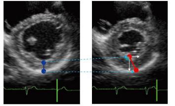

6 Background Strain = Lr-L0 / L0
7 Hypothesis We hypothesized that male false positive patients might have diastolic and subclinical systolic dysfunction, and STE could detect these dysfunction.
8 Study population 755 pts who underwent treadmill exercise test prior cardiovascular surgery, documented myocardial infarction, valvular heart diseases, complete left branch block in electrocardiography, depressed LVEF, atrial fibrillation. (All n=69) 133pts (19.4%); Positive study 553pts (80.6%); Negative study Evaluated by nuclear medicine; 30pts (22.6%) False positive; 36pts (27.1% male; 30pts, female 6pts) True positive; 67pts (50.4%) Single vessel disease; 30pts (22.6%) Multi vessel disease; 37pts (27.8%) Aged-matched control; 30pts False positive in men; 30pts True positive in men; 55pts
9 Methods Treadmill exercise test All patients underwent symptoms-limited TET according to one of the standard protocols (usually Bruce, modified Bruce, or Ramp). This study adopted the standard end points; to halt the exercise test was determined on a basis of fatigue, severe angina, ECG changes compatible with myocardial ischemia, hypertension (systolic BP 220 mmhg), hypotension (decrement of systolic BP 20 mmhg), or significant arrhythmias. Positive TET was defined as horizontal or down sloping ST depression of 1mm below the baseline or ST elevation of 1 mm above the baseline 80 ms after the J point.
10 Methods Two-dimensional echocardiography Echocardiography was performed using a commercially available system (Vivid E9 Dimension, GE Health care, Horten, Norway) equipped with a 3.5-MHz transducer within 2 days before or after TET. Global longitudinal, circumferential, and radial strain were calculated offline using EchoPack software (GE Health care, Horten, Norway) to evaluate LV systolic function.
 and systolic velocity (S ), and E/E was calculated as E")
11 Tissue Doppler Imaging S E (cm/s) S (cm/s) E/E E Tissue Doppler imaging measured peak early diastolic mitral annular velocity (E ) and systolic velocity (S ), and E/E was calculated as E divided by E.


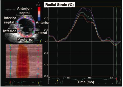
12 Speckle tracking imaging Global longitudinal strain The endocardial border was manually traced in the end-systolic frame using commercially available 2D strain software. Speckles were tracked frame by-frame throughout the LV wall during the cardiac cycle. Longitudinal strain was measured in a 16-segment LV model from apical views; 4-chamber, 2- chamber, and 3-chamber views.
 and global")

13 Global circumferential and radial strain Circumferential strain Global circumferential strain (GCS) and global radial strain (GRS) were obtained from the average of strain values, including basal, middle and apical levels. Radial strain
14 Clinical and treadmill exercise test data Variables Controls False positive P value Age 60.6± ± Body mass index 23.7± ± Hypertension (%) Smoking (%) Diabetes (%) Dyslipidemia (%) LV hypertrophy (%) Ex duration (min) 7.5± ± SBP at rest (mmhg) 128.8± ± DBP at rest (mmhg) 85.6± ± SBP peak ex (mmhg) 185.6± ± DBP peak ex (mmhg) 94.2± ± HR at rest (bpm) 79.4± ± HR peak ex (bpm) 153.8± ± Mean predicted HR (%) 92.4± ± ST level (mv) 0.08± ±0.13 <0.001
15 Conventional echocardiographic findings Controls False positive True positive P value (ANOVA) LVDd (mm) 44.4± ± ±5.1* LVDs (mm) 27.8± ± ±5.6* EDVI (ml/m 2 ) 57.1± ± ±13.6* <0.001 ESVI (ml/m2) 19.1± ± ±10.5* <0.001 EF (%) 68.0± ± ±10.2* IVSD 9.4± ± ± PWD 9.5± ± ± LV mass index (g/m 2 ) 85.6± ± ± LAVI 19.5± ± ± E/A 1.16± ± ±0.30* *; Significant difference (p<0.05) Control vs. True positive, ; Significant difference (p<0.05) False positive vs. True positive LVDd, left ventricular diastolic diameter; LVDs, left ventricular systolic diameter; EDVI, end diastolic volume index; ESVI, end systolic volume index; EF, ejection fraction; IVS, interventricular septal dimension; PWD, posterior wall dimension; LAVI, left atrial volume index.
16 Comparison of advanced echocardiographic findings P=0.002 P= P=0.006 P=ns P=ns P=ns E' (cm/s) ± ± ±2.1 E/E' ± ± ±2.1 Controls False positive True positive Controls False positive True positive P= P=0.013 P=0.215 S' (cm/s) ± ± ±1.2 Controls False positive True positive
17 Comparison of advanced echocardiographic findings Global longtudinal strain (%) P<0.001 P<0.001 P= ± ± ±2.3 Controls False positive True positive Global circumferential strain (%) P=0.043 P=ns P=ns -23.4± ± ±4.3 Controls False positive True positive Global radial strain (%) P=0.032 P=ns P=ns 43.2± ± ±6.6 Controls False positive True positive
18 Determinants of false positive TET Univariate Multivariate OR (95%CI) P value OR (95%CI) P value EF 1.03 ( ) LV mass index 0.92 ( ) E' 1.14 ( ) ( ) S 1.44 ( ) ( ) GLS 1.48 ( ) ( ) GCS 1.13 ( ) ( ) GRS 0.94 ( ) 0.488
19 Echocardiographic parameters predict positive exercise test. 100 Sensitivity (%) Specificity (%) Global longitudinal strain S E AUC Cut-off Sensitivity Specificity E' 0.75 <6.8cm/s S' 0.72 <6.7cm/s GLS 0.79 >-20.3%
20 Results The incidence of false positive results (27.1%) was similar to that of the earlier study. No significant differences in clinical and exercise characteristics except ECG changes were observed between the two groups. Both LV diastolic and subclinical systolic dysfunction were independently related to false positive TET
21 Clinically silent Clinically recognized Discussion Ischemic cascade Coronary flow reserve Endotherial function Camici PG, et al. Circulation 1992 Rigo F, et al. AJC 2002 Angina ECG abnormalities Systolic dysfunction Strain abnormalities Diastolic dysfunction Perfusion abnormalities Time/Magnitude ischemia Asimul A, Puthumana J. Echocardiography in acute coronary syndrome 2009
22 Conclusions Speckle-tracking and tissue Doppler echocardiography detected LV diastolic and subclinical systolic dysfunction in patients with false positive TET.
Global left ventricular circumferential strain is a marker for both systolic and diastolic myocardial function
 Global left ventricular circumferential strain is a marker for both systolic and diastolic myocardial function Toshinari Onishi 1, Samir K. Saha 2, Daniel Ludwig 1, Erik B. Schelbert 1, David Schwartzman
Coronary artery disease (CAD) risk factors
 risk factors") Background Coronary artery disease (CAD) risk factors CAD Risk factors Hypertension Insulin resistance /diabetes Dyslipidemia Smoking /Obesity Male gender/ Old age Atherosclerosis Arterial stiffness precedes
Background Coronary artery disease (CAD) risk factors CAD Risk factors Hypertension Insulin resistance /diabetes Dyslipidemia Smoking /Obesity Male gender/ Old age Atherosclerosis Arterial stiffness precedes
LV FUNCTION ASSESSMENT: WHAT IS BEYOND EJECTION FRACTION
 LV FUNCTION ASSESSMENT: WHAT IS BEYOND EJECTION FRACTION Jamilah S AlRahimi Assistant Professor, KSU-HS Consultant Noninvasive Cardiology KFCC, MNGHA-WR Introduction LV function assessment in Heart Failure:
Velocity Vector Imaging as a new approach for cardiac magnetic resonance: Comparison with echocardiography
 Velocity Vector Imaging as a new approach for cardiac magnetic resonance: Comparison with echocardiography Toshinari Onishi 1, Samir K. Saha 2, Daniel Ludwig 1, Erik B. Schelbert 1, David Schwartzman 1,
Conflict of interest: none declared
 The value of left ventricular global longitudinal strain assessed by three-dimensional strain imaging in the early detection of anthracycline-mediated cardiotoxicity C. Mornoş, A. Ionac, D. Cozma, S. Pescariu,
Evaluation of Left Ventricular Diastolic Dysfunction by Doppler and 2D Speckle-tracking Imaging in Patients with Primary Pulmonary Hypertension
 ESC Congress 2011.No 85975 Evaluation of Left Ventricular Diastolic Dysfunction by Doppler and 2D Speckle-tracking Imaging in Patients with Primary Pulmonary Hypertension Second Department of Internal
Right Ventricular Strain in Normal Healthy Adult Filipinos: A Retrospective, Cross- Sectional Pilot Study
 Right Ventricular Strain in Normal Healthy Adult Filipinos: A Retrospective, Cross- Sectional Pilot Study By Julius Caesar D. de Vera, MD Jonnah Fatima B. Pelat, MD Introduction Right ventricle contributes
Highlights from EuroEcho 2009 Echo in cardiomyopathies
 Highlights from EuroEcho 2009 Echo in cardiomyopathies Bogdan A. Popescu University of Medicine and Pharmacy, Bucharest, Romania ESC Congress 2010 Hypertrophic cardiomyopathy To determine the differences
Advanced Multi-Layer Speckle Strain Permits Transmural Myocardial Function Analysis in Health and Disease:
 Advanced Multi-Layer Speckle Strain Permits Transmural Myocardial Function Analysis in Health and Disease: Clinical Case Examples Jeffrey C. Hill, BS, RDCS Echocardiography Laboratory, University of Massachusetts
Heart Failure in Women: Dr Goh Ping Ping Cardiologist Asian Heart & Vascular Centre
 Heart Failure in Women: More than EF? Dr Goh Ping Ping Cardiologist Asian Heart & Vascular Centre Overview Review pathophysiology as it relates to diagnosis and management Rational approach to workup:
Left Ventricular Dyssynchrony in Patients Showing Diastolic Dysfunction without Overt Symptoms of Heart Failure
 ORIGINAL ARTICLE DOI: 10.3904/kjim.2010.25.3.246 Left Ventricular Dyssynchrony in Patients Showing Diastolic Dysfunction without Overt Symptoms of Heart Failure Jae Hoon Kim, Hee Sang Jang, Byung Seok
Dr. Dermot Phelan MB BCh BAO PhD European Society of Cardiology 2012
 Relative Apical Sparing of Longitudinal Strain Using 2- Dimensional Speckle-Tracking Echocardiography is Both Sensitive and Specific for the Diagnosis of Cardiac Amyloidosis. Dr. Dermot Phelan MB BCh BAO
The importance of left atrium in LV diastolic function
 II Baltic Heart Failure Meeting and Congress of Latvian Society of Cardiology The importance of left atrium in LV diastolic function Dr. Artem Kalinin Eastern Clinical University Hospital Riga 30.09.2010.
Μαρία Μπόνου Διευθύντρια ΕΣΥ, ΓΝΑ Λαϊκό
 Μαρία Μπόνου Διευθύντρια ΕΣΥ, ΓΝΑ Λαϊκό Diastolic HF DD: Diastolic Dysfunction DHF: Diastolic HF HFpEF: HF with preserved EF DD Pathophysiologic condition: impaired relaxation, LV compliance, LV filling
Feasibility and limitations of 2D speckle tracking echocardiography
 ORIGINAL ARTICLE 204 A prospective study in daily clinical practice Feasibility and limitations of 2D speckle tracking echocardiography Lina Melzer, Anja Faeh-Gunz, Barbara Naegeli, Burkhardt Seifert*,
Prognostic Value of Cardiopulmonary Exercise Testing in Patients with Atrial Fibrillation
 Prognostic Value of Cardiopulmonary Exercise Testing in Patients with Atrial Fibrillation Hidekazu Tsuneoka 1)2), Akira Koike 2), Osamu Nagayama 2), Koji Sakurada 2), Hitoshi Sawada 2), Kazutaka Aonuma
Restrictive Cardiomyopathy
 ESC Congress 2011, Paris Imaging Unusual Causes of Cardiomyopathy Restrictive Cardiomyopathy Kazuaki Tanabe, MD, PhD Professor of Medicine Chair, Division of Cardiology Izumo, Japan I Have No Disclosures
Alicia Armour, MA, BS, RDCS
 Alicia Armour, MA, BS, RDCS No disclosures Review 2D Speckle Strain (briefly) Discuss some various patient populations & disease pathways where Strain can be helpful Discuss how to acquire images for Strain
Quantitation of right ventricular dimensions and function
 SCCS Basics of cardiac assessment Quantitation of right ventricular dimensions and function Tomasz Kukulski, MD PhD Dept of Cardiology, Congenital Heart Disease and Electrotherapy Silesian Medical University
PRESENTER DISCLOSURE INFORMATION. There are no potential conflicts of interest regarding current presentation
 PRESENTER DISCLOSURE INFORMATION There are no potential conflicts of interest regarding current presentation Better synchrony and diastolic function for septal versus apical right ventricular permanent
Value of echocardiography in chronic dyspnea
 Value of echocardiography in chronic dyspnea Jahrestagung Schweizerische Gesellschaft für /Schweizerische Gesellschaft für Pneumologie B. Kaufmann 16.06.2016 Chronic dyspnea Shortness of breath lasting
Incorporating the New Echo Guidelines Into Everyday Practice
 Incorporating the New Echo Guidelines Into Everyday Practice Clinical Case RIGHT VENTRICULAR FAILURE Gustavo Restrepo MD President Elect Interamerican Society of Cardiology Director Fellowship Training
Strain and Strain Rate Imaging How, Why and When?
 Strain and Strain Rate Imaging How, Why and When? João L. Cavalcante, MD Advanced Cardiac Imaging Fellow Cleveland Clinic Foundation Disclosures: No conflicts of interest Movement vs Deformation Movement
Load and Function - Valvular Heart Disease. Tom Marwick, Cardiovascular Imaging Cleveland Clinic
 Load and Function - Valvular Heart Disease Tom Marwick, Cardiovascular Imaging Cleveland Clinic Indications for surgery in common valve lesions Risks Operative mortality Failed repair - to MVR Operative
Fetal cardiac function: what to use and does it make a difference?
 17 th International Conference on Prenatal Diagnosis and Therapy Lisbon, June 2013 Fetal cardiac function: what to use and does it make a difference? Fàtima Crispi Department of Maternal-Fetal Medicine,
Tissue Doppler and Strain Imaging
 Tissue Doppler and Strain Imaging Steven J. Lester MD, FRCP(C), FACC, FASE Relevant Financial Relationship(s) None Off Label Usage None 1 Objective way with which to quantify the minor amplitude and temporal
Appendix II: ECHOCARDIOGRAPHY ANALYSIS
 Appendix II: ECHOCARDIOGRAPHY ANALYSIS Two-Dimensional (2D) imaging was performed using the Vivid 7 Advantage cardiovascular ultrasound system (GE Medical Systems, Milwaukee) with a frame rate of 400 frames
Tissue Doppler and Strain Imaging. Steven J. Lester MD, FRCP(C), FACC, FASE
, FACC, FASE") Tissue Doppler and Strain Imaging Steven J. Lester MD, FRCP(C), FACC, FASE Relevant Financial Relationship(s) None Off Label Usage None a. Turn the wall filters on and turn down the receiver gain. b. Turn
Tissue Doppler and Strain Imaging Steven J. Lester MD, FRCP(C), FACC, FASE Relevant Financial Relationship(s) None Off Label Usage None a. Turn the wall filters on and turn down the receiver gain. b. Turn
Tissue Doppler and Strain Imaging
 Tissue Doppler and Strain Imaging Steven J. Lester MD, FRCP(C), FACC, FASE Relevant Financial Relationship(s) None Off Label Usage None 1 Objective way with which to quantify the minor amplitude and temporal
Rest and Exercise Echocardiography in Hypertrophic Cardiomyopathy: Determinants of Exercise Peak Gradient and Predictors of Outcome
 Rest and Exercise Echocardiography in Hypertrophic Cardiomyopathy: Determinants of Exercise Peak Gradient and Predictors of Outcome G. Deswarte, AS. Polge, N. Lamblin, A. Millaire, M. Richardson, C. Bauters,
Novel echocardiographic modalities: 3D echo, speckle tracking and strain rate imaging. Potential roles in sports cardiology. Stefano Caselli, MD, PhD
 Novel echocardiographic modalities: 3D echo, speckle tracking and strain rate imaging. Potential roles in sports cardiology. Stefano Caselli, MD, PhD Ospedale San Pietro Fatebenefratelli Rome, Italy Differential
Masked Hypertension and Aortic Coarctation: Impact on Ventricular Function and Morphology
 Masked Hypertension and Aortic Coarctation: Impact on Ventricular Function and Morphology Giovanni Di Salvo MD, PhD, MMSc, FESC BACKGROUND MASKED HYPERTENSION Masked hypertension (MH) consists of an elevated
Disclosure Information : No conflict of interest
 Intravenous nicorandil improves symptoms and left ventricular diastolic function immediately in patients with acute heart failure : a randomized, controlled trial M. Shigekiyo, K. Harada, A. Okada, N.
Aortic valve Stenosis: Insights in the evaluation of LV function. Erwan DONAL Cardiologie CHU Rennes
 Aortic valve Stenosis: Insights in the evaluation of LV function Erwan DONAL Cardiologie CHU Rennes erwan.donal@chu-rennes.fr Preload Afterload Myocardial Fiber Shortening Circumferential Longitudinal
Advanced Echocardiography in the Evaluation of Chemotherapy Patients
 Advanced Echocardiography in the Evaluation of Chemotherapy Patients Juan Carlos Plana, MD, FACC, FASE Co-Director, Cardio-Oncology Center Section of Cardiovascular Imaging Department of Cardiovascular
Adult Echocardiography Examination Content Outline
 Adult Echocardiography Examination Content Outline (Outline Summary) # Domain Subdomain Percentage 1 2 3 4 5 Anatomy and Physiology Pathology Clinical Care and Safety Measurement Techniques, Maneuvers,
SUPPLEMENTAL MATERIAL
 SUPPLEMENTAL MATERIAL Table S1: Number and percentage of patients by age category Distribution of age Age
Exercise Pulmonary Hypertension predicts the Occurrence of Symptoms in Asymptomatic Degenerative Mitral Regurgitation
 Exercise Pulmonary Hypertension predicts the Occurrence of Symptoms in Asymptomatic Degenerative Mitral Regurgitation Julien Magne, PhD, Kim O Connor, MD, Giuseppe Romano, MD, Marie Moonen, MD, Luc A.
Impact of Nicorandil on Renal Function in Patients With Acute Heart Failure and Pre-Existing Renal Dysfunction
 Impact of Nicorandil on Renal Function in Patients With Acute Heart Failure and Pre-Existing Renal Dysfunction Masahito Shigekiyo, Kenji Harada, Ayumi Okada, Naho Terada, Hiroyoshi Yoshikawa, Akira Hirono,
Degenerative Mitral Regurgitation: Etiology and Natural History of Disease and Triggers for Intervention
 Degenerative Mitral Regurgitation: Etiology and Natural History of Disease and Triggers for Intervention John N. Hamaty D.O. FACC, FACOI November 17 th 2017 I have no financial disclosures Primary Mitral
Hypertensive heart disease and failure
 Hypertensive heart disease and failure Prof. Dr. Alan Fraser Cardiff University The heart in hypertension Pathophysiology of LV adaptation Regional development of hypertrophy Stress testing - inducible
Strain Imaging: Myocardial Mechanics Simplified and Applied
 9/28/217 Strain Imaging: Myocardial Mechanics Simplified and Applied John Gorcsan III, MD Professor of Medicine Director of Clinical Research Division of Cardiology VECTORS OF CONTRACTION Shortening Thickening
Right Ventricular Systolic Dysfunction is common in Hypertensive Heart Failure: A Prospective Study in Sub-Saharan Africa
 Right Ventricular Systolic Dysfunction is common in Hypertensive Heart Failure: A Prospective Study in Sub-Saharan Africa 1 Ojji Dike B, Lecour Sandrine, Atherton John J, Blauwet Lori A, Alfa Jacob, Sliwa
Three-dimensional Wall Motion Tracking:
 Three-dimensional Wall Motion Tracking: A Novel Echocardiographic Method for the Assessment of Ventricular Volumes, Strain and Dyssynchrony Jeffrey C. Hill, BS, RDCS, FASE Jennifer L. Kane, RCS Gerard
OPTIMIZING ECHO ACQUISTION FOR STRAIN AND DIASTOLOGY
 OPTIMIZING ECHO ACQUISTION FOR STRAIN AND DIASTOLOGY October 8, 2017 Deborah Agler, ACS, RDCS, FASE Coordinator of Education and Training Cleveland Clinic General Principles Diastology Clinical Data Heart
Abstract nr AHA, Chicago November European Heart Journal Cardiovascular Imaging, in press. Nr Peter Blomstrand
 Left Ventricular Diastolic Function Assessed by Echocardiography and Tissue Doppler Imaging is a strong Predictor of Cardiovascular Events in Patients with Diabetes Mellitus Type 2 Peter Blomstrand, Martin
How To Perform Strain Imaging; Step By Step Approach. Maryam Bo Khamseen Echotechnoligist II EACVI, ARDMS, RCS King Abdulaziz Cardiac Center- Riyadh
 How To Perform Strain Imaging; Step By Step Approach Maryam Bo Khamseen Echotechnoligist II EACVI, ARDMS, RCS King Abdulaziz Cardiac Center- Riyadh Outlines: Introduction Describe the basic of myocardium
VECTORS OF CONTRACTION
 1/3/216 Strain, Strain Rate, and Torsion: Myocardial Mechanics Simplified and Applied VECTORS OF CONTRACTION John Gorcsan, MD University of Pittsburgh, Pittsburgh, PA Shortening Thickening Twisting No
Natural History and Echo Evaluation of Aortic Stenosis
 Natural History and Echo Evaluation of Aortic Stenosis Prof. Patrizio LANCELLOTTI, MD, PhD Heart Valve Clinic, University of Liège, CHU Sart Tilman, Liège, BELGIUM AORTIC STENOSIS First valvular disease
Københavns Universitet
 university of copenhagen Københavns Universitet Total average diastolic longitudinal displacement by colour tissue doppler imaging as an assessment of diastolic function de Knegt, Martina Chantal; Biering-Sørensen,
Left atrial function. Aliakbar Arvandi MD
 In the clinic Left atrial function Abstract The left atrium (LA) is a left posterior cardiac chamber which is located adjacent to the esophagus. It is separated from the right atrium by the inter-atrial
Ivana Nedeljkovic, M Ostojic, V Giga, V Stojanov, J Stepanovic, A Djordjevic Dikic, B Beleslin, M Nikolic, M Petrovic, D Popovic
 Combined cardiopulmonary exercise stress echocardiography test: New test for assessment of diastolic dysfunction in patients with hypertension Ivana Nedeljkovic, M Ostojic, V Giga, V Stojanov, J Stepanovic,
Echocardiography for the Electrophysiologist: Day-to-day practice. Emmanuel Fares, MD
 Echocardiography for the Electrophysiologist: Day-to-day practice Emmanuel Fares, MD EP and pacing service, Department of Cardiovascular Medicine, Cairo University Agenda Role of echo in arrhythmia management:
HIGHLIGHT SESSION. Imaging. J. L. Zamorano Gomez (Madrid, ES) Disclosures: Speaker Philips
 Disclosures: Speaker Philips") Imaging. J. L. Zamorano Gomez (Madrid, ES) Disclosures: Speaker Philips Agenda ECHO Diagnosis & Prognosis : Functional MR Severity Aortic Stenosis CT How to select pts for TAVI Adding prognostic info to
Imaging. J. L. Zamorano Gomez (Madrid, ES) Disclosures: Speaker Philips Agenda ECHO Diagnosis & Prognosis : Functional MR Severity Aortic Stenosis CT How to select pts for TAVI Adding prognostic info to
2/2/2011. Strain and Strain Rate Imaging How, Why and When? Movement vs Deformation. Doppler Myocardial Velocities. Movement. Deformation.
 Strain and Strain Rate Imaging How, Why and When? João L. Cavalcante, MD Advanced Cardiac Imaging Fellow Cleveland Clinic Foundation Disclosures: No conflicts of interest Movement vs Deformation Movement
Cardiac Magnetic Resonance in pregnant women
 Cardiac Magnetic Resonance in pregnant women Chen SSM, Leeton L, Dennis AT Royal Women s Hospital and The University of Melbourne, Parkville, Australia alicia.dennis@thewomens.org.au Quantification of
Echocardiography. Guidelines for Valve and Chamber Quantification. In partnership with
 Echocardiography Guidelines for Valve and Chamber Quantification In partnership with Explanatory note & references These guidelines have been developed by the Education Committee of the British Society
Indian Heart Journal
 Indian Heart Journal 70 (2018) 637 641 Contents lists available at ScienceDirect Indian Heart Journal journal homepage: www.elsevier.com/locate/ihj Original Article Global longitudinal strain, ejection
Exercise Testing/Echocardiography in Asymptomatic AS
 Exercise Testing/Echocardiography in Asymptomatic AS Raluca Dulgheru, MD Heart Valve Clinic, University of Liège, CHU Sart Tilman, BELGIUM Disclosure related to this presentation: None VALVULAR HEART DISEASE
DECLARATION OF CONFLICT OF INTEREST. None
 DECLARATION OF CONFLICT OF INTEREST None Hot Topics in Echocardiography: The position of the EAE EAE / ASE recommendation about Echo Assessment of Cardiac Mechanics Jens-Uwe Voigt Dpt. of Cardiovascular
Echocardiographic Assessment of the Left Ventricle
 Echocardiographic Assessment of the Left Ventricle Theodora Zaglavara, MD, PhD, BSCI/BSCCT Department of Cardiovascular Imaging INTERBALKAN EUROPEAN MEDICAL CENTER 2015 The quantification of cardiac chamber
My Patient Needs a Stress Test
 My Patient Needs a Stress Test Amy S. Burhanna,, MD, FACC Coastal Cardiology Cape May Court House, New Jersey Absolute and relative contraindications to exercise testing Absolute Acute myocardial infarction
Myocardial Strain Imaging in Cardiac Diseases and Cardiomyopathies.
 Myocardial Strain Imaging in Cardiac Diseases and Cardiomyopathies. Session: Cardiomyopathy Tarun Pandey MD, FRCR. Associate Professor University of Arkansas for Medical Sciences Disclosures No relevant
Cardiac Chamber Quantification by Echocardiography
 Cardiac Chamber Quantification by Echocardiography Maryam Bokhamseen, RCS, RCDS, EACVI Echotechnologist ǁ, Non invasive Cardiac Laboratory King Abdulaziz Cardiac Center. Outline: Introduction. Background
The Impact of Autonomic Neuropathy on Left Ventricular Function in Normotensive Type 1 Diabetic Patients: a Tissue Doppler Echocardiographic Study
 Diabetes Care Publish Ahead of Print, published online November 13, 2007 The Impact of Autonomic Neuropathy on Left Ventricular Function in Normotensive Type 1 Diabetic Patients: a Tissue Doppler Echocardiographic
Role of echocardiography in the assessment of ischemic heart disease 분당서울대학교병원윤연이
 Role of echocardiography in the assessment of ischemic heart disease 분당서울대학교병원윤연이 Outline Evaluation of Chest pain Evaluation of MI complications Prediction of Outcomes Evaluation of Chest pain Evaluation
Velocity, strain and strain rate: Doppler and Non-Doppler methods. Thoraxcentre, Erasmus MC,Rotterdam
 Velocity, strain and strain rate: Doppler and Non-Doppler methods J Roelandt J. Roelandt Thoraxcentre, Erasmus MC,Rotterdam Basics of tissue Doppler imaging Instantaneous annular velocity profiles IVCT
Abstract ESC Pisa
 Abstract ESC 82441 Maximal left ventricular mass-to-power output: A novel index to assess left ventricular performance and to predict outcome in patients with advanced heart failure FL. Dini 1, D. Mele
Mechanisms of heart failure with normal EF Arterial stiffness and ventricular-arterial coupling. What is the pathophysiology at presentation?
 Mechanisms of heart failure with normal EF Arterial stiffness and ventricular-arterial coupling What is the pathophysiology at presentation? Ventricular-arterial coupling elastance Central arterial pressure
Acute impairment of basal left ventricular rotation but not twist and untwist are involved in the pathogenesis of acute hypertensive pulmonary oedema
 Acute impairment of basal left ventricular rotation but not twist and untwist are involved in the pathogenesis of acute hypertensive pulmonary oedema A.D. Margulescu 1,2, R.C. Sisu 1,2, M. Florescu 2,
Heart Failure with preserved ejection fraction (HFpEF)
") Heart Failure with preserved ejection fraction (HFpEF) Dr. Pierpaolo Pellicori Hull York Medical School Kingston-upon-Hull United Kingdom Conflict of interest: none Heart failure is a contemporary problem
Heart Failure with preserved ejection fraction (HFpEF) Dr. Pierpaolo Pellicori Hull York Medical School Kingston-upon-Hull United Kingdom Conflict of interest: none Heart failure is a contemporary problem
The difficult patient with mitral regurgitation
 Clinical pathways The difficult patient with mitral regurgitation Stress echo can be the best tool Challenging cases Maria João Andrade, Lisbon PT Management of Severe Chronic Organic MR Echo Exercise
Imaging in Heart Failure: A Multimodality Approach. Thomas Ryan, MD
 Imaging in Heart Failure: A Multimodality Approach Thomas Ryan, MD Heart Failure HFrEF HFpEF EF50% Lifetime risk 20% Prevalence 6M Americans Societal costs - $30B 50% 5-year survival 1 Systolic
Assessing Function by Echocardiography in VHD Asymptomatic Severe Organic MR. Dr. Julien Magne, PhD Sart Tilman Liège, BELGIUM
 Assessing Function by Echocardiography in VHD Asymptomatic Severe Organic MR Dr. Julien Magne, PhD Sart Tilman Liège, BELGIUM Conflict of Interest Disclosure None Why to assess LV function in asymptomatic
Echocardiographic Evaluation of the Cardiomyopathies. Stephanie Coulter, MD, FACC, FASE April, 2016
 Echocardiographic Evaluation of the Cardiomyopathies Stephanie Coulter, MD, FACC, FASE April, 2016 Cardiomyopathies (CMP) primary disease intrinsic to cardiac muscle Dilated CMP Hypertrophic CMP Infiltrative
Atrial dyssynchrony syndrome: An overlooked cause of heart failure with normal ejection fraction
 Atrial dyssynchrony syndrome: An overlooked cause of heart failure with normal ejection fraction JC Eicher, G Laurent, O Barthez, A Mathé, G Bertaux, JE Wolf Heart Failure Treatment Unit, Rhythmology and
New aspects of Echocardiography in Hypertensive Heart Disease. Fausto J. Pinto, MD, PhD, FESC, FACC, FASE
 New aspects of Echocardiography in Hypertensive Heart Disease Fausto J. Pinto, MD, PhD, FESC, FACC, FASE Progressive increase in cardiovascular morbidity (left) and all-cause mortality (right) rates
10/7/2013. Systolic Function How to Measure, How Accurate is Echo, Role of Contrast. Thanks to our Course Director: Neil J.
 Systolic Function How to Measure, How Accurate is Echo, Role of Contrast Neil J. Weissman, MD MedStar Health Research Institute & Professor of Medicine Georgetown University Washington, D.C. No Disclosures
E/Ea is NOT an essential estimator of LV filling pressures
 Euroecho Kopenhagen Echo in Resynchronization in 2010 E/Ea is NOT an essential estimator of LV filling pressures Wilfried Mullens, MD, PhD December 10, 2010 Ziekenhuis Oost Limburg Genk University Hasselt
Νεότερα ςτην Υπερηχοκαρδιογραφία. Βαςίλειοσ Καμπερίδησ Clinical research fellow in Cardiology
 Νεότερα ςτην Υπερηχοκαρδιογραφία Βαςίλειοσ Καμπερίδησ Clinical research fellow in Cardiology Disclosures ESC training grant EACVI research grant HCS training grant ELIKAR research grant Evolution of Echocardiography
Left atrial mechanical function and stiffness in patients with atrial septal aneurysm: A speckle tracking study
 ORIGINAL ARTICLE Cardiology Journal 2015, Vol. 22, No. 5, 535 540 DOI: 10.5603/CJ.a2015.0033 Copyright 2015 Via Medica ISSN 1897 5593 Left atrial mechanical function and stiffness in patients with atrial
Aortic Root Dilatation as a Marker of Subclinical Left Ventricular Diastolic Dysfunction in Patients with Cardiovascular Risk Factors
 The Journal of International Medical Research 2011; 39: 64 70 Aortic Root Dilatation as a Marker of Subclinical Left Ventricular Diastolic Dysfunction in Patients with Cardiovascular Risk Factors H MASUGATA,
Evalua&on)of)Le-)Ventricular)Diastolic) Dysfunc&on)by)Echocardiography:) Role)of)Ejec&on)Frac&on)
of)Le-)Ventricular)Diastolic) Dysfunc&on)by)Echocardiography:) Role)of)Ejec&on)Frac&on)") Evalua&on)of)Le-)Ventricular)Diastolic) Dysfunc&on)by)Echocardiography:) Role)of)Ejec&on)Frac&on) N.Koutsogiannis) Department)of)Cardiology) University)Hospital)of)Patras)! I have no conflicts of interest
Evalua&on)of)Le-)Ventricular)Diastolic) Dysfunc&on)by)Echocardiography:) Role)of)Ejec&on)Frac&on) N.Koutsogiannis) Department)of)Cardiology) University)Hospital)of)Patras)! I have no conflicts of interest
Altered left ventricular geometry and torsional mechanics in high altitude-induced pulmonary hypertension:
 Altered left ventricular geometry and torsional mechanics in high altitude-induced pulmonary hypertension: a 3-D echocardiographic study B.W. De Boeck,* S. Kiencke, C. Dehnert, K. Auinger, # M. Maggiorini,
Rotation: Echocardiography: Transthoracic Echocardiography (TTE)
") Rotation: Echocardiography: Transthoracic Echocardiography (TTE) Rotation Format and Responsibilities: Fellows rotate in the echocardiography laboratory in each clinical year. Rotations during the first
Rotation: Echocardiography: Transthoracic Echocardiography (TTE) Rotation Format and Responsibilities: Fellows rotate in the echocardiography laboratory in each clinical year. Rotations during the first
HFPEF Echo with Strain vs. MRI T1 Mapping
 HFPEF Echo with Strain vs. MRI T1 Mapping Erik Schelbert, MD MS Director, Cardiovascular Magnetic Resonance Assistant Professor of Medicine Heart & Vascular Institute University of Pittsburgh Disclosures
Is normal ejection fraction equivalent to normal systolic function?
 Is normal ejection fraction equivalent to normal systolic function? D. Vinereanu University of Medicine, Bucharest, Romania EAE course, Bucharest No 2 nd criterion (out of 3) for the diagnosis of HFNEF:
Diagnosis is it really Heart Failure?
 ESC Congress Munich - 25-29 August 2012 Heart Failure with Preserved Ejection Fraction From Bench to Bedside Diagnosis is it really Heart Failure? Prof. Burkert Pieske Department of Cardiology Med.University
좌심실수축기능평가 Cardiac Function
 Basic Echo Review Course 좌심실수축기능평가 Cardiac Function Seonghoon Choi Cardiology Hallym university LV systolic function Systolic function 좌심실수축기능 - 심근의수축으로심실에서혈액을대동맥으로박출하는기능 실제임상에서 LV function 의의미 1Diagnosis
RIGHT VENTRICULAR SIZE AND FUNCTION
 RIGHT VENTRICULAR SIZE AND FUNCTION Edwin S. Tucay, MD, FPCC, FPCC, FPSE Philippine Society of Echocardiography Quezon City, Philippines Echo Mission, BRTTH, Legaspi City, July 1-2, 2016 NO DISCLOSURE
Tissue Doppler Imaging in Congenital Heart Disease
 Tissue Doppler Imaging in Congenital Heart Disease L. Youngmin Eun, M.D. Department of Pediatrics, Division of Pediatric Cardiology, Kwandong University College of Medicine The potential advantage of ultrasound
DISCLOSURE. Myocardial Mechanics. Relevant Financial Relationship(s) Off Label Usage
 Off Label Usage") 7th Annual Team Echocardiography: The Heart of Cardiovascular Medicine Tissue Doppler, Strain, Speckle: What? How? Christopher J Kramer RDCS Aurora Medical Group Advanced Cardiovascular Services, Aurora
7th Annual Team Echocardiography: The Heart of Cardiovascular Medicine Tissue Doppler, Strain, Speckle: What? How? Christopher J Kramer RDCS Aurora Medical Group Advanced Cardiovascular Services, Aurora
Chamber Quantitation Guidelines: What is New?
 Chamber Quantitation Guidelines: What is New? Roberto M Lang, MD J AM Soc Echocardiogr 2005; 18:1440-1463 1 Approximately 10,000 citations iase in itune Cardiac Chamber Quantification: What is New? Database
WHAT DO ELECTROPHYSIOLOGISTS WANT TO KNOW FROM ECHOCARDIOGRAPHERS BEFORE, DURING&AFTER CARDIAC RESYNCHRONIZATION THERAPY?
 WHAT DO ELECTROPHYSIOLOGISTS WANT TO KNOW FROM ECHOCARDIOGRAPHERS BEFORE, DURING&AFTER CARDIAC RESYNCHRONIZATION THERAPY? Mary Ong Go, MD, FPCP, FPCC, FACC OUTLINE What is CRT Who needs CRT What does the
GENERAL PRINCIPLES FOR ECHO ASSESSMENT OF DIASTOLIC FUNCTION (For full recommendation refer to the Left Ventricular Diastolic Function Guideline)
") 1 THE AMERICAN SOCIETY OF ECHOCARDIOGRAPHY RECOMMENDATIONS FOR THE EVALUATION OF LEFT VENTRICULAR DIASTOLIC FUNCTION BY ECHOCARDIOGRAPHY: A QUICK REFERENCE GUIDE FROM THE ASE WORKFLOW AND LAB MANAGEMENT
1 THE AMERICAN SOCIETY OF ECHOCARDIOGRAPHY RECOMMENDATIONS FOR THE EVALUATION OF LEFT VENTRICULAR DIASTOLIC FUNCTION BY ECHOCARDIOGRAPHY: A QUICK REFERENCE GUIDE FROM THE ASE WORKFLOW AND LAB MANAGEMENT
Effect of Heart Rate on Tissue Doppler Measures of E/E
 Cardiology Department of Bangkok Metropolitan Administration Medical College and Vajira Hospital, Bangkok, Thailand Abstract Background: Our aim was to study the independent effect of heart rate (HR) on
Nancy Goldman Cutler, MD Beaumont Children s Hospital Royal Oak, Mi
 Nancy Goldman Cutler, MD Beaumont Children s Hospital Royal Oak, Mi Identify increased LV wall thickness (WT) Understand increased WT in athletes Understand hypertrophic cardiomyopathy (HCM) Enhance understanding
Research Article Changes in Mitral Annular Ascent with Worsening Echocardiographic Parameters of Left Ventricular Diastolic Function
 Scientifica Volume 216, Article ID 633815, 4 pages http://dx.doi.org/1.1155/216/633815 Research Article Changes in Mitral Annular Ascent with Worsening Echocardiographic Parameters of Left Ventricular
LV geometric and functional changes in VHD: How to assess? Mi-Seung Shin M.D., Ph.D. Gachon University Gil Hospital
 LV geometric and functional changes in VHD: How to assess? Mi-Seung Shin M.D., Ph.D. Gachon University Gil Hospital LV inflow across MV LV LV outflow across AV LV LV geometric changes Pressure overload
ECHO HAWAII. Role of Stress Echo in Valvular Heart Disease. Not only ischemia! Cardiomyopathy. Prosthetic Valve. Diastolic Dysfunction
 Role of Stress Echo in Valvular Heart Disease ECHO HAWAII January 15 19, 2018 Kenya Kusunose, MD, PhD, FASE Tokushima University Hospital Japan Not only ischemia! Cardiomyopathy Prosthetic Valve Diastolic