2D/3D in Evaluation of Atrial Septum
|
|
|
- Beryl Bailey
- 5 years ago
- Views:
Transcription


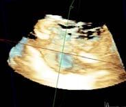
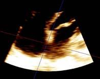
1 2D/3D in Evaluation of Atrial Septum Roberto M Lang, MD OSTIUM SECUNDUM ASD: 2D AND 3D TNSESOPHAGEAL ECHO 1


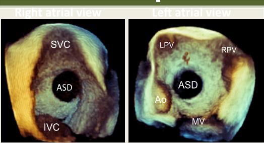
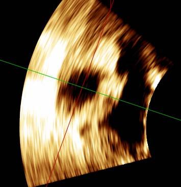
2 Biplane views D Acquisi on Acquire 3D volume Lang RM et al. JASE 2012;25:3 46. Right atrial view 3D Display Le atrial view Rotate to en face le atrial view Display with superior vena cava at 11 o clock posi on Display with right upper pulmonary vein at 1 o clock posi on Inter-atrial Septum Data Acquisition Ao PFO Ao A AS RPV SVC AS IVC S L view view A S R P 2
 Johri")

 Ao")
3 Patent foramen ovale (PFO) Johri A. Heart 2011;97:1441e1453 THE PATENT FOMEN OVALE (PFO) Ao PFO RPV A AS 3


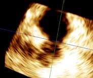
4 Inter-atrial Septum 90 % IAS FO MV LV View Sugeng L, Lang RM; J Am Coll Cardiol 4


5 Patent Foramen Ovale view IAS PFO Right atrial perspective LV view Left atrial perspective Patent Foramen Ovale 5


6 SUBTYPES OF ATRIAL SEPTAL COMMUNICATIONS <1% 2-3% 80-90% 2-3% 2-3% J Am Soc Echocardiogr 2015;28: Ostium Secundum Ostium secundum ASD Johri A. Heart 2011;97:1441e1453 6


7 OSTIUM SECUNDUM ASD: TNSTHOCIC ECHOCARDIOGPHY Apical 4-chamber Parasternal SAX Subxyphoid X The IAS is parallel to the US beam in the apical 4- CH view and PSAX view The preferred view for imaging/measuring the IAS on TTE is the subcostal view. Here the IAS is perpendicular to the US beam TNSTHOCIC ECHOCARDIOGPHY FOR INTER-ATRIAL SEPTAL DEFECTS Agitated saline + maneuvers to increase pressures (Valsalva, cough) ASD Microbubbles appear in the < 3-6 heart beats Intra-pulmonary shunting Bubbles appear > 3-6 heart beats (confirmed if bubbles seen in pulmonary veins) 7


8 ASD CHACTERISTICS THAT SHOULD BE REPORTED ASD size and dynamic nature ASD shape Rims Presence of fenestrations Presence of an ASA ASD type ASD location ASD: Surgeons View SVC AO TV 8


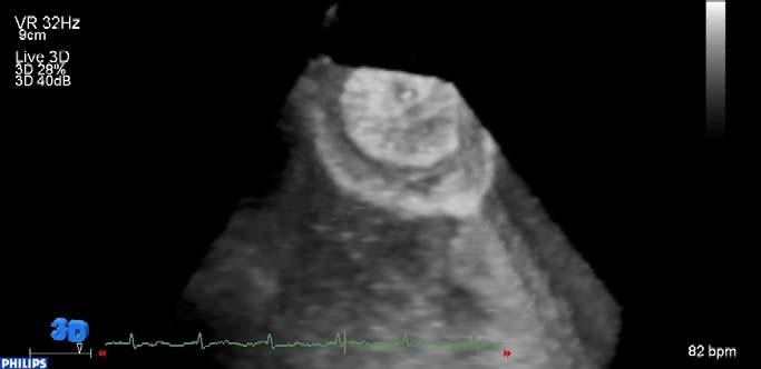
9 OSTIUM SECUNDUM ASD: 3D TEE Oval Round Small Large S L Change in size over cardiac cycle 3D full volume right side view A RV Ao PA RV Ao PA Atrial diastole Atrial systole 9


10 COMPLEX ASD SHAPE: INACCUTE ASSESSMENT WITH 2D IMAGING X SVC AO SAX IVC TV 4CH 2D TEE MPR quantitation Y Z X 10


11 ASD s : TTE Apical 4-chamber 3D zoom: perspective OSTIUM SECUNDUM ASD: 3D TNSTHOCIC ECHOCARDIOGPHY 11


12 ASD CHACTERISTICS THAT SHOULD BE REPORTED ASD size and dynamic nature ASD shape Presence of fenestrations Presence of an ASA ASD type ASD location ME Bi-Caval: SVC rim TEE 4CH: Posterior rim TEE MESAX: Aortic rim TEE 4CH: AV rim ME Bi-Caval: IVC rim Sobrino A. Arch Cardiol Mex 2012;82(1):37-47 TEE LE: CS rim 12


13 Secundum ASD rims: 1.SVC 2. Aortic 3. AV 4. IVC 5.RPV 5 SVC 1 ASD 2 Ao LPV 2 1 ASD 5 RPV 5A 4 IVC MV LV S L A en face view S R P en face view superior Secundum ASD Rim deficiencies SVC aortic Ao SVC RPA * SVC S Ao * CS R * A SVC inferior I IVC RPV RPV * * IVC 13


14 ASD CHACTERISTICS THAT SHOULD BE REPORTED ASD size and dynamic nature ASD shape Rims Presence of an ASA ASD type ASD location EHJCi


15 COMPLEX ASD REPAIR COMPLETE ASSESSMENT OF THE INTER- ATRIAL SEPTUM IS IMPORTANT Device deployed 2. Residual shunt 3. Fenestrations 3 15

16 COMPLETE ASSESSMENT OF THE INTER- ATRIAL SEPTUM IS IMPORTANT ASD CHACTERISTICS THAT SHOULD BE REPORTED ASD size and dynamic nature ASD shape Rims Presence of fenestrations ASD type ASD location 16


17 ATRIAL SEPTAL ANEURYSM Redundancy or saccular deformity of the atrial septum associated with increased mobility. Defined as an excursion of 10 mm from the plane of the atrial septum into the or or a combined excursion right and left of 15 mm ATRIAL SEPTAL ANEURYSM 17


18 ATRIAL SEPTAL ANEURYSM Amplatzer device closure ATRIAL SEPTAL ANEURYSM 1 day post device closure 18


19 RT3DE TEE GUIDANCE OF ASD CLOSURE A ASD B Guide wire C Delivery system Left atrial disc IAS IAS IAS D F Left atrial disc E I Left atrial disc F Left atrial disc IAS I Delivery system Right atrial disc Delivery system Right atrial disc PERI-INTERVENTIONAL 3D TEE PFO CLOSURE 19


20 ASD CHACTERISTICS THAT SHOULD BE REPORTED ASD size and dynamic nature ASD shape Rims Presence of fenestrations Presence of an ASA A T V D R V LVO T B E C R V LVOT Occluder Gerbode Defect 20


 Right lower and")
 An sinus")

21 SINUS VENOSUS ASD Partial/complete absence of sinus venosus septum between SVC and the right upper pulmonary vein (SVC type) Right lower and middle pulmonary veins and the (IVC type) SINUS VENOSUS ASD An sinus venosus septal defect can be difficult to detect on TTE 21


22 SINUS VENOSUS ASD An sinus venosus defect can be identified on TEE by the absence of the atrial septum immediately beneath the orifice of the SVC SUPERIOR SINUS VENOSUS ASD MRI - Coronal 3D RUPV SVC MRI - Axial RV 22


23 TEE Color Doppler 3D TEE Right Atrium View 3D TEE Left Atrium View CS RV TV CS MV CS LV Cardiac CT Surgical Right Atrium View Surgical Left Atrium View AAO CS CS CS CARDIAC CT: UNROOFED CORONARY SINUS DEFECT Normal Abnormal Ao CS Ao CS Defect 23


24 UNROOFED CORONARY SINUS DEFECT: TNSTHOCIC ECHOCARDIOGPHY RV CS Defect Wall of the coronary sinus in the is deficient or absent UNROOFED CORONARY SINUS DEFECT: 2D TNS-ESOPHAGEAL ECHO 4-chamber view CS Defect RV RV 24


25 3D TEE: THE UNROOFED CORONARY SINUS FROM LEFT TO RIGHT THE OPETING ROOM Defect CS 25
Atrial Septal Defects
 Supplementary ACHD Echo Acquisition Protocol for Atrial Septal Defects The following protocol for echo in adult patients with atrial septal defects (ASDs) is a guide for performing a comprehensive assessment
Atrial Septal Defect Closure. Stephen Brecker Director, Cardiac Catheterisation Labs
 Stephen Brecker Director, Cardiac Catheterisation Labs ADVANCED ANGIOPLASTY Incorporating The Left Main 5 Plus Course Conflicts of Interest The following companies have supported educational courses held
When Does 3D Echo Make A Difference?
 When Does 3D Echo Make A Difference? Wendy Tsang, MD, SM Assistant Professor, University of Toronto Toronto General Hospital, University Health Network 1 Practical Applications of 3D Echocardiography Recommended
INTEGRATING ECHOCARDIOGRAPHY WITH CATHETER INTERVENTIONS FOR CONGENITAL HEART DISEASE. Krishna Kumar SevenHills Hospital, Mumbai, India
 INTEGRATING ECHOCARDIOGRAPHY WITH CATHETER INTERVENTIONS FOR CONGENITAL HEART DISEASE Krishna Kumar SevenHills Hospital, Mumbai, India Why talk about it? What is the big deal? Are we not stating the obvious?
Normal TTE Examination, Doppler Echocardiography and Normal Antegrade Flow Patterns
 Normal TTE Examination, Doppler Echocardiography and Normal Antegrade Flow Patterns Pravin Patil, MD FACC FASE Associate Professor of Medicine Director, Cardiovascular Disease Training Program Lewis Katz
Normal TTE/TEE Examinations
 Normal TTE/TEE Examinations Geoffrey A. Rose, MD FACC FASE Sanger Heart & Vascular Institute Before you begin imaging... Obtain the patient s Height Weight BP PLAX View PLAX View Is apex @ 9-10 o clock?
JOINT MEETING 2 Tricuspid club Chairpersons: G. Athanassopoulos, A. Avgeropoulou, M. Khoury, G. Stavridis
 JOINT MEETING 2 Tricuspid club Chairpersons: G. Athanassopoulos, A. Avgeropoulou, M. Khoury, G. Stavridis Similarities and differences in Tricuspid vs. Mitral Valve Anatomy and Imaging. Echo evaluation
ΔΙΑΧΕΙΡΙΣΗ ΑΣΘΕΝΩΝ ΜΕ ΜΕΣΟΚΟΛΠΙΚΗ ΕΠΙΚΟΙΝΩΝΙΑ ΖΑΧΑΡΑΚΗ ΑΓΓΕΛΙΚΗ ΚΑΡΔΙΟΛΟΓΟΣ ΗΡΑΚΛΕΙΟ - ΚΡΗΤΗ
 ΔΙΑΧΕΙΡΙΣΗ ΑΣΘΕΝΩΝ ΜΕ ΜΕΣΟΚΟΛΠΙΚΗ ΕΠΙΚΟΙΝΩΝΙΑ ΖΑΧΑΡΑΚΗ ΑΓΓΕΛΙΚΗ ΚΑΡΔΙΟΛΟΓΟΣ ΗΡΑΚΛΕΙΟ - ΚΡΗΤΗ European Accreditation in TTE, TEE and CHD Echocardiography NOTHING TO DECLARE ATRIAL SEPTAL DEFECT TYPES SECUNDUM
Pulmonary arteriovenous fistula
 International Journal of Medical Imaging 2014; 2(2): 34-38 Published online April 10, 2014 (http://www.sciencepublishinggroup.com/j/ijmi) doi: 10.11648/j.ijmi.20140202.16 Pulmonary arteriovenous fistula
CMR for Congenital Heart Disease
 CMR for Congenital Heart Disease * Second-line tool after TTE * Strengths of CMR : tissue characterisation, comprehensive access and coverage, relatively accurate measurements of biventricular function/
Echocardiography in the Adult with Congenital Heart Disease
 1 1 Echocardiography in the Adult with Congenital Heart Disease Julie A. Kovach Indications for Echocardiography in the Evaluation of the Adult with Congenital Heart Disease........ 279 Indications and
Revealing new insights. irotate electronic rotation and xplane adjustable biplane imaging. Ultrasound cardiology. irotate and xplane
 Ultrasound cardiology irotate and xplane Revealing new insights irotate electronic rotation and xplane adjustable biplane imaging Annemien van den Bosch and Jackie McGhie Department of Cardiology, Erasmus
Among the congenital defects of the heart diagnosed
 Atrial Septal Defect: Anatomoechocardiographic Correlation Luis Muñóz-Castellanos, MD, Nilda Espinola-Zavaleta, MD, PhD, Magdalena Kuri-Nivón, MD, José Francisco Ruíz, MD, and Candace Keirns, MD, Mexico
The Normal Echocardiogram
 The Normal Echocardiogram Pravin V. Patil, MD FACC Lewis Katz School of Medicine at Temple University Acknowledgments Dr. Susan Wiegers Dr. Martin Keane Temple Cardiac Sonographers Disclosures No relevant
Multidetector computed tomography in the evaluation of atrial septal defects
 Multidetector computed tomography in the evaluation of atrial septal defects Poster No.: C-0502 Congress: ECR 2010 Type: Educational Exhibit Topic: Cardiac Authors: S. Espejo, R. Ysamat, B. Cajal, M. Pan,
Diversion of the inferior vena cava following repair of atrial septal defect causing hypoxemia
 Marshall University Marshall Digital Scholar Internal Medicine Faculty Research Spring 5-2004 Diversion of the inferior vena cava following repair of atrial septal defect causing hypoxemia Ellen A. Thompson
Welcome Number 6. The background of the Cardiac Sonographer Network News masthead is a diagnostic image:
 Number 6 Welcome Number 6 Welcome to the newsletter created just for you: sonographers who perform pediatric echocardiograms in primarily adult echo labs. Each issue features tips on echocardiography of
Giovanni Di Salvo MD, PhD, FESC Second University of Naples Monaldi Hospital
 Giovanni Di Salvo MD, PhD, FESC Second University of Naples Monaldi Hospital VSD is one of the most common congenital cardiac abnormalities in the newborn. It can occur as an isolated finding or in combination
Echocardiography Conference
 Echocardiography Conference David Stultz, MD Cardiology Fellow, PGY-6 September 20, 2005 Atrial Septal Aneurysm Bulging of Fossa Ovalis Associated commonly with Atrial septal defect or small perforations
Interventions in Adult Congenital Heart Disease: Role of CV Imaging. Associate Professor. ACHD mortality. Pillutla. Am Heart J 2009;158:874-9
 Interventions in Adult Congenital Heart Disease: Role of CV Imaging Sangeeta Shah MD, FACC, FASE Associate Professor ACHD mortality Pillutla. Am Heart J 2009;158:874-9 Adult Congenital Heart Disease Heterogenity
Echocardiographic Guidance During Placement of the Buttoned Double-Disk Device for Atrial Septa1 Defect Closure
 Echocardiographic Guidance During Placement of the Buttoned Double-Disk Device for Atrial Septa1 Defect Closure L. LUANN MINICH, M.D., and A. REBECCA SNIDER, M.D. Department of Pediatrics, C.S. Mott Children
ATRIAL SEPTAL CLOSURE AND LEFT ATRIAL APPENDAGE OCCLUSION: INDICATIONS AND GUIDANCE ECHOCARDIOGRAPHY IN INTERVENTIONAL CARDIOLOGY
 ATRIAL SEPTAL CLOSURE AND LEFT ATRIAL APPENDAGE OCCLUSION: INDICATIONS AND GUIDANCE Aristides G. Panlilio, MD, FPCP, FPCC,FPSE, FASE Philippine Heart Center Chinese General Hospital and Medical Center
Concomitant procedures using minimally access
 Surgical Technique on Cardiac Surgery Concomitant procedures using minimally access Nelson Santos Paulo Cardiothoracic Surgery, Centro Hospitalar de Vila Nova de Gaia, Oporto, Portugal Correspondence to:
Breakout Session: Transesophageal Echocardiography
 Breakout Session: Transesophageal Echocardiography Doris Ockert, MD Andrew Schroeder, MD University of Wisconsin School of Medicine and Public Health Jutta Novalija, MD, PhD Medical College of Wisconsin
Case Report Sinus Venosus Atrial Septal Defect as a Cause of Palpitations and Dyspnea in an Adult: A Diagnostic Imaging Challenge
 Case Reports in Medicine Volume 2015, Article ID 128462, 4 pages http://dx.doi.org/10.1155/2015/128462 Case Report Sinus Venosus Atrial Septal Defect as a Cause of Palpitations and Dyspnea in an Adult:
Trans-septal Catheterization. December 8, Jonathan Tobis, MD Professor of Medicine Interventional Cardiology, UCLA
 Trans-septal Catheterization December 8, 2015 Jonathan Tobis, MD Professor of Medicine Interventional Cardiology, UCLA No conflicts of interest for this talk BRK = Brockenbrough needle BRK may be easier
A Magnetic Resonance Imaging Method for
 Journal of Cardiovascular Magnetic Resonance, 1(1), 59-64 (1999) INVITED PAPER Use of MRI in ASD Asessment A Magnetic Resonance Imaging Method for Evaluating Atrial Septa1 Defects Godtfred Holmvang Cardiac
List of Videos. Video 1.1
 Video 1.1 Video 1.2 Video 1.3 Video 1.4 Video 1.5 Video 1.6 Video 1.7 Video 1.8 The parasternal long-axis view of the left ventricle shows the left ventricular inflow and outflow tract. The left atrium
British Society of Echocardiography
 British Society of Echocardiography Affiliated to the British Cardiac Society A Minimum Dataset for a Standard Adult Transthoracic Echocardiogram From the British Society of Echocardiography Education
Transcatheter closure of patent foramen ovale using the internal jugular venous approach
 New methods in diagnosis and therapy Transcatheter closure of patent foramen ovale using the internal jugular venous approach Przemysław Węglarz 1,2, Ewa Konarska-Kuszewska 2, Tadeusz Zębik 2, Piotr Kuszewski
Adel Hasanin Ahmed 1 ASD
 Adel Hasanin Ahmed 1 ASD Atrial septal defect (ASD) is the commonest form of congenital heart disease seen in adults. The commonest form of defect is the secundum ASD, accounting for two thirds of cases,
Device Closure of ASD in Children Using the Amplatzer Devices: Indications and catheter preparations, technique, and outcome [15]
![Device Closure of ASD in Children Using the Amplatzer Devices: Indications and catheter preparations, technique, and outcome [15]](/thumbs/73/69533656.jpg "Device Closure of ASD in Children Using the Amplatzer Devices: Indications and catheter preparations, technique, and outcome [15]") Device Closure of ASD in Children Using the Amplatzer Devices: Indications and catheter preparations, technique, and outcome [15] SCAI Fellow Course Fall 2014 Ralf J Holzer MD MSc FSCAI Medical Director
Device Closure of ASD in Children Using the Amplatzer Devices: Indications and catheter preparations, technique, and outcome [15] SCAI Fellow Course Fall 2014 Ralf J Holzer MD MSc FSCAI Medical Director
Adult Congenital Heart Disease: What All Echocardiographers Should Know Sharon L. Roble, MD, FACC Echo Hawaii 2016
 1 Adult Congenital Heart Disease: What All Echocardiographers Should Know Sharon L. Roble, MD, FACC Echo Hawaii 2016 DISCLOSURES I have no disclosures relevant to today s talk 2 Why should all echocardiographers
Patent Foramen Ovale: Diagnosis and Treatment
 Patent Foramen Ovale: Diagnosis and Treatment Anthony DeMaria Judy and Jack White Chair in Cardiology University of California, San Diego At one time or another a Grantee, Sponsored Speaker or Ad-hoc Consultant
We are IntechOpen, the world s leading publisher of Open Access books Built by scientists, for scientists. International authors and editors
 We are IntechOpen, the world s leading publisher of Open Access books Built by scientists, for scientists 3,500 108,000 1.7 M Open access books available International authors and editors Downloads Our
Clinical Value of 3D Echo: Volumes and Valves
 Clinical Value of 3D Echo: Volumes and Valves James D. Thomas, M.D., F.A.C.C. Cardiovascular Imaging Center Department of Cardiology Cleveland Clinic Foundation Cleveland, Ohio, USA Conflicts: None 3D2011:1
ARTIFACTS: THEORY AND ILLUSTRATIVE EXAMPLES
 ARTIFACTS: THEORY AND ILLUSTRATIVE EXAMPLES Robert A. Levine, M.D. Marielle Scherrer-Crosbie, M.D. Eric M. Isselbacher, M.D. No conflicts of interest Philippe Bertrand, Pieter Vendervoort, Hasselt and
ΔΙΑΔΕΡΜΙΚΗ ΑΝΤΙΜΕΤΩΠΙΣΗ ΔΟΜΙΚΩΝ ΠΑΘΗΣΕΩΝ: Ο ΡΟΛΟΣ ΤΗΣ ΑΠΕΙΚΟΝΙΣΗΣ ΣΤΟ ΑΙΜΟΔΥΝΑΜΙΚΟ ΕΡΓΑΣΤΗΡΙΟ ΣΤΗΝ ΤΟΠΟΘΕΤΗΣΗ MITRACLIP
 ΔΙΑΔΕΡΜΙΚΗ ΑΝΤΙΜΕΤΩΠΙΣΗ ΔΟΜΙΚΩΝ ΠΑΘΗΣΕΩΝ: Ο ΡΟΛΟΣ ΤΗΣ ΑΠΕΙΚΟΝΙΣΗΣ ΣΤΟ ΑΙΜΟΔΥΝΑΜΙΚΟ ΕΡΓΑΣΤΗΡΙΟ ΣΤΗΝ ΤΟΠΟΘΕΤΗΣΗ MITRACLIP ΒΛΑΣΗΣ ΝΙΝΙΟΣ MD MRCP ΚΛΙΝΙΚΗ ΑΓΙΟΣ ΛΟΥΚΑΣ ΘΕΣΣΑΛΟΝΙΚΗ CONFLICT OF INTEREST PROCTOR
ECHOCARDIOGRAPHY SERVICE OBJECTIVES FOR ECHOCARDIOGRAPHY IN THE McGILL CARDIOLOGY TRAINING PROGRAM
 ECHOCARDIOGRAPHY SERVICE OBJECTIVES FOR ECHOCARDIOGRAPHY IN THE McGILL CARDIOLOGY TRAINING PROGRAM As stipulated by Royal College training requirements, residents undergo a minimum of 6 months of training
Partial anomalous pulmonary venous connection to superior
 Cavo-Atrial Anastomosis Technique for Partial Anomalous Pulmonary Venous Connection to the Superior Vena Cava The Warden Procedure Robert A. Gustafson, MD Partial anomalous pulmonary venous connection
BASIL D. THANOPOULOS MD, PhD Associate Professor Honorary Consultant, RBH, London, UK
 TRANSCATHETER CLOSURE OF ATRIAL SEPTAL DEFECT AND PFO BASIL D. THANOPOULOS MD, PhD Associate Professor Honorary Consultant, RBH, London, UK TRANSCATHETER CLOSURE OF ATRIAL SEPTAL DEFECT AND PFO BASIL D.
Multimodality Imaging of Septal Defects
 Multimodality Imaging of Septal Defects Ohio-ACC 2018 Annual Meeting October 27, 2018 Kan N. Hor, MD Director, Cardiac Magnetic Resonance Imaging Associate Professor of Pediatrics The Heart Center, Nationwide
Surgical Management Of TAPVR. Daniel A. Velez, M.D. Congenital Cardiac Surgeon Phoenix Children s Hospital
 Surgical Management Of TAPVR Daniel A. Velez, M.D. Congenital Cardiac Surgeon Phoenix Children s Hospital No Disclosures Goals Review the embryology and anatomy Review Surgical Strategies for repair Discuss
Adult Echocardiography Examination Content Outline
 Adult Echocardiography Examination Content Outline (Outline Summary) # Domain Subdomain Percentage 1 2 3 4 5 Anatomy and Physiology Pathology Clinical Care and Safety Measurement Techniques, Maneuvers,
Cardiac ultrasound protocols
 Cardiac ultrasound protocols IDEXX Telemedicine Consultants Two-dimensional and M-mode imaging planes Right parasternal long axis four chamber Obtained from the right side Displays the relative proportions
Feasibility of real-time three-dimensional transoesophageal echocardiography for guidance of percutaneous atrial septal defect closure
 European Journal of Echocardiography (2009) 10, 543 548 doi:10.1093/ejechocard/jen337 Feasibility of real-time three-dimensional transoesophageal echocardiography for guidance of percutaneous atrial septal
THE NORMAL AND ABNORMAL INTER-ATRIAL SEPTUM
 THE NORMAL AND ABNORMAL INTER-ATRIAL SEPTUM BY REGINALD HUDSON From the Institute of Cardiology and National Heart Hospital Received April 5, 1954 This paper is an elementary study of the normal and abnormal
Ins and Outs of the Atria
 Ins and Outs of the Atria San Antonio Echocardiography Society July 2007 Joe M. Moody, Jr, MD UTHSCSA and STVAHCS Atrial Echo: The Ins and Outs Atrial Anatomic review Atrial size Atrial function by echo
Most common fetal cardiac anomalies
 Most common fetal cardiac anomalies Common congenital heart defects CHD % of cardiac defects Chromosomal Infants Fetuses anomaly (%) 22q11 deletion (%) VSD 30 5~10 20~40 10 PS 9 5 (PA w/ VSD) HLHS 7~9
Adel Hasanin Ahmed 1
 Adel Hasanin Ahmed 1 PERICARDIAL DISEASE The pericardial effusion ends anteriorly to the descending aorta and is best visualised in the PLAX. PSAX is actually very useful sometimes for looking at posterior
M-Mode Echocardiography Is it still Alive? Itzhak Kronzon, MD,FASE. Sampling Rate M-Mode: 1800 / sec 2D: 30 / sec
 M-Mode Echocardiography Is it still Alive? Itzhak Kronzon, MD,FASE Honoraria: Philips Classical M-mode Echocardiography M-Mode offers better time and image resolution. Sampling Rate M-Mode: 1800 / sec
Right Heart Evaluation ASE Guidelines Review. Chris Mann RDCS, RCS, FASE Faculty, Echocardiography Pitt Community College Greenville, NC
 Right Heart Evaluation ASE Guidelines Review Chris Mann RDCS, RCS, FASE Faculty, Echocardiography Pitt Community College Greenville, NC Objectives Briefly review right atrial and right ventricular anatomy
The Doppler Examination. Katie Twomley, MD Wake Forest Baptist Health - Lexington
 The Doppler Examination Katie Twomley, MD Wake Forest Baptist Health - Lexington OUTLINE Principles/Physics Use in valvular assessment Aortic stenosis (continuity equation) Aortic regurgitation (pressure
Transcatheter Atrial Septal Defect Closure with Right Aortic Arch Is it really difficult? M Tokue, H Hara, K Sugi, M Nakamura
 5th Asia Pacific Congenital & Structural Heart Intervention Symposium 2014 10 12 October 2014, Hong Kong Convention and Exhibition Centre Organizer: Hong Kong Society of Congenital & Structural Heart Disease
Case 47 Clinical Presentation
 93 Case 47 C Clinical Presentation 45-year-old man presents with chest pain and new onset of a murmur. Echocardiography shows severe aortic insufficiency. 94 RadCases Cardiac Imaging Imaging Findings C
Imaging Evaluation of the Ventricular Septum
 Imaging Evaluation of the Ventricular Septum Craig E Fleishman, MD FACC FASE The Heart Center at Arnold Palmer Hospital for Children, Orlando SCAI Fall Fellows Course 2013 Las Vegas Disclosure Information
Spectrum of Findings of Sinus Venosus Atrial Septal Defect: CT and MR Findings
 Spectrum of Findings of Sinus Venosus Atrial Septal Defect: CT and MR Findings Poster No.: P-0026 Congress: ESCR 2015 Type: Scientific Poster Authors: J. M. Madrid, P. J. Mergo, P. Bartolomé, J. Phelan,
Congenital heart disease. By Dr Saima Ali Professor of pediatrics
 Congenital heart disease By Dr Saima Ali Professor of pediatrics What is the most striking clinical finding in this child? Learning objectives By the end of this lecture, final year student should be able
8/31/2016. Mitraclip in Matthew Johnson, MD
 Mitraclip in 2016 Matthew Johnson, MD 1 Abnormal Valve Function Valve Stenosis Obstruction to valve flow during that phase of the cardiac cycle when the valve is normally open. Hemodynamic hallmark - pressure
Data Collected: June 17, Reported: June 30, Survey Dates 05/24/ /07/2010
 Job Task Analysis for ARDMS Pediatric Echocardiography Data Collected: June 17, 2010 Reported: Analysis Summary For: Pediatric Echocardiography Exam Survey Dates 05/24/2010-06/07/2010 Invited Respondents
Percutaneous VSD closure
 Percutaneous VSD closure Gianfranco Butera San Donato Milanese - Italy Patients selection Pts having hemodynamically significant VSD Left ventricular enlargement (left ventricular overload),defined as
The earliest application of intracardiac imaging
 Intracardiac Echocardiography for Structural Heart Defects A review of ICE innovations, devices, and techniques. BY ZAHID AMIN, MD; QI-LING CAO, MD; AND ZIYAD M. HIJAZI, MD The earliest application of
Image Library Case Listing:
 Image Library Case Listing: 1. Giant left atrial myxoma with mitral valve damage 2. Type A aortic dissection 3. Primum ASD 4. Aortic Transection from motor vehicle accident 5. Snake thrombus in right atrium
Introduction to TEE using Heartworks Echocardiography Simulator
 Introduction to TEE using Heartworks Echocardiography Simulator Steven M. Ewer, MD Assistant Professor Division of Cardiovascular Medicine University of Wisconsin School of Medicine & Public Health Version
Anatomy of Atrioventricular Septal Defect (AVSD)
") Surgical challenges in atrio-ventricular septal defect in grown-up congenital heart disease Anatomy of Atrioventricular Septal Defect (AVSD) S. Yen Ho Professor of Cardiac Morphology Royal Brompton and
Surgical challenges in atrio-ventricular septal defect in grown-up congenital heart disease Anatomy of Atrioventricular Septal Defect (AVSD) S. Yen Ho Professor of Cardiac Morphology Royal Brompton and
Γεώργιος Δ. Κατσιμαγκλής. Αν. Διευθυντής Καρδιολογικής ΚλινικήςΝΝΑ Διευθυντής Αιμοδυναμικού Εργαστηρίου ΝΝΑ
 Γεώργιος Δ. Κατσιμαγκλής Αν. Διευθυντής Καρδιολογικής ΚλινικήςΝΝΑ Διευθυντής Αιμοδυναμικού Εργαστηρίου ΝΝΑ I have no disclosures ΣΥΧΝΟΤΗΤΑ An atrial septal defect (ASD) is a deficiency of the atrial septum.
Heart and Soul Evaluation of the Fetal Heart
 Heart and Soul Evaluation of the Fetal Heart Ivana M. Vettraino, M.D., M.B.A. Clinical Associate Professor, Michigan State University College of Human Medicine Objectives Review the embryology of the formation
ASCeXAM / ReASCE. Practice Board Exam Questions Monday Morning
 ASCeXAM / ReASCE Practice Board Exam Questions Monday Morning Ultrasound Physics Artifacts Doppler Physics Imaging, Knobology, and Artifacts Echocardiographic Evaluation of the RV Tricuspid and Pulmonary
Fig.1 Normal appearance of RV in SAX:
 Tutorial 7 - Assessment of the right heart Assessment of the Right heart The right heart assessment clinically and echocardiographically is not a very important part of mainstream cardiology. In the ICU,
ECHOCARDIOGRAPHY DATA REPORT FORM
 Patient ID Patient Study ID AVM - - Date of form completion / / 20 Initials of person completing the form mm dd yyyy Study period Preoperative Postoperative Operative 6-month f/u 1-year f/u 2-year f/u
TRANSCATHETER CLOSURE OF ATRIAL SEPTAL DEFECT AND PFO
 TRANSCATHETER CLOSURE OF ATRIAL SEPTAL DEFECT AND PFO BASIL D. THANOPOULOS MD, PhD Associate Professor Agios Loukas Clinic, Thessaloniki, Greece Ares Heart Center, Bucharest, Romania Honorary Consultant,
Echocardiographic assessment in Adult Patients with Congenital Heart Diseases
 Echocardiographic assessment in Adult Patients with Congenital Heart Diseases Athanasios Koutsakis Cardiologist, Cl. Research Fellow George Giannakoulas Ass. Professor in Cardiology 1st Cardiology Department,
Intracardiac echocardiography: an ideal guiding tool for device closure of interatrial communications
 Eur J Echocardiography (2005) 6, 92e96 Intracardiac echocardiography: an ideal guiding tool for device closure of interatrial communications Thomas Bartel a, *, Thomas Konorza a, Ulrich Neudorf b, Tiko
ADULT CONGENITAL HEART DISEASE. Stuart Lilley
 ADULT CONGENITAL HEART DISEASE Stuart Lilley More adults than children have congenital heart disease Huge variety of congenital lesions from minor to major Heart failure, re-operation and arrhythmia are
What I Have Learned from 3D Imaging of Heart Valve Disease
 What I Have Learned from 3D Imaging of Heart Valve Disease Rebecca T. Hahn, MD Director of Interventional Echocardiography Columbia University Core Lab Director for multiple tricuspid device trials for
Pericardial Diseases. Smonporn Boonyaratavej, MD. Division of Cardiology, Department of Medicine Chulalongkorn University
 Pericardial Diseases Smonporn Boonyaratavej, MD Division of Cardiology, Department of Medicine Chulalongkorn University Cardiac Center, King Chulalongkorn Memorial Hospital 21 AUGUST 2016 Pericardial
Index. cardiology.theclinics.com. Note: Page numbers of article titles are in boldface type.
 Index Note: Page numbers of article titles are in boldface type. A Acute ischemic stroke TOAST classification of, 270 Acute myocardial infarction (AMI) cardioembolic stroke following, 207 208 noncardioembolic
CARDIAC AND CORONARY ARTERY ANATOMY NO DISCLOSURES. Axial Anatomy of Heart. Axial Anatomy of Heart. Axial Anatomy of Heart
 CARDIAC AND CORONARY ARTERY ANATOMY NO DISCLOSURES NASCI MEETING, ORLANDO FLORIDA 2009 KOSTAKI G. BIS, MD, FACR DEPARTMENT OF RADIOLOGY WILLIAM BEAUMONT HOSPITAL Royal Oak, Michigan OBJECTIVES CARDIAC
Glenmark Cardiac Centre Mumbai, India
 ASD device closure: Long term follow up Bharat Dalvi, MD Glenmark Cardiac Centre Mumbai, India Our experience 1998 to 2011 1566 patients 912 patients > 4 years FU Exclusive with ASO Clinical, electrocardiographic
Echocardiography in adult congenital heart disease
 S12 Department of Cardiology, Royal Hospital for Sick Children, Glasgow G3 8SJ, UK A Houston S Lilley T Richens University Department of Medicine and Therapeutics, Western Infirmary, Glasgow G11 6NT, UK
Imaging Guide Echocardiography
 Imaging Guide Guide to Small Animal Echocardiography using the Vevo Imaging Systems System Compatibility: This guide contains instructions and suggestions for work on the Vevo2100, VevoLAZR, Vevo 3100
Fetal Echocardiography and the Routine Obstetric Sonogram
 JDMS 23:143 149 May/June 2007 143 Fetal Echocardiography and the Routine Obstetric Sonogram SHELLY ZIMBELMAN, RT(R)(CT), RDMS, RDCS ASAD SHEIKH, MD, RDCS Congenital heart disease (CHD) is the most common
PFO- To Close for Comfort. By: Vincent J.Caracciolo, MD FACC
 PFO- To Close for Comfort By: Vincent J.Caracciolo, MD FACC PATENT FORAMEN OVALE PFO- congenital lesion that frequently persists into adulthood ( 25-30%)- autopsy and TEE studies. PFO prevalence higher
TEE Outside of the Cardiac OR
 TEE Outside of the Cardiac OR STEVE GIBSON MD PHD OU DEPARTMENT OF ANESTHEIOLOGY I have no financial relationships or conflicts of interest to disclose TRANSESOPHAGEAL ECHOCARDIOGRAPHY Basic principles
Back to Basics: Common Errors In Quantitation In Everyday Practice
 Back to Basics: Common Errors In Quantitation In Everyday Practice Deborah Agler, ACS, RDCS, FASE October 9, 2017 ASE: Echo Florida Rebecca T. Hahn, MD Director of Interventional Echocardiography Professor
Diagnostic approach to heart disease
 Diagnostic approach to heart disease Initial work up History Physical exam Chest radiographs ECG Special studies Echocardiography Cardiac catheterization Echocardiography principles Technique of producing
DEVELOPMENT OF THE CIRCULATORY SYSTEM L E C T U R E 5
 DEVELOPMENT OF THE CIRCULATORY SYSTEM L E C T U R E 5 REVIEW OF CARDIAC ANATOMY Heart 4 chambers Base and apex Valves Pericardial sac 3 layers: epi, myo, endo cardium Major blood vessels Aorta and its
Total Anomalous Pulmonary Venous Connections: Anatomy and Diagnostic Imaging
 Total Anomalous Pulmonary Venous Connections: Anatomy and Diagnostic Imaging Timothy Slesnick, MD March 12, 2015 Congenital Cardiac Anesthesia Society Annual Meeting Disclosures I will discuss the use
How to assess an adult with a Ventricular Septal Defect. When it should be closed and how?
 TTE AND TEE VDS ASSESSMENT. WHAT S S THE SIZE, WHERE ARE THE MARGINS? How to assess an adult with a Ventricular Septal Defect. When it should be closed and how? Dr Gianfranco Butera, MD, PhD Dr Gianfranco
Coronary Sinus Atrial Septal Defect (Unroofed Coronary Sinus) with Total Anomalous Pulmonary Venous Connection A Case Report
 with Total Anomalous Pulmonary Venous Connection A Case Report") Case Reports in Clinical Medicine, 2017, 6, 1-18 http://www.scirp.org/journal/crcm ISSN Online: 2325-7083 ISSN Print: 2325-7075 Coronary Sinus Atrial Septal Defect (Unroofed Coronary Sinus) with Total
Case Reports in Clinical Medicine, 2017, 6, 1-18 http://www.scirp.org/journal/crcm ISSN Online: 2325-7083 ISSN Print: 2325-7075 Coronary Sinus Atrial Septal Defect (Unroofed Coronary Sinus) with Total
PFO Management update
 PFO Management update May 12, 2017 Peter Casterella, MD Swedish Heart and Vascular 1 PFO Update 2017: Objectives Review recently released late outcomes of RESPECT trial and subsequent FDA approval of PFO
Echocardiography in Adult Congenital Heart Disease
 Echocardiography in Adult Congenital Heart Disease Michael Vogel Kinderherz-Praxis München CHD missed in childhood Subsequent lesions after repaired CHD Follow-up of cyanotic heart disease CHD missed in
For Personal Use. Copyright HMP 2013
 Case Report With Brief Review J INVASIVE CARDIOL 2013;25(4):E78-E80 Percutaneous Transjugular Device Closure of Postoperative Residual Atrial Septal Defect Saktheeswaran Mahesh Kumar, MD, DM, Sasidharan
Intracardiac EchoCardiography (ICE) Common Views
 Common Views") Intracardiac EchoCardiography (ICE) Common Views Introduction What is ICE? Catheter with microscopic ultrasound transducer tip and doppler capabilities inserted into the heart via the IVC (typically) or
Intracardiac EchoCardiography (ICE) Common Views Introduction What is ICE? Catheter with microscopic ultrasound transducer tip and doppler capabilities inserted into the heart via the IVC (typically) or
Left atrial function. Aliakbar Arvandi MD
 In the clinic Left atrial function Abstract The left atrium (LA) is a left posterior cardiac chamber which is located adjacent to the esophagus. It is separated from the right atrium by the inter-atrial
The Physiology of the Fetal Cardiovascular System
 The Physiology of the Fetal Cardiovascular System Jeff Vergales, MD, MS Department of Pediatrics Division of Pediatric Cardiology jvergales@virginia.edu Disclosures I serve as the medical director for
Pediatric Echocardiography Examination Content Outline
 Pediatric Echocardiography Examination Content Outline (Outline Summary) # Domain Subdomain Percentage 1 Anatomy and Physiology Normal Anatomy and Physiology 10% 2 Abnormal Pathology and Pathophysiology
Hybrid Muscular VSD Closure in Small Weight Children
 Hybrid Muscular VSD Closure in Small Weight Children Shakeel A Qureshi, on behalf of: John P. Cheatham, MD George H. Dunlap Endowed Chair in Interventional Cardiology Director Cardiac Catheterization &
Since the introduction of transesophageal echocardiography
 ASE/SCA Guidelines for Performing a Comprehensive Intraoperative Multiplane Transesophageal Echocardiography Examination: Recommendations of the American Society of Echocardiography Council for Intraoperative