25 different brand names >44 different models Sizes mm
|
|
|
- Betty Webb
- 5 years ago
- Views:
Transcription

1
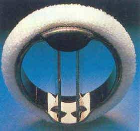
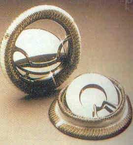
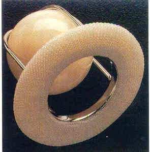
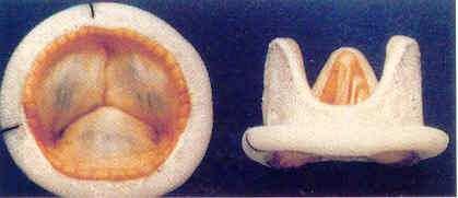
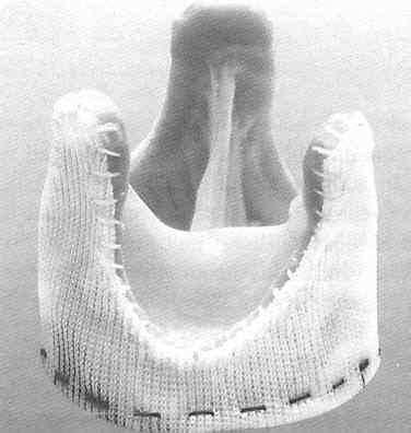
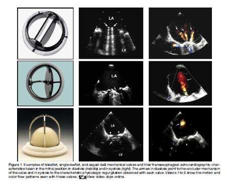
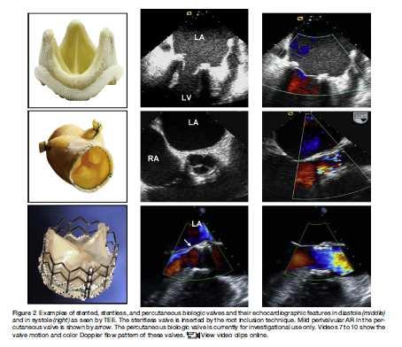
2 Types of Prosthetic Valves BIOLOGIC STENTED Porcine xenograft Pericardial xenograft STENTLESS Porcine xenograft Pericardial xenograft Homograft (allograft) Autograft PERCUTANEOUS MECHANICAL Bileaflet Single tilting disc Caged-ball 25 different brand names >44 different models Sizes mm

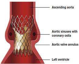
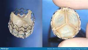
3 Types of prosthetic valves Mechanical vales Biologic (tissue) valves Percutaneous biologic valves
4 Diagnostic methods for evaluating prosthetic valve function Echocardiography with Doppler is the modality of choice for anatomic and hemodynamic evaluation of prosthetic heart valves Cine fluoroscopy Cardiac Catheterization Computed Tomography

5 3-D echocardiography and Doppler Assessment of the whole valve apparatus and the annulus MITRAL POSITION > aortic position

6

7 Cinefluoroscopy TEE sensitivity Mitral position 100% Aortic position 85% CF has a complementary diagnostic role in evaluating disc mobility of mechanical valves in the aortic position Muratori M, Montorsi P, Teruzzi G, et al: American Journal of Cardiology 2006
8

9 Computed Tomography Pannus formation Residual angle 66 degrees Intra-operative findings Tsai I-C, Lin Y-K, Chang Y, et al: Eur Radiol 2009

10 Common physiologic characteristics of prosthetic valves Stentless, cadaveric homograft and autograft aortic valves may be indistinguishable from native valves by echocardiographic imaging Stentless aortic valve 2 years post-op Native aortic valve
11 Common physiologic characteristics of prosthetic valves Acoustic shadowing Acoustic shadowing is not a significant limitation of TTE to assess prosthetic aortic valves, as it is in mitral prosthetic valves Acoustic shadowing is more prominent with mechanical valves compared to biologic valves

12 Shadowing & Flow Masking
13 Common physiologic characteristics of prosthetic valves Physiologic stenosis Physiologic regurgitation
14 Causes of physiologic stenosis Valve design / Valve type Valve size Pressure recovery phenomenon Increased flow across the valve Patient-prosthesis mismatch
15 Causes of Physiologic regurgitation Backflow from the closing movement of the ball or disc(s) Bileaflet and single disc valves have built-in, additional leakage backflow The purpose of this design is to reduce the risk of valve thrombosis Biological valves have minimal or no intrinsic regurgitation at implantation Daniels CJ, Barbetseas J, Boudoulas H: ACC SAP, version 7, 2009
16
17
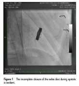
18 Early and Late Complications of Prosthetic Valves Patient-prosthesis mismatch Dehiscence Primary failure Thrombosis and thromboembolism Pannus formation Pseudoaneurysm formation Endocarditis Hemolysis

19 Distinction between thrombus and pannus

20 Aortic St Jude Medical valve Pannus formation
21 TRANSESOPHAGEAL ECHOCARDIOGRAPHY INDICATIONS OF TEE IN PROSTHETIC VALVES Valvular regurgitation Mitral and tricuspid valves > aortic valves Suspected valve obstruction Assessment of valve motion, thrombus vs pannus, candidates for thrombolysis Evaluation of associated structural abnormalities Vegetations, thrombi, ring abscess, pseudoaneurysm, fistulas Atrial, atrial appendage thrombi Inadequate transthoracic echocardiographic study Barbetseas J & Zoghbi WA: Cardiology Clinics 1998
22 Parameters in the Comprehensive Evaluation of Prosthetic Valve Function CLINICAL INFORMATION Date of valve replacement Type and size of the prosthetic valve Height, weight, body surface area Symptoms and related clinical findings Blood pressure and heart rate
23 IMAGING OF THE VALVE Motion of leaflets or occluder Presence of calcification on the leaflets or abnormal echo densities on the various components of the prosthesis Valve sewing ring integrity and motion DOPPLER ECHOCARDIOGRAPHY OF THE VALVE Contour of the jet velocity signal Peak velocity and gradient Mean pressure gradient VTI of the jet DVI Pressure half-time in MV and TV EOA Presence, location and severity of regurgitation
24 OTHER ECHOCARDIOGRAPHIC DATA LV and RV size, function, and hypertrophy LA and right atrial size Concomitant valvular disease Estimation of pulmonary artery pressure PREVIOUS POSTOPERATIVE STUDIES Comparison of above parameters is particularly helpful in suspected valvular dysfunction
25 Normal Prosthetic Valves DETERMINANTS OF VELOCITY/GRADIENT Valve size Valve type Flow rate
26

27
28 Normal Doppler echocardiographic values for PrAVs St Jude Aortic Size(mm) Peak Vel Mean Gr DVI EOA (ECHO) EOA (SR AREA) ± ±0.6 17±7 10±6 0.37± ± ± ± ± ±1.40 Chafizadeh ER & Zoghbi WA: Circulation 1991
29 Doppler Echocardiographic Evaluation of Prosthetic Aortic Valves DOPPLER ECHOCARDIOGRAPHY OF THE VALVE Peak velocity/gradient Mean gradient Contour of the jet velocity (AT) DVI EOA Presence, location, and severity of regurgitation PERTINENT CARDIAC CHAMBERS LV size, function, and hypertrophy
30
31
32
33
34

35 Normal vs obstructed prosthetic aortic valve
36 Doppler Parameters of Prosthetic Aortic Valve Function SUGGESTS STENOSIS Peak Velocity > 4 m/s Mean Gradient > 35 mmhg DVI < 0.25 EOA < 0.8 cm2 Contour of the jet Rounded, symmetrical Acceleration Time > 100 ms
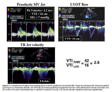
37 Echocardiographic and Doppler Parameters in Evaluation of Prosthetic Mitral Valve Function (Stenosis or Regurgitation) DOPPLER ECHOCARDIOGRAPHY OF THE VALVE OTHER PERTINENT ECHOCARDIOGRAPHIC AND DOPPLER PARAMETERS Peak early velocity Mean gradient Heart rate at the time of Doppler Pressure half-time DVI=VTI PrMV/VTI LVO EOA Presence, location, and severity of regurgitation LV size and function LA size RV size and function Estimation of PASP

38 Prosthetic Mitral Valve

39 BEFORE AND AFTER THROMBOLYSIS
40 Doppler Parameters of Prosthetic Mitral Valve Stenosis Peak Velocity Mean Gradient EOA (continuity equation) Pressure half-time SUGGESTS SIGNIFICANT STENOSIS > 2.5 m/s > 10 mmhg < 1 cm2 > 200 ms
41 Prosthetic Mitral Valve Limitations of Pressure Half-time Method Merging of E and A velocities ( PR, tachycardia) Delayed LV relaxation Decreased LV compliance Atrio-ventricular coupling Significant aortic insufficiency
42 Parameters for Evaluation of the Severity of Prosthetic Aortic Valve Regurgitation VALVE STRUCTURE AND MOTION Mechanical or bioprosthetic STUCTURAL PARAMETERS LV size DOPPLER PARAMETERS (qualitative or semiquantitative) Jet width in central jets (% LVO diameter): color Jet density: CW Doppler Jet deceleration rate (PHT, ms): CW Doppler LVO flow vs pulmonary flow: PW Doppler Diastolic flow reversal in the Desc Ao: PW Doppler DOPPLER PARAMETERS (quantitative) Regurgitant volume (ml/beat) Regurgitant fraction (%) SEVERE Abnormal Dilated Large ( 65%) Dense Steep(<200) Greatly increased Prominent, holodiastolic >60 >50

43 TEE Paravalvular significant AR
44 Transthoracic Echocardiographic Findings Suggestive of Significant Prosthetic MR in Mechanical Valves with Normal Pressure Half-time FINDING Peak mitral velocity 1.9 m/s VTI PrMV / VTI LVO 2.5 Mean gradient 5 mmhg LV stroke volume (2D or 3D imaging) is > 30% higher than systemic stroke volume by Doppler Maximal TR jet velocity > 3m/s Systolic flow convergence seen in the LV toward the prosthesis COMMENTS Also consider high flow, PPM Measurement errors increase in AF due to difficulty in matching cardiac cycles; also consider PPM At physiologic heart rate; also consider high flow, PPM Validation lacking; significant MR is suspected when LV function is normal or hyperdynamic and VTI LVO is < 16 cm Consider residual postop pulmonary hypertension or other causes Validation lacking; technically challenging to detect readily
45
46
47 Echocardiographic and Doppler Criteria for Severity of Prosthetic MR Using Findings from TTE and TEE STRUCTURAL PARAMETERS LV size Prosthetic valve DOPPLER PARAMETRS Color flow jet area Flow convergence Jet density: CW Doppler Jet contour: CW Doppler Pulmonary venous flow QUANTITATIVE PARAMETERS VC width (cm) R vol (ml/beat) RF (%) EROA (cm2) SEVERE Usually dilated Abnormal Large central jet (usually > 8 cm2 or > 40% of LA area) or variable size wall-impinging jet swirling in the left atrium Large Dense Early peaking, triangular Systolic flow reversal
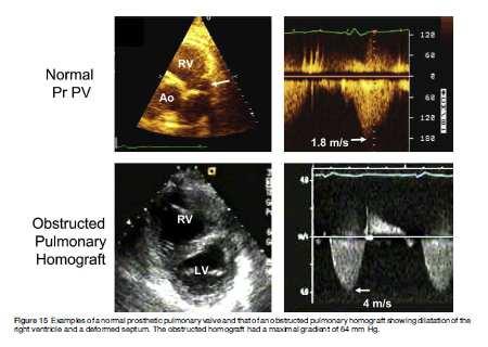
48 Imaging and Doppler Parameters in Evaluation of Prosthetic Pulmonary Valve Function DOPPLER ECHOCARDIOGRAPHY OF THE VALVE Peak velocity/peak gradient Mean gradient DVI, EOA (few data exist) Presence, location, and severity of regurgitation RELATED CARDIAC CHAMBERS RV size, function, hypertrophy, and RV systolic pressure
49 Findings Suspicious for Prosthetic Pulmonary Valve Stenosis Cusp or leaflet thickening or immobility Narrowing of forward color map Peak velocity through the prosthesis >3 m/s or 2 m/s through a homograft (suspicious but not diagnostic of stenosis) Increase in peak velocity on serial studies Impaired RV function or elevated RV systolic pressure
50
51 Evaluation of Severity of Prosthetic Pulmonary Valve Regurgitation Valve structure RV size Jet size by color Doppler (central jets) Jet density by CW Doppler Jet deceleration rate by CW Doppler Pulmonary systolic flow vs systemic flow by PW Doppler Diastolic flow reversal in the pulmonary artery SEVERE Abnormal or valve dehiscence Dilated (unless there are other reasons) Usually large, with a wide origin; jet width >50% of pulmonary annulus; may be brief in duration Dense Steep deceleration (not specific for severe PR), early termination of diastolic flow Greatly increased (cutoff values are not well validated) Present
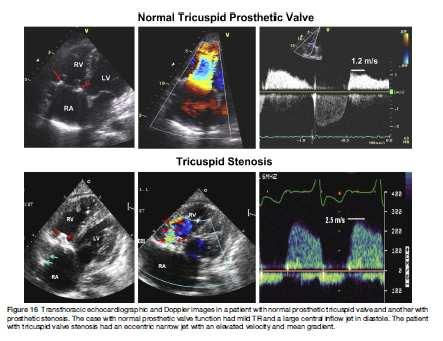
52 Echocardiographic and Doppler Parameters in Evaluation of Prosthetic Tricuspid Valve Function DOPPLER ECHOCARDIOGRAPHY OF THE VALVE Peak early velocity Mean gradient Heart rate at time of Doppler assessment Pressure half-time VTI PrTV / VTI LVO EOA Presence, location, and severity of TR RELATED CARDIAC CHAMBERS, IVC AND HEPATIC VEINS RV size and function Right atrial size Size of IVC and response to inspiration Hepatic vein flow pattern
53 Doppler Paramters of Prosthetic Tricuspid Valve Function Peak velocity (increased also with TR) Mean gradient (increased also with TR) Pressure half-time EOA and VTI PrTV/VTI LVO CONSIDER VALVE STENOSIS (average 5 cycles) > 1.7 m/s 6 mm Hg 230 ms No data yet available for tricuspid prosthesis
54
55 Echocardiographic and Doppler Parameters Used in Grading Severity of Prosthetic Tricuspid Valve Regurgitation Valve structure Jet area by color Doppler, central jets only (cm2) VC width (cm) (for valvular TR jet, extrapolated from native TR, unknown cutoffs for paravalvular TR) SEVERE Abnormal or valve dehiscence > 10 > 0.7 Jet density and contour by CW Doppler Dense with early peaking Doppler systolic hepatic flow Right atrium, right ventricle, IVC Holosystolic reversal Markedly dilated
56 Comprehensive Evaluation of Prosthetic Valve Function Clinical evaluation, know valve type and size Baseline echo/doppler study after surgery Transthoracic 2D/Doppler evaluation of structure and function is the first line diagnostic method In cases with high velocity jet/gradient and suspected stenosis or regurgitation: - Compare with previous studies - TEE, fluoroscopy (if needed for aortic mechanical prosthetic valve), CT (?)
57 THANK YOU
Echocardiographic Evaluation of Mitral Valve Prostheses
 Echocardiographic Evaluation of Mitral Valve Prostheses Dennis A. Tighe, M.D., FACC, FACP, FASE Cardiovascular Medicine University of Massachusetts Medical School Worcester, MA www.asecho.org 1 Nishimura
Prosthetic valve dysfunction: stenosis or regurgitation
 Prosthetic valve dysfunction: stenosis or regurgitation Jean G. Dumesnil MD, FRCP(C), FACC, FASE(Hon) Quebec Heart and Lung Institute, Québec, Québec No disclosures Possible Causes of High Gradients in
Echocardiographic Evaluation of Aortic Valve Prosthesis
 Echocardiographic Evaluation of Aortic Valve Prosthesis Amr E Abbas, MD, FACC, FASE, FSCAI, FSVM, RPVI Co-Director, Echocardiography, Director, Interventional Cardiology Research, Beaumont Health System
PROSTHETIC VALVE BOARD REVIEW
 PROSTHETIC VALVE BOARD REVIEW The correct answer D This two chamber view shows a porcine mitral prosthesis with the typical appearance of the struts although the leaflets are not well seen. The valve
Prosthesis-Patient Mismatch or Prosthetic Valve Stenosis?
 EuroValves 2015, Nice Prosthesis-Patient Mismatch or Prosthetic Valve Stenosis? Philippe Pibarot, DVM, PhD, FACC, FAHA, FASE FESC Canada Research Chair in Valvular Heart Diseases Université LAVAL Disclosure
Echocardiographic Evaluation of Aortic Valve Prosthesis
 Echocardiographic Evaluation of Aortic Valve Prosthesis Amr E Abbas, MD, FACC, FASE, FSCAI, FSVM, RPVI Co Director, Echocardiography, Director, Interventional Cardiology Research, Beaumont Health System
Echocardiographic Evaluation of Aortic Valve Prosthesis
 Echocardiographic Evaluation of Aortic Valve Prosthesis Amr E Abbas, MD, FACC, FASE, FSCAI, FSVM, RPVI Director, Interventional Cardiology Research, Beaumont Health System Associate Professor of Medicine,
A Practical Approach to Prosthetic Valves
 A Practical Approach to Prosthetic Valves Bonita Anderson DMU (Cardiac), MApplSc (Med Ultrasound), ACS, AMS, FASE https://doi.org/10.1161/circulationaha.108.778886 Disclosures None 1 Know the Product Know
Patient/prosthesis mismatch: how to evaluate and when to act?
 Patient/prosthesis mismatch: how to evaluate and when to act? Svend Aakhus, MD, PhD Oslo University Hospital, Norway Disclosures: No conflict of interest Types of aortic valve prostheses (AVR) Mechanical
Disclosures Rebecca T. Hahn, MD, FASE
 The New ASE Guidelines for Native Valvular Regurgitation Mitral Regurgitation The New ASE Guidelines: Role of 2D/3D and CMR (With caveats and comments from R. Hahn) William A. Zoghbi MD, FASE, MACC Professor
ICE: Echo Core Lab-CRF
 APPENDIX 1 ICE: Echo Core Lab-CRF Study #: - Pt Initials: 1. Date of study: / / D D M M M Y Y Y Y 2. Type of Study: TTE TEE 3. Quality of Study: Poor Moderate Excellent Ejection Fraction 4. Ejection Fraction
Valvular Regurgitation: Can We Do Better Than Colour Doppler?
 Valvular Regurgitation: Can We Do Better Than Colour Doppler? A/Prof David Prior St Vincent s Hospital Melbourne Sports Cardiology Valvular Regurgitation Valve regurgitation volume loads the ventricles
Bogdan A. Popescu. University of Medicine and Pharmacy Bucharest, Romania. EAE Course, Bucharest, April 2010
 Bogdan A. Popescu University of Medicine and Pharmacy Bucharest, Romania EAE Course, Bucharest, April 2010 This is how it started Mitral stenosis at a glance 2D echo narrow diastolic opening of MV leaflets
PROSTHETIC. V PROSTHETIC.V
 PROSTHETIC. V PROSTHETIC.V VALVE TYPE The valve types now implanted include: 1-bileaflet and tilting disc mechanical valves, 2-stented porcine and pericardial xenografts, 3-stentless porcine xenografts,
Comprehensive Hemodynamics By Doppler Echocardiography. The Echocardiographic Swan-Ganz Catheter.
 Comprehensive Hemodynamics By Doppler Echocardiography. The Echocardiographic Swan-Ganz Catheter. Itzhak Kronzon, MD, FASE, FACC, FESC, FAHA, FACP, FCCP North Shore HS, LIJ/Lenox Hill Hospital, New York
Echo Doppler Assessment of Right and Left Ventricular Hemodynamics.
 Echo Doppler Assessment of Right and Left Ventricular Hemodynamics. Itzhak Kronzon, MD, FASE, FACC, FESC, FAHA, FACP, FCCP Northwell, Lenox Hill Hospital, New York Professor of Cardiology Hofstra University
TSDA Boot Camp September 13-16, Introduction to Aortic Valve Surgery. George L. Hicks, Jr., MD
 TSDA Boot Camp September 13-16, 2018 Introduction to Aortic Valve Surgery George L. Hicks, Jr., MD Aortic Valve Pathology and Treatment Valvular Aortic Stenosis in Adults Average Course (Post mortem data)
Διαδερμική θεραπεία των παραβαλβιδικών διαφυγών Σ.Ράμμος
 ΔΜΙΝΑΡΙΟ ΔΠΙΣΗΜΟΝΙΚΩΝ ΔΝΩΔΩΝ ΔΛΛΗΝΙΚΟΤ ΚΟΛΛΔΓΙΟΤ ΚΑΡ ΙΟΛΟΓΙΑ 2-4.10.2015 Δπιζηημονική Ένωζη Δπεμβαηικής Καρδιολογίας Βαλβιδοπάθειες, Γιαδερμικές παρεμβάζεις Διαδερμική θεραπεία των παραβαλβιδικών διαφυγών
P = 4V 2. IVC Dimensions 10/20/2014. Comprehensive Hemodynamic Evaluation by Doppler Echocardiography. The Simplified Bernoulli Equation
 Comprehensive Hemodynamic Evaluation by Doppler Echocardiography Itzhak Kronzon, MD North Shore LIJ/ Lenox Hill Hospital New York, NY Disclosure: Philips Healthcare St. Jude Medical The Simplified Bernoulli
TAVR: Echo Measurements Pre, Post And Intra Procedure
 2017 ASE Florida, Orlando, FL October 10, 2017 8:00 8:25 AM 25 min TAVR: Echo Measurements Pre, Post And Intra Procedure Muhamed Sarić MD, PhD, MPA Director of Noninvasive Cardiology Echo Lab Associate
HEMODYNAMIC ASSESSMENT
 HEMODYNAMIC ASSESSMENT INTRODUCTION Conventionally hemodynamics were obtained by cardiac catheterization. It is possible to determine the same by echocardiography. Methods M-mode & 2D echo alone can provide
HOW IMPORTANT ARE THESE ECHO MEASUREMENTS ANYWAY?
 HOW IMPORTANT ARE THESE ECHO MEASUREMENTS ANYWAY? John D. Carroll, MD Professor, Director of Interventional Cardiology and Co-Medical Director of the Cardiac and Vascular Center, University of Colorado
ESC/EACTS Guidelines for the Management of Valvular Heart Disease
 ES/EATS Guidelines for the Management of Valvular Heart Disease European Journal of ardio-thoracic Surgery 2012 - Why do we need new guidelines on the management of valvular disease? New evidence has been
Hemodynamic Assessment. Assessment of Systolic Function Doppler Hemodynamics
 Hemodynamic Assessment Matt M. Umland, RDCS, FASE Aurora Medical Group Milwaukee, WI Assessment of Systolic Function Doppler Hemodynamics Stroke Volume Cardiac Output Cardiac Index Tei Index/Index of myocardial
MITRAL STENOSIS. Joanne Cusack
 MITRAL STENOSIS Joanne Cusack BSE Breakdown Recognition of rheumatic mitral stenosis Qualitative description of valve and sub-valve calcification and fibrosis Measurement of orifice area by planimetry
2/4/2011. Nathan Kerner, M.D.
 Nathan Kerner, M.D. Definition Elevated pressures - cut off usually >40 mmhg pulmonary artery systolic pressure (PASP) Usually associated with elevated pulmonary vascular resistance (PVR) measured in dynessec/cm
RVOTO adult and post-op
 Right ventricular outflow tract obstruction in the adult: native and post-op Helmut Baumgartner Westfälische Wilhelms-Universität Münster Adult Congenital and Valvular Heart Disease Center University of
ASE Guidelines on Aortic Regurgitation What Do I Measure? Case Studies
 ASE Guidelines on Aortic Regurgitation What Do I Measure? Case Studies Mitral Regurgitation The New ASE Guidelines: Role of 2D/3D and CMR William A. Zoghbi MD, FASE, MACC Professor and Chairman, Department
DOPPLER HEMODYNAMICS (1) QUANTIFICATION OF PRESSURE GRADIENTS and INTRACARDIAC PRESSURES
 QUANTIFICATION OF PRESSURE GRADIENTS and INTRACARDIAC PRESSURES") THORAXCENTRE DOPPLER HEMODYNAMICS (1) QUANTIFICATION OF PRESSURE GRADIENTS and INTRACARDIAC PRESSURES J. Roelandt DOPPLER HEMODYNAMICS Intracardiac pressures and pressure gradients Volumetric measurement
THORAXCENTRE DOPPLER HEMODYNAMICS (1) QUANTIFICATION OF PRESSURE GRADIENTS and INTRACARDIAC PRESSURES J. Roelandt DOPPLER HEMODYNAMICS Intracardiac pressures and pressure gradients Volumetric measurement
Quantification of Aortic Regurgitation
 Quantification of Aortic Regurgitation ASE Review 2018 Boston Susan E Wiegers, MD, FASE, FACC Professor of Medicine And thanks to Dr. Roberto Lang Disclosure None related to this presentation 1 Objectives
MITRAL REGURGITATION ECHO PARAMETERS TOOL
 Comprehensive assessment of qualitative and quantitative parameters, along with the use of standardized nomenclature when reporting echocardiographic findings, helps to better define a patient s MR and
Heart Valves: Before and after surgery
 Heart Valves: Before and after surgery Tim Sutton, Consultant Cardiologist Middlemore Hospital, Auckland Auckland Heart Group Indications for intervention in Valvular disease To prevent sudden death and
SONOGRAPHER & NURSE LED VALVE CLINICS
 SONOGRAPHER & NURSE LED VALVE CLINICS Frequency of visits and alerts AORTIC STENOSIS V max > 4.0 m/s or EOA < 1.0 cm 2 V max 3.5 4.0 m/s + Ca+ V max 3.0 4.0 m/s or EOA 1.0-1.5 cm 2 V max 2.5 3.0 m/s every
What are the best diagnostic tools to quantify aortic regurgitation?
 What are the best diagnostic tools to quantify aortic regurgitation? Agnès Pasquet, MD, PhD Pôle de Recherche Cardiovasculaire Institut de Recherche Expérimentale et Clinique Université catholique de Louvain
Cases of Abnormal Prosthetic Valves
 Cases of Abnormal Prosthetic Valves Sunil Mankad, MD, FACC, FCCP, FASE Associate Professor of Medicine Mayo Clinic College of Medicine Director, Transesophageal Echcoardiography Associate Director, Cardiology
Prof. Patrizio LANCELLOTTI, MD, PhD Heart Valve Clinic, University of Liège, CHU Sart Tilman, Liège, BELGIUM
 The Patient with Aortic Stenosis and Mitral Regurgitation Prof. Patrizio LANCELLOTTI, MD, PhD Heart Valve Clinic, University of Liège, CHU Sart Tilman, Liège, BELGIUM Aortic Stenosis + Mitral Regurgitation?
The Doppler Examination. Katie Twomley, MD Wake Forest Baptist Health - Lexington
 The Doppler Examination Katie Twomley, MD Wake Forest Baptist Health - Lexington OUTLINE Principles/Physics Use in valvular assessment Aortic stenosis (continuity equation) Aortic regurgitation (pressure
Adel Hasanin Ahmed 1
 Adel Hasanin Ahmed 1 PERICARDIAL DISEASE The pericardial effusion ends anteriorly to the descending aorta and is best visualised in the PLAX. PSAX is actually very useful sometimes for looking at posterior
Valvular Heart Disease
 Valvular Heart Disease B K Singh, MD, FACC Disclosures: None 1 CARDIAC CYCLE S2 S2=A2P2 S1=M1T1 S4 S1 S3 2 JVP Carotid S1 Slitting of S2 S3 S4 Ejection click Opening snap Dynamic Auscultation What is the
Echocardiography as a diagnostic and management tool in medical emergencies
 Echocardiography as a diagnostic and management tool in medical emergencies Frank van der Heusen MD Department of Anesthesia and perioperative Care UCSF Medical Center Objective of this presentation Indications
How to assess ischaemic MR?
 ESC 2012 How to assess ischaemic MR? Luc A. Pierard, MD, PhD, FESC, FACC Professor of Medicine Head, Department of Cardiology University Hospital Sart Tilman, Liège ESC 2012 No conflict of interest Luc
British Society of Echocardiography
 British Society of Echocardiography Affiliated to the British Cardiac Society A Minimum Dataset for a Standard Adult Transthoracic Echocardiogram From the British Society of Echocardiography Education
Adult Echocardiography Examination Content Outline
 Adult Echocardiography Examination Content Outline (Outline Summary) # Domain Subdomain Percentage 1 2 3 4 5 Anatomy and Physiology Pathology Clinical Care and Safety Measurement Techniques, Maneuvers,
April 16, 09:00-09:15 중앙대학교 윤신원
 April 16, 09:00-09:15 중앙대학교 윤신원 When to perform Echocardiography in IE? Vegetations?(pathologic Whatever the level hallmark) of suspicion Intracardiac abscess? Confirm or R/O at the Earliest opportunity.
Fig.1 Normal appearance of RV in SAX:
 Tutorial 7 - Assessment of the right heart Assessment of the Right heart The right heart assessment clinically and echocardiographically is not a very important part of mainstream cardiology. In the ICU,
Focused. se with 2008 F. lar Heart Diseas. date. ents With Valvul. Upd. gement of Patie. lines for Manag. HA 2006 Guidel ACC/AH. Fig.
 ACC/AH HA 2006 Guidel nic severe AI (Fig. 4). ned by age, ay also be helpful nd echo. For AI, ollow up may be or MRI rather than mension; SD, end lines for Manag gement of Patie Upd ents With Valvul date
CHAPTER 4 AN EFFICACIOUS APPROACH FOR THE QUANTIFICATION OF MITRAL REGURGITATION USING IMAGE PROCESSING AND PROXIMAL FLOW CONVERGENCE METHOD
 CHAPTER 4 AN EFFICACIOUS APPROACH FOR THE QUANTIFICATION OF MITRAL REGURGITATION USING IMAGE PROCESSING AND PROXIMAL FLOW CONVERGENCE METHOD 4.1. Introduction Mitral Regurgitation also called Mitral Insufficiency
PRINCIPLES OF ENDOCARDITIS
 015 // Endocarditis CONTENTS 140 Principles of Endocarditis 141 Native Valve Endocarditis 143 Complications of Native Valve Endocarditis 145 Right Heart Endocarditis 145 Prosthetic Valve Endocarditis 146
How to Assess and Treat Obstructive Lesions
 How to Assess and Treat Obstructive Lesions Erwin Oechslin, MD, FESC, FRCPC, Director, Congenital Cardiac Centre for Adults Peter Munk Cardiac Centre University Health Network/Toronto General Hospital
Pulmonary Hypertension: Echocardiographic Evaluation of Pulmonary Hypertension and Right Ventricular Function. Irmina Gradus-Pizlo, MD
 Pulmonary Hypertension: Echocardiographic Evaluation of Pulmonary Hypertension and Right Ventricular Function Irmina Gradus-Pizlo, MD Disclosures: Nothing to disclose Overview Is pulmonary hypertension
Echocardiographic evaluation of mitral stenosis
 Echocardiographic evaluation of mitral stenosis Euroecho 2011 Philippe Unger, MD, FESC Erasme Hospital, ULB, Brussels, Belgium I have nothing to declare EuroHeart Survey Etiology of single native left-sided
MR echo case. N.Koutsogiannis Department of Cardiology University Hospital Of Patras
 MR echo case N.Koutsogiannis Department of Cardiology University Hospital Of Patras Case A 35 years old male came to the echo lab for a third opinion for his valvulopathy. He reports a long standing MR
Echo Assessment Pre-TAVI
 Disclosure Statement of Financial Interest Within the past 12 months, I or my spouse/partner have had a financial Interest /arrangement or affiliation with the organization(s) listed below Echocardiographic
Prof. JL Zamorano Hospital Universitario Ramón y Cajal
 Prof. JL Zamorano Hospital Universitario Ramón y Cajal Should we forget TR? Nath J et al. Impact of tricuspid regurgitation on long-term survival. J Am Coll Cardiol. 2004; 43:405-409 Why is it difficult
ASCeXAM / ReASCE. Practice Board Exam Questions Monday Morning
 ASCeXAM / ReASCE Practice Board Exam Questions Monday Morning Ultrasound Physics Artifacts Doppler Physics Imaging, Knobology, and Artifacts Echocardiographic Evaluation of the RV Tricuspid and Pulmonary
Section 1: Initial Evaluation for Valvular Heart Disease Table 1: Initial Evaluation of an Asymptomatic Patient
 Section 1: Initial Evaluation for Valvular Heart Disease Table 1: Initial Evaluation of an Asymptomatic Patient Indication 1. Unexplained murmur or abnormal heart sounds 2. Reasonable suspicion of valvular
Echocardiography. Guidelines for Valve and Chamber Quantification. In partnership with
 Echocardiography Guidelines for Valve and Chamber Quantification In partnership with Explanatory note & references These guidelines have been developed by the Education Committee of the British Society
Michigan Society of Echocardiography 30 th Year Jubilee
 Michigan Society of Echocardiography 30 th Year Jubilee Stress Echocardiography in Valvular Heart Disease Moving Beyond CAD Karthik Ananthasubramaniam, MD FRCP (Glas) FACC FASE FASNC Associate Professor
Uncommon Doppler Echocardiographic Findings of Severe Pulmonic Insufficiency
 Uncommon Doppler Echocardiographic Findings of Severe Pulmonic Insufficiency Rahul R. Jhaveri, MD, Muhamed Saric, MD, PhD, FASE, and Itzhak Kronzon, MD, FASE, New York, New York Background: Two-dimensional
8/31/2016. Mitraclip in Matthew Johnson, MD
 Mitraclip in 2016 Matthew Johnson, MD 1 Abnormal Valve Function Valve Stenosis Obstruction to valve flow during that phase of the cardiac cycle when the valve is normally open. Hemodynamic hallmark - pressure
A Surgeon s Perspective Guidelines for the Management of Patients with Valvular Heart Disease Adapted from the 2006 ACC/AHA Guideline Revision
 A Surgeon s Perspective Guidelines for the Management of Patients with Valvular Heart Disease Adapted from the 2006 ACC/AHA Guideline Revision Prof. Pino Fundarò, MD Niguarda Hospital Milan, Italy Introduction
Diastolic Heart Function: Applying the New Guidelines Case Studies
 Diastolic Heart Function: Applying the New Guidelines Case Studies Mitral Regurgitation The New ASE William Guidelines: A. Zoghbi Role MD, of FASE, 2D/3D MACCand CMR Professor and Chairman, Department
Doppler Basic & Hemodynamic Calculations
 Doppler Basic & Hemodynamic Calculations August 19, 2017 Smonporn Boonyaratavej MD Division of Cardiology, Department of Medicine Chulalongkorn University Cardiac Center, King Chulalongkorn Memorial Hospital
The production of murmurs is due to 3 main factors:
 Heart murmurs The production of murmurs is due to 3 main factors: high blood flow rate through normal or abnormal orifices forward flow through a narrowed or irregular orifice into a dilated vessel or
JOINT MEETING 2 Tricuspid club Chairpersons: G. Athanassopoulos, A. Avgeropoulou, M. Khoury, G. Stavridis
 JOINT MEETING 2 Tricuspid club Chairpersons: G. Athanassopoulos, A. Avgeropoulou, M. Khoury, G. Stavridis Similarities and differences in Tricuspid vs. Mitral Valve Anatomy and Imaging. Echo evaluation
TAVR TTE INTERROGATION BY ALAN MATTHEWS
 TAVR TTE INTERROGATION BY ALAN MATTHEWS KEYS TO ACCURATE ASSESSMENT EDWARDS SAPIEN VALVE 3 PHASES OF TAVR TTE Evaluation (Qualifying) Placement (Intraoperative) Follow-up (Post-Op) GOALS High quality TTE
ECHOCARDIOGRAPHY DATA REPORT FORM
 Patient ID Patient Study ID AVM - - Date of form completion / / 20 Initials of person completing the form mm dd yyyy Study period Preoperative Postoperative Operative 6-month f/u 1-year f/u 2-year f/u
Basic Approach to the Echocardiographic Evaluation of Ventricular Diastolic Function
 Basic Approach to the Echocardiographic Evaluation of Ventricular Diastolic Function J A F E R A L I, M D U N I V E R S I T Y H O S P I T A L S C A S E M E D I C A L C E N T E R S T A F F C A R D I O T
Management of Difficult Aortic Root, Old and New solutions
 Management of Difficult Aortic Root, Old and New solutions Hani K. Najm MD, Msc, FRCSC,, FACC, FESC Chairman, Pediatric and Congenital Heart Surgery Cleveland Clinic Conflict of Interest None Difficult
3D Printing & Echocardiography
 ASE SOTA Feb 19, 2018 3D Printing & Echocardiography Stephen H. Little, MD John S. Dunn Chair in Cardiovascular Research and Education, Associate professor, Weill Cornell Medicine Disclosures Personal
Aortic Valve Practice Guidelines: What Has Changed and What You Need to Know
 Aortic Valve Practice Guidelines: What Has Changed and What You Need to Know James F. Burke, MD Program Director Cardiovascular Disease Fellowship Lankenau Medical Center Disclosure Dr. Burke has no conflicts
Experience with 500 Stentless Aortic Valve Replacements
 Experience with 500 Stentless Aortic Valve Replacements Dimitrios C. Iliopoulos, MD Cardiac Surgeon Ass. Professor of Surgery University of Athens, School of Medicine I declare no conflict of interest
Course Learning Objectives Sunday, February 17 Friday, February 22
 Course Learning Objectives Sunday, February 17 Friday, February 22 1. Define the physical principles of ultrasound technology and its applications for two and threedimensional (2D, 3D) imaging and use
Diastolic Function: What the Sonographer Needs to Know. Echocardiographic Assessment of Diastolic Function: Basic Concepts 2/8/2012
 Diastolic Function: What the Sonographer Needs to Know Pat Bailey, RDCS, FASE Technical Director Beaumont Health System Echocardiographic Assessment of Diastolic Function: Basic Concepts Practical Hints
Case # 1. Page: 8. DUKE: Adams
 Case # 1 Page: 8 1. The cardiac output in this patient is reduced because of: O a) tamponade physiology O b) restrictive physiology O c) coronary artery disease O d) left bundle branch block Page: 8 1.
Giovanni Di Salvo MD, PhD, FESC Second University of Naples Monaldi Hospital
 Giovanni Di Salvo MD, PhD, FESC Second University of Naples Monaldi Hospital VSD is one of the most common congenital cardiac abnormalities in the newborn. It can occur as an isolated finding or in combination
Advanced Applica,on of Point- of- Care Echocardiography in Cri,cal Care. Dr. Mark Tutschka Dr. Rob ArnAield
 Advanced Applica,on of Point- of- Care Echocardiography in Cri,cal Care Dr. Mark Tutschka Dr. Rob ArnAield OBJECTIVES Provide an overview of common advanced echocardiographic techniques suitable for use
Adult Cardiac Surgery
 Adult Cardiac Surgery Mahmoud ABU-ABEELEH Associate Professor Department of Surgery Division of Cardiothoracic Surgery School of Medicine University Of Jordan Adult Cardiac Surgery: Ischemic Heart Disease
Cardiovascular Structure & Function
 Cardiovascular Structure & Function Cardiovascular system: The heart Arteries Veins Capillaries Lymphatic vessels Weighting of the heart ceremony: Ancient Egyptians William Harvey and Blood Flow April
NEW GUIDELINES. A Guideline Protocol for the Assessment of Aortic Regurgitation From the British Society of Echocardiography Education Committee
 NEW GUIDELINES A Guideline Protocol for the Assessment of Aortic Regurgitation From the British Society of Echocardiography Education Committee Gill Wharton, Prathap Kanagala (Lead Authors) Richard Steeds
ASCeXAM / ReASCE. Practice Board Exam Questions. Monday Afternoon
 ASCeXAM / ReASCE Practice Board Exam Questions Monday Afternoon Aortic Valve Prostheses Aortic Regurgitation Stress Echocardiography Ischemic Heart Disease and Myocardial Infarction 3D Echocardiography
Case Reviews: Hemodynamic Calculations in Valvular Regurgitation
 Case Reviews: Hemodynamic Calculations in Valvular Regurgitation Case 5 History: 69-year-old man with orthotopic heart transplant 15 years ago. Inferior MI several years ago. Recurrent CHF. Currently dyspneic
Echo Week - Learning Objectives
 Echo Week - Learning Objectives Sunday, February 25 5-7 pm Physics Review Moderator: Mark Taylor, MD 1. Understand the ultrasound physics and apply it to image creation and optimization 2. Infer the interaction
Evaluation of Left Ventricular Diastolic Dysfunction by Doppler and 2D Speckle-tracking Imaging in Patients with Primary Pulmonary Hypertension
 ESC Congress 2011.No 85975 Evaluation of Left Ventricular Diastolic Dysfunction by Doppler and 2D Speckle-tracking Imaging in Patients with Primary Pulmonary Hypertension Second Department of Internal
Right Heart Hemodynamics: Echo-Cath Discrepancies
 Department of cardiac, thoracic and vascular sciences University of Padua, School of Medicine Padua, Italy Right Heart Hemodynamics: Echo-Cath Discrepancies Luigi P. Badano, MD, PhD, FESC, FACC **Dr. Badano
Imaging Assessment of Aortic Stenosis/Aortic Regurgitation
 Imaging Assessment of Aortic Stenosis/Aortic Regurgitation Craig E Fleishman, MD FACC FASE The Heart Center at Arnold Palmer Hospital for Children, Orlando SCAI Fall Fellows Course 2014 Las Vegas Disclosure
Valvular disease : Ο ρόλος του CMR. Sophie Mavrogeni MD FESC. Onassis Cardiac Surgery Center Athens Greece
 Valvular disease : Ο ρόλος του CMR Sophie Mavrogeni MD FESC Onassis Cardiac Surgery Center Athens Greece Aortic Valve CMR EVALUATION OF AORTIC VALVE Phase-contrast CMR of the aorta to determine aortic
Rotation: Echocardiography: Transthoracic Echocardiography (TTE)
") Rotation: Echocardiography: Transthoracic Echocardiography (TTE) Rotation Format and Responsibilities: Fellows rotate in the echocardiography laboratory in each clinical year. Rotations during the first
Rotation: Echocardiography: Transthoracic Echocardiography (TTE) Rotation Format and Responsibilities: Fellows rotate in the echocardiography laboratory in each clinical year. Rotations during the first
Appendix II: ECHOCARDIOGRAPHY ANALYSIS
 Appendix II: ECHOCARDIOGRAPHY ANALYSIS Two-Dimensional (2D) imaging was performed using the Vivid 7 Advantage cardiovascular ultrasound system (GE Medical Systems, Milwaukee) with a frame rate of 400 frames
Marti McCulloch, BS, MBA, RDCS, FASE Houston, Texas
 Marti McCulloch, BS, MBA, RDCS, FASE Houston, Texas Mitral Regurgitation What to Expect Review Specific Signs of Severity Supportive Signs of Severity Qualitative Parameters Structural Doppler Quantitative
What echo measurements are key prior to MitraClip?
 APHP CHU Bichat - Claude Bernard What echo measurements are key prior to MitraClip? Eric Brochet,MD Cardiology Department Hopital Bichat Paris France No disclosure Conflict of interest Case 69 y.o man
Goals of Reporting. Background of the STS/ACC TVT Registry. TVT Registry TAVR and MitraClip 10/11/ /11/2016 9:10 9:30 AM
 10/11/2016 9:10 9:30 AM Rebecca T. Hahn, MD Director of Interventional Echocardiography Columbia University Medical College Background of the STS/ACC TVT Registry The STS/ACC TVT Registry is a benchmarking
Echocardiography: Guidelines for Valve Quantification
 Echocardiography: Guidelines for Echocardiography: Guidelines for Chamber Quantification British Society of Echocardiography Education Committee Richard Steeds (Chair), Gill Wharton (Lead Author), Jane
Aortic Regurgitation & Aorta Evaluation
 VALVULAR HEART DISEASE Regurgitation Valvular Lessions 2017 Aortic Regurgitation & Aorta Evaluation Jorge Eduardo Cossío-Aranda MD, FACC Chairman of Outpatient Care Department Instituto Nacional de Cardiología
DISCLOSURE. Echocardiography in Systemic Diseases: Questions. Relevant Financial Relationship(s) None. Off Label Usage None 5/7/2018
 None. Off Label Usage None 5/7/2018") Echocardiography in Systemic Diseases: Questions Sunil Mankad, MD, FACC, FCCP, FASE Associate Professor of Medicine Mayo Clinic College of Medicine Director, Transesophageal Echocardiography Associate
Echocardiography in Systemic Diseases: Questions Sunil Mankad, MD, FACC, FCCP, FASE Associate Professor of Medicine Mayo Clinic College of Medicine Director, Transesophageal Echocardiography Associate
가천의대길병원소아심장과최덕영 PA C IVS THE EVALUATION AND PRINCIPLES OF TREATMENT STRATEGY
 가천의대길병원소아심장과최덕영 PA C IVS THE EVALUATION AND PRINCIPLES OF TREATMENT STRATEGY PA c IVS (not only pulmonary valve disease) Edwards JE. Pathologic Alteration of the right heart. In: Konstam MA, Isner M, eds.
2017 Cardiovascular Symposium CARDIAC SURGERY UPDATE: SMALLER INCISIONS AND LESS COUMADIN DAVID L. SAINT, MD
 2017 Cardiovascular Symposium CARDIAC SURGERY UPDATE: SMALLER INCISIONS AND LESS COUMADIN DAVID L. SAINT, MD David L Saint M.D. Tallahassee Memorial Hospital Southern Medical Group Division of Cardiothoracic
Tricuspid and Pulmonary Valve Disease
 Tricuspid and Pulmonary Valve Disease Lawrence Rudski MD FRCPC FACC FASE Professor of Medicine Director, Division of Cardiology Jewish General Hospital McGill University Question 1 All of the following
ACUTE CENTRAL PERIFERALEMBOLISM
 EAE TEACHING COURSE 2010 Belgrade, Serbia October 22-23, 2010 ACUTE CENTRAL and PERIFERALEMBOLISM Maria João Andrade Lisbon, PT BACKGROUND Stroke is a leading cause of mortality and long-term disability
The production of murmurs is due to 3 main factors:
 Heart murmurs The production of murmurs is due to 3 main factors: high blood flow rate through normal or abnormal orifices forward flow through a narrowed or irregular orifice into a dilated vessel or