Biology 212: Anatomy and Physiology II. Lab #5: Physiology of the Cardiovascular System For Labs Associated With Dr. Thompson s Lectures
|
|
|
- Loreen McCormick
- 5 years ago
- Views:
Transcription
1 Biology 212: Anatomy and Physiology II Lab #5: Physiology of the Cardiovascular System For Labs Associated With Dr. Thompson s Lectures References: Saladin, KS: Anatomy and Physiology, The Unity of Form and Function 8 th (2018). Required reading before beginning this lab: Chapters 19 and 20 Introduction: Your heart pumps blood throughout your body and must last your entire life without failure or fatigue. A heart rate of 72 cardiac cycles per minute (which is average) means that your heart must contract and relax more than four thousand times per hour, one hundred thousand times per day, or nearly thirty-eight million times each year, rarely skipping a beat and never being allowed to rest for more than a fraction of a second at a time. It must also be able to respond to differing physical demands which the body places on it. It can speed up or slow down. It can pump more or less blood with each contraction. It can pump this blood under higher or lower pressure. Similarly, your blood vessels must distribute that blood to all other organs and tissues of the body, then return it to the heart. However, it would be a mistake to assume that they are just passive tubes. As noted in the previous lab exercise, each artery, vein, and capillary has a specific structure, and your arteries and veins are constantly, on a second-by-second basis, adjusting their size ensue that the blood is sent to the right places at the right times and at the right pressures. For example: constriction (narrowing) of the femoral artery in the anterior thigh will reduce the amount of blood delivered to capillaries throughout the lower limb, decrease the blood pressure throughout the limb and thus the perfusion of blood through capillaries, and increase the resistance or back-pressure against which the heart has to push. Dilation (widening) of this artery will have the opposite effects: more blood will flow into the limb, the heart will not have to push as hard to get it there, and blood will flow into capillaries with more pressure. Changes in the size of the femoral vein, obviously, will have similar effects on blood flowing OUT of the limb. Obviously, the heart and the blood vessels must be able to respond to changes in each other s physiology in order to function properly. In this lab exercise, we will examine some important aspects of that physiology of the human circulatory system. Learning Objectives: Upon completion of this lab exercise, students will be able to: Describe the correlation between cardiac contraction and pulse rate, how these change with exercise, and how they are used to determine cardiac output. Describe the cardiac conduction system of the human heart. Identify waves, intervals, and segments on and electrocardiogram and correlate these with depolarization and repolarization events in the heart. Analyze an electrocardiogram to determine heart rate, the length of the PR interval, and the length of the QT interval. 1
2 ANATOMY OF THE HEART AND BLOOD FLOW Exercise 1: Review the anatomy of the heart using the models. Be sure you can identify the apex, right atrium, right ventricle, left atrium, left ventricle, interatrial septum, and interventricular septum. Using the diagrams in your Saladin text, be sure you understand the pattern of blood flow through the heart from the superior or inferior vena cava until it enters the aorta. Diagram that here, identifying in the proper order each chamber, valve, and great vessel the blood passes through as well as the lungs. Questions for Discussion Based On Your Reading of Chapters 19 and 20 Before Coming to Lab: The heart is often described as a double pump, sending blood simultaneously through two circuits. What does this mean? From which chamber of the heart is blood pumped under the highest pressure? Why? Define systole, diastole, and cardiac cycle for your lab partners. Be sure you understand that both systole and diastole are necessary for proper heart function. During each cardiac cycle, each chamber of the heart must relax and fill with blood before it can contract and eject that blood into another chamber or into an artery. Exercise 2: Review the histologic structure of arteries and veins as shown in Figure 20.2 of your Saladin text. Be sure you understand how the contraction of smooth muscle in the tunica media of smaller arteries and arterioles will cause constriction (narrowing) of those vessels, thus increasing the blood pressure necessary to force blood through them. MEASUREMENTS OF CARDIAC CONTRACTION - PULSE RATE It is important that the various organs of the body get blood pumped to them at the proper rate: too much blood flow can be just as damaging as too little. The heart can regulate the amount of blood it pumps by changing two factors: its heart rate, or the number of times it contracts per minute, and its stroke volume, or the amount of blood it pumps with each contraction. An increase in either or both of these will increase the rate of blood flow throughout the body, while a decrease in either or both of these will decrease the amount of blood it pumps. Multiplying heart rate (number of cardiac cycles per minute) and stroke volume (amount of blood pumped per systole) produces a very useful clinical measurement called the cardiac output of the heart. 2
3 While we can t easily measure stroke volume in this lab (that would require either very sophisticated instruments or the injection of a dye into your blood), we CAN easily measure heart rate and the effect of exercise on it. Exercise 3: Using your third or fourth fingers, find the arterial pulse of the radial artery in your wrist. This should be just lateral to the tendon of your flexor carpi radialis muscle - Figures and B.19 in your Saladin textbook will help you find these. Now find the pulse of the common carotid artery on one side of your neck in the depression between your trachea and your sternocleidomastoid muscle -Figures and should help you do this. Using the second hand of your watch or the clock in the lab, count the number of pulses you feel in your radial artery in one minute Count the number of pulses you feel in your carotid artery in one minute Next, attach a finger-tip pulse oximeter to the end of the second (index) finger of either hand and press the small button to turn it on. Rest this hand on your lab table so you can read the display. After a few seconds it should display your pulse rate ( PR ) it will also display the oxygen saturation of your blood ( %SpO 2 ), which you can ignore. Questions for Discussion Based On Your Reading of Chapters 19 and 20 Before Coming to Lab: Are the pulses in your radial and carotid arteries the same? Are these the same as the pulse rate on the pulse oximeter? Would you expect them to be? Why or why not? Exercise 4: Have one volunteer from your lab group sit as quietly as possible for two or three minutes with her or his eyes closed. Measure the pulse rate Now have this person step quickly up and down from the platform or concrete block for one minute. Immediately measure the pulse rate Have this person sit quietly again for three or four minutes. Measure the pulse rate You should have noticed that exercise significantly increased the pulse rate, which then returned to its resting rate after exercise stopped. This, obviously, would allow the heart to deliver more blood each minute to the muscles while they are active. 3
4 Assuming that the stroke volume remained unchanged during exercise (it didn t, but let s pretend it did), how much more blood was pumped by the heart each minute during exercise than when your lab partner was resting? For example: if the heart rate increased from 60 beats per minute at rest to 80 beats per minute during exercise, this would be an increase of 20 beats per minute. Dividing this by the resting rate, 20/60, would be a 33% increase. If it went from 60 to 90, this would be a 30/60 = 50% increase. Repeat this experiment on another member of your lab group. Be sure you understand the relationship between heart rate, stroke volume, and cardiac output. You should also realize that each chamber of the heart does not empty completely with each systole - a little bit of blood gets left behind. Be sure you understand (from your reading) ejection fraction and end-systolic volume. Questions for Discussion Based On Your Reading of Chapters 19 and 20 Before Coming to Lab: Explain to your lab partners why, at the cellular level, your skeletal muscles need to] have an increased cardiac output (and thus increased blood flow) when they are active. Explain what effect it would have on your body if you were unable to increase or decrease your heart rate (and thus your cardiac output) during exercise. Explain what effect it would have on your body if your heart rate (and thus cardiac output) increased during exercise but then was unable to return to normal as you rested. Explain what ejection fraction, end-systolic volume, and end-diastolic volume are. THE CARDIAC CONDUCTION SYSTEM Unlike skeletal muscle, which contracts only when stimulated by a somatic motor neuron (it is neurogenic), cardiac muscle cells are myogenic, meaning that they will spontaneously depolarize and contract. This occurs first at one specific region of the heart, then spreads along specialized cardiac myocytes before it is widely spread throughout the myocardium of the heart. This results in a coordinated pattern of depolarization of the myocytes, stimulating them to contract and relax in this same coordinated pattern. Anything that stops this myogenic signal will therefore stop the heart, and anything which interferes with its coordinated spread among myocytes will similarly interfere with their coordinated contraction. Either condition is lethal. 4
5 Exercise 5: Let's examine the components of the cardiac conduction system. Using Figure and the accompanying text, identify the following. 1. The Sinoatrial (SA) node is a group of myocytes in the right atrium near the opening of the superior vena cava. This is the pacemaker for the heart, where depolarization and thus contraction of myocytes begins. The action potentials (depolarization followed by repolarization) of these cells spread across the atria, causing them to contract 2. The atrioventricular (AV) node is located at the lower end of the interatrial septum near the right atrioventricular valve. It receives the action potential which has traveled from the sinoatrial node along the wall of the atrium, then after a brief delay relays it to the atrioventricular bundle. The fibrous tissues separating the atria from the ventricles acts as an insulator which prevents the action potential from passing from the atria to the ventricles unless it passes through this node. The AV node is thus the gateway that allows for signal transmission to the ventricles. 3. The atrioventricular bundle leaves the atrioventricular node and enters the interventricular septum. where it divides into right and left bundle branches which send independent signals to each of the ventricles. As the action potential is traveling along the bundle and its branches, it is electrically isolated from nearby myocytes and does not stimulate them to contract - the signal is just passing through on its way to the apex of the heart. 4. As each bundle branch reaches the apex of the heart, it divides into numerous Purkinje fibers. These distribute the action potential to individual myocytes, causing them to contract. Since cardiac myocytes are electrically connected through gap junctions at their intercalated discs, this electrical stimulus can rapidly spread to all myocytes in the myocardium of the ventricles, and they contract simultaneously. Questions for Discussion Based On Your Reading of Chapters 19 and 20 Before Coming to Lab: Explain to your lab partners what is happening in a myocyte when it depolarizes and when it repolarizes, and how these form an action potential which can lead to its contraction. Explain to your lab partners how cardiac myocytes are connected to each other by intercalated disks, and how the gap junctions in these intercalated disks allow the depolarization of one cardiac myocyte to be quickly spread to adjacent myocytes. Exercise 6: On the models of the heart, identify where each of the components of the cardiac conduction system would be located: the sinoatrial node, the atrioventricular node, the atrioventricular bundle, the right and left bundle branches, and the Purkinje fibers. They are probably not marked on the models, but you need to know where they are and the sequence in which the electrical signal (the action potential of depolarization followed by repolarization) travels through them. 5

6 ELECTRICAL ACTIVITY OF THE HEART - THE ELECTROCARDIOGRAM As noted above, the cardiac muscle of the heart contracts in response to electrical signals which are generated by the sinoatrial node, transmitted through the cardiac conduction system, and passed to millions of individual myocytes which then depolarize and repolarize together. As these electrical signals spread across the heart they are also conducted through all other fluids of the body, and they can be detected on the surface of the skin by an electrocardiograph. The output, or record, is a graph called an electrocardiogram (ECG or EKG) which records the electrical differences between two electrodes, one positive and one negative, placed on opposite sides of the heart, often on the limbs. Changes in these electrical differences, measured as thousandths of a volt or millivolts (mv), are recorded as the heart goes through each cardiac cycle. The time variable is plotted on the X-axis and the voltage difference is on the Y-axis as shown on this EKG. If the electrical signal, either a depolarization or a repolarization, is moving from the negative electrode towards the positive electrode you will see a positive or upward EKG deflection. When the electrical signal moves away from the positive electrode, the recording will be a negative or downward deflection. If the electrical signal moves perpendicular to the positive and negative electrodes, or if there is no electrical activity occurring, the EKG tracing will remain flat. Notice that all three types of movement are evident on the EKG above. In general, depolarizations and repolarizations of the cardiac conduction system (sinoatrial node, atrioventricular node, atrioventricular bundle and its branches, Purkinje fibers) are too small to be detected with an EKG, so the only electrical activity we see on the graph is the depolarization and repolarization of millions of cardiac myocytes in the atria and ventricles. As noted on the electrocardiogram, specific types of electrical changes within these myocytes produce specific upward and downward deflections called waves. Exercise 7: Examine Figures and and identify each of the following parts of an EKG: 1. The P wave and is produced when the depolarization signals spread from the sinoatrial node across the atria and causes the myocytes of those chambers to depolarize. This will ultimately signal these cells to contract, but remember that the EKG measures only the electrical signal, not the contraction which will start about 0.1 seconds later. 2. The PR interval is measured from the beginning of the P wave to the beginning of the QRS complex. This interval represents the time it takes the electrical impulse to travel from the sinoatrial node to the ventricles, including the P wave showing depolarization of the atrial myocytes. It is called the PR interval because the Q wave is often absent. Normal values lie between 0.12 and 0.20 seconds. Part of the PR interval is the PQ segment (also called the PR segment) from the end of the P-wave to the beginning of the QRS complex. This represents the time it takes for the electrical impulses to travel from the atrioventricular node (after atrial depolarization) through the atrioventricular bundle and its branches to the Purkinje fibers and then to the myocytes of both ventricles. 6
7 3. The QRS complex consists of 3 deflections: a small downward deflection (Q wave, often missing), a sharp rapid upward deflection (R wave) and a second small downward deflection (S wave). The entire complex marks depolarization of cardiac myocytes in the right and left ventricles and is typically about 0.08 to 0.12 seconds in duration. The large size (i.e., large voltage change) of this complex reflects the large muscle mass of the ventricles. The right and left atria also repolarize during the time of the QRS complex, but this relatively small electrical signal is masked by the much greater electrical signal caused by millions of myocytes depolarizing. 4. The T wave is the final deflection you see, showing repolarization of the myocytes in the ventricles. 5. The QT interval, measured from the beginning of the QRS complex to the end of the T wave, shows the time it takes the ventricles to depolarize and repolarize and has a typical duration of 0.35 to 0.43 seconds. The part of this called the ST segment, from the end of the QRS complex to the beginning of the T wave, shows the time after depolarization of the ventricular myocytes but before their repolarization. The ventricles are actually contracting during this time and forcing blood out the pulmonary trunk (from the right ventricle) and the aorta (from the left ventricle). Notice that this pattern repeats itself again and again with each cardiac cycle. There is a relatively long period of time when the EKG tracing is flat from the T-wave of one cardiac cycle to the P-wave of the next one, indicating no electrical activity in the heart. There is also no contraction during this time - the heart gets a short rest between each contraction. Notice also that the atria depolarize (indicated by the P-wave) first, followed by a pause (the flat line of the PQ segment) before the ventricles depolarize (QRS complex). This means that the atria are indeed stimulated to contract first while the ventricles remain relaxed, then the ventricles are stimulated to contract while the atria are relaxing. If you think about it, this pattern is necessary for proper cardiac function. First, the right atrium fills with blood from the vena cavae and the left atrium fills with blood from the pulmonary veins as these two chambers relax. They contract while the ventricles are relaxed and thus have room for the blood pushed out by the atria. As the ventricles contract a fraction of a second later, the atria are already relaxing and filling with blood for the next cycle. Then the ventricles relax and the atria contract to fill them with blood, and the pattern repeats itself over and over again. Exercise 8: In the space below, draw a EKG trace for three cardiac cycles. Label the waves and intervals discussed above. 7
8 Exercise 9: It s important to remember that an electrocardiogram only shows the movement of ions and their electrical charges (that is, depolarization and repolarization of cardiac myocytes) during each cardiac cycle. It does NOT show the actual contraction of the heart. If you are healthy, of course, that electrical activity will cause contraction of your cardiac myocytes a fraction of a second later to generate the pressure which moves blood through your circulatory system. However, if your heart is very weak or if it is not receiving enough oxygen it is possible for the cardiac myocytes to depolarize and repolarize without that contraction. Examine Figure in your Saladin textbook and compare the middle graph showing the ECG/EKG with the upper graph of Pressure (mm Hg). a) Note how left ventricular pressure (the black line) increases when the cardiac myocytes in that chamber contract a fraction of a second after they depolarize as shown by the QRS complex on the EKG. Note how this ventricular pressure decreases when its myocytes relax a fraction of a second after they repolarize as shown by the T-wave on the EKG. b) The same thing happens in the left atrium, shown by the lower red line on that upper graph. Note how left atrial pressure rises (caused by contraction of the atrial myocytes) a fraction of a second after the P-wave shows their depolarization. Note then how atrial pressure returns to zero when its myocytes repolarize and thus relax, but this repolarization is not seen on the EKG because it is happening at the same time that the QRS complex shows ventricular depolarization. Exercise 10: Your instructor will now lead you through a demonstration of an ECG on a member of the class. While you do not need to know how to set up the instrumentation to do this, you should pay attention to how electrodes are placed on this person and how she or he is positioned in order to get a good electrical signal with minimal electrical interference from contraction of skeletal muscles. 1. The volunteers should remove all metallic jewelry from her or his wrists and ankles. Using the sticky electrode pads, one lead (wire) will be attached to the left arm, one lead will be attached to the right arm, and the third, or electrical ground, lead will be attached to one leg. The closer these are to the heart, the better, but a good ECG tracing is usually obtained when they are placed on the forearms and lower leg. 2. The volunteer should lie quietly on the lab bench with arms and hands at the sides, not contacting any other part of the body. Be sure legs are supported on a chair if necessary and thus relaxed. The person should not be using any muscles except those necessary for breathing. 3. Your instructor will then start the software, and you should see an ECG tracing appear on the screen. If all of the leads (wires) were attached correctly, this should appear similar to Figure and the one you drew in Exercise 5, with the P, R, and T waves shown as upward deflections from the line and the Q and S waves as downward deflections. 4. The ECG tracing may appear ragged. The instructor will have the volunteer open and close his or her fists, then move her or his arms. Notice that the trace moves around the screen and the ECG is distorted. This tells you how important it is to keep still and relaxed when recording the ECG. 8
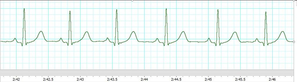
9 The EKG you obtain should resemble figure or 19.17a of your text, except much longer and probably not as smooth unless your volunteer was VERY relaxed and held VERY still. Your instructor will demonstrate how to measure certain intervals on that tracing, then you will do these calculations on the two EKGs which are attached. Exercise 11: Note that the X-axis of the tracing shows the number of seconds which have elapsed since the tracing began, and the grid pattern behind the tracing is divided into 10 boxes for each second. This makes it very easy to calculate the time it took for various events to occur - since the tracing moved ten boxes in one second, then each box is 1/10 th of a second. By counting the boxes, you can determine how many seconds (or fractions of a second) each interval lasted. 1. On one of your printed EKGs, measure the interval (in seconds) from the R-wave of one QRS complex to the R-wave of the next QRS complex, then do the same thing for the next two R-R intervals. Record your three R-R intervals here: seconds seconds Average your three measurements to calculate the average R-R interval and thus the average time between ventricular depolarizations (QRS complexes): seconds seconds Since each R-wave occurs exactly once per cardiac cycle, you can use this information to calculate heart rate For example: If the average R-R interval was 0.4 seconds - that is, a cardiac cycle occurred every 4/10 of a second - then the heart rate can be calculated as 1 cardiac cycle 60 sec. 1 cardiac cycle 60 sec. 60 cardiac cycles 150 cardiac cycles 0.4 sec. minute 0.4 sec. minute 0.4 minute 1 minute Using your measurements of R-R intervals, calculate the heart rate for the individual from whom this EKG was taken: cardiac cycles per minute On a printed EKG, measure the PR interval (in seconds) for three cycles seconds seconds Average your measurements to calculate seconds the average length of the PR interval: seconds
10 3. On a printed EKG, measure the QT interval (in seconds) for three cycles seconds seconds Average your measurements to calculate seconds the average length of the QT interval: seconds Questions for Discussion Based On Your Reading of Chapters 19 and 20 Before Coming to Lab: Explain to members of your lab group what it would mean if your EKG showed no P-waves. Explain to members of your lab group what it would mean if your EKG showed no QRS complex. Explain to members of your lab group what it would mean if your EKG showed no T-waves. Explain to members of your lab group what it would mean if the interval between QRS complexes became shorter. Explain to other members of your lab group what is happening in the heart during the PR interval. What would a very long P-R interval indicate about the functioning of your heart? Explain to other members of your lab group what is happening in the heart during the QT interval. What would a very long QT interval indicate about the functioning of your heart? Exercise 11: A second printed EKG is attached to this exercise. Repeat your measurements on this one at home or during open lab to be sure you understand how to do it. 10
11 11
12 12
CARDIOVASCULAR SYSTEM
 CARDIOVASCULAR SYSTEM Overview Heart and Vessels 2 Major Divisions Pulmonary Circuit Systemic Circuit Closed and Continuous Loop Location Aorta Superior vena cava Right lung Pulmonary trunk Base of heart
Cardiovascular Physiology
 Cardiovascular Physiology The mammalian heart is a pump that pushes blood around the body and is made of four chambers: right and left atria and right and left ventricles. The two atria act as collecting
37 1 The Circulatory System
 H T H E E A R T 37 1 The Circulatory System The circulatory system and respiratory system work together to supply cells with the nutrients and oxygen they need to stay alive. a) The respiratory system:
Cardiac Conduction System
 Cardiac Conduction System What causes the Heart to Beat? Heart contracts by electrical signals! Cardiac muscle tissue contracts on its own an electrical signal is sent out by the heart so that all cells
Cardiovascular System Notes: Physiology of the Heart
 Cardiovascular System Notes: Physiology of the Heart Interesting Heart Fact Capillaries are so small it takes ten of them to equal the thickness of a human hair. Review What are the 3 parts of the cardiovascular
Anatomy Review: The Heart Graphics are used with permission of A.D.A.M. Software, Inc. and Benjamin/Cummings Publishing Co.
 Anatomy Review: The Heart Graphics are used with permission of A.D.A.M. Software, Inc. and Benjamin/Cummings Publishing Co. Anatomy Views Label the diagrams of the heart below: Interactive Physiology Study
Lab #3: Electrocardiogram (ECG / EKG)
") Lab #3: Electrocardiogram (ECG / EKG) An introduction to the recording and analysis of cardiac activity Introduction The beating of the heart is triggered by an electrical signal from the pacemaker. The
Lab #3: Electrocardiogram (ECG / EKG) An introduction to the recording and analysis of cardiac activity Introduction The beating of the heart is triggered by an electrical signal from the pacemaker. The
Electrical Conduction
 Sinoatrial (SA) node Electrical Conduction Sets the pace of the heartbeat at 70 bpm AV node (50 bpm) and Purkinje fibers (25 40 bpm) can act as pacemakers under some conditions Internodal pathway from
Outline. Electrical Activity of the Human Heart. What is the Heart? The Heart as a Pump. Anatomy of the Heart. The Hard Work
 Electrical Activity of the Human Heart Oguz Poroy, PhD Assistant Professor Department of Biomedical Engineering The University of Iowa Outline Basic Facts about the Heart Heart Chambers and Heart s The
Anatomy of the Heart
 Biology 212: Anatomy and Physiology II Anatomy of the Heart References: Saladin, KS: Anatomy and Physiology, The Unity of Form and Function 8 th (2018). Required reading before beginning this lab: Chapter
Biology 212: Anatomy and Physiology II Lab #4: CARDIOVASCULAR PHYSIOLOGY AND THE ELECTROCARDIOGRAM
 Biology 212: Anatomy and Physiology II Lab #4: CARDIOVASCULAR PHYSIOLOGY AND THE ELECTROCARDIOGRAM References: Saladin, KS: Anatomy and Physiology, The Unity of Form and Function 7 th (2015). Be sure you
Cardiovascular System
 Cardiovascular System The Heart Cardiovascular System The Heart Overview What does the heart do? By timed muscular contractions creates pressure gradients blood moves then from high pressure to low pressure
The HEART. What is it???? Pericardium. Heart Facts. This muscle never stops working It works when you are asleep
 This muscle never stops working It works when you are asleep The HEART It works when you eat It really works when you exercise. What is it???? Located between the lungs in the mid thoracic region Apex
Chapter 20: Cardiovascular System: The Heart
 Chapter 20: Cardiovascular System: The Heart I. Functions of the Heart A. List and describe the four functions of the heart: 1. 2. 3. 4. II. Size, Shape, and Location of the Heart A. Size and Shape 1.
Cardiac Cycle. Each heartbeat is called a cardiac cycle. First the two atria contract at the same time.
 The Heartbeat Cardiac Cycle Each heartbeat is called a cardiac cycle. First the two atria contract at the same time. Next the two ventricles contract at the same time. Then all the chambers relax. http://www.youtube.com/watch?v=frd3k6lkhws
Lab 7. Physiology of Electrocardiography
 7.1 Lab 7. Physiology of Electrocardiography The heart is a muscular pump that circulates blood throughout the body. To efficiently pump the blood, cardiac contractions must be coordinated and are regulated
10. Thick deposits of lipids on the walls of blood vessels, called, can lead to serious circulatory issues. A. aneurysm B. atherosclerosis C.
 Heart Student: 1. carry blood away from the heart. A. Arteries B. Veins C. Capillaries 2. What is the leading cause of heart attack and stroke in North America? A. alcohol B. smoking C. arteriosclerosis
The Heart. Happy Friday! #takeoutyournotes #testnotgradedyet
 The Heart Happy Friday! #takeoutyournotes #testnotgradedyet Introduction Cardiovascular system distributes blood Pump (heart) Distribution areas (capillaries) Heart has 4 compartments 2 receive blood (atria)
Unit 10 ~ Learning Guide
 Unit 10 ~ Learning Guide Name: INSTRUCTIONS Complete the following notes and questions as you work through the related lessons. You are required to have this package completed BEFORE you write your unit
THE CARDIOVASCULAR SYSTEM. Heart 2
 THE CARDIOVASCULAR SYSTEM Heart 2 PROPERTIES OF CARDIAC MUSCLE Cardiac muscle Striated Short Wide Branched Interconnected Skeletal muscle Striated Long Narrow Cylindrical PROPERTIES OF CARDIAC MUSCLE Intercalated
Large Arteries of Heart
 Cardiovascular System (Part A-2) Module 5 -Chapter 8 Overview Arteries Capillaries Veins Heart Anatomy Conduction System Blood pressure Fetal circulation Susie Turner, M.D. 1/5/13 Large Arteries of Heart
Chapter 18 - Heart. I. Heart Anatomy: size of your fist; located in mediastinum (medial cavity)
") Chapter 18 - Heart I. Heart Anatomy: size of your fist; located in mediastinum (medial cavity) A. Coverings: heart enclosed in double walled sac called the pericardium 1. Fibrous pericardium: dense connective
Chapter 18 - Heart I. Heart Anatomy: size of your fist; located in mediastinum (medial cavity) A. Coverings: heart enclosed in double walled sac called the pericardium 1. Fibrous pericardium: dense connective
Heart. Heart 2-Tunica media: middle layer (media ='middle') muscle fibers (smooth or cardiac).
 muscle fibers (smooth or cardiac).") t. innermost lumenal General Circulatory system heart and blood vessels walls have 3 layers (inside to outside) 1-Tunica interna: aka tunica intima layer--lumenal layer epithelium--endothelium simple squamous
t. innermost lumenal General Circulatory system heart and blood vessels walls have 3 layers (inside to outside) 1-Tunica interna: aka tunica intima layer--lumenal layer epithelium--endothelium simple squamous
The cardiovascular system is composed of the heart and blood vessels that carry blood to and from the body s organs. There are 2 major circuits:
 1 The cardiovascular system is composed of the heart and blood vessels that carry blood to and from the body s organs. There are 2 major circuits: pulmonary and systemic. The pulmonary goes out to the
Cardiovascular System
 Cardiovascular System Purpose Transport oxygen and nutrients Take waste products away from tissues & organs Things we learned Blood pressure: the force of blood pushing against the walls of blood vessels
4. The two inferior chambers of the heart are known as the atria. the superior and inferior vena cava, which empty into the left atrium.
 Answer each statement true or false. If the statement is false, change the underlined word to make it true. 1. The heart is located approximately between the second and fifth ribs and posterior to the
Practice Exercises for the Cardiovascular System
 Practice Exercises for the Cardiovascular System On the diagram below, color the oxygen-rich blood red and the oxygen-poor blood blue. Label the parts: Continued on the next page... Label the parts on
Chapter 20 (2) The Heart
 The Heart") Chapter 20 (2) The Heart ----------------------------------------------------------------------------------------------------------------------------------------- Describe the component and function of
Chapter 20 (2) The Heart ----------------------------------------------------------------------------------------------------------------------------------------- Describe the component and function of
12.2 Monitoring the Human Circulatory System
 12.2 Monitoring the Human Circulatory System Video 1: 3D Animation of Heart Pumping Blood blood flow through the heart... Video 2: Hank Reviews Everything on the Heart https://www.youtube.com/watch?v=x9zz6tcxari
Health Science 20 Circulatory System Notes
 Health Science 20 Circulatory System Notes Functions of the Circulatory System The circulatory system functions mainly as the body s transport system. It transports: o Oxygen o Nutrients o Cell waste o
Cardiovascular system
 BIO 301 Human Physiology Cardiovascular system The Cardiovascular System: consists of the heart plus all the blood vessels transports blood to all parts of the body in two 'circulations': pulmonary (lungs)
The Heart. Size, Form, and Location of the Heart. 1. Blunt, rounded point; most inferior part of the heart.
 12 The Heart FOCUS: The heart is composed of cardiac muscle cells, which are elongated, branching cells that appear striated. Cardiac muscle cells behave as a single electrical unit, and the highly coordinated
HUMAN ANATOMY AND PHYSIOLOGY
 HUMAN ANATOMY AND PHYSIOLOGY NAME Detection of heart sounds. Clean the ear pieces of the stethoscope before using. The ear pieces should be pointing slightly forward when inserted into the ears because
11/10/2014. Muscular pump Two atria Two ventricles. In mediastinum of thoracic cavity 2/3 of heart's mass lies left of midline of sternum
 It beats over 100,000 times a day to pump over 1,800 gallons of blood per day through over 60,000 miles of blood vessels. During the average lifetime, the heart pumps nearly 3 billion times, delivering
The Mammalian Circulatory System
 The Mammalian Heart The Mammalian Circulatory System Recall: What are the 3 cycles of the mammalian circulatory system? What are their functions? What are the three main vessel types in the mammalian circulatory
Chapter 13 The Cardiovascular System: Cardiac Function
 Chapter 13 The Cardiovascular System: Cardiac Function Overview of the Cardiovascular System The Path of Blood Flow through the Heart and Vasculature Anatomy of the Heart Electrical Activity of the Heart
PART I. Disorders of the Heart Rhythm: Basic Principles
 PART I Disorders of the Heart Rhythm: Basic Principles FET01.indd 1 1/11/06 9:53:05 AM FET01.indd 2 1/11/06 9:53:06 AM CHAPTER 1 The Cardiac Electrical System The heart spontaneously generates electrical
CRITICAL THINKING QUESTIONS AND ANSWERS AND CYCLE 2 LAB EXAM TEMPLATE. There are two main mechanisms that work in conjunction to return the blood
 CRITICAL THINKING QUESTIONS AND ANSWERS AND CYCLE 2 LAB EXAM TEMPLATE There are two main mechanisms that work in conjunction to return the blood THE CARDIAC PUMP 1) The forward pull(vis a fronte) This
Science in Sport. 204a ECG demonstration (Graph) Read. The Electrocardiogram. ECG Any 12 bit EASYSENSE. Sensors: Loggers: Logging time: 10 seconds
 Read. The Electrocardiogram. ECG Any 12 bit EASYSENSE. Sensors: Loggers: Logging time: 10 seconds") Sensors: Loggers: ECG Any 12 bit EASYSENSE Science in Sport Logging time: 10 seconds 204a ECG demonstration (Graph) Read Regular medical check ups are essential part of the life of a professional sports
Sensors: Loggers: ECG Any 12 bit EASYSENSE Science in Sport Logging time: 10 seconds 204a ECG demonstration (Graph) Read Regular medical check ups are essential part of the life of a professional sports
Pearson's Comprehensive Medical Assisting Administrative and Clinical Competencies
 Pearson's Comprehensive Medical Assisting Administrative and Clinical Competencies THIRD EDITION CHAPTER 27 The Cardiovascular System Lesson 1: Overview of the Cardiovascular System Lesson Objectives Upon
Lab #3: Electrocardiogram (ECG / EKG)
") Lab #3: Electrocardiogram (ECG / EKG) An introduction to the recording and analysis of cardiac activity Introduction The beating of the heart is triggered by an electrical signal from the pacemaker. The
Lab #3: Electrocardiogram (ECG / EKG) An introduction to the recording and analysis of cardiac activity Introduction The beating of the heart is triggered by an electrical signal from the pacemaker. The
Warm Up- Monday -AND- Setup Cornell Notes.
 Warm Up- Monday Brainstorm in your notebook: If the heart sends blood to all organs, how and where does the heart get blood to provide oxygen for its muscles? -AND- Setup Cornell Notes. Announcements Unit
ECG. Prepared by: Dr.Fatima Daoud Reference: Guyton and Hall Textbook of Medical Physiology,12 th edition Chapters: 11,12,13
 ECG Prepared by: Dr.Fatima Daoud Reference: Guyton and Hall Textbook of Medical Physiology,12 th edition Chapters: 11,12,13 The Concept When the cardiac impulse passes through the heart, electrical current
BIO 360: Vertebrate Physiology Performing and analyzing an EKG Lab 11: Performing and analyzing an EKG Lab report due April 17 th
 BIO 60: Vertebrate Physiology Lab : Lab report due April 7 th All muscles produce an electrical current when they contract. The heart is no exception. An electrocardiogram (ECG or EKG) is a graphical recording
Major Function of the Cardiovascular System. Transportation. Structures of the Cardiovascular System. Heart - muscular pump
 Structures of the Cardiovascular System Heart - muscular pump Blood vessels - network of tubes Blood - liquid transport vehicle brachiocephalic trunk superior vena cava right pulmonary arteries right pulmonary
Cardiac Telemetry Self Study: Part One Cardiovascular Review 2017 THINGS TO REMEMBER
 Please review the above anatomy of the heart. THINGS TO REMEMBER There are 3 electrolytes that affect cardiac function o Sodium, Potassium, and Calcium When any of these electrolytes are out of the normal
Lab 16. The Cardiovascular System Heart and Blood Vessels. Laboratory Objectives
 Lab 16 The Cardiovascular System Heart and Blood Vessels Laboratory Objectives Describe the anatomical structures of the heart to include the pericardium, chambers, valves, and major vessels. Describe
Cardiovascular System Notes: Heart Disease & Disorders
 Cardiovascular System Notes: Heart Disease & Disorders Interesting Heart Facts The Electrocardiograph (ECG) was invented in 1902 by Willem Einthoven Dutch Physiologist. This test is still used to evaluate
Lab Activity 23. Cardiac Anatomy. Portland Community College BI 232
 Lab Activity 23 Cardiac Anatomy Portland Community College BI 232 Cardiac Muscle Histology Branching cells Intercalated disc: contains many gap junctions connecting the adjacent cell cytoplasm, creates
The Heart and Cardiovascular System
 The Heart and Cardiovascular System What you will learn The location of the heart 3 layers and covering of the heart Explain the function of the heart as 2 separate pumps Identify the 4 chambers of the
CARDIAC CYCLE CONTENTS. Divisions of cardiac cycle 11/13/13. Definition. Badri Paudel GMC
 CARDIAC CYCLE Badri Paudel GMC CONTENTS Ø DEFINATION Ø DIVISION OF CARDIAC CYCLE Ø SUB DIVISION AND DURATION OF CARDIAC CYCLE Ø SYSTOLE Ø DIASTOLE Ø DESCRIPTION OF EVENTS OF CARDIAC CYCLE Ø SUMMARY Ø ELECTROCARDIOGRAPHY
IB TOPIC 6.2 THE BLOOD SYSTEM
 IB TOPIC 6.2 THE BLOOD SYSTEM THE BLOOD SYSTEM TERMS TO KNOW circulation ventricle artery vein 6.2.U1 - Arteries convey blood at high pressure from the ventricles to the tissues of the body Circulation
Collin County Community College. ! BIOL Anatomy & Physiology! WEEK 5. The Heart
 Collin County Community College! BIOL. 2402 Anatomy & Physiology! WEEK 5 The Heart 1 (1578-1657) A groundbreaking work in the history of medicine, English physician William Harvey s Anatomical Essay on
The Cardiovascular System
 Essentials of Human Anatomy & Physiology Elaine N. Marieb Slides 11.1 11.19 Seventh Edition Chapter 11 The Cardiovascular System Functions of the Cardiovascular system Function of the heart: to pump blood
Unit 6: Circulatory System. 6.2 Heart
 Unit 6: Circulatory System 6.2 Heart Functions of Circulatory System 1. The heart is the pump necessary to circulate blood to all parts of the body 2. Arteries, veins and capillaries are the structures
Chapter 14. The Cardiovascular System
 Chapter 14 The Cardiovascular System Introduction Cardiovascular system - heart, blood and blood vessels Cardiac muscle makes up bulk of heart provides force to pump blood Function - transports blood 2
THE HEART. A. The Pericardium - a double sac of serous membrane surrounding the heart
 THE HEART I. Size and Location: A. Fist-size weighing less than a pound (250 to 350 grams). B. Located in the mediastinum between the 2 nd rib and the 5 th intercostal space. 1. Tipped to the left, resting
Ch 19: Cardiovascular System - The Heart -
 Ch 19: Cardiovascular System - The Heart - Give a detailed description of the superficial and internal anatomy of the heart, including the pericardium, the myocardium, and the cardiac muscle. Trace the
Lab 2. The Intrinsic Cardiac Conduction System. 1/23/2016 MDufilho 1
 Lab 2 he Intrinsic Cardiac Conduction System 1/23/2016 MDufilho 1 Figure 18.13 Intrinsic cardiac conduction system and action potential succession during one heartbeat. Superior vena cava ight atrium 1
Chp. 5 The cardiovascular system. What are the function of the cardiovascular system? Arteries and arterioles:
 5.1 Overview of the cardiovascular system Chp. 5 The cardiovascular system Includes the heart and blood vessels Brings nutrients to cells and helps get rid of wastes Blood is refreshed in the lung, kidneys,
Electrocardiography I Laboratory
 Introduction The body relies on the heart to circulate blood throughout the body. The heart is responsible for pumping oxygenated blood from the lungs out to the body through the arteries and also circulating
CIRCULATORY SYSTEM BLOOD VESSELS
 Name: Block: CIRCULATORY SYSTEM Multicellular organisms (above the level of roundworms) rely on a circulatory system to bring nutrients to, and take wastes away from, cells. In higher organisms such as
IB TOPIC 6.2 THE BLOOD SYSTEM
 IB TOPIC 6.2 THE BLOOD SYSTEM TERMS TO KNOW circulation ventricle artery vein THE BLOOD SYSTEM 6.2.U1 - Arteries convey blood at high pressure from the ventricles to the tissues of the body Circulation
ELECTROCARDIOGRAPHY (ECG)
") ELECTROCARDIOGRAPHY (ECG) The heart is a muscular organ, which pumps blood through the blood vessels of the circulatory system. Blood provides the body with oxygen and nutrients, as well as assists in
ELECTROCARDIOGRAPHY (ECG) The heart is a muscular organ, which pumps blood through the blood vessels of the circulatory system. Blood provides the body with oxygen and nutrients, as well as assists in
The Cardiovascular System
 11 PART A The Cardiovascular System PowerPoint Lecture Slide Presentation by Jerry L. Cook, Sam Houston University ESSENTIALS OF HUMAN ANATOMY & PHYSIOLOGY EIGHTH EDITION ELAINE N. MARIEB The Cardiovascular
STRUCTURES OF THE CARDIOVASCULAR SYSTEM
 STRUCTURES OF THE CARDIOVASCULAR SYSTEM CARDIOVASCULAR SYSTEM Also called the circulatory system Consists of the heart, arteries, veins, and capillaries Main function is to pump/circulate oxygenated blood
Objectives of the Heart
 Objectives of the Heart Electrical activity of the heart Action potential EKG Cardiac cycle Heart sounds Heart Rate The heart s beat separated into 2 phases Relaxed phase diastole (filling of the chambers)
BME 5742 Bio-Systems Modeling and Control. Lecture 41 Heart & Blood Circulation Heart Function Basics
 BME 5742 Bio-Systems Modeling and Control Lecture 41 Heart & Blood Circulation Heart Function Basics Dr. Zvi Roth (FAU) 1 Pumps A pump is a device that accepts fluid at a low pressure P 1 and outputs the
Figure 1 muscle tissue to its resting state. By looking at several beats you can also calculate the rate for each component.
 ANALYZING THE HEART WITH EKG WITH LABQUEST LAB From Human Physiology with Vernier Westminster College INTRODUCTION An electrocardiogram (ECG or EKG) is a graphical recording of the electrical events occurring
The Cardiovascular System
 The Cardiovascular System The Cardiovascular System A closed system of the heart and blood vessels The heart pumps blood Blood vessels allow blood to circulate to all parts of the body The function of
Circulation. Circulation = is a process used for the transport of oxygen, carbon! dioxide, nutrients and wastes through-out the body
 Circulation Circulation = is a process used for the transport of oxygen, carbon! dioxide, nutrients and wastes through-out the body Heart = muscular organ about the size of your fist which pumps blood.
Cardiovascular System. Biology 105 Lecture 15 Chapter 12
 Cardiovascular System Biology 105 Lecture 15 Chapter 12 Outline I. Functions of cardiovascular system II. Components of the cardiovascular system: I. Blood vessels II. Heart III. Regulation of the heartbeat
Chapter 27 -The Heart & Blood Vessels
 Chapter 27 -The Heart & Blood Vessels 3.2 Learning Objectives 3.2.2 Organisational Complexity of the human 1. Describe the structures and organisation of tissues in the closed circulatory system. 2. Discuss
Analyzing the Heart with EKG
 Analyzing the Heart with EKG LabQuest An electrocardiogram (ECG or EKG) is a graphical recording of the electrical events occurring within the heart. In a healthy heart there is a natural pacemaker in
CIRCULATION & GAS EXCHANGE
 AP BIOLOGY ACTIVITY2.13 Text:Campbell,v.8,chapter42 NAME DATE HOUR CIRCULATION & GAS EXCHANGE 1. In general, what is the function of transport systems? 2. What method/structure do most invertebrates use
Aim: Transport- Why is it so important to multicellular organisms?
 Aim: Transport- Why is it so important to multicellular organisms? I.Transportthe absorption and circulation that allows substances to pass into or out of cells and move throughout the organism. A. absorptionsubstances
3/26/15 HTEC 91. EKG Sign-in Book. The Cardiac Cycle. Parts of the ECG. Waves. Waves. Review of protocol Review of placement of chest leads (V1, V2)
") EKG Sign-in Book HTEC 91 Review of protocol Review of placement of chest leads (V1, V2) Medical Office Diagnostic Tests Week 2 http://www.cvphysiology.com/arrhythmias/a013c.htm The Cardiac Cycle Represents
EKG Sign-in Book HTEC 91 Review of protocol Review of placement of chest leads (V1, V2) Medical Office Diagnostic Tests Week 2 http://www.cvphysiology.com/arrhythmias/a013c.htm The Cardiac Cycle Represents
By the end of this lecture, you will be able to: Understand the 12 lead ECG in relation to the coronary circulation and myocardium Perform an ECG
 By the end of this lecture, you will be able to: Understand the 12 lead ECG in relation to the coronary circulation and myocardium Perform an ECG recording Identify the ECG changes that occur in the presence
Principles of Anatomy and Physiology
 Principles of Anatomy and Physiology 14 th Edition CHAPTER 20 The Cardiovascular System: The Heart Introduction The purpose of the chapter is to: 1. Learn about the components of the cardiovascular system
The Circulatory System
 The Circulatory System Key Questions What are the functions of the circulatory system? How does the heart pump blood through the body? What are three types of blood vessels? Vocabulary myocardium atrium
the Cardiovascular System I
 the Cardiovascular System I By: Dr. Nabil A Khouri MD, MsC, Ph.D MEDIASTINUM 1. Superior Mediastinum 2. inferior Mediastinum Anterior mediastinum. Middle mediastinum. Posterior mediastinum Anatomy of
Cardiovascular System- Heart. Miss Wheeler Unit 8
 Cardiovascular System- Heart Miss Wheeler Unit 8 Overview CARDIOVASCULAR SYSTEM heart vessels Made up of heart, blood vessels, and blood Functions Heart- pump blood Vessels- (veins, arteries, capillaries)
Introduction to Lesson 2 - Heartbeat
 Introduction to Lesson 2 - Heartbeat Activity: Locate your pulse at rest. Count how many times it beats in 15 seconds (look at a clock), then multiply this number by 4. This is your pulse rate Approximately
Full file at
 MULTIPLE CHOICE. Choose the one alternative that best completes the statement or answers the question. 1) What electrical event must occur for atrial kick to occur? 1) A) Atrial repolarization B) Ventricular
Your heart is a muscular pump about the size of your fist, located
 How Your Heart Works Your heart is a muscular pump about the size of your fist, located slightly to the left and behind your breastbone. Its function is to pump blood throughout your body. As your heart
Cardiac physiology. b. myocardium -- cardiac muscle and fibrous skeleton of heart
 I. Heart anatomy -- general gross. A. Size/orientation - base/apex B. Coverings D. Chambers 1. parietal pericardium 2. visceral pericardium 3. Layers of heart wall a. epicardium Cardiac physiology b. myocardium
Cardiovascular Physiology. Heart Physiology. Introduction. The heart. Electrophysiology of the heart
 Cardiovascular Physiology Heart Physiology Introduction The cardiovascular system consists of the heart and two vascular systems, the systemic and pulmonary circulations. The heart pumps blood through
Chapter 3 Biological measurement 3.1 Nerve conduction
 Chapter 3 Biological measurement 3.1 Nerve conduction Learning objectives: What is in a nerve fibre? How does a nerve fibre transmit an electrical impulse? What do we mean by action potential? Nerve cells
CIRCULATORY SYSTEM TASK CARDS Worksheet
 CIRCULATORY SYSTEM TASK CARDS Worksheet Name: Date: Instructions: Put the answers to each task card in the numbered boxes on the chart. 1 a) left semilunar valve / aortic valve b) blood would backflow
Test Review Circulatory System Chapters
 Test Review Circulatory System Chapters 13-2010 1. The tissue that forms the tight fitting sac around the heart is the a. parietal pericardium c. myocardium b. visceral pericardium d. endocardium 2. Which
Cardiovascular System
 Cardiovascular System BELLWORK: Define using technology angio hemo/hema cardio brady as in bradycardia tachy as in tachycardia Standards 8) Outline basic concepts of normal structure and function of all
CIRCULATION. Cardiovascular & lymphatic systems Functions. Transport Defense / immunity Homeostasis
 CIRCULATION CIRCULATION Cardiovascular & lymphatic systems Functions Transport Defense / immunity Homeostasis 2 Types of Circulatory Systems Open circulatory system Contains vascular elements Mixing of
CARDIOVASCULAR SYSTEM Worksheet
 CARDIOVASCULAR SYSTEM Worksheet NAME Section A: Blood Basics http://www.psbc.org/hematology/01_index.htm Although blood appears to be red liquid it is actually composed of yellowish liquid called plasma
Section 5.1 The heart and heart disease
 Section 5.1 The heart and heart disease Mammals are too large to rely on diffusion. They need a circulatory system to move substances around the body. Blood moves down pressure gradients, from high to
The Cardiovascular System (Heart)
") The Cardiovascular System The Cardiovascular System (Heart) A closed system of the heart and blood vessels The heart pumps blood Blood vessels allow blood to circulate to all parts of the body The function
The Cardiovascular System The Cardiovascular System (Heart) A closed system of the heart and blood vessels The heart pumps blood Blood vessels allow blood to circulate to all parts of the body The function
CAMOSUN COLLEGE BIOLOGY 144 (2010) LABS
 LABS") LAB 8: CARDIOVASCULAR PHYSIOLOGY PART 1. HEART SOUNDS AND PULSE DETERMINATIONS Introduction Two distinct sounds can be heard during each cardiac cycle. These sounds are commonly described as lub and dup
LAB 8: CARDIOVASCULAR PHYSIOLOGY PART 1. HEART SOUNDS AND PULSE DETERMINATIONS Introduction Two distinct sounds can be heard during each cardiac cycle. These sounds are commonly described as lub and dup
10/23/2017. Muscular pump Two atria Two ventricles. In mediastinum of thoracic cavity 2/3 of heart's mass lies left of midline of sternum
 It beats over 100,000 times a day to pump over 1,800 gallons of blood per day through over 60,000 miles of blood vessels. During the average lifetime, the heart pumps nearly 3 billion times, delivering
A. Incorrect! The left ventricle receives oxygenated blood from the lungs via the left atrium.
 Anatomy and Physiology - Problem Drill 16: The Cardiovascular System No. 1 of 10 Instruction: (1) Read the problem statement and answer choices carefully (2) Work the problems on paper as needed (3) Pick
current, and acting like
 Heart 10 IV. HEART PHYSIOLOGY - How the heart beats. How the heart depolarizes the myocardium, which leads to a contraction. A) INTRINSIC CONTROL - Heart controls its own rhythm. HOW? The presence of gap