Noninvasive Coronary Angiography Using Multislice Computed Tomography (MSCT) Jeffrey M. Schussler, MD, Paul A. Grayburn, MD
|
|
|
- Amos Osborne
- 5 years ago
- Views:
Transcription
1 Heart Online First, published on December 30, 2005 as /hrt Noninvasive Coronary Angiography Using Multislice Computed Tomography (MSCT) Jeffrey M. Schussler, MD, Paul A. Grayburn, MD Department of Internal Medicine Division of Cardiology Baylor University Medical Center / Baylor Jack and Jane Hamilton Heart and Vascular Hospital Dallas, TX Key Words: Cardiac Imaging, Coronary Arteries, CT Coronary Angiography, Computed Tomography Correspondence to: Paul A. Grayburn, MD Baylor Heart and Vascular Institute 621 N. Hall St., Suite H030 Dallas, TX paulgr@baylorhealth.edu Heart: first published as /hrt on 30 December Downloaded from on 24 July 2018 by guest. Protected by copyright. Copyright Article author (or their employer) Produced by BMJ Publishing Group Ltd (& BCS) under licence. 1
2 Invasive coronary angiography has long been the reference standard for the detection of coronary atherosclerosis. It is generally safe, with a serious complication rate of < 1/1000.[1] At present, over 4 million cardiac catheterizations are performed yearly in the United States. Of these, up to 27% have either angiographically normal coronary arteries or minimal atherosclerosis.[2] Given the expense of cardiac catheterization and the desire to use this valuable resource for therapeutic rather than diagnostic purposes, there is a strong impetus to develop noninvasive means of accurately detecting coronary atherosclerosis. This review focuses on multislice computed tomography (MSCT) for coronary artery imaging. Calcium Scoring Electron beam computed tomography (EBCT) has long been available for detection of coronary calcium deposits as a surrogate marker for coronary atherosclerosis. EBCT uses a rotating electron beam and a stationary tungsten target, which allows very fast scan times, and prospective ECG gating. This is important because the heart, unlike other organs, is in constant motion. EBCT does not require iodinated contrast and therefore cannot distinguish between soft plaque, the arterial wall, and the blood within the lumen of the artery. The only identifiable part of the coronary artery is calcium within an atherosclerotic plaque. This is identified by high x-ray attenuation values (Hounsfield units >130). While high calcium scores are sensitive for the presence of significant coronary artery disease (CAD), an American College of Cardiology/American Heart Association Consensus Panel did "not recommend EBCT for diagnosing obstructive CAD because of its low specificity." Furthermore, calcium scoring cannot identify "soft" plaque, which may be more prone to plaque rupture and acute coronary syndromes. Accordingly, calcium scoring by EBCT is probably best used as a risk factor for CAD, rather than as a diagnostic test. With the advent of 16 and now 64 slice scanners, the temporal resolution of multislice computed tomography (MSCT) is now on par with EBCT, and can substitute for EBCT in the evaluation of coronary calcium [3]. In head-tohead comparisons, MSCT tends to have better spatial resolution and contrast-to-noise ratios, which leads to improved detection of pathology in smaller and more distal portions of the coronary tree.[4, 5, 6] Multislice Computed Tomography (MSCT) MSCT scanners employ an x-ray beam and multiple detectors. The patient is placed on a table, which moves through the rotating x-ray beam, producing a spiral scan. Early MSCT instruments used 4 detectors; current clinical MSCT scanners use 16 or 64 detectors. These acquire volumetric data with a nearly isotropic voxel of roughly 0.4 X 0.4 X 0.5 mm, exceeding current MRI scanners in spatial resolution. Post-processing of acquired raw data is typically performed after the scan by the interpreting physician. Several techniques for reconstruction of the data can be used including curved reformat, three dimensional rendering, as well as standard oblique, saggital, and coronal views. Axial (i.e. raw) data can also be evaluated if there is any question as to whether artifact has crept into the reconstruction algorhythms (Figure 1). Since many of these views are Heart: first published as /hrt on 30 December Downloaded from on 24 July 2018 by guest. Protected by copyright. 2
3 reconstructions of a raw data-set, there are opportunities for artifacts to appear in the final images. Technique / Protocol for Scanning The predominant issues with MSCT imaging of the coronary arteries are all motion related. Images must be acquired at a portion of the cardiac cycle where it is most motionless, typically during diastole. With EBCT, there is the ability to turn the scanner off and on with such rapidity that it is possible to prospectively gate the scan and only acquire images during a specified part of the heart cycle. Multislice coronary CT usually utilizes retrospective gating, in which the entire cardiac cycle is acquired and then only enddiastolic images are reconstructed. The advantage to prospective imaging is involves less radiation exposure to the patient. The advantage to retrospective gating is that since images have been acquired during the entire cardiac cycle it is possible to evaluate cardiac function (i.e. ejection fraction). Current MSCT scanners have the option of modulating the amount of radiation given during a particular part of the cardiac cycle, allowing reduction of the total radiation dose while still acquiring information throughout the whole cycle. Controlling the heart rate and rhythm are of paramount importance to the acquisition of a CT coronary angiogram. With newer 64-detector instruments, excellent images can be obtained with heart rates < 80 min -1. However, optimal images are obtained with a heart rate < 60 min -1. This may require IV beta-blockade at the time of scanning. Even more important than heart rate is heart rhythm. Irregular rhythms confound the ability of the scanner to gate to the cardiac cycle, especially with retrospective gating. Thus, patients with atrial fibrillation or frequent atrial or ventricular ectopy are prone to motion artifacts. Breath holding is mandatory for coronary CT scanning. With coronary CT imaging, overlapping slices are required since only a portion of the cardiac cycle is used. This mandates breath holds of approximately 25 seconds for 16-detector scanners. With 64- slice technology, this breath hold is reduced to approximately 5 seconds. The shorter scant time reduces the likelihood of poor scan quality due to respiratory motion artifact, as well as potential patient movement and heart rate variability.(figure 2) CT coronary angiography involves at least 2 separate scans, with separate breath holds of varying lengths. The first scan is a scout image, which is taken without contrast. It is a relatively low resolution (and low-radiation) scan which is obtained at a slice thickness of several millimeters. Using this initial scan, the location of the left main coronary can be determined. At this point, timing of the contrast bolus can be achieved one of two ways. A test bolus of ~10 ml of iodinated contrast can be given, while a region of interest (ROI) is placed in the aorta at the level of the left main coronary. Continuous low-level scanning is performed, and a contrast timing peak is then obtained which determines the transit time from the intravenous line (typically a large-bore, antecubital IV) to the arrival at the level of the coronaries. Using this timing-bolus, the final scan is performed with the true contrast bolus. Alternatively, continuous low-level scanning can be Heart: first published as /hrt on 30 December Downloaded from on 24 July 2018 by guest. Protected by copyright. 3
4 performed at the level of the left main coronary. Contrast is injected into the venous system, and the full scan is triggered once a peak level is obtained. In our experience, a single sublingual nitroglycerine tablet (0.4 mg) given a few minutes prior to the contrast injection improves image quality, and so this is given prior to the final coronary scan. Some institutions perform calcium scoring as part of their routine protocol prior to contrast injection. Although this does incur slightly more radiation dose to the patient, it can be used instead of the scout images, and can give additional prognostic information. Calcium scores cannot be obtained from a contrast study, so if one is desired as part of the patient s workup, it needs to be performed as a separate scan.[7] Accuracy of MSCT in Detecting Coronary Stenosis The sensitivity and specificity and predictive values of multislice CT coronary angiography in published studies are shown in Table 1. In most of these studies, a significant coronary lesion is defined as 50% diameter stenosis by quantitative coronary angiography (QCA). Sensitivity and positive predictive value is good in most studies [8, 9, 10, 11, 12, 13, 14, 15, 16, 17, 18, 19, 20, 21, 22, 23, 24, 25, 26, 27]. Moreover, there is a very high specificity and negative predictive value. This is very important, because it suggests that CT coronary angiography can rule out severe coronary atherosclerosis in the face of symptoms or equivocal functional tests. It must be recognized that these good results are obtained when technically inadequate scans or patients with rapid heart rates or arrhythmias are excluded. Nevertheless, it appears that in patients with good quality studies, the test is very accurate for detection of significant coronary artery disease compared to invasive coronary angiography. (Figure 3) There is currently no long term data regarding outcomes for patients who have negative tests, but these will be forthcoming given time. Clinical Applications for MSCT There are currently no published guidelines for the clinical application of CT coronary angiography. Patients who have chest pain, but who are low-to-moderate risk or equivocal stress tests are ideal candidates for CT coronary angiography as are patients who are asymptomatic but who have very high risk for early or severe cardiovascular disease. For the particularly anxious patient with atypical chest pain, the combination of a low-risk stress test and a negative CT coronary angiogram is a powerful tool for reassurance.(figure 4)(Figure 5) Often more important, the ability to demonstrate mild coronary atherosclerosis can lead to lifestyle and diet changes, as well as encouraging compliance with medical therapy. In our institution, we have found that approximately 80% of patients who are sent for MSCT imaging of their coronaries for the diagnosis of chest pain do not have subsequent catheterization. The majority of these patients have had stress tests which were equivocal, or technically positive. For example, a 45 year old woman with risk factors for coronary disease and sharp, stabbing chest pain may have a stress echocardiogram as their next test of choice. If they are unable to reach target heart rate, but have 1.5 mm of Heart: first published as /hrt on 30 December Downloaded from on 24 July 2018 by guest. Protected by copyright. 4
5 ST depression at peak exercise, even with normal wall motion, they are likely to have an invasive cardiac catheterization. In our experience, these types of intermediate probability patients are ideal for MSCT coronary angiography to triage them to further invasive testing or medical management. We do not currently use this test as a stand-alone pre-operative modality. However, it is possible that in the future, the pre-operative evaluation for non-cardiac surgery could rely heavily on a negative CT coronary angiogram. In addition, patients at low risk for CAD who are scheduled for valve surgery or repair of congenital heart disease could potentially undergo MSCT instead of invasive catheterization. Finally, it may be useful in situations where traditional stress imaging methods are notoriously difficult, such as left bundle branch block. There are situations in which CT coronary angiography is useful after an invasive catheterization has already been performed. MSCT is particularly well suited for defining the exact origin and course of anomalous coronary arteries identified by conventional coronary angiography.[28](figure 6) Like intravascular ultrasound, MSCT is able to demonstrate pathology in the wall of the artery, and not just define obstruction of the lumen. In this manner, CTCA can often give a more total view of coronary pathology, including evaluation of the lumen, the amount of soft, fatty plaque, as well as calcified plaque burden in the coronary vasculature.[29, 30, 31] MSCT is also very well suited to evaluating patency of coronary bypass grafts(figure 7), and in some cases may obviate the need for stress testing in some patients who are post-bypass surgery.[21, 32, 33, 34, 35, 36] Future Directions Scanners which are fast enough to acquire gated scans of the heart in 5 seconds are fast enough to acquire gated scans of the entire thorax in ~15 seconds. This opens the door for the so-called triple rule out. In essence, a physician could diagnose coronary disease, aortic dissection, and pulmonary embolus with a single MSCT scan. In addition, it would be possible to diagnose pericardial effusion and pulmonary disease (i.e. pneumonia, fibrosis) at the same time (a quintuple rule out ). The major problems with this type of scan are related to the timing of the scan relative to the contrast bolus, and the need for the interpreting physician to be familiar with cardiac, pulmonary, mediastinal, and vascular pathology. Future developments in technology may include larger (e.g. 256-detector) detector arrays or so-called flat panel scanners, all of which will have increased spatial resolution.[37] They would theoretically be able to image the entire heart in a single revolution, which would allow the entire coronary tree to be imaged in a single heartbeat. Controversies There are several controversies which have been ignited by the advent of CT coronary angiography. The most prominent is the question of competence. Different medical subspecialties have stepped forward and promoted themselves as the logical owners of this type of imaging, including specialists in cardiology, radiology, and nuclear medicine. Each type of physician brings different expertise to the interpretation of these Heart: first published as /hrt on 30 December Downloaded from on 24 July 2018 by guest. Protected by copyright. 5
6 images, and it is currently unclear if any of these groups have a clear advantage in their interpretation. Cardiologists have a large body of knowledge of plaque characteristics, and a wealth of information concerning the clinical care of these types of patients. In addition, they are the most knowledgeable when it comes to coronary anatomy, normal variations, and anomalies. Radiologists have the most experience with the technology itself, and are expert in the evaluation of axial anatomy, and have an in-depth knowledge of radiation safety and CT physics. Nuclear medicine specialists who have particular training in cardiac imaging are also interested in this technology as a powerful adjunct to functional cardiac testing. There is a great deal of overlap with cardiologists and radiologists, many of whom read nuclear perfusion studies. A recent ACC / AHA task force published a set of guidelines for those practitioners who are interested in level 1, level 2, or level 3 training in the area of CT and MR cardiac imaging.[38] Conclusion Coronary angiography by MSCT is a promising technology that already appears to have superb sensitivity and specificity for detecting significant obstruction in the large epicardial coronary arteries. Because of its high spatial resolution and ability to image the arterial lumen and wall, further technological development will likely make this an important diagnostic tool for cardiovascular pathophysiology. Acknowledgements/Disclosures: Dr. Schussler is a speaker/consultant for GE Healthcase. Dr. Grayburn receives grant support from Bristol-Myers Squibb, Guidant, Medtronic, POINT Biomedical, Evalve Inc, and the National Institutes of Health of the United States of America. The Corresponding Author has the right to grant on behalf of all authors and does grant on behalf of all authors, an exclusive licence (or non-exclusive for government employees) on a worldwide basis to the BMJ Publishing Group Ltd and its Licensees to permit this article to be published in Heart editions and any other BMJPGL products to exploit all subsidiary rights, as set out in our licence. Heart: first published as /hrt on 30 December Downloaded from on 24 July 2018 by guest. Protected by copyright. 6
7 Table 1. Accuracy of MSCT in detecting >50% diameter stenosis. Analysis by Segment Analysis by Patient Author Number of Type of patients Sens Spec PPV NPV Sens Spec PPV NPV pts/segs 16-slice MSCT Achenbach [8] 22/83 No symptoms NR NR NR NR Herzog*[9] 38 Chest pain NR NR NR NR Schuijf[10] 45/450 Chest pain NR NR NR NR Mollet[11] 128/1384 Stable angina Hoffman[12] 33/530 Known CAD Achenbach[13] 50/128 Suspected CAD NR NR Kuettner[14] 60/780 Suspected CAD NR NR NR NR Ropers[15] 77/62E Suspected CAD NR NR Hoffman[16] 103/1384 Suspected CAD Kopp[17] 102/1200 Suspected CAD NR NR NR NR Nieman[18] 58/231 Suspected CAD NR NR NR NR Schuijf[19] 31/277 HTN Burgstahler[20] 10/21 Prior CABG NR NR NR NR Schlosser[21] 48/131 Prior CABG NR NR NR NR Martuscelli[22] 93/285 Prior CABG NR NR NR NR NR NR Burgstahler[23] 13/43 Prior CABG NR NR NR NR 64-slice MSCT Leber[24] 55/798 Stable angina NR NR NR NR Leschka[25] 67/1005 Suspected CAD Raff[26] 70/1065 Suspected CAD Pugliese[27] 35/494 Stable angina * used a definition of 75% diameter stenosis E evaluable segments (excluding artifacts) NR not reported Heart: first published as /hrt on 30 December Downloaded from on 24 July 2018 by guest. Protected by copyright. 7
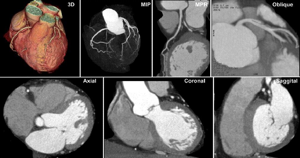
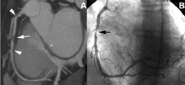
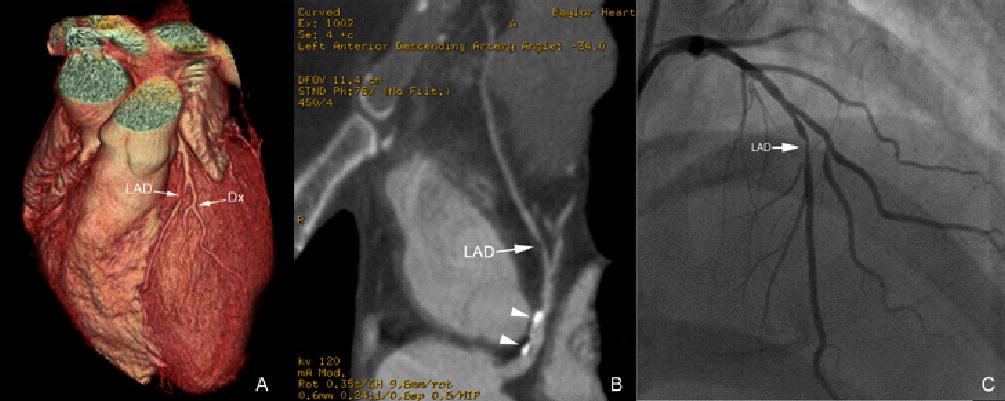
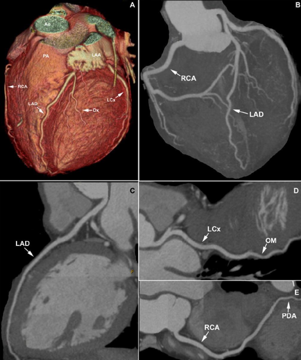
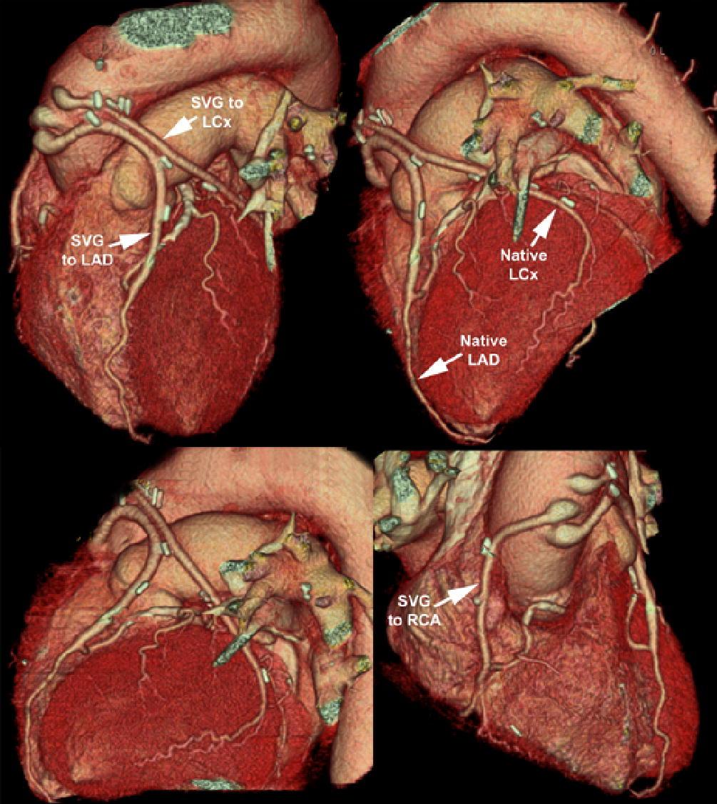
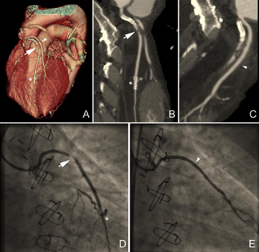
8 Figure Legends Figure slice cardiac images of a normal heart and coronary arteries. These include three dimensional (3D), maximum intensity pixel (MIP), as well as curved reformat and oblique views of the left main and LAD. Axial, coronal and saggital views give information concerning chamber size, wall thickness, and (to a lesser degree) valvular structure. Figure Slice coronary CT (A) demonstrating a "high-grade" lesion (arrow) in the mid right coronary artery (RCA). Note that there are two areas of registration artifact (arrowheads) which could be mistaken for stenoses. Figure Slice coronary CT of a patient with typical angina. Three dimensional reconstruction (A) as well as curved reformatted images (B) demonstrate a high grade lesion which is entirely of soft plaque (arrow) in the LAD. Calcific plaque is seen more proximally in the vessel (arrowheads). Invasive angiography (C) confirmed these findings. Figure Slice coronary CT images of a normal coronary tree. Multiple reconstructions are seen: three dimensional (A) which gives a good overview of the course of the arterial tree in relation to the larger structures of the heart; maximum intensity pixel (MIP) view (B) which can highlight areas of calcified plaque; curved reformatted views (C, D, and E) which are primarily used to determine extent and severity of both soft as well as calcific plaque. Figure slice cardiac CT with multiple 3D reconstructions. Patent grafts to the left anterior descending (LAD), circumflex (LCx) as well as right coronary artery (RCA) are seen. Three dimensional reconstruction is the primary tool used to evluate the overall patentcy of coroanry bypass grafts with CT coronary angiography. Figure 6. Anomalous right coronary artery (RCA): The invasive coronary angiogram was unsatisfactory (A-C) due to extreme anterior takeoff of the RCA and takeoff from the left coronary cusp. CT coronary angiogram clearly demonstrates the exact location of the takeoff of the RCA ostium. Axial slices (D) were extremely helpful in localizing the separate ostium of the RCA, and it's distance from the left main coronary. Three dimensional reconstruction (E, F) show the RCA in relation to the left main (*). Reconstruction of the pulmonary artery (PA) trunk (G) allows additional localization of the proximal course of the RCA, which is between the aorta (Ao) and PA. Figure Slice images of a post-coronary bypass patient. Three dimensional reconstruction (A) clearly demonstrates bypass grafts to the LAD (arrow) and diagonal branch (arrowhead). Curved reformatted images of the grafts show a "high-grade" lesion in the graft to the LAD (B) and a patent graft to the diagonal branch (C). A patent stent (asterisk) is seen on the 3D as well as the curved reformatted images. Invasive angiography (D and E) confirm these findings. Heart: first published as /hrt on 30 December Downloaded from on 24 July 2018 by guest. Protected by copyright. 8
9 References 1 Madigan NP, Sanfelippo JF, Curtis JJ, et al. Coronary angiography reviewed. Part II. Complication rate and management of the 'high risk' patient. Mo Med 1980;77: Scanlon PJ, Faxon DP, Audet AM, et al. ACC/AHA guidelines for coronary angiography. A report of the American College of Cardiology/American Heart Association Task Force on practice guidelines (Committee on Coronary Angiography). Developed in collaboration with the Society for Cardiac Angiography and Interventions. J Am Coll Cardiol 1999;33: Nasir K, Budoff MJ, Post WS, et al. Electron beam CT versus helical CT scans for assessing coronary calcification: current utility and future directions. Am Heart J 2003;146: Achenbach S, Giesler T, Ropers D, et al. Comparison of image quality in contrastenhanced coronary-artery visualization by electron beam tomography and retrospectively electrocardiogram-gated multislice spiral computed tomography. Invest Radiol 2003;38: Leber AW, Knez A, Becker C, et al. Non-invasive intravenous coronary angiography using electron beam tomography and multislice computed tomography. Heart 2003;89: Lembcke A, Wiese TH, Schnorr J, et al. Image quality of noninvasive coronary angiography using multislice spiral computed tomography and electron-beam computed tomography: intraindividual comparison in an animal model. Invest Radiol 2004;39: Muhlenbruch G, Wildberger JE, Koos R, et al. Coronary calcium scoring using 16- row multislice computed tomography: nonenhanced versus contrast-enhanced studies in vitro and in vivo. Invest Radiol 2005;40: Achenbach S, Moselewski F, Ropers D, et al. Detection of calcified and noncalcified coronary atherosclerotic plaque by contrast-enhanced, submillimeter multidetector spiral computed tomography: a segment-based comparison with intravascular ultrasound. Circulation 2004;109: Herzog C, Britten M, Balzer JO, et al. Multidetector-row cardiac CT: diagnostic value of calcium scoring and CT coronary angiography in patients with symptomatic, but atypical, chest pain. Eur Radiol 2004;14: Schuijf JD, Bax JJ, Salm LP, et al. Noninvasive coronary imaging and assessment of left ventricular function using 16-slice computed tomography. Am J Cardiol 2005;95: Mollet NR, Cademartiri F, Nieman K, et al. Multislice spiral computed tomography coronary angiography in patients with stable angina pectoris. J Am Coll Cardiol 2004;43: Hoffmann MH, Shi H, Schmid FT, et al. Noninvasive coronary imaging with MDCT in comparison to invasive conventional coronary angiography: a fast-developing technology. AJR Am J Roentgenol 2004;182: Achenbach S, Ropers D, Pohle FK, et al. Detection of coronary artery stenoses using multi-detector CT with 16 x 0.75 collimation and 375 ms rotation. Eur Heart J 2005;26: Kuettner A, Trabold T, Schroeder S, et al. Noninvasive detection of coronary lesions using 16-detector multislice spiral computed tomography technology: initial clinical results. J Am Coll Cardiol 2004;44: Heart: first published as /hrt on 30 December Downloaded from on 24 July 2018 by guest. Protected by copyright. 9
10 15 Ropers D, Baum U, Pohle K, et al. Detection of coronary artery stenoses with thinslice multi-detector row spiral computed tomography and multiplanar reconstruction. Circulation 2003;107: Hoffmann MH, Shi H, Schmitz BL, et al. Noninvasive coronary angiography with multislice computed tomography. Jama 2005;293: Kopp AF, Schroeder S, Kuettner A, et al. Non-invasive coronary angiography with high resolution multidetector-row computed tomography. Results in 102 patients. Eur Heart J 2002;23: Nieman K, Cademartiri F, Lemos PA, et al. Reliable noninvasive coronary angiography with fast submillimeter multislice spiral computed tomography. Circulation 2002;106: Schuijf JD, Bax JJ, Jukema JW, et al. Noninvasive evaluation of the coronary arteries with multislice computed tomography in hypertensive patients. Hypertension 2005;45: Burgstahler C, Kuettner A, Kopp AF, et al. Non-invasive evaluation of coronary artery bypass grafts using multi-slice computed tomography: initial clinical experience. Int J Cardiol 2003;90: Schlosser T, Konorza T, Hunold P, et al. Noninvasive visualization of coronary artery bypass grafts using 16-detector row computed tomography. J Am Coll Cardiol 2004;44: Martuscelli E, Romagnoli A, D'Eliseo A, et al. Evaluation of venous and arterial conduit patency by 16-slice spiral computed tomography. Circulation 2004;110: Burgstahler C, Beck T, Kuettner A, et al. Non-invasive evaluation of coronary artery bypass grafts using 16-row multi-slice computed tomography with 188 ms temporal resolution. Int J Cardiol 2006;106: Leber AW, Knez A, von Ziegler F, et al. Quantification of obstructive and nonobstructive coronary lesions by 64-slice computed tomography: a comparative study with quantitative coronary angiography and intravascular ultrasound. J Am Coll Cardiol 2005;46: Leschka S, Alkadhi H, Plass A, et al. Accuracy of MSCT coronary angiography with 64-slice technology: first experience. Eur Heart J 2005;26: Raff GL, Gallagher MJ, O'Neill WW, et al. Diagnostic accuracy of noninvasive coronary angiography using 64-slice spiral computed tomography. J Am Coll Cardiol 2005;46: Pugliese F, Mollet NR, Runza G, et al. Diagnostic accuracy of non-invasive 64-slice CT coronary angiography in patients with stable angina pectoris. Eur Radiol 2005: Shi H, Aschoff AJ, Brambs HJ, et al. Multislice CT imaging of anomalous coronary arteries. Eur Radiol 2004;14: Schoenhagen P, White RD, Nissen SE, et al. Coronary imaging: angiography shows the stenosis, but IVUS, CT, and MRI show the plaque. Cleve Clin J Med 2003;70: Schoenhagen P, Tuzcu EM, Stillman AE, et al. Non-invasive assessment of plaque morphology and remodeling in mildly stenotic coronary segments: comparison of 16-slice computed tomography and intravascular ultrasound. Coron Artery Dis 2003;14: Komatsu S, Hirayama A, Omori Y, et al. Detection of coronary plaque by computed tomography with a novel plaque analysis system, 'Plaque Map', and comparison with intravascular ultrasound and angioscopy. Circ J 2005;69:72-7. Heart: first published as /hrt on 30 December Downloaded from on 24 July 2018 by guest. Protected by copyright. 10
11 32 Rossi R, Chiurlia E, Ratti C, et al. Noninvasive assessment of coronary artery bypass graft patency by multislice computed tomography. Ital Heart J 2004;5: Demaria RG, Piciche M, Vernhet H, et al. Internal thoracic arterial grafts evaluation by multislice CT scan: a preliminary study. J Card Surg 2004;19: Willmann JK, Weishaupt D, Kobza R, et al. Coronary artery bypass grafts: ECGgated multi-detector row CT angiography--influence of image reconstruction interval on graft visibility. Radiology 2004;232: Chiurlia E, Menozzi M, Ratti C, et al. Follow-up of coronary artery bypass graft patency by multislice computed tomography. Am J Cardiol 2005;95: Ratti C, Barbieri A, Ligabue G, et al. Non-invasive, three-dimensional visualization of coronary artery bypass grafts by multislice spiral computed tomography. Int J Cardiol 2005;99: Mahnken AH, Seyfarth T, Flohr T, et al. Flat-panel detector computed tomography for the assessment of coronary artery stents: phantom study in comparison with 16-slice spiral computed tomography. Invest Radiol 2005;40: Budoff MJ, Cohen MC, Garcia MJ, et al. ACCF/AHA clinical competence statement on cardiac imaging with computed tomography and magnetic resonance: a report of the American College of Cardiology Foundation/American Heart Association/American College of Physicians Task Force on Clinical Competence and Training. J Am Coll Cardiol 2005;46: Heart: first published as /hrt on 30 December Downloaded from on 24 July 2018 by guest. Protected by copyright. 11
12
13
14
15
16

17
18
Chapter 4. Department of Cardiology, Leiden University Medical Center, Leiden, The Netherlands. Department of Radiology,
 Chapter 4 Impact of Coronary Calcium Score on Diagnostic Accuracy of Multislice Computed Tomography Coronary Angiography for Detection of Coronary Artery Disease Gabija Pundziute, 1,3 Joanne D. Schuijf,
Spiral Multislice Computed Tomography Coronary Angiography: A Current Status Report
 Clin. Cardiol. 30, 437 442 (2007) Spiral Multislice Computed Tomography Coronary Angiography: A Current Status Report P. J. De Feyter, M.D., PH.D., W. B. Meijboom, M.D., A. Weustink, M.D., C. Van Mieghem,
Non-invasive Coronary Angiography: the Role, Limitations and Future of 64-Slice Spiral Computed Tomography Coronary Angiography
 HOSPITAL CHRONICLES 2009, 4(3): 105 109 Review Non-invasive Coronary Angiography: the Role, Limitations and Future of 64-Slice Spiral Computed Tomography Coronary Angiography Arkadios C. Roussakis, MD
Diagnostic Accuracy of Noninvasive Coronary Angiography Using 64-Slice Spiral Computed Tomography
 Journal of the American College of Cardiology Vol. 46, No. 3, 2005 2005 by the American College of Cardiology Foundation ISSN 0735-1097/05/$30.00 Published by Elsevier Inc. doi:10.1016/j.jacc.2005.05.056
The Final 10-Year Follow-up Results from the Bari Randomized Trial J Am Coll Cardiol (2007) 49;1600-6
 49;1600-6") The Final 10-Year Follow-up Results from the Bari Randomized Trial J Am Coll Cardiol (2007) 49;1600-6 n&list_uids=17433949 64-Multislice Detector Computed Tomography Coronary Angiography as Potential Alternative
The Final 10-Year Follow-up Results from the Bari Randomized Trial J Am Coll Cardiol (2007) 49;1600-6 n&list_uids=17433949 64-Multislice Detector Computed Tomography Coronary Angiography as Potential Alternative
Improvement of Image Quality with ß-Blocker Premedication on ECG-Gated 16-MDCT Coronary Angiography
 16-MDCT Coronary Angiography Shim et al. 16-MDCT Coronary Angiography Sung Shine Shim 1 Yookyung Kim Soo Mee Lim Received December 1, 2003; accepted after revision June 1, 2004. 1 All authors: Department
Studies with electron beam computed tomography (EBCT) Imaging
 Imaging") Imaging Predictive Value of 16-Slice Multidetector Spiral Computed Tomography to Detect Significant Obstructive Coronary Artery Disease in Patients at High Risk for Coronary Artery Disease Patient- Versus
Imaging Predictive Value of 16-Slice Multidetector Spiral Computed Tomography to Detect Significant Obstructive Coronary Artery Disease in Patients at High Risk for Coronary Artery Disease Patient- Versus
Improved Noninvasive Assessment of Coronary Artery Bypass Grafts With 64-Slice Computed Tomographic Angiography in an Unselected Patient Population
 Journal of the American College of Cardiology Vol. 49, No. 9, 2007 2007 by the American College of Cardiology Foundation ISSN 0735-1097/07/$32.00 Published by Elsevier Inc. doi:10.1016/j.jacc.2006.10.066
A Noninvasive Assessment of CAD
 : A Noninvasive Assessment of CAD In this article, Dr. Heilbron and Dr. Forster look at the noninvasive assessment of coronary artery disease (CAD), by means of coronary computed tomography angiography
The diagnostic evaluation of dual-source CT (DSCT) in the diagnosis of coronary artery stenoses
 in the diagnosis of coronary artery stenoses") Original Article Open Access The diagnostic evaluation of dual-source CT (DSCT) in the diagnosis of coronary artery stenoses Ziqiao Lei 1, Jin Gu 2, Qing Fu 3, Heshui Shi 4, Haibo Xu 5, Ping Han 6, Jianming
Original Article Open Access The diagnostic evaluation of dual-source CT (DSCT) in the diagnosis of coronary artery stenoses Ziqiao Lei 1, Jin Gu 2, Qing Fu 3, Heshui Shi 4, Haibo Xu 5, Ping Han 6, Jianming
Cardiac Computed Tomography
 Cardiac Computed Tomography Authored and approved by Koen Nieman Stephan Achenbach Francesca Pugliese Bernard Cosyns Patrizio Lancellotti Anastasia Kitsiou Contents CARDIAC COMPUTED TOMOGRAPHY Page 1.
Perspectives of new imaging techniques for patients with known or suspected coronary artery disease
 Perspectives of new imaging techniques for patients with known or suspected coronary artery disease Department of Cardiology, Leiden University Medical Center, Leiden, The Netherlands Correspondence: Jeroen
Coronary CT Angiography
 Coronary CT Angiography Byoung Wook Choi, M.D. Department of Diagnostic Radiology Yonsei University College of Medicine, Severance Hospital E mail : bchoi@yumc.yonsei.ac.kr Abstract With the advent of
Accuracy of Multislice Computed Tomography in the Preoperative Assessment of Coronary Disease in Patients With Aortic Valve Stenosis
 Journal of the American College of Cardiology Vol. 47, No. 10, 2006 2006 by the American College of Cardiology Foundation ISSN 0735-1097/06/$32.00 Published by Elsevier Inc. doi:10.1016/j.jacc.2005.11.085
Cardiac computed tomography: indications, applications, limitations, and training requirements
 European Heart Journal (2008) 29, 531 556 doi:10.1093/eurheartj/ehm544 SPECIAL ARTICLE Cardiac computed tomography: indications, applications, limitations, and training requirements Report of a Writing
SYMPOSIA. Coronary CTA. Indications, Patient Selection, and Clinical Implications
 SYMPOSIA Indications, Patient Selection, and Clinical Implications Christian Thilo, MD,* Mark Auler, MD,* Peter Zwerner, MD,w Philip Costello, MD,* and U. Joseph Schoepf, MD* Abstract: Recent technical
Image quality and diagnostic accuracy of 16-slice multidetector computed tomography for the detection of coronary artery disease in obese patients
 (2006) 30, 569 573 & 2006 Nature Publishing Group All rights reserved 0307-0565/06 $30.00 www.nature.com/ijo ORIGINAL ARTICLE Image quality and diagnostic accuracy of 16-slice multidetector computed tomography
What every radiologist should know about cardiac CT: A case-based pictorial review
 What every radiologist should know about cardiac CT: A case-based pictorial review Poster No.: C-0555 Congress: ECR 2010 Type: Educational Exhibit Topic: Cardiac Authors: C. M. Capuñay, P. Carrascosa,
2004;77:800 4 MSCT OF CORONARY ARTERY BYPASS GRAFTS. Results. CABG With Adequate Diagnostic Quality
 Isotropic Half-Millimeter Angiography of Coronary Artery Bypass Grafts With 16-Slice Computed Tomography Marc Dewey, MD, Alexander Lembcke, MD, Christian Enzweiler, MD, Bernd Hamm, MD, and Patrik Rogalla,
ROLE OF MULTISLICE COMPUTED TOMOGRAPHY IN CARDIAC IMAGING
 ROLE OF MULTISLICE COMPUTED TOMOGRAPHY IN CARDIAC IMAGING Non-invasive coronary angiography along with multidetector computed tomography or magnetic resonance imaging is attracting increasing interest
Impact of 64-Slice Multidetector Computed Tomography on Other Diagnostic Studies for Coronary Artery Disease
 CLINICAL RESEARCH STUDY Impact of 64-Slice Multidetector Computed Tomography on Other Diagnostic Studies for Coronary Artery Disease Alex J. Auseon, DO, Sunil S. Advani, MD, Charles A. Bush, MD, Subha
Coronary Artery Imaging. Suvipaporn Siripornpitak, MD Inter-hospital Conference : Rajavithi Hospital
 Coronary Artery Imaging Suvipaporn Siripornpitak, MD Inter-hospital Conference : Rajavithi Hospital Larger array : cover scan area Detector size : spatial resolution Rotation speed : scan time Retrospective
Ultrasound. Computed tomography. Case studies. Utility of IQon Spectral CT in. cardiac imaging
 Ultrasound Computed tomography Case studies Utility of IQon Spectral CT in cardiac imaging Cardiac imaging is a challenging procedure where it is necessary to image a motion-free heart. This requires a
Chapter. Non-Invasive Coronary Imaging and Assessment of Left Ventricular Function using 16-slice Computed Tomography
 Chapter 3 Non-Invasive Coronary Imaging and Assessment of Left Ventricular Function using 16-slice Computed Tomography Joanne D. Schuijf, Jeroen J. Bax, Liesbeth P. Salm, J. Wouter Jukema, Hildo J. Lamb,
Radiation Dose Reduction and Coronary Assessability of Prospective Electrocardiogram-Gated Computed Tomography Coronary Angiography
 Journal of the American College of Cardiology Vol. 52, No. 18, 2008 2008 by the American College of Cardiology Foundation ISSN 0735-1097/08/$34.00 Published by Elsevier Inc. doi:10.1016/j.jacc.2008.07.048
Diagnostic and Prognostic Value of Coronary Ca Score
 Diagnostic and Prognostic Value of Coronary Ca Score Dr. Ghormallah Alzahrani Cardiac imaging division, Adult Cardiology department Prince Sultan Cardiac Center ( PSCC) Madina, June 2 Coronary Calcium
TITLE: Multi-Slice Computed Tomography Coronary Angiography for Coronary Artery Disease: A Review of the Clinical Effectiveness and Guidelines
 TITLE: Multi-Slice Computed Tomography Coronary Angiography for Coronary Artery Disease: A Review of the Clinical Effectiveness and Guidelines DATE: 25 February 2009 CONTEXT AND POLICY ISSUES: Coronary
Clinical Medicine Insights: Cardiology
 Open Access: Full open access to this and thousands of other papers at http://www.la-press.com. Clinical Medicine Insights: Cardiology Supplementary Issue: Cardiovascular Imaging: Current Developments
RAMA-EGAT Risk Score for Predicting Coronary Artery Disease Evaluated by 64- Slice CT Angiography
 RAMA-EGAT Risk Score for Predicting Coronary Artery Disease Evaluated by 64- Slice CT Angiography Supalerk Pattanaprichakul, MD 1, Sutipong Jongjirasiri, MD 2, Sukit Yamwong, MD 1, Jiraporn Laothammatas,
, David Stultz, MD. Cardiac CT. David Stultz, MD Cardiology Fellow, PGY 6 March 28, 2006
 Cardiac CT David Stultz, MD Cardiology Fellow, PGY 6 March 28, 2006 Courtesy Tom Kracus Courtesy Kettering Tom Medical Kracus Cente Kettering Medical Center 2003-2006, David Stultz, MD Courtesy Tom Kracus
Dr Felix Keng. Imaging of the heart is technically difficult because: Role of Cardiac MSCT. Current: Cardiac Motion Respiratory Motion
 Siemens Philips Dr Felix Keng GE Toshiba Role of Cardiac MSCT Current: Structural / congenital heart imaging Extra-cardiac / Great vessel imaging Volumes and ejection fractions (cine + gating) Calcium
M Marwan, D Ropers, T Pflederer, W G Daniel, S Achenbach
 Department of Cardiology, University of Erlangen, Erlangen, Germany Correspondence to: Dr M Marwan, Innere Medizin II, Ulmenweg 18, 91054 Erlangen, Germany; mohamed.marwan@ uk-erlangen.de Accepted 17 November
Cardiac CT Angiography
 Cardiac CT Angiography Dr James Chafey, Radiologist Why do we need a better test for C.A.D? 1. CAD is the leading cause of death in the US CAD 31% Cancer 23% Stroke 7% 2. The prevalence of atherosclerosis
Disclosure Information
 Coronary CTA Pearls and Pitfalls Ricardo C. Cury, MD, FSCCT, FAHA, FACC Chairman of Radiology Radiology Associates of South Florida Director of Cardiac Imaging Miami Cardiac and Vascular Institute Past-President
Optimal image reconstruction intervals for non-invasive coronary angiography with 64-slice CT
 Eur Radiol (2006) 16: 1964 1972 DOI 10.1007/s00330-006-0262-x CARDIAC Sebastian Leschka Lars Husmann Lotus M. Desbiolles Oliver Gaemperli Tiziano Schepis Pascal Koepfli Thomas Boehm Borut Marincek Philipp
Horizon Scanning Technology Summary. Magnetic resonance angiography (MRA) imaging for the detection of coronary artery disease
 imaging for the detection of coronary artery disease") Horizon Scanning Technology Summary National Horizon Scanning Centre Magnetic resonance angiography (MRA) imaging for the detection of coronary artery disease April 2007 This technology summary is based
Horizon Scanning Technology Summary National Horizon Scanning Centre Magnetic resonance angiography (MRA) imaging for the detection of coronary artery disease April 2007 This technology summary is based
b. To facilitate the management decision of a patient with an equivocal stress test.
 National Imaging Associates, Inc. Clinical guidelines EBCT HEART CT & HEART CT CONGENITAL CCTA CPT4 Codes: 75571 EBCT 75572, 75573 Heart CT & Heart CT Congenital 75574 - CCTA LCD ID Number: L33559 J K
Noninvasive Coronary Imaging: Plaque Imaging by MDCT
 Coronary Physiology & Imaging Summit 2007 Noninvasive Coronary Imaging: Plaque Imaging by MDCT Byoung Wook Choi Department of Radiology Yonsei University, Seoul, Korea Stary, H. C. et al. Circulation
Improving Diagnostic Accuracy of MDCT Coronary Angiography in Patients with Mild Heart Rhythm Irregularities Using ECG Editing
 Cademartiri et al. Heart Rhythm Irregularities on MDCT Angiography Cardiac Imaging Original Research A C M E D E N T U R I C A L I M A G I N G AJR 2006; 186:634 638 0361 803X/06/1863 634 American Roentgen
Correlation of Cardiac CTA to Conventional Cardiac Angiography in Diagnosing Coronary Artery Stenosis in a Community Based Center
 Correlation of Cardiac CTA to Conventional Cardiac Angiography in Diagnosing Coronary Artery Stenosis in a Community Based Center Mathieu Sabbagh, R3 Michigan State University Radiology Garden City Hospital
Angio-CT: heart and coronary arteries
 European Journal of Radiology 45 (2003) S32/S36 www.elsevier.com/locate/ejrad Angio-CT: heart and coronary arteries Andreas F. Kopp * Tübingen University Hospital, Tübingen, Germany Received 22 November
MEDICAL POLICY. Proprietary Information of Excellus Health Plan, Inc. A nonprofit independent licensee of the BlueCross BlueShield Association
 MEDICAL POLICY SUBJECT: CARDIAC COMPUTED TOMOGRAPHIC PAGE: 1 OF: 7 If the member's subscriber contract excludes coverage for a specific service it is not covered under that contract. In such cases, medical
Cardiac Imaging Tests
 Cardiac Imaging Tests http://www.medpagetoday.com/upload/2010/11/15/23347.jpg Standard imaging tests include echocardiography, chest x-ray, CT, MRI, and various radionuclide techniques. Standard CT and
Non-invasive intravenous coronary angiography using electron beam tomography and multislice computed tomography
 633 CARDIOVASCULAR MEDICINE Non-invasive intravenous coronary angiography using electron beam tomography and multislice computed tomography A W Leber, A Knez, C Becker, A Becker, C White, C Thilo, M Reiser,
Coronary revascularization treatment based on dual-source computed tomography
 Eur Radiol (2008) 18: 1800 1808 DOI 10.1007/s00330-008-0959-0 CARDIAC R. Dikkers T. P. Willems L. H. Piers G. J. de Jonge R. A. Tio H. J. van der Zaag-Loonen P. M. A. van Ooijen F. Zijlstra M. Oudkerk
MEDICAL POLICY. Proprietary Information of Excellus Health Plan, Inc. A nonprofit independent licensee of the BlueCross BlueShield Association
 MEDICAL POLICY SUBJECT: CARDIAC/CORONARY COMPUTED TOMOGRAPHIC ANGIOGRAPHY PAGE: 1 OF: 6 If a product excludes coverage for a service, it is not covered, and medical policy criteria do not apply. If a commercial
Diagnostic accuracy of dual-source computed tomography in the detection of coronary chronic total occlusion: Comparison with invasive angiography
 African Journal of Biotechnology Vol. 10(19), pp. 3854-3858, 9 May, 2011 Available online at http://www.academicjournals.org/ajb DOI: 10.5897/AJB10.983 ISSN 1684 5315 2011 Academic Journals Full Length
Computed tomography in coronary imaging: current status
 7 Computed tomography in coronary imaging: current status ARJUN NAIR AND ANAND DEVARAJ Recent technological advances have led to improvements in the use of computerised tomography for coronary imaging.
Cardiac CT angiography (CTA) has emerged as a
 has emerged as a") Clinical Utility of Cardiac CTA The current role and cost effectiveness of this modality. BY SUNIL MIRCHANDANI, MD, AND JAMES K. MIN, MD Cardiac CT angiography (CTA) has emerged as a highly accurate tool
Clinical Utility of Cardiac CTA The current role and cost effectiveness of this modality. BY SUNIL MIRCHANDANI, MD, AND JAMES K. MIN, MD Cardiac CT angiography (CTA) has emerged as a highly accurate tool
Bypass Graft and Native Postanastomotic Coronary Artery Patency: Assessment With Computed Tomography
 Bypass Graft and Native Postanastomotic Coronary Artery Patency: Assessment With Computed Tomography Daniele Andreini, MD, Gianluca Pontone, MD, Giovanni Ballerini, MD, Erika Bertella, MD, Enrica Nobili,
Advances in the Noninvasive Evaluation of Coronary Artery Disease With Multislice Computed Tomography
 Chapter 1 Advances in the Noninvasive Evaluation of Coronary Artery Disease With Multislice Computed Tomography Gabija Pundziute, 1,3 Joanne D. Schuijf, 1,4 J. Wouter Jukema, 1,4 Albert de Roos, 2 Ernst
Coronary artery bypass grafting has been a historically. Multislice CT Evaluation of Coronary Artery Bypass Graft Patients SYMPOSIA
 SYMPOSIA Multislice CT Evaluation of Coronary Artery Bypass Graft Patients Robert Chapman Gilkeson, MD* and Alan H. Markowitz, MDw Abstract: Continuous improvement in multislice computed tomography technology
Fundamentals, Techniques, Pitfalls, and Limitations of MDCT Interpretation and Measurement
 Fundamentals, Techniques, Pitfalls, and Limitations of MDCT Interpretation and Measurement 3 rd Annual Imaging & Physiology Summit November 20-21, 21, 2009 Seoul, Korea Wm. Guy Weigold, MD, FACC Cardiovascular
Sixty-four-slice multidetector computed tomography: the future of ED cardiac care
 Thomas Jefferson University Jefferson Digital Commons Department of Radiology Faculty Papers Department of Radiology October 2006 Sixty-four-slice multidetector computed tomography: the future of ED cardiac
General Cardiovascular Magnetic Resonance Imaging
 2 General Cardiovascular Magnetic Resonance Imaging 19 Peter G. Danias, Cardiovascular MRI: 150 Multiple-Choice Questions and Answers Humana Press 2008 20 Cardiovascular MRI: 150 Multiple-Choice Questions
Zurich Open Repository and Archive
 University of Zurich Zurich Open Repository and Archive Winterthurerstr. 190 CH-8057 Zurich http://www.zora.uzh.ch Year: 2008 Combining dual-source computed tomography coronary angiography and calcium
Adapted Transfer Function Design for Coronary Artery Evaluation
 Adapted Transfer Function Design for Coronary Artery Evaluation Sylvia Glaßer 1, Steffen Oeltze 1, Anja Hennemuth 2, Skadi Wilhelmsen 3, Bernhard Preim 1 1 Department of Simulation and Graphics, University
Computed Tomography Imaging of the Coronary Arteries
 Chapter 6 Computed Tomography Imaging of the Coronary Arteries G.J. Pelgrim, M. Oudkerk and R. Vliegenthart Additional information is available at the end of the chapter http://dx.doi.org/10.5772/54044
Computed Tomography of the Coronary Arteries
 NORMAL TC-99m MIBI MYOCARDIAL PERFUSION CT ANGIOGRAPHY Computed Tomography of the Coronary Arteries Authors: P.J. de Feyter MD, A. Weustink MD, F. Alberghina MD, K. Gruszczynska MD, N. van Pelt MD, F.
Noninvasive Evaluation With Multislice Computed Tomography in Suspected Acute Coronary Syndrome
 Journal of the American College of Cardiology Vol. 52, No. 3, 2008 2008 by the American College of Cardiology Foundation ISSN 0735-1097/08/$34.00 Published by Elsevier Inc. doi:10.1016/j.jacc.2008.04.012
Diagnostic Accuracy of Multidetector Computed Tomography Coronary Angiography in Patients With Dilated Cardiomyopathy
 Journal of the American College of Cardiology Vol. 49, No. 20, 2007 2007 by the American College of Cardiology Foundation ISSN 0735-1097/07/$32.00 Published by Elsevier Inc. doi:10.1016/j.jacc.2007.01.086
Impact of SSF on diagnostic performance of coronary CT angiography within one heart beat in patients with high heart rate using a 256-row detector CT
 Impact of SSF on diagnostic performance of coronary CT angiography within one heart beat in patients with high heart rate using a 256-row detector CT Junfu Liang 1,2, Hui Wang 1, Lei Xu 1, Li Dong 1, Zhanming
Multidetector CT Angiography for the Detection of Left Main Coronary Artery Disease. Rani K. Hasan, M.D. Intro to Clinical Research July 22 nd, 2011
 Multidetector CT Angiography for the Detection of Left Main Coronary Artery Disease Rani K. Hasan, M.D. Intro to Clinical Research July 22 nd, 2011 Outline Background Hypothesis Study Population Methodology
CT Imaging of Atherosclerotic Plaque. William Stanford MD Professor-Emeritus Radiology University of Iowa College of Medicine Iowa City, IA
 CT Imaging of Atherosclerotic Plaque William Stanford MD Professor-Emeritus Radiology University of Iowa College of Medicine Iowa City, IA PREVALENCE OF CARDIOVASCULAR DISEASE In 2006 there were 80 million
Cardiac CT: Your chest pain patient CAN be discharged from the Emergency Department
 Cardiac CT: Your chest pain patient CAN be discharged from the Emergency Department Asif Serajian, DO FACC FSCAI Co-director of Cardiac CT Elmhurst Memorial Hospital 1 No financial disclosures relevant
Fellows on this rotation are expected to attend nuclear conferences and multimodality imaging conference.
 Rotation: Imaging 1 Imaging 1 provides COCATS Level 1 experience for nuclear cardiology (including SPECT and PET) and cardiac CT. Fellows will administer, process, and read cardiac nuclear studies with
Multidetector Computed Tomography (MDCT) in Coronary Surgery: First Experiences With a New Tool for Diagnosis of Coronary Artery Disease
 in Coronary Surgery: First Experiences With a New Tool for Diagnosis of Coronary Artery Disease") Multidetector Computed Tomography (MDCT) in Coronary Surgery: First Experiences With a New Tool for Diagnosis of Coronary Artery Disease Hendrik Treede, MD, Christoph Becker, MD, Hermann Reichenspurner,
Multidetector Computed Tomography (MDCT) in Coronary Surgery: First Experiences With a New Tool for Diagnosis of Coronary Artery Disease Hendrik Treede, MD, Christoph Becker, MD, Hermann Reichenspurner,
Soft and Intermediate Plaques in Coronary Arteries: How Accurately Can We Measure CT Attenuation Using 64-MDCT?
 64-MDCT Measurement of Coronary Artery Plaques Cardiac Imaging Original Research Jun Horiguchi 1 Chikako Fujioka 1 Masao Kiguchi 1 Yun Shen 2 Christian E. Althoff 3,4 Hideya Yamamoto 5 Katsuhide Ito 3
Recent developments in cardiac CT
 REVIEW Recent developments in cardiac CT With the introduction of 64-multidetector row CT, coronary CT angiography has become a clinical tool, owing to improved image quality and reduced breath-hold time,
Imaging the Vulnerable Plaque. David A. Dowe, MD Atlantic Medical Imaging
 Imaging the Vulnerable Plaque David A. Dowe, MD Atlantic Medical Imaging Why is this so important? The Acute Situation Coronary disease-important Diagnosis of cardiovascular disease cost $148 billion in
Use of Nuclear Cardiology in Myocardial Viability Assessment and Introduction to PET and PET/CT for Advanced Users
 Use of Nuclear Cardiology in Myocardial Viability Assessment and Introduction to PET and PET/CT for Advanced Users February 1 5, 2011 University of Santo Tomas Hospital Angelo King A-V Auditorium Manila,
MEDICAL POLICY. Proprietary Information of YourCare Health Plan
 TOMOGRAPHIC ANGIOGRAPHY (CARDIAC CTA): CONTRAST- MEDICAL POLICY PAGE: 1 OF: 7 If the member's subscriber contract excludes coverage for a specific service it is not covered under that contract. In such
Computed Tomography of the Coronary Arteries
 Cardiology Update DAVOS 2011 Computed Tomography of the Coronary Arteries Anders Persson M.D., Ph.D Director, Assoc. Professor Center for Medical Image Science and Visualization Linköping University SWEDEN
Calcium Scoring and Cardiac CT
 Calcium Scoring and Cardiac CT John C. Finley, MD, FACC, FASE Medical Director, CT Department; Alaska Heart and Vascular Institute February 9, 2018 1. Calcium Scoring 2. CT Coronary Angiography 3. Use
Is computed tomography angiography really useful in. of coronary artery disease?
 Is computed tomography angiography really useful in screening patients with high risk of coronary artery disease? Myeong-Ki Hong, M.D. Ph D Professor of Medicine Division of Cardiology, Severance Cardiovascular
Assessment of Non-Calcified Coronary Plaques Using 64-Slice Computed Tomography: Comparison With Intravascular Ultrasound
 ORIGINAL ARTICLE DOI 10.4070 / kcj.2009.39.3.95 Print ISSN 1738-5520 / On-line ISSN 1738-5555 Copyright c 2009 The Korean Society of Cardiology Assessment of Non-Calcified Coronary Plaques Using 64-Slice
Eur Heart J. 2011;32:637-45
 Diagnostic Performance of Non-Invasive Multidetector Computed Tomography Coronary Angiography to Detect Coronary Artery Disease using Different Endpoints: Detection of Significant Stenosis versus Detection
Coronary angiography is the standard way of visualizing
 Clinical Investigation and Reports Coronary Artery Fly-Through Using Electron Beam Computed Tomography Peter M.A. van Ooijen, MSc; Matthijs Oudkerk, MD, PhD; Robert J.M. van Geuns, MD; Benno J. Rensing,
Accuracy of dual-source CT coronary angiography: first experience in a high pre-test probability population without heart rate control
 Eur Radiol (2006) 16: 2739 2747 DOI 10.1007/s00330-006-0474-0 CARDIAC Hans Scheffel Hatem Alkadhi André Plass Robert Vachenauer Lotus Desbiolles Oliver Gaemperli Tiziano Schepis Thomas Frauenfelder Thomas
Head-to-Head Comparison of Coronary Plaque Evaluation Between Multislice Computed Tomography and Intravascular Ultrasound Radiofrequency Data Analysis
 JACC: CARDIOVASCULAR INTERVENTIONS VOL. 1, NO. 2, 2008 2008 BY THE AMERICAN COLLEGE OF CARDIOLOGY FOUNDATION ISSN 1936-8798/08/$34.00 PUBLISHED BY ELSEVIER INC. DOI: 10.1016/j.jcin.2008.01.007 Head-to-Head
Chapter 5 Section 1.1. Diagnostic Radiology (Diagnostic Imaging)
") Radiology Chapter 5 Section 1.1 Issue Date: March 7, 1986 Authority: 32 CFR 199.4(a), (b)(2)(x), (c)(2)(viii), (e)(14) and 32 CFR 199.6(d)(2) 1.0 CPT 1 PROCEDURE CODES 70010-72292, 73000-76499, 77071-77084,
Radiology Chapter 5 Section 1.1 Issue Date: March 7, 1986 Authority: 32 CFR 199.4(a), (b)(2)(x), (c)(2)(viii), (e)(14) and 32 CFR 199.6(d)(2) 1.0 CPT 1 PROCEDURE CODES 70010-72292, 73000-76499, 77071-77084,
Triple Rule-out using 320-row-detector volume MDCT: A comparison of the wide volume and helical modes
 Triple Rule-out using 320-row-detector volume MDCT: A comparison of the wide volume and helical modes Poster No.: C-0488 Congress: ECR 2012 Type: Authors: Keywords: DOI: Scientific Exhibit E.-J. Kang,
SPECT-CT: Τι πρέπει να γνωρίζει ο Καρδιολόγος
 SPECT-CT: Τι πρέπει να γνωρίζει ο Καρδιολόγος Δρ Αναστασία Κίτσιου Διευθύντρια, Καρδιολογική Κλινική, Σισμανόγλειο ΓΝΑ Chair, Education Committee, Section on Nuclear Cardiology & Cardiac CT, EACVI, ESC
I have no financial disclosures
 Manpreet Singh MD I have no financial disclosures Exercise Treadmill Bicycle Functional capacity assessment Well validated prognostic value Ischemic assessment ECG changes ST segments Arrhythmias Hemodynamic
Cardiac CT imaging in coronary artery disease: Current status and future directions
 Research Highlight Cardiac CT imaging in coronary artery disease: Current status and future directions Zhonghua Sun Discipline of Medical Imaging, Department of Imaging and Applied Physics, Curtin University,
Sixty four slice Computed Tomography Scan (64-slice
 Kathmandu University Medical Journal (2008), Vol. 6, No. 2, Issue 22, 257-261 Review Article 64-Slice CT Scan in Kathmandu Medical College Teaching Hospital Karki D 1, Neopane A 2, Regmi S 3, Acharya S
Purpose. Methods and Materials
 Comparison of iterative and filtered back-projection image reconstruction techniques: evaluation of heavily calcified vessels with coronary CT angiography Poster No.: C-1644 Congress: ECR 2011 Type: Scientific
An estimated 1.46 million diagnostic cardiac catheterizations
 Computed tomographic coronary angiography: experience at Baylor University Medical Center/Baylor Jack and Jane Hamilton Heart and Vascular Hospital JEFFREY M. SCHUSSLER, MD, WILLIAM D. DOCKERY, MD, TIMOTHY
CT Coronary Angiography - Indications: From the guidelines to clinical practice
 CT Coronary Angiography - Indications: From the guidelines to clinical practice Multimodality Working Group of Cardiovascular Imaging (Nuc C, CCT CMR) Hellenic Cardiology Society Seminars, Thessaloniki,
THE ROLE OF HIGH END MULTI DETECTOR CT IN CORONARY IMAGING ESSAY
 THE ROLE OF HIGH END MULTI DETECTOR CT IN CORONARY IMAGING ESSAY Submitted for partial fulfillment of Master degree in Radiodiagnosis By Ahmed Yehia Ahmed (M.B.B.Ch., Cairo University) Supervisors Prof.
Εξελίξεις και νέες προοπτικές στην καρδιαγγειακή απεικόνιση CT. Σταμάτης Κυρζόπουλος Ωνάσειο Καρδιοχειρουργικό Κέντρο
 Εξελίξεις και νέες προοπτικές στην καρδιαγγειακή απεικόνιση CT Σταμάτης Κυρζόπουλος Ωνάσειο Καρδιοχειρουργικό Κέντρο No conflict of interest to disclose Noninvasive Cardiac Imaging Unresolved Issues-Future
Detection of Coronary Artery Stenoses with Thin Slice Multi Detector Row Spiral Computed Tomography Angiography
 Med. J. Cairo Univ., Vol. 79, No. 1, Sep. 389-406, 2011 www.medicaljournalofcairouniversity.com Detection of Coronary Artery Stenoses with Thin Slice Multi Detector Row Spiral Computed Tomography Angiography
Feasibility of contrast agent volume reduction on 640-slice CT coronary angiography in patients with low heart rate
 Feasibility of contrast agent volume reduction on 640-slice CT coronary angiography in patients with low heart rate Poster No.: B-0742 Congress: ECR 2013 Type: Authors: Keywords: DOI: Scientific Paper
Coronary artery disease remains
 CT scanning of the coronary arteries: How to do it and how to interpret it Leo P. Lawler, MD, FRCR Coronary artery disease remains one of the leading killers in the western world. Given that many of those
Interpreting CT Angiography: Three-Dimensional Reconstruction Techniques
 4 Interpreting CT Angiography: Three-Dimensional Reconstruction Techniques Matthew J. Budoff This chapter will take you through the performance and evaluation of the non-invasive coronary angiogram. Using
Coronary Artery Anomalies from Birth to Adulthood; the Role of CT Coronary Angiography in Sudden Cardiac Death Screening
 Coronary Artery Anomalies from Birth to Adulthood; the Role of CT Coronary Angiography in Sudden Cardiac Death Screening E O Dwyer 1, C O Brien 1, B Loo 1, A Snow Hogan 1, O Buckley1 2, B 1. Department
ADVANCED CARDIOVASCULAR IMAGING. Medical Knowledge. Goals and Objectives PF EF MF LF Aspirational
 Medical Knowledge Goals and Objectives PF EF MF LF Aspirational Know the basic principles of magnetic resonance imaging (MRI) including the role of the magnetic fields and gradient coil systems, generation
Disclosures. GETTING TO THE HEART OF THE MATTER WITH MULTIMODALITY CARDIAC IMAGING Organ Review Meeting 25 September. Overview
 GETTING TO THE HEART OF THE MATTER WITH MULTIMODALITY CARDIAC IMAGING Organ Review Meeting 25 September Disclosures None relevant to this presentation Mini Pakkal Assistant Professor of Radiology University
Low-dose CT coronary angiography in the step-andshoot mode: diagnostic performance
 1 Institute of Diagnostic Radiology, University Hospital Zurich, Zurich, Switzerland; 2 Clinic for Cardiovascular Surgery, University Hospital Zurich, Zurich, Switzerland; 3 Cardiovascular Centre, University
Recent Advancement of Cardiac CT
 Recent Advancement of Cardiac CT Mario J Garcia, MD, FACC, FACP Director, Non-Invasive Cardiology Professor of Medicine and Radiology Mount Sinai School of Medicine * Disclosure: grant support from Philips