The Fetus: Five Top Do Not Miss Diagnoses. Doppler Ultrasound
|
|
|
- Derek Booker
- 5 years ago
- Views:
Transcription
1 The Fetus: Five Top Do Not Miss Diagnoses Doppler Ultrasound Giancarlo Mari, MD, MBA Professor and Chair Department of Obstetrics and Gynecology University of Tennessee Health Science Center Memphis, TN
2 Five Top Do Not Miss Diagnoses Head Compression Fetal Anemia IUGR and Preeclampsia Ductal constriction Ductus venosus and impending fetal demise
3 A Few Concepts in Doppler Ultrasound

4 Doppler Formula Fd = 2(Fc x V x cos α) C Christian J. Doppler was an Austrian physicist who described the Doppler effect in 1842
 they are backscattered and return to the transducer at a different frequency. This different frequency is the Doppler shift (Fd).")
5 Doppler Effect: Application to Obstetrics From a transducer, ultrasounds are emitted at a frequency Fc. When they hit a structure that moves (for example, blood flow) they are backscattered and return to the transducer at a different frequency. This different frequency is the Doppler shift (Fd). The Doppler shift increases as the velocity of the blood flow increases (V) and as the cosine of the angle (A) between the ultrasound beam and the direction of the blood flow increases. C is a constant (velocity of the ultrasound in water: 1540 m/sec)

6 The Doppler shift arrives to the transducer. The information is analyzed, and it is presented as waveforms. Time On the y-axis, there is the velocity value. Some of the old ultrasound equipment reported the Doppler shift on the y-axis.
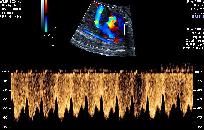
7 Types of Doppler Used in Obstetrics There are 4 types of Doppler ultrasound Spectral Doppler. There are two types of spectral Doppler: Pulsed and Continuous Color flow Doppler Power Doppler Tissue Doppler shows tissue motion such as the cardiac wall movements

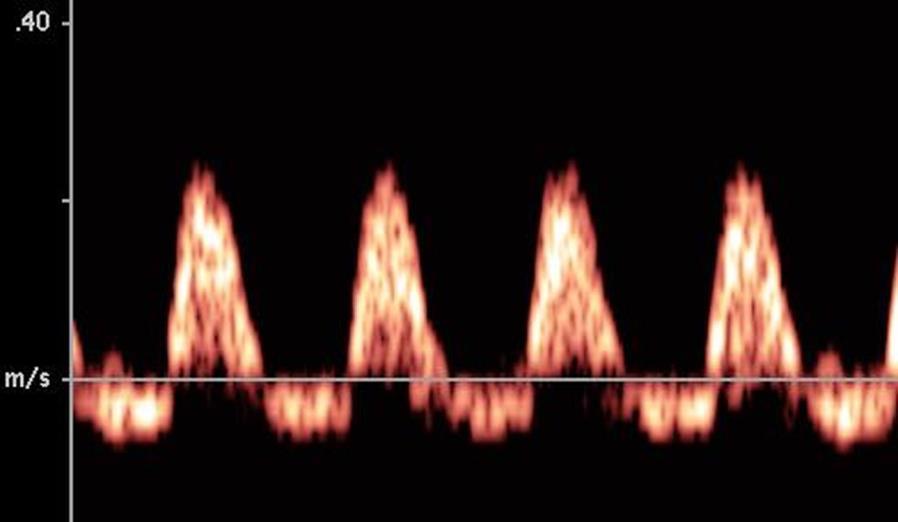
8 Direction of Blood Flow Toward the Transducer The waveforms are represented above the baseline


9 Direction of Blood Flow Away from the Transducer The waveforms are represented below the baseline
10 Does the velocity value reported on the y-axis of this set of waveforms reflect the real velocity of the blood flow? Based on what we said about the angle and the velocity, the answer is: We do not know. If the angle between the ultrasound beam and the direction of the blood flow was 0, the answer is YES. If the angle was not close to 0, the answer is NO.

11 Angle Dependence Fd = 2(Fc x V x cos α) C This slide shows the cos α values (horizontal lines) at different angles. When the angle is 90, the cos α = 0. Therefore, the value of the Doppler shift becomes 0. If this value is 0, there is no waveform generated, and no velocity can be measured.
12 It is not always easy to get an angle of 0 between the ultrasound beam and the direction of the blood flow; therefore, the velocity cannot be accurately measured in all of the cases. This is the reason why we often use angle-independent indices to quantify the waveforms.
13 Angle-Independent Indices A B = A/B ratio (Stuart et al, 1980) A - B B A - B Mean = = Resistance index (Pourcelot, 1974) Pulsatility index (Gosling and King, 1975)
14 Angle-Independent Indices These indices are independent of the angle. Therefore, the values do not change significantly when the angle changes The following slides provide a few examples


15 Angle Dependence Angle 45 Angle close to 0 o


16 Angle Dependence Flow is perpendicular to angle of incidence (cos 90 o = 0)
17 Five Top Do Not Miss Diagnoses Head Compression
18

19 MCA and Reversed Flow
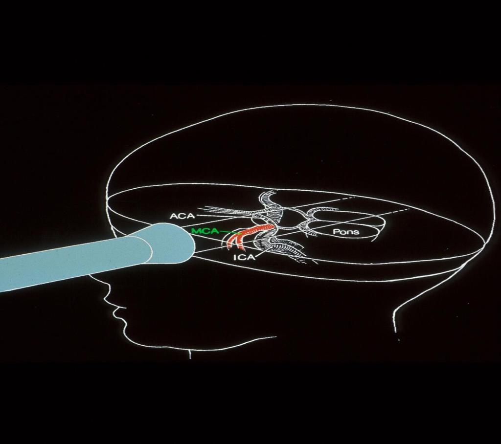
20 Middle Cerebral Artery Reversed flow at the MCA often is not pathologic; rather, it is due to compression of the transducer on the fetal head. Mari G. Am J Obstet Gynecol 2009 February 5 (epub)
21 Causes of Reversal of Flow in the MCA Excessive Transducer pressure Vyas et al Br J Obstet Gynaecol 1990;97:740 Impending fetal death Sepulveda et al. Am J Obstet Gynecol 1996;174:1645 Cardiac anomalies (unpublished data) Following fetal heart rate decelerations Late and variables
22 Five Top Do Not Miss Diagnoses Fetal Anemia

23 Waveforms of the Circle of Willis Doppler waveforms obtained in the same fetus at: A, middle cerebral artery; B and C, middle cerebral artery and anterior cerebral artery at their origin from the internal carotid artery; D and E, anterior cerebral artery; F, posterior communicating artery; G, posterior cerebral artery. The values indicate the pulsatility index. Mari G. J Ultrasound Med 1994; 13:


24 Circle of Willis The most studied artery of the Circle of Willis is the middle cerebral artery (MCA)

25 Mari G, et al. Am J Obstet Gynecol 1992;166:1262
 is the highest point of the waveform.")
26 Middle Cerebral Artery Peak Systolic Velocity The middle cerebral artery can be easily sampled with an angle of 0, and the true velocity of the blood flow can be obtained. The peak systolic velocity (PSV) is the highest point of the waveform. Therefore, for the MCA, we can easily obtain the PI (angle independent) and the PSV (an angle close to 0 is needed).

27 A C E B D F Middle Cerebral Artery Peak Systolic Velocity Steps for the correct sampling of middle cerebral artery peak systolic velocity. The use of an angle corrector increases the intra- and inter-observer variability. Therefore, its use is not recommended. Mari G, et al. J Ultrasound Med 2005; 24:425
28 Where Do We Need to Sample the MCA? Mari G, et al. J Ultrasound Med 2005; 24:425

29 Where Do We Need to Sample the MCA? The sample volume should be taken soon after the origin of the middle cerebral artery from the internal carotid artery. The artery should be visualized for its entire length, and an angle corrector should not be used. Mari G, et al. Ultrasound Obstet Gynecol 1995; 5:400

30 Middle Cerebral Artery Mari G, Deter RL. Am J Obstet Gynecol 1992;166:1262

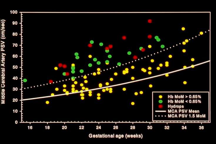
31 Middle Cerebral Artery Peak Systolic Velocity This graph represents the reference range of the middle cerebral artery PSV throughout gestation. Mari G, et al. Ultrasound Obstet Gynecol 1995; 5:400
32
33 Hemoglobin (gr/dl) Mild Anemia Moderate Anemia Severe Anemia Gestational Age (weeks) Mari G. et al. N Engl J Med 2000; 342:
34 Hemoglobin (gr/dl) Mild Anemia Moderate Anemia Severe Anemia Severe Anemia with risk of Hydrops Gestational Age (weeks) Mari G. et al. N Engl J Med 2000; 342:9
35
36 1.5 MM Mari G, et al N Engl J Med 2000; 342:9
37 Multicenter study in 5 tertiary referral centers 125 fetuses at risk for anemia MCA-PSV used for timing a cordocentesis Zimmermann R, et al. Br J Obstet Gynaecol 2002;109:746
38 Five Top Do Not Miss Diagnoses Prediction of Preeclampsia and IUGR

39 Uterine Artery Normal flow velocity waveforms of the uterine artery obtained in a normal pregnancy at 22 weeks gestation. We consider normal a pulsatility index < 1.41 at weeks gestation. Mari G. Am J Obstet Gynecol 2009 February 5 (epub)
40 Uterine Artery By ~18 weeks, trophoblasts: Invade the inner 1/3 of the myometrium Migrate through spiral arterioles Spiral arterioles lose the elastic layer Vessels become maximally dilated Brosens et al. Obstet Gynecol Annu 1972, 1:177 Pijnenborg et al. Placenta 1980; 1:3
41 Utero-Placental Vessels Normal pregnancy Preeclamptic and/or IUGR pregnancy Khong TJ et al, Br J Obstet Gynaecol, 1986;93:1049

42 Uterine Artery A B A, Normal. B, Abnormal. The arrows indicate the notching that is considered abnormal. However, the following slides will clarify why an index is preferable to the notching. Mari G. Am J Obstet Gynecol 2009 February 5 (epub)


43 Uterine Artery A B Q: What is the difference between A and B? Mari G. Am J Obstet Gynecol 2009 February 5 (epub)

44 Uterine Artery A B Q: What is the difference between A and B? A: Speed of recording. Same patient shown; however, a notch appears in B but not in A. Mari G. Am J Obstet Gynecol 2009 February 5 (epub)
45 1) At what GA should we perform Uterine artery Doppler? weeks gestation; 1 st trimester 2) How do we evaluate the results? NPV very high PPV better for high risk patients Bower S, et al. Br J Obstet Gynaecol, 1993;100:989 Harrington K, et al. Ultrasound Obstet Gynecol,1996 7:182 Coleman et al, Ultrasound Obstet Gynecol,2000; 15:7
46 L Velauthar, et al UOG 2014;43: Meta-analysis (1866 citation-18 studies) women 1 st trimester uterine artery -Preeclampsia and IUGR Sensitivity (95% CI) Specificity (95% CI) Early PE 48% (39-56) 92% (89-95) Early IUGR 39% (26-54) 93% (91-95) Any PE 26% (22-31) 93% (90-95) Any IUGR 15% (12-19) 93% (91-95)
47 Five Top Do Not Miss Diagnoses Ductal constriction
48
49 Indomethacin Ductal constriction and tricuspid regurgitation Oligohydramnios

50 Mari G, et al. J Clin Ultrasound 1996; 24:185-96
51 Ductus Arterius Constriction It occurs in 50% of patients treated with indomethacin In 10% of the cases, the effect is severe The ductal constriction is reversible Mari G, et al. J Clin Ultrasound 1996; 24:
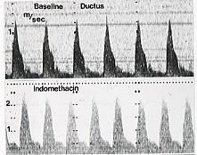
52

53 Mari G, et al. Am J Obstet Gynecol 1989; 161:1528
54 Five Top Do Not Miss Diagnoses IUGR Breathing Transitional phase Umbilical vein and Ductus venosus Ductus venosus and sovrahepatic veins


55 Umbilical Artery Flow velocity waveforms of the umbilical artery in a normal fetus from 11 to 40 weeks. Note the diastole that increases with advancing gestation. This indicates that the placental vascular resistance decreases in the normal fetus with advancing gestation. Reference ranges for the umbilical artery RI, A/B ratio, and PI.


56 Umbilical Artery: High placental vascular resistance

57 Mari and Deter. Am J Obstet Gynecol 1992;166:
58 MCA pulsatility index Mari and Deter. Am J Obstet Gynecol 1992;166: !! Gestational age (weeks)

59 MCA Waveforms at 24 Weeks A = Normal B = Brain sparing effect

60 Central Venous Circulation Ductus Venosus Biphasic Doppler Waveform First phase ~ ventricular systole Second phase ~ early diastole Nadir ~ late diastole (atrial kick)
61

62 S D S D a a Ductus venosus Hemodynamically, these phases (S, D, a) reflect the rapid chronologic change in pressure gradients between the umbilical vein and the right atrium

63 Fetal Breathing

64


65 Umbilical Vein Quantitative assessment: Velocity Qualitative assessment: Pulsation
66 What are the arrows pointing to?

67 S D E A a PIV = S a

68 Central Venous Circulation Ductus Venosus Doppler index is S/A or S-A/A Reflect RV preload
69 Ductus Venosus
70 Is Ductus venosus reversed flow an indication for delivery?
71 DV Transitional Phase DV RF 1 hour later Picconi J, et al. Am J Perinatol 2008; 25:

72 DV Transitional Phase Forward Flow Transitional Phase Reversed Flow Picconi J, et al. Am J Perinatol 2008; 25:


73 DV RF 21 days before IUFD DV RF 9 days before delivery


74 What is the difference among the different sets of waveforms? A B C

75 a. What is the SIA index and b. What does it indicate? a. Peak systolic velocity Isovolumetric relaxation + a-wave b. Myocardial function Picconi et al J Ultrasound Med 2008;27:1283

76 Picconi et al J Ultrasound Med 2008;27:1283
39 th Annual Perinatal Conference Vanderbilt University December 6, 2013 IUGR. Diagnosis and Management
 39 th Annual Perinatal Conference Vanderbilt University December 6, 2013 IUGR Diagnosis and Management Giancarlo Mari, M.D., M.B.A. Professor and Chair Department of Obstetrics and Gynecology University
Diagnosis and Management of the Early Growth Restricted Fetus
 11 th Congress of Maternal Fetal Medicine and Perinatology Society of Turkey Diagnosis and Management of the Early Growth Restricted Fetus Giancarlo Mari, MD, MBA, FACOG, FAIUM Professor and Chair Department
4/19/2018. St. Cloud Hospital Perinatology Kristin Olson, RDMS, RVT
 St. Cloud Hospital Perinatology Kristin Olson, RDMS, RVT Review Fetal Circulation Provide Indications for Umbilical Artery, Middle Cerebral Artery, and Ductus Venosus Doppler studies. Demonstrate normal
Optimising your Doppler settings for an accurate PI. Alison McGuinness Mid Yorks Hospitals
 Optimising your Doppler settings for an accurate PI Alison McGuinness Mid Yorks Hospitals Applications Both maternal uterine and fetal circulations can be studied with doppler sonography Uterine arteries
Editorial. Color and pulsed Doppler in fetal echocardiography A. ABUHAMAD
 Ultrasound Obstet Gynecol 2004; 24: 1 9 Published online in Wiley InterScience (www.interscience.wiley.com). DOI: 10.1002/uog.1096 Editorial Color and pulsed Doppler in fetal echocardiography A. ABUHAMAD
Basic Doppler Assessment of Fetal Distress
 Basic Doppler Assessment of Fetal William J. Polzin, M.D. Co-Director, Fetal Care Center of Cincinnati Director, Division of Maternal-Fetal Medicine Good Samaritan Hospital Cincinnati, OH No Relevant Disclosures
PIAF study: Placental insufficiency and aortic isthmus flow Jean-Claude Fouron, MD
 Dear colleagues, I would like to thank you very sincerely for agreeing to participate in our multicentre study on the clinical significance of recording fetal aortic isthmus flow during placental circulatory
First Trimester Fetal Echocardiography: Insight Into the Fetal Circulation
 First Trimester Fetal Echocardiography: Insight Into the Fetal Circulation Lisa K. Hornberger, MD Fetal & Neonatal Cardiology Program Department of Pediatrics, Division of Cardiology Department of Obstetrics
What effects will proximal or distal disease have on a waveform?
 Spectral Doppler Interpretation Director of Ultrasound Education & Quality Assurance Baylor College of Medicine Division of Maternal-Fetal Medicine Maternal Fetal Center Imaging Manager Texas Children
Appendix II: ECHOCARDIOGRAPHY ANALYSIS
 Appendix II: ECHOCARDIOGRAPHY ANALYSIS Two-Dimensional (2D) imaging was performed using the Vivid 7 Advantage cardiovascular ultrasound system (GE Medical Systems, Milwaukee) with a frame rate of 400 frames
Key issues in (early and late) IUGR
 IUGR") Key issues in (early and late) IUGR Eduard Gratacós Maternal-Fetal Medicine Department, Hospital Clínic, University of Barcelona www.fetalmedicinebarcelona.org (early-onset) IUGR vs SGA: the era of UA
Key issues in (early and late) IUGR Eduard Gratacós Maternal-Fetal Medicine Department, Hospital Clínic, University of Barcelona www.fetalmedicinebarcelona.org (early-onset) IUGR vs SGA: the era of UA
Assessment of fetal heart function and rhythm
 Assessment of fetal heart function and rhythm The fetal myocardium Early Gestation Myofibrils 30% of myocytes Less sarcoplasmic reticula Late Gestation Myofibrils 60% of myocytes Increased force per unit
A (quasi)evidence-based approach to the management of early-onset IUGR
evidence-based approach to the management of early-onset IUGR") A (quasi)evidence-based approach to the management of early-onset IUGR Eduard Gratacós Barcelona Center for Maternal-Fetal and Neonatal Medicine Hospital Clínic and Hospital Sant Joan de Deu, University
A (quasi)evidence-based approach to the management of early-onset IUGR Eduard Gratacós Barcelona Center for Maternal-Fetal and Neonatal Medicine Hospital Clínic and Hospital Sant Joan de Deu, University
The role of Doppler studies in predicting individual intrauterine fetal demise after laser therapy for twin twin transfusion syndrome
 Ultrasound Obstet Gynecol 2003; 22: 246 251 Published online in Wiley InterScience (www.interscience.wiley.com). DOI: 10.1002/uog.215 The role of Doppler studies in predicting individual intrauterine fetal
Normal pericardial fluid in the fetus: color and spectral Doppler analysis
 Ultrasound Obstet Gynecol 2001; 18: 248 252 Normal pericardial fluid in the fetus: color and spectral Blackwell ORIGINAL Science, PAPERLtd Doppler analysis S.-J. YOO*, J.-Y. MIN and Y.-H. LEE *Department
Management of IUGR Prof. Dr. Acar KOÇ
 Management of IUGR Prof. Dr. Acar KOÇ Ankara University School of Medicine Department of OB&GYN Department of Perinatology Definition and Diagnosis: SGA IUGR EFW: < 10th percentile EFW: < 10th percentile
First-Trimester Fetal Cardiac Function
 CME Article First-Trimester Fetal Cardiac Function Noirin E. Russell, MRCPI, Fionnuala M. McAuliffe, MD, FRCPI, MRCOG Objective. The purpose of this study was to establish normal values for fetal heart
Doppler Basic & Hemodynamic Calculations
 Doppler Basic & Hemodynamic Calculations August 19, 2017 Smonporn Boonyaratavej MD Division of Cardiology, Department of Medicine Chulalongkorn University Cardiac Center, King Chulalongkorn Memorial Hospital
Ductus Venosus Doppler Ultrasound in Diabetic Pregnancies
 International Journal of Basic and Applied Sciences. Vol. 5 No. 2. 2016. Copyright by. All Rights Reserved Full Length Research Paper Ductus Venosus Doppler Ultrasound in Diabetic Pregnancies Alaa Ibrahim
What effects will proximal or distal disease have on an waveform?
 Spectral Doppler Interpretation Director Director of of Ultrasound Ultrasound Education Education & & Quality Quality Assurance Assurance Baylor Baylor College College of of Medicine Medicine Division
Evaluation of Fetal Pulmonary Veins During Early Gestation by Pulsed Doppler Ultrasound: A Feasibility Study
 J. Fetal Med. (March 2015) 2:27 32 DOI 10.1007/s40556-015-0038-y ORIGINAL ARTICLE Evaluation of Fetal Pulmonary Veins During Early Gestation by Pulsed Doppler Ultrasound: A Feasibility Study Aldo L. Schenone
Bits and Bobs secondary causes of heart problems. Dr Angela McBrien 9 th September 2017
 Bits and Bobs secondary causes of heart problems Dr Angela McBrien 9 th September 2017 Not the heart Dextroposition Heart in the right chest with the apex to the left Often caused by left sided chest mass
Failing right ventricle
 Failing right ventricle U. Herberg 1, U. Gembruch 2 1 Pediatric Cardiology, 2 Prenatal Diagnostics and Fetal Therapy, University of Bonn, Germany Prenatal Physiology Right ventricle dominant ventricle
Relevance of measuring diastolic time intervals in the ductus venosus during the early stages of twin twin transfusion syndrome
 Ultrasound Obstet Gynecol 2007; 30: 983 987 Published online 15 November 2007 in Wiley InterScience (www.interscience.wiley.com). DOI: 10.1002/uog.5161 Relevance of measuring diastolic time intervals in
FETAL ECHO IN TWIN PREGNACY: MONOCHORIONIC TWINS DELHI CHILD HEART CENTER & INDRAPRASTHA APOLLO HOSPITAL NEW DELHI
 FETAL ECHO IN TWIN PREGNACY: MONOCHORIONIC TWINS DELHI CHILD HEART CENTER & INDRAPRASTHA APOLLO HOSPITAL NEW DELHI Scope of this talk Twin to Twin Transfusion TRAP Sequence Congenital Heart Defects in
Right Ventricle Steven J. Lester MD, FACC, FRCP(C), FASE Mayo Clinic, Arizona
, FASE Mayo Clinic, Arizona") Right Ventricle Steven J. Lester MD, FACC, FRCP(C), FASE Mayo Clinic, Arizona 1. In which scenario will applying the simplified Bernoulli equation to the peak tricuspid regurgitation velocity and adding
Right Ventricle Steven J. Lester MD, FACC, FRCP(C), FASE Mayo Clinic, Arizona 1. In which scenario will applying the simplified Bernoulli equation to the peak tricuspid regurgitation velocity and adding
Fetal cardiovascular parameters for the prediction of postnatal cardiovascular risk in intrauterine growth-restriction?
 17 th International Conference on Prenatal Diagnosis and Therapy Lisbon, June 2013 Fetal cardiovascular parameters for the prediction of postnatal cardiovascular risk in intrauterine growth-restriction?
SWISS SOCIETY OF NEONATOLOGY. Prenatal closure of the ductus arteriosus
 SWISS SOCIETY OF NEONATOLOGY Prenatal closure of the ductus arteriosus March 2007 Leone A, Fasnacht M, Beinder E, Arlettaz R, Neonatal Intensive Care Unit (LA, AR), University Hospital Zurich, Cardiology
COMPREHENSIVE EVALUATION OF FETAL HEART R. GOWDAMARAJAN MD
 COMPREHENSIVE EVALUATION OF FETAL HEART R. GOWDAMARAJAN MD Disclosure No Relevant Financial Relationships with Commercial Interests Fetal Echo: How to do it? Timing of Study -optimally between 22-24 weeks
Diastolic Function: What the Sonographer Needs to Know. Echocardiographic Assessment of Diastolic Function: Basic Concepts 2/8/2012
 Diastolic Function: What the Sonographer Needs to Know Pat Bailey, RDCS, FASE Technical Director Beaumont Health System Echocardiographic Assessment of Diastolic Function: Basic Concepts Practical Hints
Doppler assessment of fetal aortic isthmus blood flow in two different sonographic planes during the second half of gestation
 Ultrasound Obstet Gynecol 2005; 26: 170 174 Published online in Wiley InterScience (www.interscience.wiley.com). DOI: 10.1002/uog.1955 Doppler assessment of fetal aortic isthmus blood flow in two different
Venous Doppler Evaluation of the Growth-Restricted Fetus
 Venous Doppler Evaluation of the Growth-Restricted Fetus Ahmet Alexander Baschat, MD KEYWORDS Fetal growth restriction Doppler Ductus venosus Venous circulation Fetal surveillance Integrated testing The
Hemodynamic Assessment. Assessment of Systolic Function Doppler Hemodynamics
 Hemodynamic Assessment Matt M. Umland, RDCS, FASE Aurora Medical Group Milwaukee, WI Assessment of Systolic Function Doppler Hemodynamics Stroke Volume Cardiac Output Cardiac Index Tei Index/Index of myocardial
Summary. HVRA s Cardio Vascular Genetic Detailed L2 Obstetrical Ultrasound. CPT 76811, 76825, _ 90% CHD detection. _ 90% DS detection.
 What is the role of fetal echocardiography (2D 76825, cardiovascular color flow mapping 93325) as performed in conjunction with detailed fetal anatomy scan (CPT 76811) now that AIUM requires limited outflow
Circulatory System MARE HEIFER. aorta cvc. iia eia ipa. aorta. dca. (uov) oa. uboa. uma. ubva va. bua. iia. uma. ubva. eia. ua bua. cvc.
 oa. uboa. uma. ubva va. bua. iia. uma. ubva. eia. ua bua. cvc.") Circulatory System 13 MARE aorta cvc ov (uov) oa dca uboa iia eia ipa ua uma bua ubva va HEIFER iia aorta cvc eia oa uma ua bua ubva va ov (uov) uboa 18 Chapter 1 Hemodynamics Plug flow Natural disturbed
Circulatory System 13 MARE aorta cvc ov (uov) oa dca uboa iia eia ipa ua uma bua ubva va HEIFER iia aorta cvc eia oa uma ua bua ubva va ov (uov) uboa 18 Chapter 1 Hemodynamics Plug flow Natural disturbed
Correlation analysis of ductus venosus velocity indices and fetal cardiac function
 Ultrasound Obstet Gynecol 2014; 43: 515 519 Published online 3 April 2014 in Wiley Online Library (wileyonlinelibrary.com). DOI: 10.1002/uog.13242 Correlation analysis of ductus venosus velocity indices
Heart and Soul Evaluation of the Fetal Heart
 Heart and Soul Evaluation of the Fetal Heart Ivana M. Vettraino, M.D., M.B.A. Clinical Associate Professor, Michigan State University College of Human Medicine Objectives Review the embryology of the formation
Diagnosis of Congenital Cardiac Defects Between 11 and 14 Weeks Gestation in High-Risk Patients
 Article Diagnosis of Congenital Cardiac Defects Between 11 and 14 Weeks Gestation in High-Risk Patients Zeev Weiner, MD, Abraham Lorber, MD, Eliezer Shalev, MD Objective. To examine the feasibility of
Opinion. Technical aspects of aortic isthmus Doppler velocimetry in human fetuses
 Ultrasound Obstet Gynecol 2009; 33: 628 633 Published online in Wiley InterScience (www.interscience.wiley.com). DOI: 10.1002/uog.6406 Opinion Technical aspects of aortic isthmus Doppler velocimetry in
Fetal Cardiac Function and Venous Circulation - Experiences with Velocity Vector Imaging
 Fetal Cardiac Function and Venous Circulation - Experiences with Velocity Vector Imaging Dahlbäck, Charlotte Published: 2015-01-01 Link to publication Citation for published version (APA): Dahlbäck, C.
Anatomy & Physiology
 1 Anatomy & Physiology Heart is divided into four chambers, two atrias & two ventricles. Atrioventricular valves (tricuspid & mitral) separate the atria from ventricles. they open & close to control flow
Volume Flow. Volume Flow
 Volume Flow Jonathan M. Rubin, M.D., Ph.D. Department of Radiology Volume Flow Technique initially described by Hottenger and Meindl in 1974 Describes method for measuring the total flux across a flow
Heart and Lungs. LUNG Coronal section demonstrates relationship of pulmonary parenchyma to heart and chest wall.
 Heart and Lungs Normal Sonographic Anatomy THORAX Axial and coronal sections demonstrate integrity of thorax, fetal breathing movements, and overall size and shape. LUNG Coronal section demonstrates relationship
Fetal cardiac function: what to use and does it make a difference?
 17 th International Conference on Prenatal Diagnosis and Therapy Lisbon, June 2013 Fetal cardiac function: what to use and does it make a difference? Fàtima Crispi Department of Maternal-Fetal Medicine,
Redistribution of arterial circulation has been documented
 Ultrasonographic and Biochemical Markers of Human Fetal Cardiac Dysfunction in Placental Insufficiency Kaarin Mäkikallio, MD; Olli Vuolteenaho, MD; Pentti Jouppila, MD; Juha Räsänen, MD Background Placental
Guide to Small Animal Vascular Imaging using the Vevo 770 Micro-Ultrasound System
 Guide to Small Animal Vascular Imaging using the Vevo 770 Micro-Ultrasound System January 2007 Objectives: After completion of this module, the participant will be able to accomplish the following: Understand
Case Report Right Ventricular Outflow Tract Obstruction in Monochorionic Twins with Selective Intrauterine Growth Restriction
 Case Reports in Pediatrics Volume 2012, Article ID 426825, 4 pages doi:10.1155/2012/426825 Case Report Right Ventricular Outflow Tract Obstruction in Monochorionic Twins with Selective Intrauterine Growth
Evaluation of normal fetal pulmonary veins from the early second trimester by enhanced-flow (e-flow) echocardiography
 echocardiography") Ultrasound Obstet Gynecol 211; 38: 652 657 Published online 1 November 211 in Wiley Online Library (wileyonlinelibrary.com). DOI: 1.12/uog.8965 Evaluation of normal fetal pulmonary veins from the early
Ultrasound Obstet Gynecol 211; 38: 652 657 Published online 1 November 211 in Wiley Online Library (wileyonlinelibrary.com). DOI: 1.12/uog.8965 Evaluation of normal fetal pulmonary veins from the early
PART II ECHOCARDIOGRAPHY LABORATORY OPERATIONS ADULT TRANSTHORACIC ECHOCARDIOGRAPHY TESTING
 PART II ECHOCARDIOGRAPHY LABORATORY OPERATIONS ADULT TRANSTHORACIC ECHOCARDIOGRAPHY TESTING STANDARD - Primary Instrumentation 1.1 Cardiac Ultrasound Systems SECTION 1 Instrumentation Ultrasound instruments
Introduction to Fetal Medicine. Lloyd R. Feit M.D. Associate Professor of Pediatrics Warren Alpert Medical School Brown University
 Associate Professor of Pediatrics Warren Alpert Medical School Brown University Fetal Cardiology Important in evaluation of high risk pregnancies. Information obtainable in > 95% of patients attempted.
Myocardial Velocities, Dynamics of the Septum Primum, and Placental Dysfunction in Fetuses with Growth Restriction
 138 Myocardial Velocities, Dynamics of the Septum Primum, and Placental Dysfunction in Fetuses with Growth Restriction Alexandre Antonio Naujorks, MD, PhD, Paulo Zielinsky, MD, PhD, Caroline Klein, MD,
Adel Hasanin Ahmed 1
 Adel Hasanin Ahmed 1 PERICARDIAL DISEASE The pericardial effusion ends anteriorly to the descending aorta and is best visualised in the PLAX. PSAX is actually very useful sometimes for looking at posterior
COPYRIGHTED MATERIAL. The fetal circulation CHAPTER 1. Postnatal circulation
 1 CHAPTER 1 The fetal circulation The circulation in the fetus differs from that in the adult. Knowledge of the course and distribution of the fetal circulation is important to our understanding of the
Twin-reversed arterial perfusion sequence: pre- and postoperative cardiovascular findings in the pump twin
 Ultrasound Obstet Gynecol 2009; 34: 550 555 Published online 24 September 2009 in Wiley InterScience (www.interscience.wiley.com). DOI: 10.1002/uog.6431 Twin-reversed arterial perfusion sequence: pre-
Modified myocardial performance index for evaluation of fetal cardiac function in pre-eclampsia
 Ultrasound Obstet Gynecol 2009; 33: 51 57 Published online 11 December 2008 in Wiley InterScience (www.interscience.wiley.com). DOI: 10.1002/uog.6272 Modified myocardial performance index for evaluation
A lthough changes in cardiac morphology during organogenesis
 334 CARDIOVASCULAR MEDICINE Human fetal cardiac function during the first trimester of pregnancy K Mäkikallio, P Jouppila, J Räsänen... See end of article for authors affiliations... Correspondence to:
AOGS ORIGINAL RESEARCH ARTICLE
 AOGS ORIGINAL RESEARCH ARTICLE Ventricular outputs, central blood flow distribution and flow pattern through the aortic isthmus of fetuses with simple transposition of the great arteries JULIE BLANC 1,2,
Introduction to Fetal Doppler Echocardiography
 Chapter 32 Introduction to Fetal Doppler Echocardiography Dev Maulik Introduction Evaluation of the fetal heart constitutes one of the critical areas of prenatal diagnosis. Advances in diagnostic medical
Effect of physiological heart rate changes on left ventricular dimensions and mitral blood flow velocities in the normal fetus
 ELSEVIER Early Human Development 40 (1995) 109-114 Effect of physiological heart rate changes on left ventricular dimensions and mitral blood flow velocities in the normal fetus P.B. Tsyvian a, K.V. Malkin
ULTRASOUND OF THE FETAL HEART
 ULTRASOUND OF THE FETAL HEART Cameron A. Manbeian, MD Disclosure Statement Today s faculty: Cameron Manbeian, MD does not have any relevant financial relationships with commercial interests or affiliations
The cerebroplacental Doppler ratio predicts postnatal outcome in fetuses with congenital heart block
 ORIGINAL ARTICLE The cerebroplacental Doppler ratio predicts postnatal outcome in fetuses with congenital heart block GA Fleming 1, A Bircher 2, A Kavanaugh-McHugh 1 and MR Liske 1 (2008) 28, 791 796 r
The Cardiac Cycle Clive M. Baumgarten, Ph.D.
 The Cardiac Cycle Clive M. Baumgarten, Ph.D. OBJECTIVES: 1. Describe periods comprising cardiac cycle and events within each period 2. Describe the temporal relationships between pressure, blood flow,
Mild tricuspid regurgitation: a benign fetal finding at various stages of pregnancy
 Ultrasound Obstet Gynecol 2005; 26: 606 610 Published online 7 October 2005 in Wiley InterScience (www.interscience.wiley.com). DOI: 10.1002/uog.1999 Mild tricuspid regurgitation: a benign fetal finding
Continuous wave Doppler ultrasound in evaluation of cerebral blood flow in neonates
 Archives of Disease in Childhood, 1983, 58, 677-681 Continuous wave Doppler ultrasound in evaluation of cerebral blood flow in neonates P H GRAY, E A GRIFFIN, J E DRUMM, D E FITZGERALD, AND N M DUIGNAN
Adult Echocardiography Examination Content Outline
 Adult Echocardiography Examination Content Outline (Outline Summary) # Domain Subdomain Percentage 1 2 3 4 5 Anatomy and Physiology Pathology Clinical Care and Safety Measurement Techniques, Maneuvers,
Estimated cardiac output and cardiovascular profile score in fetuses with high cardiac output lesions
 Ultrasound Obstet Gynecol 213; 41: 54 58 Published online in Wiley Online Library (wileyonlinelibrary.com). DOI: 1.12/uog.1239 Estimated cardiac output and cardiovascular profile score in fetuses with
Tissue Doppler Imaging
 Cronicon OPEN ACCESS Hesham Rashid* Tissue Doppler Imaging CARDIOLOGY Editorial Department of Cardiology, Benha University, Egypt *Corresponding Author: Hesham Rashid, Department of Cardiology, Benha University,
HDlive Silhouette Mode With Spatiotemporal Image Correlation for Assessment of the Fetal Heart
 ORIGINAL RESEARCH HDlive Silhouette Mode With Spatiotemporal Image Correlation for Assessment of the Fetal Heart Toshiyuki Hata, MD, PhD, Mohamed Ahmed Mostafa AboEllail, MD, Suraphan Sajapala, MD, Mari
DEVELOPMENT OF THE CIRCULATORY SYSTEM L E C T U R E 5
 DEVELOPMENT OF THE CIRCULATORY SYSTEM L E C T U R E 5 REVIEW OF CARDIAC ANATOMY Heart 4 chambers Base and apex Valves Pericardial sac 3 layers: epi, myo, endo cardium Major blood vessels Aorta and its
Prediction of acidemia at birth by Doppler assessment of fetal cerebral transverse sinus in pregnancies with placental insufficiency
 Ultrasound Obstet Gynecol 2009; 33: 188 192 Published online 6 October 2008 in Wiley InterScience (www.interscience.wiley.com). DOI: 10.1002/uog.6130 Prediction of acidemia at birth by Doppler assessment
Uncommon Doppler Echocardiographic Findings of Severe Pulmonic Insufficiency
 Uncommon Doppler Echocardiographic Findings of Severe Pulmonic Insufficiency Rahul R. Jhaveri, MD, Muhamed Saric, MD, PhD, FASE, and Itzhak Kronzon, MD, FASE, New York, New York Background: Two-dimensional
UPDATE ON DIAGNOSIS AND MANAGEMENT OF FETAL GROWTH RESTRICTION
 UPDATE ON DIAGNOSIS AND MANAGEMENT OF FETAL GROWTH RESTRICTION Eduard Gratacos Servicio de Medicina Maternofetal Hospital Clinic y Hospital Sant Joan de Deu - Universidad de Barcelona www.fetalmedicinebarcelona.org
Congenital heart disease in twin-to-twin transfusion syndrome treated with fetoscopic laser surgery
 Chapter 10 Congenital heart disease in twin-to-twin transfusion syndrome treated with fetoscopic laser surgery Enrico Lopriore MD Regina Bökenkamp MD Marry Rijlaarsdam MD Marieke Sueters MD Frank PHA Vandenbussche
Diagnostic approach to heart disease
 Diagnostic approach to heart disease Initial work up History Physical exam Chest radiographs ECG Special studies Echocardiography Cardiac catheterization Echocardiography principles Technique of producing
An update on technique of fetal echocardiography with emphasis on anomalies detectable in four chambered view.
 An update on technique of fetal echocardiography with emphasis on anomalies detectable in four chambered view. Dr. Ranjitha.G Specialist Radiologist NMC-SH Al ain, UAE Fetal echocardiography is an essential
AN INTRODUCTION TO DOPPLER. Sarah Gardner, Clinical lead, Tissue viability service. Oxford Health NHS Foundation Trust.
 AN INTRODUCTION TO DOPPLER Sarah Gardner, Clinical lead, Tissue viability service. Oxford Health NHS Foundation Trust. THE DOPPLER EFFECT The Doppler Principle was described by Physicist and mathematician
The short-term effect of nifedipine tocolysis on placental, fetal cerebral and atrioventricular Doppler waveforms
 Ultrasound Obstet Gynecol 004; 4: 761 765 Published online 6 October 004 in Wiley InterScience (www.interscience.wiley.com). DOI: 10.100/uog.1770 The short-term effect of nifedipine tocolysis on placental,
Assessment of Cardiac Dysfunction in the Intrauterine Growth-restricted Fetuses from Pre-eclamptic Mothers
 Assessment of Cardiac Dysfunction in the Intrauterine Growth-restricted 10.5005/jp-journals-10009-1346 Fetuses from Pre-eclamptic Mothers Original Article Assessment of Cardiac Dysfunction in the Intrauterine
Vascular Sonography Examination
 Vascular Sonography Examination The purpose of The American Registry of Radiologic Technologists (ARRT ) Vascular Sonography Examination is to assess the knowledge and cognitive skills underlying the intelligent
Carlos Eduardo Suaide Silva, Luiz Darcy Cortez Ferreira, Luciana Braz Peixoto, Claudia Gianini Monaco, Manuel Adán Gil, Juarez Ortiz
 Silva et al Original Article Arq Bras Cardiol Study of the Myocardial Contraction and Relaxation Velocities through Doppler Tissue Imaging Echocardiography. A New Alternative in the Assessment of the Segmental
1. Which of the following blood vessels has a thin elastic layer? A. Aorta. B. Pulmonary artery. C. Posterior vena cava. D. Mesenteric capillary.
 CIRCULATORY SYSTEM 1. Which of the following blood vessels has a thin elastic layer? A. Aorta. B. Pulmonary artery. C. Posterior vena cava. D. Mesenteric capillary. 2. Capillary beds are equipped with
How to recognise a congenitally infected fetus? Dr. Amar Bhide Consultant in Obstetrics and Fetal Medicine
 How to recognise a congenitally infected fetus? Dr. Amar Bhide Consultant in Obstetrics and Fetal Medicine Scope Cytomegalovirus Parvovirus Varicella Toxoplasma Rubella Clinical scenarios Maternal exposure
2. capillaries - allow exchange of materials between blood and tissue fluid
 Chapter 19 - Vascular System A. categories and general functions: 1. arteries - carry blood away from heart 2. capillaries - allow exchange of materials between blood and tissue fluid 3. veins - return
Cardiac ultrasound protocols
 Cardiac ultrasound protocols IDEXX Telemedicine Consultants Two-dimensional and M-mode imaging planes Right parasternal long axis four chamber Obtained from the right side Displays the relative proportions
Transcranial Doppler (Basic Step) Dae-il Chang, M.D., Sung Sang Yoon, M.D. Department of Neurology, College of Medicine, Kyunghee university
 Dae-il Chang, M.D., Sung Sang Yoon, M.D. Department of Neurology, College of Medicine, Kyunghee university") Transcranial Doppler (Basic Step) Dae-il Chang, M.D., Sung Sang Yoon, M.D. Department of Neurology, College of Medicine, Kyunghee university Principles of Doppler Ultrasonography Major target Speed & direction
Transcranial Doppler (Basic Step) Dae-il Chang, M.D., Sung Sang Yoon, M.D. Department of Neurology, College of Medicine, Kyunghee university Principles of Doppler Ultrasonography Major target Speed & direction
Assessment of LV systolic function
 Tutorial 5 - Assessment of LV systolic function Assessment of LV systolic function A knowledge of the LV systolic function is crucial in the undertanding of and management of unstable hemodynamics or a
STRUCTURED EDUCATION REQUIREMENTS IMPLEMENTATION DATE: JULY 1, 2016
 STRUCTURED EDUCATION REQUIREMENTS Vascular Sonography The purpose of structured education is to provide the opportunity for individuals to develop mastery of discipline-specific knowledge that, when coupled
Doppler changes in the main fetal brain arteries at different stages of hemodynamic adaptation in severe intrauterine growth restriction
 Ultrasound Obstet Gynecol 2007; 30: 297 302 Published online 30 July 2007 in Wiley InterScience (www.interscience.wiley.com). DOI: 10.1002/uog.4084 Doppler changes in the main fetal brain arteries at different
Prospect Cardiac Packages. S-Sharp
 Prospect Cardiac Packages S-Sharp B mode: Teichholz: Teichholz formula LV Volume 2D: modified Simpson's rule method ALM: area length method LV Volume (Intg.): integral method M mode: Long axis: Teichholz
Cardiac Cycle MCQ. Professor of Cardiovascular Physiology. Cairo University 2007
 Cardiac Cycle MCQ Abdel Moniem Ibrahim Ahmed, MD Professor of Cardiovascular Physiology Cairo University 2007 1- Regarding the length of systole and diastole: a- At heart rate 75 b/min, the duration of
VFI Technology to Change the Way You Work
 Analogic Ultrasound VFI Technology to Change the Way You Work Vascular Ultrasound Made Easier Vector Flow Imaging VFI VFI is a ground-breaking technology that can revolutionize the workflow for many Doppler
Maternal and Fetal Physiology
 Background Maternal and Fetal Physiology Anderson Lo, DO Fellow, Maternal-Fetal Medicine Wayne State University School of Medicine SEMCME Fetal Assessment Course July 20, 2018 Oxygen pathway Mother Placenta
Epidemiological studies have demonstrated fetal growth
 Pediatric Cardiology Fetal Hemodynamic Adaptive Changes Related to Intrauterine Growth The Generation R Study Bero O. Verburg, MD, PhD; Vincent W.V. Jaddoe, MD, PhD; Juriy W. Wladimiroff, MD, PhD; Albert
Circulatory System Review
 Circulatory System Review 1. Know the diagrams of the heart, internal and external. a) What is the pericardium? What is myocardium? What is the septum? b) Explain the 4 valves of the heart. What is their
UPDATE ON DIAGNOSIS AND MANAGEMENT OF FETAL GROWTH RESCTRICTION
 UPDATE ON DIAGNOSIS AND MANAGEMENT OF FETAL GROWTH RESCTRICTION Eduard Gratacos Servicio de Medicina Maternofetal Hospital Clinic y Hospital Sant Joan de Deu - Universidad de Barcelona www.fetalmedicinebarcelona.org
Endothelial function is impaired in women who had pre-eclampsia
 Endothelial function is impaired in women who had pre-eclampsia Christian Delles, Catriona E Brown, Joanne Flynn, David M Carty Institute of Cardiovascular and Medical Sciences University of Glasgow United
Introduction to Biomedical Imaging
 Alejandro Frangi, PhD Computational Imaging Lab Department of Information & Communication Technology Pompeu Fabra University www.cilab.upf.edu Basic principles. Comparison to X-rays Ultrasound > 20kHz
Cardiovascular system
 Cardiovascular system L-4 Blood pressure & special circulation Dr Than Kyaw 27 February 2012 Blood Pressure (BP) Pressure generation and flow Blood is under pressure within its closed system. Pressure
8/20/18. The Doppler Effect. Objectives. What is the Doppler Effect. Doppler principles. Spectral Waveform. Image recognition. Vascular Ultrasound
 Vascular Ultrasound: Physics and Haemodynamics Objectives Doppler principles Spectral Waveform Key factors Haemodynamics: Stenosis Waveforms Image recognition Vascular Ultrasound: A flawed paradigm What