Practical Histology. Cardiovascular System. Dr Narmeen S. Ahmad
|
|
|
- Ellen Blankenship
- 5 years ago
- Views:
Transcription
1 Practical Histology Cardiovascular System Dr Narmeen S. Ahmad
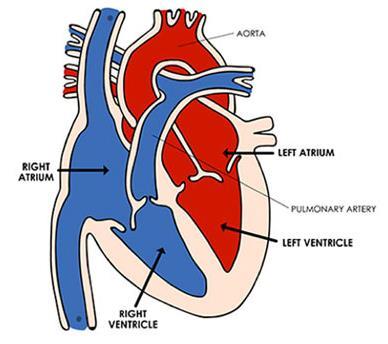
2 The Cardiovascular System A closed system of the heart and blood vessels Functions of cardiovascular system: Transport nutrients, hormones Remove waste products Gaseous exchange Immunity
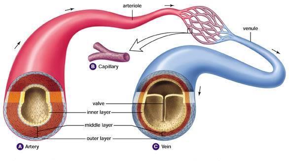
3 The Cardiovascular System A- Heart: Highly modified, muscular blood vessel specialized for pumping the blood composed of two atria and two ventricles. B- Closed circuit of vessels: The vessels are listed below in the order that blood would follow as it leaves the heart. 1. Elastic arteries (e.g., aorta and pulmonary arteries). 2. Muscular arteries (medium arteries). 3. Small arteries and arterioles. 4. Capillaries. 5. Venules and small veins. 6. Medium veins 7. Large veins (e.g., venue cavae return blood to the heart).
4

5 Basic Structural Organization: The walls of the entire cardiovascular system, consists of three concentric layers or tunics that are continuous between both the heart and vessels. The constituents and thickness of these layers vary depending on the mechanical and metabolic functions of the vessel. 1. Tunica interna or intima 2. Tunica media 3. Tunica externa or adventitia
6 Inner tunic In the heart, this layer is called the endocardium; in vessels it is termed the tunica intima. Composition: I. Simple squamous epithelium (endothelium). II. Varying amounts and types of connective tissue. III. In the largest vessels, smooth muscle may be present in the connective tissue layer.
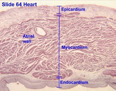
7 Middle tunic In the heart this layer is composed of cardiac muscle and is called the myocardium. In vessels this layer is composed of circularly oriented smooth muscle plus connective tissue and is called the tunica media.
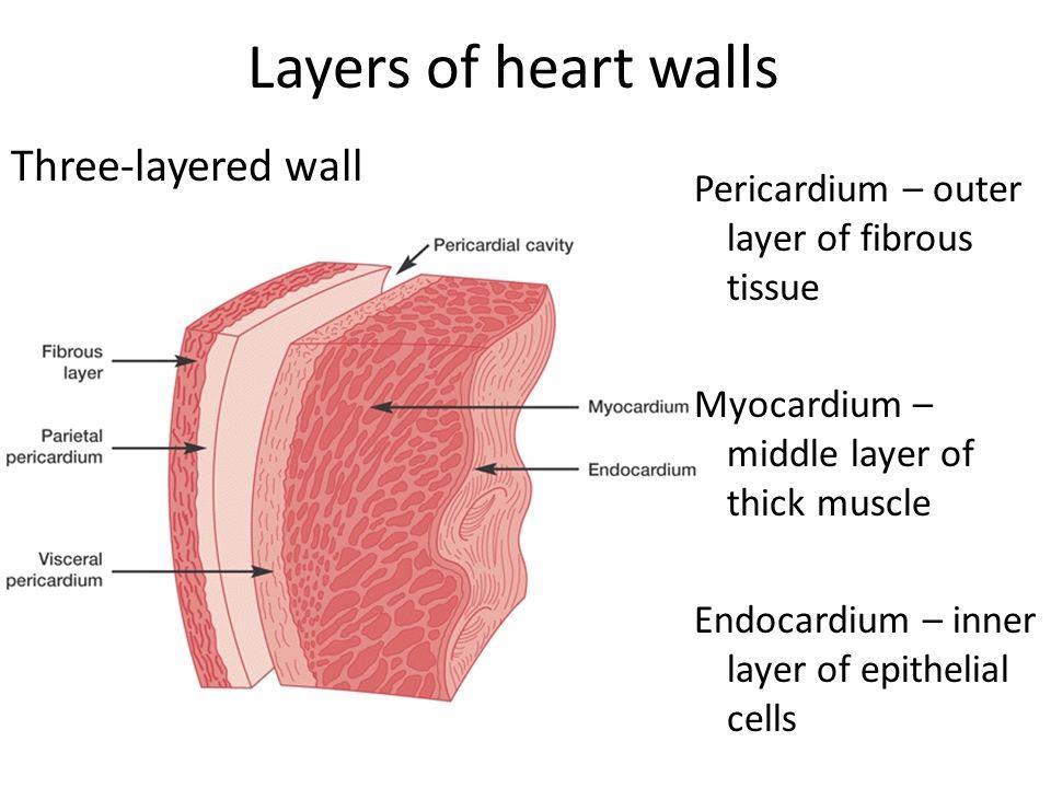
8 Outer tunic In the heart, this layer consists of a serous membrane, called the epicardium (visceral pericardium) composed of connective tissue covered with a simple squamous epithelium (mesothelium). In vessels, this layer is called the tunica adventitia (externa) and is composed of connective tissue; variable amount of longitudinally arranged smooth muscle is present in this layer in the largest veins.
9 The Heart: Chambers Right and left side act as separate pumps Four chambers Atria Receiving chambers Right atrium Left atrium Ventricles Discharging chambers Right ventricle Left ventricle

10 Innermost layer of heart: Endocardium Consists of an endothelium (simple squamous epithelium) plus underlying connective tissue. Cardiac valves: Folds of the endocardium. Semilunar valves at the base of the aortic and pulmonary trunks prevent backflow of blood into the heart. Atrioventricular valves (bicuspid and tricuspid) prevent backflow of blood from the ventricles into the atria. Bicuspid valve
11 Middle layer of heart: Myocardium Composed of cardiac muscle. Thickest layer of the heart. Variation in thickness depends on the function of each chamber; thicker in ventricles than atria and thicker in left ventricle than right ventricle. Exernal layer of heart: Epicardium (visceral pericardium): Serous membrane on the surface of the myocardium. Consists of a simple squamous epithelium and a loose connective tissue, with adipocytes, adjacent to the myocardium.
12
13
 arteries (aorta, pulmonary arteries): The tunica intima: formed of simple squamuse epithelium called the endothelium rest on basement membrane of elastic fibers Tunica")
14 Arteries Carry blood away from the heart and toward capillary beds Have thicker walls and smaller lumens than veins of similar size. Tunica media is the predominate tunic. Types: 1- Elastic (large) arteries (aorta, pulmonary arteries): The tunica intima: formed of simple squamuse epithelium called the endothelium rest on basement membrane of elastic fibers Tunica media: It is thick layer formed of smooth muscle with elastic fibers distributed between it Tunica adventitia: formed of collagen fibers

15 2- Muscular (medium arteries): Tunica intima: formed of endothelium separated from the tunica media by the internal elastic layer Tunica media: It is muscular layer of smooth muscle it is thin than of the large arteries separated from the adventitia by the external elastic layer Tunica adventitia: formed of collagenous fibers


16 3- Small arteries and arterioles: Tunica intima: formed of endothelium Tunica media: formed of smooth muscle arranged into six layers Tunica adventitia: formed of collagen fibers fused with the surrounded structure.


17 Capillaries Function to exchange oxygen and carbon dioxide and nutrients and metabolic wastes between blood and cells. Lumen is approximately 8 microns in diameter, thus only large enough for RBCs to move through in a single row. Composed of the endothelium (simple squamous epithelium) and its underlying basal lamina.


18 Veins Return blood from capillary beds to the heart. Have thinner walls and larger lumens than arteries of similar size. Tunica adventitia is the predominate tunic. Larger veins possess valves that are extensions of the tunica intima that serve to prevent back-flow of blood. Types: 1- Small vein or venule: tunica intima: formed of endothelium and not contain elastic fibers tunica media: formed of tow layers of smooth muscle and it is very thin. tunica adventitia: it is formed of collagen fibers connected the vein with the surrounded structures


19 2- Medium veins: The medium veins formed by the collection of the small veins and drainage into the large veins The histological structure is similar to the small veins but the tunica media is thicker and contain elastic fibers 3- large vein It is structure similar to the medium vein but the tunica media very thick and contain collagen fibers and the internal elastic layer may present Medium vein Large vein

20 SYSTEMIC AND PULMONARY CIRCULATION Pulmonary circulation The flow of blood between the heart and lungs. Systemic circulation The flow of blood between the heart and the cells of the body.
The cardiovascular system
 The cardiovascular system Components of the Cardiovascular system Heart Vessels: Arteries Capillaries Veins Functions of CVS: Transportation system where blood is the transporting vehicle Carries oxygen,
Chapter 14. The Cardiovascular System
 Chapter 14 The Cardiovascular System Introduction Cardiovascular system - heart, blood and blood vessels Cardiac muscle makes up bulk of heart provides force to pump blood Function - transports blood 2
STRUCTURES OF THE CARDIOVASCULAR SYSTEM
 STRUCTURES OF THE CARDIOVASCULAR SYSTEM CARDIOVASCULAR SYSTEM Also called the circulatory system Consists of the heart, arteries, veins, and capillaries Main function is to pump/circulate oxygenated blood
Cardiovascular System. I. Structures of the heart A. : Pericardium sack that surrounds the heart
 Cardiovascular System I. Structures of the heart A. : Pericardium sack that surrounds the heart 1. : Pericardial Cavity serous fluid filled space between the heart and the pericardium B. Heart Wall 1.
SCPA602 Cardiovascular System
 SCPA602 Cardiovascular System Associate Professor Dr. Wannee Jiraungkoorskul Department of Pathobiology, Faculty of Science, Mahidol University Tel: 02-201-5563, E-mail: wannee.jir@mahidol.ac.th 1 Objectives
Cardiovascular System
 Cardiovascular System Purpose Transport oxygen and nutrients Take waste products away from tissues & organs Things we learned Blood pressure: the force of blood pushing against the walls of blood vessels
Major Function of the Cardiovascular System. Transportation. Structures of the Cardiovascular System. Heart - muscular pump
 Structures of the Cardiovascular System Heart - muscular pump Blood vessels - network of tubes Blood - liquid transport vehicle brachiocephalic trunk superior vena cava right pulmonary arteries right pulmonary
Heart. Heart 2-Tunica media: middle layer (media ='middle') muscle fibers (smooth or cardiac).
 muscle fibers (smooth or cardiac).") t. innermost lumenal General Circulatory system heart and blood vessels walls have 3 layers (inside to outside) 1-Tunica interna: aka tunica intima layer--lumenal layer epithelium--endothelium simple squamous
t. innermost lumenal General Circulatory system heart and blood vessels walls have 3 layers (inside to outside) 1-Tunica interna: aka tunica intima layer--lumenal layer epithelium--endothelium simple squamous
The Cardiovascular System
 Essentials of Human Anatomy & Physiology Elaine N. Marieb Seventh Edition Chapter 11 The Cardiovascular System Slides 11.1 11.19 Lecture Slides in PowerPoint by Jerry L. Cook The Cardiovascular System
Anatomy: Cardiovascular System
 In the name of God Anatomy: Cardiovascular System Moradian MD, MPH, PhD candidate Tehran University of Medical Sciences drmoradian@sums.ac.ir 2015 The Cardiovascular System A closed system of the heart
Histology of the Cardiac System. Dr. Nabil Khoury Anatomy Department
 Histology of the Cardiac System Dr. Nabil Khoury Anatomy Department Objectives 1. Identify the 3 layers of the heart endocardium, myocardium, epicardium 2. Differentiate cardiacmuscle 3. Define intercalated
The Cardiovascular System
 The Cardiovascular System The Cardiovascular System A closed system of the heart and blood vessels The heart pumps blood Blood vessels allow blood to circulate to all parts of the body The function of
THE HEART. Unit 3: Transportation and Respiration
 THE HEART Unit 3: Transportation and Respiration The Circulatory System Also called the Cardiovascular System Circulates blood in the body Transports nutrients, oxygen, carbon dioxide, hormones, and blood
Cardiovascular Anatomy Dr. Gary Mumaugh
 Cardiovascular Anatomy Dr. Gary Mumaugh Location of Heart Approximately the size of your fist Location o Superior surface of diaphragm o Left of the midline in mediastinum o Anterior to the vertebral column,
Figure ) The specific chamber of the heart that is indicated by letter A is called the. Diff: 1 Page Ref: 364
 The specific chamber of the heart that is indicated by letter A is called the. Diff: 1 Page Ref: 364") Essentials of Anatomy and Physiology, 9e (Marieb) Chapter 11 The Cardiovascular System Short Answer Figure 11.1 Using Figure 11.1, identify the following: 1) The Purkinje fibers are indicated by label.
Essentials of Anatomy and Physiology, 9e (Marieb) Chapter 11 The Cardiovascular System Short Answer Figure 11.1 Using Figure 11.1, identify the following: 1) The Purkinje fibers are indicated by label.
The Cardiovascular System
 11 PART A The Cardiovascular System PowerPoint Lecture Slide Presentation by Jerry L. Cook, Sam Houston University ESSENTIALS OF HUMAN ANATOMY & PHYSIOLOGY EIGHTH EDITION ELAINE N. MARIEB The Cardiovascular
2. capillaries - allow exchange of materials between blood and tissue fluid
 Chapter 19 - Vascular System A. categories and general functions: 1. arteries - carry blood away from heart 2. capillaries - allow exchange of materials between blood and tissue fluid 3. veins - return
Unit 10 Cardiovascular System
 Unit 10 Cardiovascular System I. Functions Deliver nutrients to cells > O 2, sugars, amino acids, lipids, ions, H 2 O... Remove waste from cells > CO 2, pathogens, toxins, lactic acid... Fight off infection
The Cardiovascular and Lymphatic Systems Cardiovascular System Blood Vessels Blood Vessels Arteries Arteries Arteries
 CH 12 The Cardiovascular and s The Cardiovascular and s OUTLINE: Cardiovascular System Blood Vessels Blood Pressure Cardiovascular System The cardiovascular system is composed of Blood vessels This system
The Circulatory System
 The Circulatory System Dr. Sami Zaqout The circulatory system Circulatory system Blood vascular systems Lymphatic vascular systems Blood vascular systems Blood vascular systems The circulatory system Circulatory
Cardiovascular Physiology
 Cardiovascular Physiology Lecture 1 objectives Explain the basic anatomy of the heart and its arrangement into 4 chambers. Appreciate that blood flows in series through the systemic and pulmonary circulations.
Blood Vessels. Types of Blood Vessels Arteries carry blood away from the heart Capillaries smallest blood vessels. Veins carry blood toward the heart
 C H A P T E R Blood Vessels 20 Types of Blood Vessels Arteries carry blood away from the heart Capillaries smallest blood vessels The site of exchange of molecules between blood and tissue fluid Veins
Blood flows away from the heart in arteries, to the capillaries and back to the heart in the veins
 Cardiovascular System Summary Notes The cardiovascular system includes: The heart, a muscular pump The blood, a fluid connective tissue The blood vessels, arteries, veins and capillaries Blood flows away
THE HEART OBJECTIVES: LOCATION OF THE HEART IN THE THORACIC CAVITY CARDIOVASCULAR SYSTEM
 BIOLOGY II CARDIOVASCULAR SYSTEM ACTIVITY #3 NAME DATE HOUR THE HEART OBJECTIVES: Describe the anatomy of the heart and identify and give the functions of all parts. (pp. 356 363) Trace the flow of blood
Approximately the size of your fist Location Superior surface of diaphragm Left of the midline in mediastinum Anterior to the vertebral column,
 Dr. Gary Mumaugh Approximately the size of your fist Location Superior surface of diaphragm Left of the midline in mediastinum Anterior to the vertebral column, posterior to the sternum Posteriorly the
Cardiac Conduction System
 Cardiac Conduction System What causes the Heart to Beat? Heart contracts by electrical signals! Cardiac muscle tissue contracts on its own an electrical signal is sent out by the heart so that all cells
Health Science 20 Circulatory System Notes
 Health Science 20 Circulatory System Notes Functions of the Circulatory System The circulatory system functions mainly as the body s transport system. It transports: o Oxygen o Nutrients o Cell waste o
Lab Activity 25. Blood Vessels & Circulation. Portland Community College BI 232
 Lab Activity 25 Blood Vessels & Circulation Portland Community College BI 232 Artery and Vein Histology Walls have 3 layers: Tunica intima Tunica media Tunica externa 2 Tunica Intima Is the innermost layer
Cardiovascular System Blood Vessels
 Cardiovascular System Blood Vessels Structure of Blood Vessels The three layers (tunics) Tunica intima composed of simple squamous epithelium Tunica media sheets of smooth muscle Contraction vasoconstriction
Blood and Heart. Student Learning Objectives:
 Blood and Heart Student Learning Objectives: Identify the major components of the blood. Identify the primary structures associated with the heart Follow the blood through the path of the circulation.
a) Endocardium The endocardium is the innermost layer of the heart wall. It forms the internal lining of the atria and ventricles.
 Endocardium The endocardium is the innermost layer of the heart wall. It forms the internal lining of the atria and ventricles.") Chapter 11 Circulatory System The circulatory system consists of a muscular, pulsing heart and a system of blood vessels.the blood vessels include: Arteries which will carry the blood and it's dissolved
Chapter 11 Circulatory System The circulatory system consists of a muscular, pulsing heart and a system of blood vessels.the blood vessels include: Arteries which will carry the blood and it's dissolved
Cardiovascular System Notes: Physiology of the Heart
 Cardiovascular System Notes: Physiology of the Heart Interesting Heart Fact Capillaries are so small it takes ten of them to equal the thickness of a human hair. Review What are the 3 parts of the cardiovascular
2. right heart = pulmonary pump takes blood to lungs to pick up oxygen and get rid of carbon dioxide
 A. location in thorax, in inferior mediastinum posterior to sternum medial to lungs superior to diaphragm anterior to vertebrae orientation - oblique apex points down and to the left 2/3 of mass on left
Scrub In: Red blood cells are called: Which component of blood is necessary for the initiation of the blood clotting process:
 Scrub In: Red blood cells are called: a. erythrocytes b. leukocytes c. melanocytes d. thrombocytes Which component of blood is necessary for the initiation of the blood clotting process: a. erythrocytes
The Cardiovascular and Lymphatic Systems
 BIOLOGY OF HUMANS Concepts, Applications, and Issues Fifth Edition Judith Goodenough Betty McGuire 12 The Cardiovascular and Lymphatic Systems Lecture Presentation Anne Gasc Hawaii Pacific University and
Cardiovascular System. Heart Anatomy
 Cardiovascular System Heart Anatomy 1 The Heart Location & general description: Atria vs. ventricles Pulmonary vs. systemic circulation Coverings Walls The heart is found in the mediastinum, the medial
The Circulatory System. The Heart, Blood Vessels, Blood Types
 The Circulatory System The Heart, Blood Vessels, Blood Types The Closed Circulatory System Humans have a closed circulatory system, typical of all vertebrates, in which blood is confined to vessels and
Circulation. Sinoatrial (SA) Node. Atrioventricular (AV) Node. Cardiac Conduction System. Cardiac Conduction System. Linked to the nervous system
 Node. Atrioventricular (AV) Node. Cardiac Conduction System. Cardiac Conduction System. Linked to the nervous system") Circulation Cardiac Conduction System AHS A H S Your body resembles a large roadmap. There are routes or arteries that take you downtown to the heart of the city and veins that take you to the outskirts
Circulation Cardiac Conduction System AHS A H S Your body resembles a large roadmap. There are routes or arteries that take you downtown to the heart of the city and veins that take you to the outskirts
Human Anatomy and Physiology Chapter 19 Worksheet 1- The Heart
 Human Anatomy and Physiology Chapter 19 Worksheet 1- The Heart Name Date Period 1. The "double pump" function of the heart includes the right side, which serves as the circuit pump, while the left side
10. Thick deposits of lipids on the walls of blood vessels, called, can lead to serious circulatory issues. A. aneurysm B. atherosclerosis C.
 Heart Student: 1. carry blood away from the heart. A. Arteries B. Veins C. Capillaries 2. What is the leading cause of heart attack and stroke in North America? A. alcohol B. smoking C. arteriosclerosis
Heart Anatomy. 7/5/02 Stephen G Davenport 1
 Heart Anatomy Copyright 1999, Stephen G. Davenport, No part of this publication may be reproduced, stored in a retrieval system, or transmitted, in any form without prior written permission. 7/5/02 Stephen
CIRCULATORY SYSTEM BLOOD VESSELS
 Name: Block: CIRCULATORY SYSTEM Multicellular organisms (above the level of roundworms) rely on a circulatory system to bring nutrients to, and take wastes away from, cells. In higher organisms such as
UNIT 4: BLOOD VESSELS
 UNIT 4: BLOOD VESSELS Dr. Moattar Raza Rizvi NRS237, Physiology Generalized Structure of Blood Vessels 1 Tunica interna (tunica intima) Endothelial layer that lines the lumen of all vessels In vessels
1. What kind of blood is found in the rt. atrium? (oxygenated or deoxygenated)
") Carl Christennsen, PhD Chap. 19, 20, & 21 - Circulatory System Bio. 2304 Human Anatomy HEART 1. What kind of blood is found in the rt. atrium? (oxygenated or deoxygenated) Where does this blood come from?
Carl Christennsen, PhD Chap. 19, 20, & 21 - Circulatory System Bio. 2304 Human Anatomy HEART 1. What kind of blood is found in the rt. atrium? (oxygenated or deoxygenated) Where does this blood come from?
THE CARDIOVASCULAR SYSTEM. Part 1
 THE CARDIOVASCULAR SYSTEM Part 1 CARDIOVASCULAR SYSTEM Blood Heart Blood vessels What is the function of this system? What other systems does it affect? CARDIOVASCULAR SYSTEM Functions Transport gases,
Histology of the myocardium and blood vessels. Prof. Abdulameer Al-Nuaimi
 Histology of the myocardium and blood vessels Prof. Abdulameer Al-Nuaimi E-mail: a.al-nuaimi@sheffield.ac.uk E-mail: abdulameerh@yahoo.com Histology of blood vessels The walls of arteries and veins are
Pearson's Comprehensive Medical Assisting Administrative and Clinical Competencies
 Pearson's Comprehensive Medical Assisting Administrative and Clinical Competencies THIRD EDITION CHAPTER 27 The Cardiovascular System Lesson 1: Overview of the Cardiovascular System Lesson Objectives Upon
The Heart. Happy Friday! #takeoutyournotes #testnotgradedyet
 The Heart Happy Friday! #takeoutyournotes #testnotgradedyet Introduction Cardiovascular system distributes blood Pump (heart) Distribution areas (capillaries) Heart has 4 compartments 2 receive blood (atria)
Cardivascular System Module 5: Structure and Function of Blood Vessels *
 OpenStax-CNX module: m49689 1 Cardivascular System Module 5: Structure and Function of Blood Vessels * Donna Browne Based on Structure and Function of Blood Vessels by OpenStax This work is produced by
Microscopic Anatomy CARDIOVASCULAR SYSTEM
 Microscopic Anatomy CARDIOVASCULAR SYSTEM I. Introduction The cardiovascular system is a closed system consisting of a pump, the heart, and a series of tubular blood vessels that interconnect all body
Remember: the CVS system is deriving from the mesenchyma and is lined by simple squamous epithelium called the endothelium
 Lecture 10 General med_2nd semester Microscopic anatomy and embryology of cardiovascular system Microscopic structure of the heart, excitomotoric system - its structural peculiarities Blood vessels - arteries
AN ATOMY OF THE CARDIOVASCULAR SYSTEM
 Student Name CHAPTER 18 AN ATOMY OF THE CARDIOVASCULAR SYSTEM T he heart is actually two pumps one moves blood to the lungs, the other pushes it out into the body. These two functions seem rather elementary
The Cardiovascular System (Heart)
") The Cardiovascular System The Cardiovascular System (Heart) A closed system of the heart and blood vessels The heart pumps blood Blood vessels allow blood to circulate to all parts of the body The function
The Cardiovascular System The Cardiovascular System (Heart) A closed system of the heart and blood vessels The heart pumps blood Blood vessels allow blood to circulate to all parts of the body The function
the Cardiovascular System I
 the Cardiovascular System I By: Dr. Nabil A Khouri MD, MsC, Ph.D MEDIASTINUM 1. Superior Mediastinum 2. inferior Mediastinum Anterior mediastinum. Middle mediastinum. Posterior mediastinum Anatomy of
LECTURE 5. Anatomy of the heart
 LECTURE 5. Anatomy of the heart Main components of the CVS: Heart Blood circulatory system arterial compartment haemomicrocirculatory (=microvascular) compartment venous compartment Lymphatic circulatory
(2) (1) (3) (4) BLOOD PATHWAY ASSESSMENT RUBRIC
 (1) (3) (4) BLOOD PATHWAY ASSESSMENT RUBRIC") BLOODPATHWAYASSESSMENT(4) BLOOD%PATHWAY%ASSESSMENT%(3)% BLOODPATHWAYASSESSMENT(3) (4) (3) (2) (1) Using a completely blank diagram of the heart, all valves, chambers, great vessels, and direction of blood
BLOODPATHWAYASSESSMENT(4) BLOOD%PATHWAY%ASSESSMENT%(3)% BLOODPATHWAYASSESSMENT(3) (4) (3) (2) (1) Using a completely blank diagram of the heart, all valves, chambers, great vessels, and direction of blood
Blood Functions. Blood and the Cardiovascular System. Blood. Plasma. Erythrocytes (RBCs) Erythrocytes (RBCs) 4/7/2017
 Erythrocytes (RBCs) 4/7/2017") Blood Functions Blood and the Cardiovascular System Distribution Delivery of oxygen and nutrients to all body cells; Transport of wastes to lungs and excretory organs; Transport of hormones Regulation
Blood Functions Blood and the Cardiovascular System Distribution Delivery of oxygen and nutrients to all body cells; Transport of wastes to lungs and excretory organs; Transport of hormones Regulation
Chapter 20 (1) The Heart
 The Heart") Chapter 20 (1) The Heart Learning Objectives Describe the location and structure of the heart Describe the path of a drop of blood from the superior vena cava or inferior vena cava through the heart out
Chapter 20 (1) The Heart Learning Objectives Describe the location and structure of the heart Describe the path of a drop of blood from the superior vena cava or inferior vena cava through the heart out
Blood Vessels. Over view. We have about 60,000 miles of blood vessels!
 Blood Vessels Over view 3 types of blood vessels arteries - carry blood away from heart "branch", "diverge", and "fork" veins - carry blood toward heart "join", "merge", and "converge" capillaries - site
Cardiovascular System
 Cardiovascular System Function - Func&ons to carry oxygen and nutrients to the cells of the body and carry carbon dioxide and other waste away from the cells of the body. Structures - Heart - pumps blood
Anatomy Review: The Heart Graphics are used with permission of A.D.A.M. Software, Inc. and Benjamin/Cummings Publishing Co.
 Anatomy Review: The Heart Graphics are used with permission of A.D.A.M. Software, Inc. and Benjamin/Cummings Publishing Co. Anatomy Views Label the diagrams of the heart below: Interactive Physiology Study
Human Anatomy, First Edition
 Human Anatomy, First Edition McKinley & O'Loughlin Chapter 22 : Heart 1 Functions of the Heart Center of the cardiovascular system, the heart. Connects to blood vessels that transport blood between the
The blood returns from the body and enters right atrium using the vena cava. It passes through the tricuspid valve to the right ventricle.
 The blood returns from the body and enters right atrium using the vena cava. It passes through the tricuspid valve to the right ventricle. From this camber, it passes through the pulmonary semilunar valve
2.01 Remember the structures of the circulatory system
 2.01 Remember the structures of the circulatory system Essential questions What are the structures of blood? What are the structures of the circulatory system? circulatory system 2 Structures of the circulatory
Function: Transportation of. Oxygen Nutrients Waste Hormones gases
 Function: Transportation of Oxygen Nutrients Waste Hormones gases Pericardium: double sac of serous membrane filled with fluid (pericardial fluid to be exact) that surrounds the heart. Parietal pericardium:
Vessels by Design: Basic Vessel Anatomy. Student Information Page 3A
 Vessels by Design: Basic Vessel Anatomy Student Information Page 3A Activity Introduction: Once you get home from running around all day, your throat is probably a little dry. You go to your kitchen, get
2/28/18. Cardiovascular System. Introduction. Anatomy. Chapter 26. Body is 60% to 80% fluid (by volume) Systems responsible for fluid movement are:
 Systems responsible for fluid movement are:") Cardiovascular System Chapter 26 1 Introduction Body is 60% to 80% fluid (by volume) Systems responsible for fluid movement are: - Cardiovascular helps move fluid - Lymphatic Both called pick-up and delivery
Cardiovascular System Chapter 26 1 Introduction Body is 60% to 80% fluid (by volume) Systems responsible for fluid movement are: - Cardiovascular helps move fluid - Lymphatic Both called pick-up and delivery
Lecture name: blood 2 & The Circulatory System Edited by: Buthainah Al masaeed & Yousef Qandeel
 Lecture name: blood 2 & The Circulatory System Edited by: Buthainah Al masaeed & Yousef Qandeel Now we will take about A granulocytes : Lymphocyte Monocytes 1- Lymphocyte - The second major type of presence
Cardiovascular (Circulatory) System
 System") Cardiovascular (Circulatory) System Piryaei May 2011 Circulatory System Heart Blood Vessels Macrovasculature (More than 0.1mm) Elastic Artery Muscular (Distributing) Artery Large Arteriol Small Vein Muscular
Cardiovascular (Circulatory) System Piryaei May 2011 Circulatory System Heart Blood Vessels Macrovasculature (More than 0.1mm) Elastic Artery Muscular (Distributing) Artery Large Arteriol Small Vein Muscular
Heart Dissection. 5. Locate the tip of the heart or the apex. Only the left ventricle extends all the way to the apex.
 Heart Dissection Page 1 of 6 Background: The heart is a four-chambered, hollow organ composed primarily of cardiac muscle tissue. It is located in the center of the chest in between the lungs. It is the
CVS HISTOLOGY. Dr. Nabil Khouri.
 CVS HISTOLOGY Dr. Nabil Khouri http://anatomy.kmu.edu.tw/blockhis/block3/slides/block4_24.html The Heart Wall Contract as a single unit Cardiac Muscle Simultaneous contraction due to depolarizing at the
The heart & Cardiovascular system
 The heart & Cardiovascular system The heart s continuous pulse create a base for our understanding of rhythms in everyday life. Bonnie Bainbridge Cohen The heart constantly beats throughout our lives never
Cardiovascular System
 Cardiovascular System The Heart Cardiovascular System The Heart Overview What does the heart do? By timed muscular contractions creates pressure gradients blood moves then from high pressure to low pressure
The circulatory system transports blood to deliver important substances, such as oxygen, to cells and to remove wastes, such as carbon dioxide.
 Section 1: The circulatory system transports blood to deliver important substances, such as oxygen, to cells and to remove wastes, such as carbon dioxide. K What I Know W What I Want to Find Out L What
THE CIRCULATORY SYSTEM
 Biology 30S THE CIRCULATORY SYSTEM Name: This module adapted from bblearn.merlin.mb.ca 1 Introduction to Circulation The first organ to form, and the last organ to die. The heart is the pump of life. The
4. The two inferior chambers of the heart are known as the atria. the superior and inferior vena cava, which empty into the left atrium.
 Answer each statement true or false. If the statement is false, change the underlined word to make it true. 1. The heart is located approximately between the second and fifth ribs and posterior to the
Ch 9 Transport of substances in humans
 Ch 9 Transport of substances in humans Think about (Ch 9, p.2) 1. Blood transports various substances and distributes heat around the body. It also plays a role in body defence. 2. Blood is a liquid tissue
Chapter 12 Cardiovascular System
 Chapter 12 Cardiovascular System Cardiovascular System Includes Heart and Blood Vessels Transports, nutrients and wastes to and from the tissues 1 The Blood Vessels Three Types of Blood Vessels Arteries:
CIE Biology GCSE. 9: Transport in animals. Notes.
 CIE Biology GCSE 9: Transport in animals Notes The circulatory system acts as the main transport system in animals. It is made up of blood vessels such as arteries, veins and capillaries, in which blood
Anatomy of the Heart. Figure 20 2c
 Anatomy of the Heart Figure 20 2c Pericardium & Myocardium Remember, the heart sits in it s own cavity, known as the mediastinum. The heart is surrounded by the Pericardium, a double lining of the pericardial
Extra notes for lab- 1 histology. Slide 1 : cross section in the elastic artery ( aortic arch, ascending aorta, descending aorta )
") Extra notes for lab- 1 histology Slide 1 : cross section in the elastic artery ( aortic arch, ascending aorta, descending aorta ) - twin of ascending aorta is the pulmonary trunk. Ascending aorta represents
Extra notes for lab- 1 histology Slide 1 : cross section in the elastic artery ( aortic arch, ascending aorta, descending aorta ) - twin of ascending aorta is the pulmonary trunk. Ascending aorta represents
Cardiovascular System Module 3: Heart Anatomy *
 OpenStax-CNX module: m49683 1 Cardiovascular System Module 3: Heart Anatomy * Donna Browne Based on Heart Anatomy by OpenStax This work is produced by OpenStax-CNX and licensed under the Creative Commons
Chapter 27 -The Heart & Blood Vessels
 Chapter 27 -The Heart & Blood Vessels 3.2 Learning Objectives 3.2.2 Organisational Complexity of the human 1. Describe the structures and organisation of tissues in the closed circulatory system. 2. Discuss
Cardiovascular System
 Cardiovascular System I. Structure of the Heart A. Average adult heart is 14 cm long and 9 cm wide. B. Lies in the mediastinum. C. Enclosed in the pericardium. 1. Fibrous pericardium- Outer, tough connective
Mr. Epithelium s Anatomy and Physiology Test SSSS
 Mr. Epithelium s Anatomy and Physiology Test SSSS You have 50 minutes to complete this test packet. One 8.5 x 11 cheat sheet is allowed, along with 1 non-programmable calculator dedicated to computation.
Cardiovascular System Notes: Heart Disease & Disorders
 Cardiovascular System Notes: Heart Disease & Disorders Interesting Heart Facts The Electrocardiograph (ECG) was invented in 1902 by Willem Einthoven Dutch Physiologist. This test is still used to evaluate
TOPIC 6: HUMAN HEALTH AND PHYSIOLOGY
 TOPIC 6: HUMAN HEALTH AND PHYSIOLOGY 6.2 Transport System/Circulatory Draw and label a diagram of the heart showing the four chambers, associated blood vessels, valves and the route of blood through the
IB TOPIC 6.2 THE BLOOD SYSTEM
 IB TOPIC 6.2 THE BLOOD SYSTEM TERMS TO KNOW circulation ventricle artery vein THE BLOOD SYSTEM 6.2.U1 - Arteries convey blood at high pressure from the ventricles to the tissues of the body Circulation
learningobjectives At the end of the lesson, you should be able to: - Humans Explain the need for a transport system in multicellular
 transport in humans learningobjectives At the end of the lesson, you should be able to: - Humans Explain the need for a transport system in multicellular organisms Identify the parts of the human circulatory
Heart. Structure Physiology of blood pressure and heartbeat
 Heart Structure Physiology of blood pressure and heartbeat Location and Anatomy Location and Anatomy Pericardial cavity: surrounds, isolates, and anchors heart Parietal pericardium lined with serous membrane
The Cardiovascular System home study course
 The Cardiovascular System home study course harmony house holistic therapy treatment centre and training academy www.harmony-house.org 1 Copyright 2010 by Mark and Katy Rogers All rights reserved. No part
Biology Unit 3 The Human Heart P
 Biology 2201 Unit 3 The Human Heart P 314-321 Structure and Function of the Human Heart Structure of the Human Heart Has four Chambers (2 Atria and 2 Ventricles) Made of Cardiac Muscle Found in Chest Cavity
1. Which of the following blood vessels has a thin elastic layer? A. Aorta. B. Pulmonary artery. C. Posterior vena cava. D. Mesenteric capillary.
 CIRCULATORY SYSTEM 1. Which of the following blood vessels has a thin elastic layer? A. Aorta. B. Pulmonary artery. C. Posterior vena cava. D. Mesenteric capillary. 2. Capillary beds are equipped with
THE HEART. A. The Pericardium - a double sac of serous membrane surrounding the heart
 THE HEART I. Size and Location: A. Fist-size weighing less than a pound (250 to 350 grams). B. Located in the mediastinum between the 2 nd rib and the 5 th intercostal space. 1. Tipped to the left, resting
Chapter 21. Blood Vessels and Circulation
 Chapter 21 Openstax: Chapter 20 Blood Vessels and Circulation Chapter 21 Learning Outcomes After completing Chapter 21, you will be able to: 1. Distinguish among the types of blood vessels based on their
Anatomy of the Heart
 Biology 212: Anatomy and Physiology II Anatomy of the Heart References: Saladin, KS: Anatomy and Physiology, The Unity of Form and Function 8 th (2018). Required reading before beginning this lab: Chapter
Cardiovascular System. Blood Vessel anatomy Physiology & regulation
 Cardiovascular System Blood Vessel anatomy Physiology & regulation Path of blood flow Aorta Arteries Arterioles Capillaries Venules Veins Vena cava Vessel anatomy: 3 layers Tunica externa (adventitia):
Circulation. Circulation = is a process used for the transport of oxygen, carbon! dioxide, nutrients and wastes through-out the body
 Circulation Circulation = is a process used for the transport of oxygen, carbon! dioxide, nutrients and wastes through-out the body Heart = muscular organ about the size of your fist which pumps blood.
Animal Physiology Prof. Mainak Das Department of Biological Sciences and Bioengineering Indian Institute of Technology, Kanpur. Module - 1 Lecture 7
 Animal Physiology Prof. Mainak Das Department of Biological Sciences and Bioengineering Indian Institute of Technology, Kanpur Module - 1 Lecture 7 Welcome back to the lectures in Animal Physiology in
Chapter 27 The Heart and Blood Vessels
 Chapter 27 The Heart and Blood Vessels Most animals have a closed blood system. The blood flows continuously in vessels back to the heart. In an open system the blood is pumped into open ended tubes and