Chapter 12: Cardiovascular Physiology System Overview
|
|
|
- Hubert Pierce
- 5 years ago
- Views:
Transcription
1 Chapter 12: Cardiovascular Physiology System Overview Components of the cardiovascular system: Heart Vascular system Blood

2 Figure 12-1 Plasma includes water, ions, proteins, nutrients, hormones, wastes, etc. The hematocrit is a rapid assessment of blood composition. It is the percent of the blood volume that is composed of RBCs (red blood cells).

3 Figure 12-2 The heart is the pump that propels the blood through the systemic and pulmonary circuits. Red color indicates blood that is fully oxygenated. Blue color represents blood that is only partially oxygenated.

4 Figure 12-3 The distribution of blood in a comfortable, resting person is shown here. Dynamic adjustments in blood delivery allow a person to respond to widely varying circumstances, including emergencies.
5
 Brain natriuretic peptide")
6 Functions of the heart Pumping Endocrine Atrial natriuretic peptide (ANP) Brain natriuretic peptide (BNP)

7 Figure 12-6 The Heart The major external and internal parts of the heart are shown in this diagram. The black arrows indicate the route taken by the blood as it is pumped along.

8 Valves of the heart


9 Figure 12-8 The general route of the blood through the body is shown, including passage through the heart (colored box).
10 Three major types of cardiac muscle: Atrial muscle Ventricular muscle Contractile cells Specialized excitatory and conductive muscle Autorhythmic cells

11 Cardiac muscle

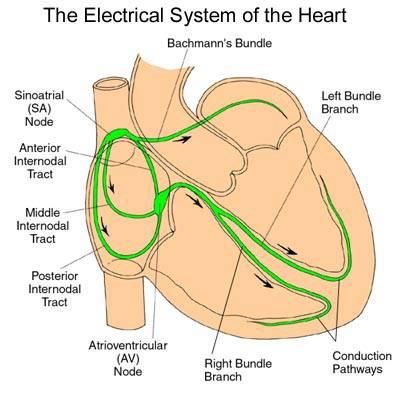
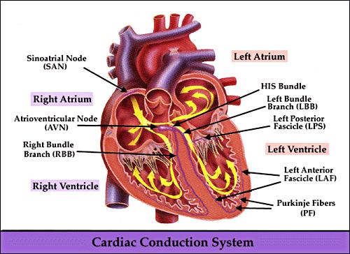
12 Figure Conducting system of the heart

13 Figure Sequence of cardiac excitation The sinoatrial node is the heart s pacemaker because it initiates each wave of excitation with atrial contraction. The Bundle of His and other parts of the conducting system deliver the excitation to the apex of the heart so that ventricular contraction occurs in an upward sweep.
14 General process of excitation and contraction of cardiac muscle Initiation of action potentials in sinoatrial node Conduction of action potentials along specialized conductive system Excitation-contraction coupling Muscle contraction
15 Click here to play the Conducting System of the Heart Flash Animation
16
17
18

19 Scaling from the level of the organelle to the organ
20 Transmembrane potentials recorded in different heart regions
21 Transmembrane potentials in epicardium and endocardium
22 Transmembrane potential of ventricular cells and its ionic mechanisms Resting Potential: -90 mv Action Potential Phase 0: Depolarization Phase 1: Early phase of rapid repolarization Phase 2: Plateau Phase 3: Late phase of rapid repolarization Phase 4: Resting phase
23 Ionic mechanisms Resting potential K + equilibrium potential Na + -inward background current Electrogenic Na + -K + pump
24
25 The action potential of a myocardial pumping cell. Phase 0 Threshold potential (-70mV) Opening of fast Na + channel Regenerative cycle Figure 12-13
26 Phase 1 Transient outward current, Ito K+ current activated at 20 mv opening for 5~10 ms Figure 12-12
27 Phase 2 Inward current Outward current (Ca 2+ & Na + ) (K + current) Figure 12-12
28 Types of Ca 2+ channels in cardiac cells: (1) L-type (long-lasting) (Nowycky, 1985) (2) T-type (transient) (Nowycky, 1985)
29 Ca 2+ channels L-type T-type Duration of current long-lasting transient Activation kinetics slower faster Inactivation kinetics slower faster Threshold high (-35mV) Low (-60mV) camp/cgmp-regulated Yes No Phosphorylation-regulated Yes No Openers Bay-K Blockers varapamil Tetramethrin nifedipine, diltiazem Ni 2+ Inactivation by [Ca 2+ ] i Yes slight Patch-clamp recording run-down relatively stable
30 Outward current (K + current): (1) inward rectifier K + current (I K1 ) (2) delayed rectifier K + current (I K )
31 Phase 3 Inactivation of Ca 2+ channel Outward K + current dominates I K : Progressively increased I K1 : Regenerative K+ Outward Current Figure 12-12
32 Phase 4 Na + -Ca 2+ exchange Sarcolemmal Ca 2+ pump SR Ca 2+ pump Na + -K + pump Figure 12-12
33
 mediate K+ efflux from the cell; Na+ channels (purple) and Ca2+ channels (yellow) mediate Na+ and Ca2+ influx, respectively.")
34 a, The key ion channels (and an electrogenic transporter) in cardiac cells. K+ channels (green) mediate K+ efflux from the cell; Na+ channels (purple) and Ca2+ channels (yellow) mediate Na+ and Ca2+ influx, respectively. The Na+/Ca2+ exchanger (red) is electrogenic, as it transports three Na+ ions for each Ca2+ ion across the surface membrane. b, Ionic currents and genes underlying the cardiac action potential. Top, depolarizing currents as functions of time, and their corresponding genes; centre, a ventricular action potential; bottom, repolarizing currents and their corresponding genes. From the following article: Cardiac channelopathies Eduardo Marbán Nature 415, (10 January 2002) doi: /415213a
35 Click here to play the Action Potential in Cardiac Muscle Cell Flash Animation
36 Transmembrane potentials recorded in different heart regions
37 Transmembrane potential of autorhythmic cells and its ionic mechanisms Contractile cells Autorhythmic cells Phase 4 stable potential Phase 4 spontaneous depolarization Resting potential Maximal repolarization potential

38 Purkinje cells
39 Purkinje cells: Fast response autorhythmic cells 4
40 Ionic mechanism Phase 0~3 similar to ventricular cells Phase 4 (1) I f Funny current, Pacemaker current (2) I k Decay
41 Characteristics of I f channel Na +, K + Voltage- & time-dependent Activation Repolarized to -60mV Full activation Hyperpolarized to -100mV Inactivation Depolarized to -50mV Blocked by Cs, not by TTX
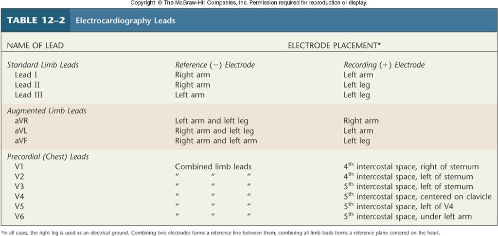
42 Sinoatrial cells
43 Sinoatrial cells: Slow response autorhythmic cells Maximal repolarization potential -70mV 0 3 Threshold potential -40mV 4 Phase 0, 3, 4
44 Ionic mechanism Phase 0: I Ca (I Ca,L ) 0 3 4
45 Phase 3: Inactivation of L-type Ca 2+ channel 0 3 Outward K + current (I k ) 4
46 Phase 4 I k decay Inactivated when repolarized to -60mV I Ca,T I f Activated when depolarized to -50mV

47 The action potential of an autorhythmic cardiac cell. Figure 12-13
48 Click here to play the Action Potential in SA Node Flash Animation


49 Electrocardiogram (ECG) The electrocardiogram (ECG) measures changes in skin electrical voltage/potential caused by electrical currents generated by the heart
50 Figure The relationship between the electrocardiogram (ECG), recorded as the difference between currents at the left and right wrists, and an action potential typical of ventricular myocardial cells. Electrocardiogram (ECG)

51 Placement of electrodes in electrocardiography
 (avr, avl, avf) V1 V2 V3 V4 V5 V6 Chest leads (Unipolar) (V1, V2, V3, V4, V5, V6) II avf")
52 The standard 12 lead ECG Einthoven s Triangle I avr avl Limb leads (Bipolar) (I, II, III) Augmented limb leads (Unipolar) (avr, avl, avf) V1 V2 V3 V4 V5 V6 Chest leads (Unipolar) (V1, V2, V3, V4, V5, V6) II avf III
53

54 Normal ECG 0.04 sec ECG interpretation Measurements Rhythm analysis Conduction analysis Waveform description Comparison with previous ECG (if any)
55 P wave: the sequential depolarization of the right and left atria QRS complex: right and left ventricular depolarization ST-T wave: ventricular repolarization U wave: origin for this wave is not clear - but probably represents "afterdepolarizations" in the ventricles
56 PR interval: time interval from onset of atrial depolarization (P wave) to onset of ventricular depolarization (QRS complex) QT interval: duration of ventricular depolarization and repolarization ST segment: the time period between the end of the QRS complex and the beginning of the T wave, during which each myocyte is in the plateau phase (phase 2) of the action potential
57 Normal Partial block Complete block Figure 12-16
58 The End.
59 Physiological properties of cardiac cells Excitability Autorhythmicity Conductivity Electrophysiological properties Contractility Mechanical property
60 Excitability Factors affecting excitability Resting potential Threshold potential Status of Na + or Ca 2+ channels

61 Periodic changes in excitability
62

63 Postrepolarization refractoriness of slow response cells

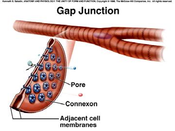
64 Valuable protective mechanism The long refractory period means that cardiac muscle cannot be restimulated until contraction is almost over & this makes summation & tetanus of cardiac muscle impossible

65 Premature systole & compensatory pause (extrasystole)
66 Autorhythmicity

67 Autorhythmicity SA node 100 times/min AV node 50 times/min Bundle of His 40 times/min Purkinje fibers 25 times/min
68 Normal pacemaker SA node Latent pacemaker (Ectopic pacemaker under pathophysiological conditions) AV node Bundle of His Purkinje fibers
69 The mechanisms of SA node to control latent pacemakers Capture Overdrive suppression

70 Factors Affecting Autorhythmicity Maximal repolarization potential Threshold potential The rate of phase 4 spontaneous depolarization
71 Pacemaker
72
73 Conductivity

74 Gap junction
75 Conducting velocity SA node Atria A-V node 0.05 m/s 0.4 m/s 0.02~0.05 m/s His bundle Purkinje fiber Ventricle 1.2~2.0 m/s 2.0~4.0 m/s 1.0 m/s Atrioventricular delay: Asynchronization of atrial and ventricular depolarization to provide adequate cardiac output
76 Factors Affecting Conductivity Structural factors Diameter of cardiac cells Gap junctions at Intercalated disk Physiological factors The velocity and amplitude of phase 0 depolarization Excitability of adjacent region
77 Reentry
Cardiac physiology. b. myocardium -- cardiac muscle and fibrous skeleton of heart
 I. Heart anatomy -- general gross. A. Size/orientation - base/apex B. Coverings D. Chambers 1. parietal pericardium 2. visceral pericardium 3. Layers of heart wall a. epicardium Cardiac physiology b. myocardium
ELECTROCARDIOGRAPHY (ECG)
") ELECTROCARDIOGRAPHY (ECG) The heart is a muscular organ, which pumps blood through the blood vessels of the circulatory system. Blood provides the body with oxygen and nutrients, as well as assists in
ELECTROCARDIOGRAPHY (ECG) The heart is a muscular organ, which pumps blood through the blood vessels of the circulatory system. Blood provides the body with oxygen and nutrients, as well as assists in
Chapter 13 The Cardiovascular System: Cardiac Function
 Chapter 13 The Cardiovascular System: Cardiac Function Overview of the Cardiovascular System The Path of Blood Flow through the Heart and Vasculature Anatomy of the Heart Electrical Activity of the Heart
Electrical Conduction
 Sinoatrial (SA) node Electrical Conduction Sets the pace of the heartbeat at 70 bpm AV node (50 bpm) and Purkinje fibers (25 40 bpm) can act as pacemakers under some conditions Internodal pathway from
Introduction. Circulation
 Introduction Circulation 1- Systemic (general) circulation 2- Pulmonary circulation carries oxygenated blood to all parts of the body carries deoxygenated blood to the lungs From Lt. ventricle aorta From
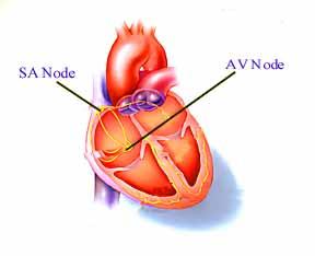
Where are the normal pacemaker and the backup pacemakers of the heart located?
 CASE 9 A 68-year-old woman presents to the emergency center with shortness of breath, light-headedness, and chest pain described as being like an elephant sitting on her chest. She is diagnosed with a
Ask Mish. EKG INTERPRETATION part i
 EKG INTERPRETATION part i What is EKG? EKG or ECG= electrocardiogram(~graphy) means the recording of the heart electrical activity from Greek kardio= heart, graphein= to write cardiac cell physiology Cardiac
CARDIOVASCULAR SYSTEM
 CARDIOVASCULAR SYSTEM Overview Heart and Vessels 2 Major Divisions Pulmonary Circuit Systemic Circuit Closed and Continuous Loop Location Aorta Superior vena cava Right lung Pulmonary trunk Base of heart
Circulatory system ( 循环系统 )
") Circulatory system ( 循环系统 ) Circulatory system: heart + blood vessels Function: nutrient transportation and metabolites returning Blood: carrier Heart: pump Blood vessels: route, substance communications
Circulatory system ( 循环系统 ) Circulatory system: heart + blood vessels Function: nutrient transportation and metabolites returning Blood: carrier Heart: pump Blood vessels: route, substance communications
Cardiac Properties MCQ
 Cardiac Properties MCQ Abdel Moniem Ibrahim Ahmed, MD Professor of Cardiovascular Physiology Cairo University 2007 1- Cardiac Valves: a- Prevent backflow of blood from the ventricles to the atria during
Cardiovascular system
 BIO 301 Human Physiology Cardiovascular system The Cardiovascular System: consists of the heart plus all the blood vessels transports blood to all parts of the body in two 'circulations': pulmonary (lungs)
Collin County Community College. ! BIOL Anatomy & Physiology! WEEK 5. The Heart
 Collin County Community College! BIOL. 2402 Anatomy & Physiology! WEEK 5 The Heart 1 (1578-1657) A groundbreaking work in the history of medicine, English physician William Harvey s Anatomical Essay on
Collin County Community College
 Collin County Community College BIOL. 2402 Anatomy & Physiology WEEK 5 The Heart 1 The Heart Beat and the EKG 2 1 The Heart Beat and the EKG P-wave = Atrial depolarization QRS-wave = Ventricular depolarization
QUIZ/TEST REVIEW NOTES SECTION 1 CARDIAC MYOCYTE PHYSIOLOGY [CARDIOLOGY]
![QUIZ/TEST REVIEW NOTES SECTION 1 CARDIAC MYOCYTE PHYSIOLOGY [CARDIOLOGY]](/thumbs/96/126998162.jpg "QUIZ/TEST REVIEW NOTES SECTION 1 CARDIAC MYOCYTE PHYSIOLOGY [CARDIOLOGY]") QUIZ/TEST REVIEW NOTES SECTION 1 CARDIAC MYOCYTE PHYSIOLOGY [CARDIOLOGY] Learning Objectives: Describe the ionic basis of action potentials in cardiac contractile and autorhythmic cells Explain the relationship
QUIZ/TEST REVIEW NOTES SECTION 1 CARDIAC MYOCYTE PHYSIOLOGY [CARDIOLOGY] Learning Objectives: Describe the ionic basis of action potentials in cardiac contractile and autorhythmic cells Explain the relationship
Department of medical physiology 7 th week and 8 th week
 Department of medical physiology 7 th week and 8 th week Semester: winter Study program: Dental medicine Lecture: RNDr. Soňa Grešová, PhD. Department of medical physiology Faculty of Medicine PJŠU Cardiovascular
Electrocardiography Normal 5. Faisal I. Mohammed, MD, PhD
 Electrocardiography Normal 5 Faisal I. Mohammed, MD, PhD 1 Objectives 2 1. Describe the different waves in a normal electrocardiogram. 2. Recall the normal P-R and Q-T interval time of the QRS wave. 3.
Electrocardiography Abnormalities (Arrhythmias) 7. Faisal I. Mohammed, MD, PhD
 7. Faisal I. Mohammed, MD, PhD") Electrocardiography Abnormalities (Arrhythmias) 7 Faisal I. Mohammed, MD, PhD 1 Causes of Cardiac Arrythmias Abnormal rhythmicity of the pacemaker Shift of pacemaker from sinus node Blocks at different
Electrocardiography Abnormalities (Arrhythmias) 7 Faisal I. Mohammed, MD, PhD 1 Causes of Cardiac Arrythmias Abnormal rhythmicity of the pacemaker Shift of pacemaker from sinus node Blocks at different
Circulatory System. Functions and Components of the Circulatory System. Chapter 13 Outline. Chapter 13
 Circulatory System Chapter 13 Chapter 13 Outline Functions and Components of the Circulatory System Composition of Blood Structure of the Heart Cardiac Cycle and Heart Sounds Electrical Activity of the
PART I. Disorders of the Heart Rhythm: Basic Principles
 PART I Disorders of the Heart Rhythm: Basic Principles FET01.indd 1 1/11/06 9:53:05 AM FET01.indd 2 1/11/06 9:53:06 AM CHAPTER 1 The Cardiac Electrical System The heart spontaneously generates electrical
Cardiovascular Physiology
 Cardiovascular Physiology The mammalian heart is a pump that pushes blood around the body and is made of four chambers: right and left atria and right and left ventricles. The two atria act as collecting
THE CARDIOVASCULAR SYSTEM. Heart 2
 THE CARDIOVASCULAR SYSTEM Heart 2 PROPERTIES OF CARDIAC MUSCLE Cardiac muscle Striated Short Wide Branched Interconnected Skeletal muscle Striated Long Narrow Cylindrical PROPERTIES OF CARDIAC MUSCLE Intercalated
ECG. Prepared by: Dr.Fatima Daoud Reference: Guyton and Hall Textbook of Medical Physiology,12 th edition Chapters: 11,12,13
 ECG Prepared by: Dr.Fatima Daoud Reference: Guyton and Hall Textbook of Medical Physiology,12 th edition Chapters: 11,12,13 The Concept When the cardiac impulse passes through the heart, electrical current
The Normal Electrocardiogram
 C H A P T E R 1 1 The Normal Electrocardiogram When the cardiac impulse passes through the heart, electrical current also spreads from the heart into the adjacent tissues surrounding the heart. A small
INTRODUCTION TO ECG. Dr. Tamara Alqudah
 INTRODUCTION TO ECG Dr. Tamara Alqudah Excitatory & conductive system of the heart + - The ECG The electrocardiogram, or ECG, is a simple & noninvasive diagnostic test which records the electrical
Conduction System of the Heart 4. Faisal I. Mohammed, MD, PhD
 Conduction System of the Heart 4 Faisal I. Mohammed, MD, PhD 1 Objectives List the parts that comprise the conduction system Explain the mechanism of slow response action potential (pacemaker potential)
CASE 10. What would the ST segment of this ECG look like? On which leads would you see this ST segment change? What does the T wave represent?
 CASE 10 A 57-year-old man presents to the emergency center with complaints of chest pain with radiation to the left arm and jaw. He reports feeling anxious, diaphoretic, and short of breath. His past history
Birmingham Regional Emergency Medical Services System
 Birmingham Regional Emergency Medical Services System 2018 ALCTE Summer Conference EKG Basics Brian Gober, MAT, ATC, NRP, CSCS Education Services Manager ECC Training Center Coordinator Birmingham Regional
Chapter 20: Cardiovascular System: The Heart
 Chapter 20: Cardiovascular System: The Heart I. Functions of the Heart A. List and describe the four functions of the heart: 1. 2. 3. 4. II. Size, Shape, and Location of the Heart A. Size and Shape 1.
Lab 2. The Intrinsic Cardiac Conduction System. 1/23/2016 MDufilho 1
 Lab 2 he Intrinsic Cardiac Conduction System 1/23/2016 MDufilho 1 Figure 18.13 Intrinsic cardiac conduction system and action potential succession during one heartbeat. Superior vena cava ight atrium 1
This presentation will deal with the basics of ECG description as well as the physiological basics of
 Snímka 1 Electrocardiography basics This presentation will deal with the basics of ECG description as well as the physiological basics of Snímka 2 Lecture overview 1. Cardiac conduction system functional
Conduction System of the Heart. Faisal I. Mohammed, MD, PhD
 Conduction System of the Heart Faisal I. Mohammed, MD, PhD 1 Objectives l List the parts that comprise the conduction system l Explain the mechanism of slow response action potential (pacemaker potential)
(D) (E) (F) 6. The extrasystolic beat would produce (A) increased pulse pressure because contractility. is increased. increased
 (E) (F) 6. The extrasystolic beat would produce (A) increased pulse pressure because contractility. is increased. increased") Review Test 1. A 53-year-old woman is found, by arteriography, to have 5% narrowing of her left renal artery. What is the expected change in blood flow through the stenotic artery? Decrease to 1 2 Decrease
Review Test 1. A 53-year-old woman is found, by arteriography, to have 5% narrowing of her left renal artery. What is the expected change in blood flow through the stenotic artery? Decrease to 1 2 Decrease
Lab 4: Introduction to Physiological Measurements - Cardiovascular
 Lab 4: Introduction to Physiological Measurements - Cardiovascular INTRODUCTION: This lab will demonstrate cardiovascular measurements by creating an ECG with instruments used in previous labs. Students
5- The normal electrocardiogram (ECG)
") 5- The (ECG) Introduction Electrocardiography is a process of recording electrical activities of heart muscle at skin surface. The electrical current spreads into the tissues surrounding the heart, a small
5- The (ECG) Introduction Electrocardiography is a process of recording electrical activities of heart muscle at skin surface. The electrical current spreads into the tissues surrounding the heart, a small
Lab 7. Physiology of Electrocardiography
 7.1 Lab 7. Physiology of Electrocardiography The heart is a muscular pump that circulates blood throughout the body. To efficiently pump the blood, cardiac contractions must be coordinated and are regulated
Sheet 5 physiology Electrocardiography-
 *questions asked by some students Sheet 5 physiology Electrocardiography- -why the ventricles lacking parasympathetic supply? if you cut both sympathetic and parasympathetic supply of the heart the heart
CIRCULATION. Cardiovascular & lymphatic systems Functions. Transport Defense / immunity Homeostasis
 CIRCULATION CIRCULATION Cardiovascular & lymphatic systems Functions Transport Defense / immunity Homeostasis 2 Types of Circulatory Systems Open circulatory system Contains vascular elements Mixing of
Cardiac Cycle. Each heartbeat is called a cardiac cycle. First the two atria contract at the same time.
 The Heartbeat Cardiac Cycle Each heartbeat is called a cardiac cycle. First the two atria contract at the same time. Next the two ventricles contract at the same time. Then all the chambers relax. http://www.youtube.com/watch?v=frd3k6lkhws
Full file at
 MULTIPLE CHOICE. Choose the one alternative that best completes the statement or answers the question. 1) What electrical event must occur for atrial kick to occur? 1) A) Atrial repolarization B) Ventricular
Cardiac Telemetry Self Study: Part One Cardiovascular Review 2017 THINGS TO REMEMBER
 Please review the above anatomy of the heart. THINGS TO REMEMBER There are 3 electrolytes that affect cardiac function o Sodium, Potassium, and Calcium When any of these electrolytes are out of the normal
Electrocardiogram ECG. Hilal Al Saffar FRCP FACC College of medicine,baghdad University
 Electrocardiogram ECG Hilal Al Saffar FRCP FACC College of medicine,baghdad University Tuesday 29 October 2013 ECG introduction Wednesday 30 October 2013 Abnormal ECG ( ischemia, chamber hypertrophy, heart
Anatomy Review: The Heart Graphics are used with permission of A.D.A.M. Software, Inc. and Benjamin/Cummings Publishing Co.
 Anatomy Review: The Heart Graphics are used with permission of A.D.A.M. Software, Inc. and Benjamin/Cummings Publishing Co. Anatomy Views Label the diagrams of the heart below: Interactive Physiology Study
ECG and Cardiac Electrophysiology
 ECG and Cardiac Electrophysiology Simon Some very basic electrophysiology Intracellular fluid: 10 mm Na, 140 mm K, etc. K Na-K ATPase Extracellular fluid: 140mM Na, 4mM K, etc. Na Ion gradient plus selective
Electrocardiography I Laboratory
 Introduction The body relies on the heart to circulate blood throughout the body. The heart is responsible for pumping oxygenated blood from the lungs out to the body through the arteries and also circulating
Cardiovascular System
 Cardiovascular System The Heart Cardiovascular System The Heart Overview What does the heart do? By timed muscular contractions creates pressure gradients blood moves then from high pressure to low pressure
CRC 431 ECG Basics. Bill Pruitt, MBA, RRT, CPFT, AE-C
 CRC 431 ECG Basics Bill Pruitt, MBA, RRT, CPFT, AE-C Resources White s 5 th ed. Ch 6 Electrocardiography Einthoven s Triangle Chest leads and limb leads Egan s 10 th ed. Ch 17 Interpreting the Electrocardiogram
Cardiovascular System Notes: Physiology of the Heart
 Cardiovascular System Notes: Physiology of the Heart Interesting Heart Fact Capillaries are so small it takes ten of them to equal the thickness of a human hair. Review What are the 3 parts of the cardiovascular
BIPN100 F15 Human Physiology I (Kristan) Problem set #5 p. 1
 Problem set #5 p. 1") BIPN100 F15 Human Physiology I (Kristan) Problem set #5 p. 1 1. Dantrolene has the same effect on smooth muscles as it has on skeletal muscle: it relaxes them by blocking the release of Ca ++ from the
BIPN100 F15 Human Physiology I (Kristan) Problem set #5 p. 1 1. Dantrolene has the same effect on smooth muscles as it has on skeletal muscle: it relaxes them by blocking the release of Ca ++ from the
BIOL 219 Spring Chapters 14&15 Cardiovascular System
 1 BIOL 219 Spring 2013 Chapters 14&15 Cardiovascular System Outline: Components of the CV system Heart anatomy Layers of the heart wall Pericardium Heart chambers, valves, blood vessels, septum Atrioventricular
4. The two inferior chambers of the heart are known as the atria. the superior and inferior vena cava, which empty into the left atrium.
 Answer each statement true or false. If the statement is false, change the underlined word to make it true. 1. The heart is located approximately between the second and fifth ribs and posterior to the
Understanding basics of EKG
 Understanding basics of EKG By Alula A.(R III) www.le.ac.uk Topic for discussion Understanding of cellular electrophysiology Basics Rate Rhythm Axis Intervals P wave QRS ST/T wave Abnormal EKGs Understanding
Lab #3: Electrocardiogram (ECG / EKG)
") Lab #3: Electrocardiogram (ECG / EKG) An introduction to the recording and analysis of cardiac activity Introduction The beating of the heart is triggered by an electrical signal from the pacemaker. The
Lab #3: Electrocardiogram (ECG / EKG) An introduction to the recording and analysis of cardiac activity Introduction The beating of the heart is triggered by an electrical signal from the pacemaker. The
Practice Exercises for the Cardiovascular System
 Practice Exercises for the Cardiovascular System On the diagram below, color the oxygen-rich blood red and the oxygen-poor blood blue. Label the parts: Continued on the next page... Label the parts on
Introduction to Electrocardiography
 Introduction to Electrocardiography Class Objectives: Introduction to ECG monitoring Discuss principles of interpretation Identify the components and measurements of the ECG ECG analysis ECG Monitoring
CARDIOCOMP Electrocardiography Primer
 TM CARDIOCOMP Electrocardiography Primer Richmond, VA, USA 23230 CARDIOCOMP 1 Version 8 Electrocardiography Primer Revised: June 2015 Copyright 2015. Phipps & Bird, Inc. All Rights Reserved. This is a
Circulatory System Function Move circulatory fluid (blood) around body Gas Transport Nutrient Transport Excretory Product Transport
 around body Gas Transport Nutrient Transport Excretory Product Transport") Lecture 37 Introduction to Circulation BY DR QAZI IMTIAZ RASOOL OBJECTIVES Functions of the Heart Generating blood pressure Routing blood: separates pulmonary and systemic circulations Ensuring one-way
Lecture 37 Introduction to Circulation BY DR QAZI IMTIAZ RASOOL OBJECTIVES Functions of the Heart Generating blood pressure Routing blood: separates pulmonary and systemic circulations Ensuring one-way
BASIC PRINCIPLES OF ECG INTERPRETATION
 Chapter 1 BASIC PRINCIPLES OF ECG INTERPRETATION Cardiac rhythm analysis may be accomplished informally via cardiac monitoring and more diagnostically via a 12-lead electrocardiogram (ECG). An electrocardiogram
*Generating blood pressure *Routing blood: separates. *Ensuring one-way blood. *Regulating blood supply *Changes in contraction
 *Generating blood pressure *Routing blood: separates pulmonary and systemic circulations *Ensuring one-way blood flow: valves *Regulating blood supply *Changes in contraction rate and force match blood
Introduction to ECG Gary Martin, M.D.
 Brief review of basic concepts Introduction to ECG Gary Martin, M.D. The electrical activity of the heart is caused by a sequence of rapid ionic movements across cell membranes resulting first in depolarization
Chapter 20 (2) The Heart
 The Heart") Chapter 20 (2) The Heart ----------------------------------------------------------------------------------------------------------------------------------------- Describe the component and function of
Chapter 20 (2) The Heart ----------------------------------------------------------------------------------------------------------------------------------------- Describe the component and function of
Interpreting Electrocardiograms (ECG) Physiology Name: Per:
 Physiology Name: Per:") Interpreting Electrocardiograms (ECG) Physiology Name: Per: Introduction The heart has its own system in place to create nerve impulses and does not actually require the brain to make it beat. This electrical
Interpreting Electrocardiograms (ECG) Physiology Name: Per: Introduction The heart has its own system in place to create nerve impulses and does not actually require the brain to make it beat. This electrical
Human Anatomy and Physiology II Laboratory Cardiovascular Physiology
 Human Anatomy and Physiology II Laboratory Cardiovascular Physiology 1 This lab involves two exercises: 1) Conduction System of the Heart and Electrocardiography and 2) Human Cardiovascular Physiology:
Outline. Electrical Activity of the Human Heart. What is the Heart? The Heart as a Pump. Anatomy of the Heart. The Hard Work
 Electrical Activity of the Human Heart Oguz Poroy, PhD Assistant Professor Department of Biomedical Engineering The University of Iowa Outline Basic Facts about the Heart Heart Chambers and Heart s The
Atlantic Health System
 Atlantic Health System Morristown Medical Center Newton Medical Center Overlook Medical Center Basic Dysrhythmia Course Day 1 1 2 Chapter 1 Anatomy and Physiology Learning Objectives 1) Identify electrophysiology
Principles of Anatomy and Physiology
 Principles of Anatomy and Physiology 14 th Edition CHAPTER 20 The Cardiovascular System: The Heart Introduction The purpose of the chapter is to: 1. Learn about the components of the cardiovascular system
Storage is accomplished through the following mechanisms:
 NROSCI/BIOSC 1070 and MSNBIO 2070 September 13, 2017 Examples of Coordinated Autonomic and Motor Responses and Return to the Cardiovascular System 1) Micturition Micturition, or the process of emptying
- why the T wave is deflected upwards although it's a repolarization wave?
 Cardiac Electrograph: - why the T wave is deflected upwards although it's a repolarization wave? After depolarization the ventricle contracts but since the heart is a volume conductor (3D not 2D), when
Biology 212: Anatomy and Physiology II. Lab #5: Physiology of the Cardiovascular System For Labs Associated With Dr. Thompson s Lectures
 Biology 212: Anatomy and Physiology II Lab #5: Physiology of the Cardiovascular System For Labs Associated With Dr. Thompson s Lectures References: Saladin, KS: Anatomy and Physiology, The Unity of Form
Biology 212: Anatomy and Physiology II Lab #4: CARDIOVASCULAR PHYSIOLOGY AND THE ELECTROCARDIOGRAM
 Biology 212: Anatomy and Physiology II Lab #4: CARDIOVASCULAR PHYSIOLOGY AND THE ELECTROCARDIOGRAM References: Saladin, KS: Anatomy and Physiology, The Unity of Form and Function 7 th (2015). Be sure you
The cardiovascular system is composed of the heart and blood vessels that carry blood to and from the body s organs. There are 2 major circuits:
 1 The cardiovascular system is composed of the heart and blood vessels that carry blood to and from the body s organs. There are 2 major circuits: pulmonary and systemic. The pulmonary goes out to the
11/10/2014. Muscular pump Two atria Two ventricles. In mediastinum of thoracic cavity 2/3 of heart's mass lies left of midline of sternum
 It beats over 100,000 times a day to pump over 1,800 gallons of blood per day through over 60,000 miles of blood vessels. During the average lifetime, the heart pumps nearly 3 billion times, delivering
DR QAZI IMTIAZ RASOOL OBJECTIVES
 PRACTICAL ELECTROCARDIOGRAPHY DR QAZI IMTIAZ RASOOL OBJECTIVES Recording of electrical events in heart Established electrode pattern results in specific tracing pattern Health of heart i. e. Anatomical
Chapter 20b Cardiac Physiology
 Chapter 20b Cardiac Physiology Heart Valve Mechanics The heart valve openand close because of pressure gradients. When pressure on one side is greater than the other, it pushes the valve open. For example,
Heart. Structure Physiology of blood pressure and heartbeat
 Heart Structure Physiology of blood pressure and heartbeat Location and Anatomy Location and Anatomy Pericardial cavity: surrounds, isolates, and anchors heart Parietal pericardium lined with serous membrane
Cardiovascular System Notes: Heart Disease & Disorders
 Cardiovascular System Notes: Heart Disease & Disorders Interesting Heart Facts The Electrocardiograph (ECG) was invented in 1902 by Willem Einthoven Dutch Physiologist. This test is still used to evaluate
The Cardiovascular System
 Chapter 18 Part A The Cardiovascular System 1/19/16 1 Annie Leibovitz/Contact Press Images Similarities of Cardiac and Skeletal Muscle RMP Ion concentration Deploarization Action Potential Repolarization
Lecture outline. Electrical properties of the heart. Automaticity. Excitability. Refractoriness. The ABCs of ECGs Back to Basics Part I
 Lecture outline The ABCs of ECGs Back to Basics Part I Meg Sleeper VMD, DACVIM (cardiology) University of Florida Veterinary School Electrical properties of the heart Action potentials Normal intracardiac
10/23/2017. Muscular pump Two atria Two ventricles. In mediastinum of thoracic cavity 2/3 of heart's mass lies left of midline of sternum
 It beats over 100,000 times a day to pump over 1,800 gallons of blood per day through over 60,000 miles of blood vessels. During the average lifetime, the heart pumps nearly 3 billion times, delivering
Cardiovascular System
 Cardiovascular System Purpose Transport oxygen and nutrients Take waste products away from tissues & organs Things we learned Blood pressure: the force of blood pushing against the walls of blood vessels
Electrocardiogram and Heart Sounds
 Electrocardiogram and Heart Sounds Five physiologic properties of cardiac muscle Automaticity: SA node is the primary pacemaker of the heart, but any cells in the conduction system can initiate their
ECG ABNORMALITIES D R. T AM A R A AL Q U D AH
 ECG ABNORMALITIES D R. T AM A R A AL Q U D AH When we interpret an ECG we compare it instantaneously with the normal ECG and normal variants stored in our memory; these memories are stored visually in
current, and acting like
 Heart 10 IV. HEART PHYSIOLOGY - How the heart beats. How the heart depolarizes the myocardium, which leads to a contraction. A) INTRINSIC CONTROL - Heart controls its own rhythm. HOW? The presence of gap
The HEART. What is it???? Pericardium. Heart Facts. This muscle never stops working It works when you are asleep
 This muscle never stops working It works when you are asleep The HEART It works when you eat It really works when you exercise. What is it???? Located between the lungs in the mid thoracic region Apex
The Electrocardiogram
 The Electrocardiogram Chapters 11 and 13 AUTUMN WEDAN AND NATASHA MCDOUGAL The Normal Electrocardiogram P-wave Generated when the atria depolarizes QRS-Complex Ventricles depolarizing before a contraction
Heart: Cardiac Function & ECGs. The Heart and Circulatory System
 Heart: Cardiac Function & ECGs Adapted From: Textbook Of Medical Physiology, 11 th Ed. Arthur C. Guyton, John E. Hall Chapters 9, 10, 11, 12, & 13 John P. Fisher The Heart and Circulatory System Introduction
Objectives of the Heart
 Objectives of the Heart Electrical activity of the heart Action potential EKG Cardiac cycle Heart sounds Heart Rate The heart s beat separated into 2 phases Relaxed phase diastole (filling of the chambers)
1. Which of the following conditions will result in a dilated, flaccid heart?
 1. Which of the following conditions will result in a dilated, flaccid heart? A) Excess calcium ions in the blood B) Excess potassium ions in the blood C) Excess sodium ions in the blood D) Increased sympathetic
ECG Interpretation Cat Williams, DVM DACVIM (Cardiology)
") ECG Interpretation Cat Williams, DVM DACVIM (Cardiology) Providing the best quality care and service for the patient, the client, and the referring veterinarian. GOAL: Reduce Anxiety about ECGs Back to
ECG Interpretation Cat Williams, DVM DACVIM (Cardiology) Providing the best quality care and service for the patient, the client, and the referring veterinarian. GOAL: Reduce Anxiety about ECGs Back to
CARDIAC PHYSIOLOGY. Amelyn U. Ramos-Rafael,M.D. Functional Anatomy of the Heart
 CARDIAC PHYSIOLOGY Amelyn U. Ramos-Rafael,M.D. Functional Anatomy of the Heart 1 Functional Anatomy of The Heart The Atria relatively thin walled The Ventricles ventricular walls thicker than atrial walls
Heart. Heart 2-Tunica media: middle layer (media ='middle') muscle fibers (smooth or cardiac).
 muscle fibers (smooth or cardiac).") t. innermost lumenal General Circulatory system heart and blood vessels walls have 3 layers (inside to outside) 1-Tunica interna: aka tunica intima layer--lumenal layer epithelium--endothelium simple squamous
t. innermost lumenal General Circulatory system heart and blood vessels walls have 3 layers (inside to outside) 1-Tunica interna: aka tunica intima layer--lumenal layer epithelium--endothelium simple squamous
The conduction system
 The conduction system In today s lecture we will discuss the conducting system of the heart. If we placed the heart in a special solution that contains Ca+ it will keep on contracting, keep in mind that
Lab Activity 24 EKG. Portland Community College BI 232
 Lab Activity 24 EKG Reference: Dubin, Dale. Rapid Interpretation of EKG s. 6 th edition. Tampa: Cover Publishing Company, 2000. Portland Community College BI 232 Graph Paper 1 second equals 25 little boxes
Cardiac Muscle and Electrical Activity *
 OpenStax-CNX module: m60384 1 Cardiac Muscle and Electrical Activity * Suzanne Wakim Based on Cardiac Muscle and Electrical Activity by OpenStax This work is produced by OpenStax-CNX and licensed under
Chapter 22. Lesson /10/2012. Cardiology. Cardiovascular Disease Risk Factors, Heart Anatomy, and Physiology
 1 Chapter 22 Cardiology 2 Lesson 22.1 Cardiovascular Disease Risk Factors, Heart Anatomy, and Physiology 3 1 Learning Objectives Identify risk factors and prevention strategies associated with cardiovascular
The Function of an ECG in Diagnosing Heart Conditions. A useful guide to the function of the heart s electrical system for patients receiving an ECG
 The Function of an ECG in Diagnosing Heart Conditions A useful guide to the function of the heart s electrical system for patients receiving an ECG Written by Erhan Selvi July 28, 2014 Audience and Scope
WHAT S THAT RHYTHM I AM HEARING? GUIDE TO AUSCULTATION OF ARRHYTHMIAS IN HORSES
 WHAT S THAT RHYTHM I AM HEARING? GUIDE TO AUSCULTATION OF ARRHYTHMIAS IN HORSES Michelle Henry Barton DVM, PhD, DACVIM University of Georgia, Athens, GA INTRODUCTION The purpose of this talk is to review
Cardiac arrhythmias. Janusz Witowski. Department of Pathophysiology Poznan University of Medical Sciences. J. Witowski
 Cardiac arrhythmias Janusz Witowski Department of Pathophysiology Poznan University of Medical Sciences A 68-year old man presents to the emergency department late one evening complaining of increasing
Cardiovascular System: The Heart
 Cardiovascular System: The Heart I. Anatomy of the Heart (See lab handout for terms list) A. Describe the size, shape and location of the heart B. Describe the structure and function of the pericardium
ELECTROCARDIOGRAPHY, ECG
 ELECTROCARDIOGRAPHY, ECG Introduction: Electrocardiography is a method to register the heart electric activity, e.g. electric potentials from outside of body. It is possible to record the changes of these
Understanding the 12-lead ECG, part II
 Bundle-branch blocks Understanding the 12-lead ECG, part II Most common electrocardiogram (ECG) abnormality Appears as a wider than normal S complex Occurs when one of the two bundle branches can t conduct