Catheter Ablation of VT Without Structural Heart Disease 성균관의대 온영근
|
|
|
- Juliet Stanley
- 5 years ago
- Views:
Transcription
1 Catheter Ablation of VT Without Structural Heart Disease 성균관의대 온영근
2 Idiopathic Monomorphic Ventricular Tachycardia Adenosine-sensitive Verapamil-sensitive Propranolol-sensitive Mech (Triggered activity) (Fascicular reentry) (Automaticity) 1) Exercise-induced Fascicular 1) Exercise-induced 2) Repetitive monomorphic 2) Incessant Induction PES c/s cathecholamine PES c/s cathecholamine Cathecholamine ECG LBBB with inferior axis RBBB with superior axis RBBB, LBBB, Polymorphic RBBB with inferior axis RBBB with rt inferior axis Origin RVOT/LVOT Lt posterior fascicle RV/LV Lt anterior fascicle Entrainment No Yes No Adenosine Terminate No effect Transient suppression Verapamil Terminate Terminate No effect Propranolol Terminate No effect Terminate/Transient supp

3 Ventricular Outflow Tract Tachycardia

4 ages of 30~50 yrs RVOT Tachycardia More frequent in women LBBB-like complex with tall R-waves in the inferior leads. 70~90% of VT patients with a structurally normal heart. Arrhythmia episodes : rare or frequent isolated PVCs, bursts of nonsustained VT, or sustained tachycardia often facilitated by catecholamines. : Exercise/emotion induced Symptoms; ranging from none to palpitations, lightheadedness, dyspnea, presyncope, or syncope.

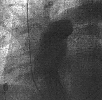
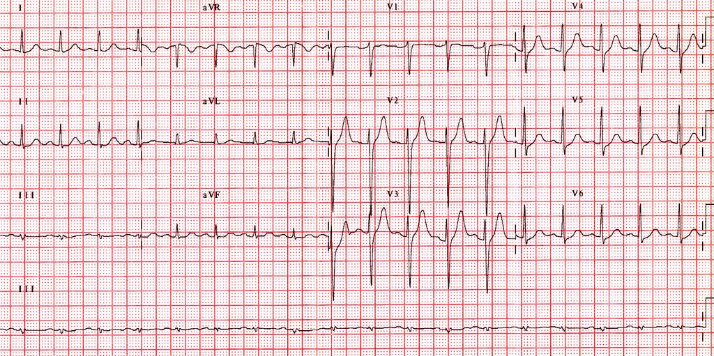
5 Case 48/M recurrent palpitation

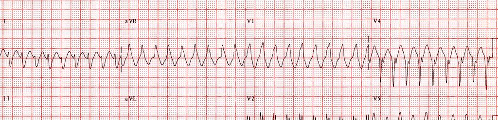
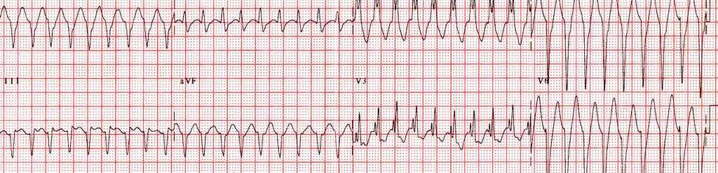
6 Exercise induced VT
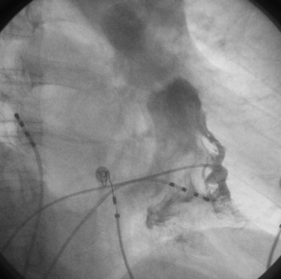
7

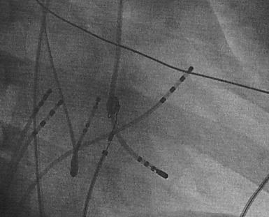
8 3D mapping 3D mapping system ; RVOT VT RF ablation
9 Evaluation of RVOT Tachycardia Exclude structural heart disease - Physical examination - ECG - Echo - SAECG - MRI - RV angiogram and biopsy Rare evolution to cardiomyopathy
10 RVOT VT No evidence of underlying structural heart disease. : generally benign, It must be distinguished from other disorders associated with RV VT, such as RV dysplasia and sarcoidosis. Patients with symptoms not readily treated with medications are candidates for ablation. An ECG showing PVCs or VT can suggest the likely region of origin of the arrhythmia to assist in mapping. Mapping based on earliest activation
11 RVOT Orientation

12 RVOT Distribution Joshi et al, JCE 2005;16suppl:S52

13 RVOT Localization Lead I: Anterior vs Posterior Dixit et al, JCE 2003;14:1 Joshi et al, JCE 2005;16suppl:S52
14 RVOT Localization QRS: Free wall vs Septal QRS duration 140 msec QRS notching in inferior leads Lead V 3 R/S ratio 1 Dixit et al, JCE 2003;14:1 Joshi et al, JCE 2005;16suppl:S52
15 Relationship between RVOT and LVOT
16 Monomorphic ventricular tachycardia with LBBB morphology and an inferior axis. : DDx of RVOT and ASC origin A Total QRS duration(ms) B R-wave duration (ms) C R-wave amplitude (mv) D S-wave amplitude (mv) Ouyang F, et al. J Am Coll Cardiol 2002;39:500
17
18
19 LBBB morphologies with right inferior axis : VT arising from the anterior septal side of the RVOT, from the right or left coronary cusp, and from the pulmonary artery. - R-wave progression : LV or the aortic cusp - R waves in V1 and V2 and a transition by lead V3 : left-sided outflow tract VT, - Later transitions at V3 and V4 : RVOT or the pulmonary artery
20 RBBB morphology : VT arising in the mitral annulus adjacent to the aortic valve or from the epicardium at the outflow tract. - RBBB patterns with dominant R waves across the precordium : mitral annulus
 Larger R/S ratio in V2 0.32 vs 0.")
 Timmermans C, et al. Circulation. 2003;108:1960 Sekiguchi, et al.")
21 Pulmonary artery VT PV PV RAO LAO Taller R in II, III, avf 1.89~1.92 mv vs 1.49~1.57 mv (PA VT vs RVOT VT) Larger R/S ratio in V vs 0.17 (PA VT vs RVOT VT) avl/avr ratio of Q-wave amplitude >1 in the PA (1.11 vs 0.88 :RVOT VT) Timmermans C, et al. Circulation. 2003;108:1960 Sekiguchi, et al. J Am Coll Cardiol 2005;45:887
22 Anatomic location of the successful ablation sites in the pulmonary artery group. The successful ablation sites were located 1.18 ± 0.43 cm above the pulmonary valve and mostly along the septum. Sekiguchi Y, et al. J Am Coll Cardiol 2005;45:887
23 VT with LBBB morphology and inferior axis RV OT PA LVOT ASV LV epi CS Total Ito S 55(69%) 7(9%) 11(14%) 7(9%) 80 Tanner 20(61%) 1(3%) 5(15%) 2(6%) 2(6%) 3(9%) 33 Sekiguchi Y 92(72%) 24(19%) 11(9%) 148 Iwai S 100(82%) 22(18%) (70%) 25(7%) 58(15%) 12(3%) 383 (100%) Ito S, et al. J Cardiovasc Electrophysiol. 2003;14:1280 Tanner H, et al. J Am Coll Cardiol 2005;45:418 Sekiguchi Y, et al. J Am Coll Cardiol 2005;45:887 Iwai S, et al. J Cardiovasc Electrophysiol, Vol. 2006;17:1
24 EPS PES Burst pacing of 200~400 msec Isoproterenol Epinephrine, phenylephrine, aminophylline, Atropine, Ca infusion, edrophonium Adenosine sensitivity Mapping: stepwise mapping RVOT, PA, CS, LVOT, ASV, and epicardial

25 RVOT Cardiac vein LVOT LAO
26 Pace Mapping Single point mapping to obtain 11/12 morphologic match of the 12-lead ECG paced QRS complex to the tachycardia QRS complex. Successful ablation sites with identical/near identical matches However, Even a perfect pace match (12/12) defines a relatively broad area of interest of ~2 cm 2. QRS morphology may be similar over 15 mm separation. Sites within 5 mm may generate differences. Kadish et al. JACC 1991, J Electrocardiol 1998 Clyne CA, et al. PACE 2007
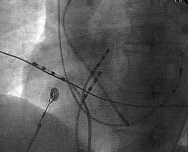
27 Pace Mapping
28 Activation Mapping Endocardial electrogram timing compared to the surface ECG. To detect the earliest endocardial activation time during tachycardia. 10~60 msec (mean 26~46 msec) prior to onset of the surface QRS
. Azegami K, et al.")
29 3D electroanatomical Mapping The mean area of myocardium activated within the first 10 msec was 3.0 ± 1.6 cm 2 (1.3~6.4 cm 2 ). Azegami K, et al. J Cardiovasc Electrophysiol 2005;16:823
30 Idiopathic Epicardial LV VT Perivascular sites of origin Catecholamine enhanced, adenosine sensitive 5~10% of idiopathic VT Daniels DV, et al. Circulation. 2006;113:1659

31 Epicardial origin of LV VT Multipolar catheter in Ant. Interventricular Vein * * Zipes. Cardiac Electrophysiology. 4 th ed
32 ECG of Idiopathic Epicardial LV VT Precordial MDI >0.55 reliably identified EPI VT. MDI : the maximum deflection index TMD: time to maximum deflection in precordial lead Daniels DV, et al. Circulation. 2006;113:1659
33 RF ablation Ablation with power settings of 50 W, a target temperature of 55~70 C and duration of 30~60 seconds. No change in the arrhythmia after 15 seconds of power delivery, it should be stopped and catheter contact and stability reassessed. Nonspecific response : Acceleration/gradual slowing Repetitive response
34 Complication Myocardial perforation with cardiac tamponade Heart block due to inadvertent slippage of the catheter toward the His bundle Injury to the LAD or left main coronary arteries Death, rare
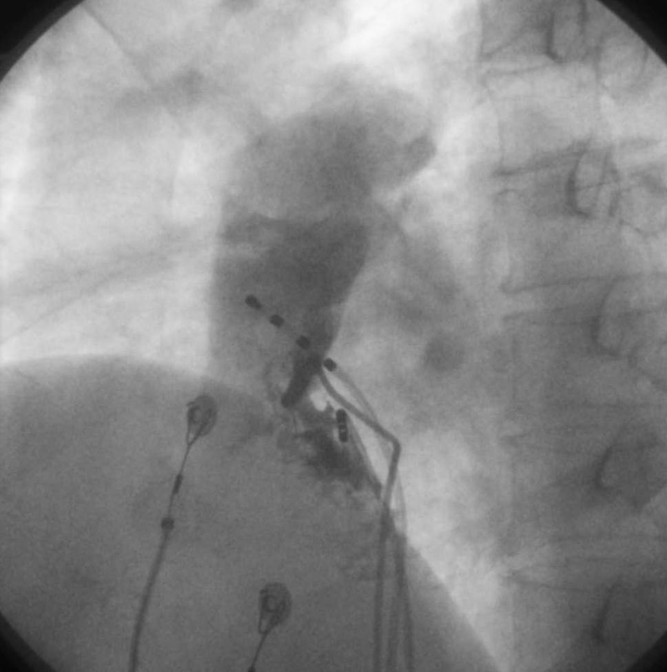
35 F/51 Case

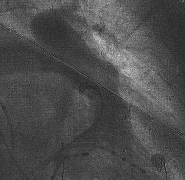
36 RAO Superior anteroseptum of RVOT LAO
37 Pace Mapping I II III avr avl avf V1 V2 V3 V4 V5 V6
38 Activation Mapping

39 Normal Heart Valves and Coronary arteries


40 Major coronary arteries lie in close proximity to the RVOT. Vaseghi M, et al. J Cardiovasc Electrophysiol, 2006;17:632

41 Vaseghi M, et al. J Cardiovasc Electrophysiol, 2006;17:632
42 Outcome of RFCA in Patients with Idiopathic RVOT Tachycardia Joshi et al, JCE 2005;16suppl:S52
43 Idiopathic Left Ventricular Tachycardia Fascicular Tachycardia (52%) Posterior fascicular reentry Anterior fascicular reentry Fascicular automaticity ASOV Tachycardia (10%) LV Endocardial Tachycardia (20%) Aortic root, basal septum (LVOT) Mitral annular Tachycardia Epicardial Tachycardia (15%) Anterior interventricular vein Middle cardiac vein Great cardiac vein Bundle Branch reentry (3%)
44 LV Fascicular Tachycardia ages of 15~40 yrs More frequent in men RBBB with left superior axis: Lt posterior fascicle (90~95%) inferoposterior LV septum RBBB with right inferior axis: Lt anterior fascicle Arrhythmia episodes anterosuperior LV septum ; sensitive to catecholamines(exercise or postexercise) or emotional stress

45 ILVT reentry may be a small macroreentrant circuit. Anterograde limb: abnormal Purkinje tissue, slow decremental conduction, verapamil-sensitive diastolic potential along the midseptum Retrograde limb : Purkinje tissue from the left posterior fascicle, Purkinje potential
46 Late Diastolic Potential Preceding Purkinje Potential in Idiopathic LV Tachycardia LDP recording sites and earliest ventricular activation sites (EAS) Tsuchiya T, et al. Circulation. 1999;99:2408

47 Diastolic potential (P1) and presystolic Purkinje potential (P2) While P1 was recorded earlier from the proximal than the distal electrodes, P2 was recorded earlier from the distal than the proximal electrodes. Nogami A, et al. J Am Coll Cardiol 2000;36:811
48 P1-P2 interval was gradually prolonged, and VT was terminated by block between P1 and P2. After ablation the P1 occurred after the QRS complex during sinus rhythm. Nogami A, et al. J Am Coll Cardiol 2000;36:811
49 Before ablation. Diastolic potential was not observed during sinus rhythm. After ablation, the P1 occurred after the QRS complex. Nogami A, et al. J Am Coll Cardiol 2000;36:811
 Ouyang F, et al. Circulation.")
50 During tachycardia the LV was initially activated at the sites with DPs, then at the posterior fascicle, then at the His bundle region, and progressively at the anterior fascicle before the entire left ventricle is finally activated. Area of slow conduction (zigzag arrow) Ouyang F, et al. Circulation. 2002;105:462
51 Optimal site for catheter ablation of verapamil-sensitive ILVT 1. When diastolic potential and presystolic Purkinje potential are recorded from the midseptal area during VT, this site should be targeted. 2. If such a diastolic potential cannot be detected, the application of RF current to the earliest ventricular activation with a fused Purkinje potential may be carried out. 3. The appearance of diastolic potential after the QRS complex during sinus rhythm appeared to be a useful marker for the effective RF application.
52 Case 36세남자 CC; palpitation PI; 2005년 6월등산직후 palpitation 발생 30분지속 2005년 9월 25일등산중 palpitation 발생 1시간지속 2006년 6월 28일샤워후 palpitation 발생 1시간지속응급실방문가족력 ; 없음.
53
54
55 Inferoapical septum RAO LAO
56 EPS P-potential
57 RFCA
58 Approach to ILVT Narrow QRS( 140 msec), RBBB with superior axis - Probable fascicular tachycardia - Activation mapping during tachycardia - Earliest diastolic potentials (pacemap may be poor.) - Fused double potentials (earliest P-potential, pacemap may be good.)
59 Approach to ILVT Wider QRS (>140 msec), inferior axis - Atypical LB more likely on septum, aortic root or aortic SOV. - Monophasic R in V1 with late or no transition, more likely mitral annulus - Consider epicardial origin If : delayed MDI(>0.55) in precordial leads short presystolic endocardial activation times poor endocardial pacemap matches at all sites failed ablation at best endocardial target site.
60 Classification of Idiopathic Monomorphic VT - Adenosine-sensitive (RVOT/LVOT) - Verapamil-sensitive (Fascicular reentry) - Propranolol-sensitive (Automaticity) Ventricular Outflow Tract Tachycardia - Evaluation of RVOT tachycardia - Localization of RVOT tachycardia by ECG - Anatomy of RVOT and LVOT - Pace mapping, Activation mapping, 3D mapping LV Fascicular Tachycardia - Purkinje Potential, Late Diastolic Potential - Fascicular reentry Approach to ILVT Summary
Ablative Therapy for Ventricular Tachycardia
 Ablative Therapy for Ventricular Tachycardia Nitish Badhwar, MD, FACC, FHRS 2 nd Annual UC Davis Heart and Vascular Center Cardiovascular Nurse / Technologist Symposium May 5, 2012 Disclosures Research
Advances in Ablation Therapy for Ventricular Tachycardia
 Advances in Ablation Therapy for Ventricular Tachycardia Nitish Badhwar, MD, FACC, FHRS Director, Cardiac Electrophysiology Training Program University of California, San Francisco For those of you who
Title. CitationJournal of Electrocardiology, 43(5): Issue Date Doc URL. Type. File Information.
: Issue Date Doc URL. Type. File Information.") Title Pleomorphic ventricular tachycardia originating from Author(s)Yokoshiki, Hisashi; Mitsuyama, Hirofumi; Watanabe, M CitationJournal of Electrocardiology, 43(5): 452-458 Issue Date 2010-09 Doc URL
Title Pleomorphic ventricular tachycardia originating from Author(s)Yokoshiki, Hisashi; Mitsuyama, Hirofumi; Watanabe, M CitationJournal of Electrocardiology, 43(5): 452-458 Issue Date 2010-09 Doc URL
Medicine. Dynamic Changes of QRS Morphology of Premature Ventricular Contractions During Ablation in the Right Ventricular Outflow Tract
 Medicine CLINICAL CASE REPORT Dynamic Changes of QRS Morphology of Premature Ventricular Contractions During Ablation in the Right Ventricular Outflow Tract A Case Report Li Yue-Chun, MD, Lin Jia-Feng,
VENTRICULAR TACHYCARDIA IN THE ABSENCE OF STRUCTURAL HEART DISEASE
 VENTRICULAR TACHYCARDIA IN THE ABSENCE OF STRUCTURAL HEART DISEASE Dimosthenis Avramidis, MD. Consultant Mitera Children s Hospital Athens Greece Scientific Associate 1st Cardiology Dpt Evangelismos Hospital
Mapping and Ablation of Challenging Outflow Tract VTs: Pulmonary Artery, LVOT, Epicardial
 Mapping and Ablation of Challenging Outflow Tract VTs: Pulmonary Artery, LVOT, Epicardial Samuel J. Asirvatham, MD Mayo Clinic Rochester California Heart Rhythm Symposium San Francisco, CA September 8,
Ventricular Tachycardia in Normal Heart: Approach and Management
 ndian Journal of Cardiology SSN-0972-1622 20 ] 2012 by the ndian Society of Cardiology Vol. 15, (3-4), 20-26 Review Article Ventricular Tachycardia in Normal Heart: Approach and Management S.K. Chutani
NAAMA s 24 th International Medical Convention Medicine in the Next Decade: Challenges and Opportunities Beirut, Lebanon June 26 July 2, 2010
 NAAMA s 24 th International Medical Convention Medicine in the Next Decade: Challenges and Opportunities Beirut, Lebanon June 26 July 2, 2010 I have a financial interest/arrangement or affiliation with
Premature ventricular complexes or contractions
 CLINICAL STUDY Analysis of Morphological Characteristics and Origins of Idiopathic Premature Ventricular Contractions Under a 12-Lead Electrocardiogram in Children with Structurally Normal Hearts Jianbin
Characteristics of systolic and diastolic potentials recorded in the left interventricular septum in verapamil-sensitive left ventricular tachycardia
 CASE REPORT Cardiology Journal 2012, Vol. 19, No. 4, pp. 418 423 10.5603/CJ.2012.0075 Copyright 2012 Via Medica ISSN 1897 5593 Characteristics of systolic and diastolic potentials recorded in the left
Tachy. Induction tachycardia lead ECG during Tachy /25/2009. Sinus Rhythm Single His
 12-lead ECG during Tachy 10.30.31 Sinus Rhythm Single His 11.20.02 Induction tachycardia 11.23.23 Tachy 11.25.23 1 I This finding excludes: (a) AVNRT (either typical or atypical) Tachy: Alternating cycle
Φαρμακεσηική αγωγή ζηις ιδιοπαθείς κοιλιακές αρρσθμίες. Άννα Κωζηοπούλοσ Επιμελήηρια Α Ωνάζειο Καρδιοτειροσργικό Κένηρο
 Φαρμακεσηική αγωγή ζηις ιδιοπαθείς κοιλιακές αρρσθμίες Άννα Κωζηοπούλοσ Επιμελήηρια Α Ωνάζειο Καρδιοτειροσργικό Κένηρο Όλες οι κοιλιακές αρρσθμίες δεν είναι ίδιες Υπάρτοσν διαθορές ζηον πληθυσμό, ηον μηχανισμό
Ablation of Ventricular Tachycardia in Non-Ischemic Cardiomyopathy
 Ablation of Ventricular Tachycardia in Non-Ischemic Cardiomyopathy Fermin C Garcia, MD University of Pennsylvania Cardiac Electrophysiology Philadelphia, PA Nothing to disclose No conflict of interest
Map-Guided Ablation of Non-ischemic VT. Takashi Nitta Cardiovascular Surgery, Nippon Medical School Tokyo, JAPAN
 Map-Guided Ablation of Non-ischemic VT Takashi Nitta Cardiovascular Surgery, Nippon Medical School Tokyo, JAPAN nothing Declaration of Interest Catheter Ablation of Non-ischemic VT Sarcoidosis, 13, 6%
Idiopathic Ventricular Tachycardia Need for an Update in EHRA/HRS Consensus?
 Idiopathic Ventricular Tachycardia Need for an Update in EHRA/HRS Consensus? Arash Arya, M.D. Department of Interventional Electrophysiology Heart Center University of Leipzig Disclosures: NONE Idiopathic
Long-Term Follow -Up After Radiofrequency Catheter Ablation of Fascicular Ventricular Tachycardia at National Institute of Cardiovascular Diseases
 Long-Term Follow -Up After Radiofrequency Catheter Ablation of Fascicular Ventricular Tachycardia at National Institute of Cardiovascular Diseases MA Ali 1, MM Hossain 1, S Hashem 1, MA Jami 1, A Hossain
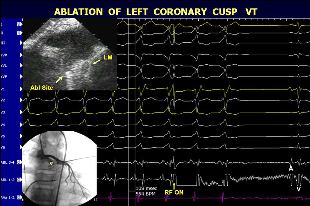
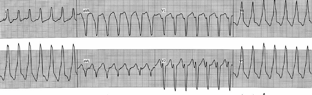
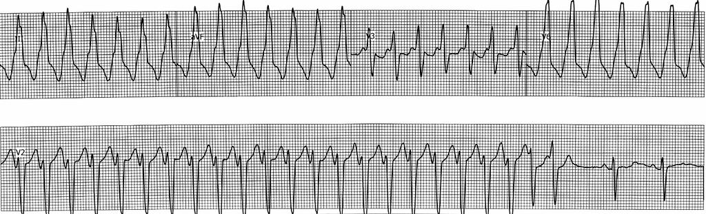
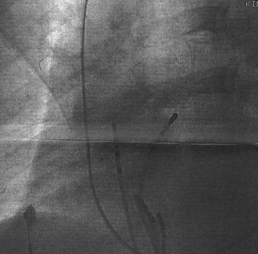
Noncontact mapping to idiopathic VT from LCC
 Narita S Noncontact mapping to idiopathic VT from LCC Case Report Radiofrequency Catheter Ablation with the Use of a Noncontact Mapping System for Ventricular Tachycardia Originating from the Aortic Sinus
Catheter Ablation of Idiopathic Premature Ventricular Contractions and Ventricular Tachycardias Originating from Right Ventricular Septum
 Catheter Ablation of Idiopathic Premature Ventricular Contractions and Ventricular Tachycardias Originating from Right Ventricular Septum Wu Lian-Pin., Li Yue-Chun., Zhao Jing-Lin, Zheng Cheng, Chen Jun-Hua,
INTRODUCTION. left ventricular non-compaction is a sporadic or familial cardiomyopathy characterized by
 A Rare Case of Arrhythmogenic Right Ventricular Cardiomyopathy Co-existing with Isolated Left Ventricular Non-compaction NS Yelgeç, AT Alper, Aİ Tekkeşin, C Türkkan INTRODUCTION Arrhythmogenic right ventricular
Original Article. Introduction. Korean Circulation Journal
 Original Article Print ISSN 1738-5520 On-line ISSN 1738-5555 Korean Circulation Journal Electrophysiological Characteristics Related to Outcome after Catheter Ablation of Idiopathic Ventricular Arrhythmia
Purkinje-related Arrhythmias
 J Arrhythmia Vol 27 No 1 2011 Review Article Purkinje-related Arrhythmias Akihiko Nogami MD Department of Heart Rhythm Management, Yokohama Rosai Hospital, Yokohama, Japan The Purkinje system has been
Case Report. Sumito Narita MD 1;3, Takeshi Tsuchiya MD, PhD 1, Hiroya Ushinohama MD, PhD 2, Shin-ichi Ando MD, PhD 3
 Case Report Identification and Radiofrequency Catheter Ablation of a Nonsustained Atrial Tachycardia at the Septal Mitral Annulus with the Use of a Noncontact Mapping System: A Case Report Sumito Narita
Benign RVOT Ectopy and RV dysplasia
 Heart Rhythm Congress Birmingham October 2009 How to distinguish between... Benign RVOT Ectopy and RV dysplasia in the child... Dr Graham Stuart 14yr old boy asymptomatic irregular pulse picked up by GP
Ankara, Turkey 2 Department of Cardiology, Division of Arrhythmia and Electrophysiology, Yuksek Ihtisas
 258 Case Report Electroanatomic Mapping-Guided Radiofrequency Ablation of Adenosine Sensitive Incessant Focal Atrial Tachycardia Originating from the Non-Coronary Aortic Cusp in a Child Serhat Koca, MD
Basic Electrophysiology Protocols
 Indian Journal of Cardiology ISSN-0972-1622 2012 by the Indian Society of Cardiology Vol. 15, (3-4), 27-37 [ 27 Review Article Shomu Bohora Assistant Professor, Deptt. of Cardiology, U.N. Mehta Institute
In recent years, much attention has been given to cardiac
 Idiopathic Left Bundle-Branch Block Shaped Ventricular Tachycardia May Originate Above the Pulmonary Valve Carl Timmermans, MD; Luz-Maria Rodriguez, MD; Harry J.G.M. Crijns, MD; Antoon F.M. Moorman, PhD;
Conventional Mapping. Introduction
 Conventional Mapping Haitham Badran Ain Shams University it Introduction The mapping approach used to guide ablation depends on the type of arrhythmia being assessed. Simple fluoroscopic anatomy is essential
Ablation of Left Ventricular Epicardial Outflow Tract Tachycardia From the Distal Great Cardiac Vein
 Journal of the American College of Cardiology Vol. 48, No. 9, 2006 2006 by the American College of Cardiology Foundation ISSN 0735-1097/06/$32.00 Published by Elsevier Inc. doi:10.1016/j.jacc.2006.06.006
Reentrant Ventricular Tachycardia Originating in the Right Ventricular Outflow Tract
 Circ J 2008; 72: 855 860 Reentrant Ventricular Tachycardia Originating in the Right Ventricular Outflow Tract Slow Conduction Identified by Right Coronary Artery Ostium Pacing Emi Nakano, MD; Tomoo Harada,
Evaluation of Morphology of Premature Ventricular Contraction on 12-Lead Electrocardiogram
 Original Article Evaluation of Morphology of Premature Ventricular Contraction on 12-Lead Electrocardiogram Umme Habiba Ferdaushi 1, M. Atahar Ali 2, Shaila Nabi 3, Mainul Islam 4, Md. Shamshul Alam 5,
Circulation: Arrhythmia and Electrophysiology CHALLENGE OF THE WEEK
 A 14-year-old girl with Wolff-Parkinson-White syndrome and recurrent paroxysmal palpitations due to atrioventricular reentry tachycardia had undergone two prior failed left lateral accessory pathway ablations
VENTRICULAR TACHYCARDIA WITH HEMODYNAMIC INSTABILITY REFRACTORY TO CARDIOVERSION: A CASE REPORT
 VENTRCULAR TACHYCARDA WTH HEMODYNAMC NSTABLTY REFRACTORY TO CARDOVERSON: A CASE REPORT Chun-Jen Chou, 1 Chee-Siong Lee, 2,3 and Wen-Ter Lai 2,3 1 Department of Emergency Medicine, Kaohsiung Municipal Hsiao-Kang
The left ventricular outflow tract (LVOT) is the most common. Original Article
 is the most common. Original Article") Original Article Prevalence and Electrocardiographic and Electrophysiological Characteristics of Idiopathic Ventricular Arrhythmias Originating From Intramural Foci in the Left Ventricular Outflow Tract
Original Article Prevalence and Electrocardiographic and Electrophysiological Characteristics of Idiopathic Ventricular Arrhythmias Originating From Intramural Foci in the Left Ventricular Outflow Tract
The V 2 Transition Ratio
 Journal of the American College of Cardiology Vol. 57, No. 22, 2011 2011 by the American College of Cardiology Foundation ISSN 0735-1097/$36.00 Published by Elsevier Inc. doi:10.1016/j.jacc.2011.01.035
Epicardial VT Ablation The Cleveland Clinic Experience
 Epicardial VT Ablation The Cleveland Clinic Experience Walid Saliba, MD, FHRS Director, EP Lab Cardiac Electrophysiology Heart and Vascular Institute Epicardial Access in the EP Lab Why Epicardial Special
EHRA Accreditation Exam - Sample MCQs Invasive cardiac electrophysiology
 EHRA Accreditation Exam - Sample MCQs Invasive cardiac electrophysiology Dear EHRA Member, Dear Colleague, As you know, the EHRA Accreditation Process is becoming increasingly recognised as an important
Ventricular arrhythmias
 Ventricular arrhythmias Assoc.Prof. Lucie Riedlbauchová, MD, PhD Department of Cardiology University HospitalMotol and2nd FacultyofMedicine, Charles University in Prague Definition and classification Ventricular
Case 1 Left Atrial Tachycardia
 Case 1 Left Atrial Tachycardia A 16 years old woman was referred to our institution because of recurrent episodes of palpitations and dizziness despite previous ablation procedure( 13 years ago) of postero-septal
Positive QRS Complex in Lead I as a Malignant Sign in Right Ventricular Outflow Tract Tachycardia
 Circulation Journal Official Journal of the Japanese Circulation Society http://www.j-circ.or.jp ORIGINAL ARTICLE Arrhythmia/Electrophysiology Positive QRS Complex in Lead I as a Malignant Sign in Right
Septal ventricular arrhythmias in the presence of structural
 Intramural Idiopathic Ventricular Arrhythmias Originating in the Intraventricular Septum Mapping and Ablation Miki Yokokawa, MD; Eric Good, DO; Aman Chugh, MD; Frank Pelosi, Jr, MD; Thomas Crawford, MD;
Case Report Coexistence of Atrioventricular Nodal Reentrant Tachycardia and Idiopathic Left Ventricular Outflow-Tract Tachycardia
 www.ipej.org 149 Case Report Coexistence of Atrioventricular Nodal Reentrant Tachycardia and Idiopathic Left Ventricular Outflow-Tract Tachycardia Majid Haghjoo, M.D, Arash Arya, M.D, Mohammadreza Dehghani,
Ventricular Tachycardia Substrate. For the ablationist. Stanley Tung, MD FRCPC Arrhythmia Service/St Paul Hospital University of British Columbia
 Ventricular Tachycardia Substrate For the ablationist Stanley Tung, MD FRCPC Arrhythmia Service/St Paul Hospital University of British Columbia Two Attitudes of Ventricular Tachycardia Ablation 1 2C:\Documents
ΔΠΔΜΒΑΣΙΚΗ ΘΔΡΑΠΔΙΑ ΚΟΙΛΙΑΚΩΝ ΑΡΡΤΘΜΙΩΝ
 ΔΠΔΜΒΑΣΙΚΗ ΘΔΡΑΠΔΙΑ ΚΟΙΛΙΑΚΩΝ ΑΡΡΤΘΜΙΩΝ ΣΔΛΙΟ ΠΑΡΑΚΔΤΑÏΓΗ ΓΙΔΤΘΤΝΣΗ ΔΤ Α Καρδιολογική Κλινική ΑΠΘ, Νοζοκομείο ΑΧΕΠΑ, Θεζζαλονίκη NO CONFLICT OF INTEREST INTRODUCTION Sustained VT is an important cause
Urgent VT Ablation in a Patient with Presumed ARVC
 Urgent VT Ablation in a Patient with Presumed ARVC Mr Alex Cambridge, Chief Cardiac Physiologist, St. Barts Hospital, London, UK The patient, a 52 year-old male, attended the ICD clinic without an appointment
Outflow Tract Ventricular Tachycardia Always Benign?
 Outflow Tract Ventricular Tachycardia Always Benign? Arash Arya, M.D. Department of Interventional Electrophysiology Heart Center University of Leipzig Disclosures: NONE Outflow Ventricular Tachycardia
LONG RP TACHYCARDIA MAPPING AND RF ABLATION
 LONG RP TACHYCARDIA MAPPING AND RF ABLATION Dr. Hayam Eldamanhoury Ain shams univeristy Arrhythmia is a too broad topic SVT is broadly defined as narrow complex ( unless aberrant conduction ) Requires
Myocardial Infarction. Reading Assignment (p66-78 in Outline )
") Myocardial Infarction Reading Assignment (p66-78 in Outline ) Objectives 1. Why do ST segments go up or down in ischemia? 2. STEMI locations and culprit vessels 3. Why 15-lead ECGs? 4. What s up with avr?
Myocardial Infarction Reading Assignment (p66-78 in Outline ) Objectives 1. Why do ST segments go up or down in ischemia? 2. STEMI locations and culprit vessels 3. Why 15-lead ECGs? 4. What s up with avr?
Ectopic Atrial Tachycardia
 Europace Madrid, 26-29 June 2011 Ectopic Atrial Tachycardia P. Loh, MD, PhD University of Utrecht Division Heart & Lungs Epidemiology Nonsustained atrial tachycardia Frequent finding on holter registrations
Idiopathic Ventricular Premature Contraction and Ventricular Tachycardia: Distribution of the Origin, Diagnostic Algorithm, and Catheter Ablation
 Review Idiopathic Ventricular Premature Contraction and Ventricular Tachycardia: Distribution of the Origin, Diagnostic Algorithm, and Catheter Ablation Yoshinori Kobayashi From the Department of Internal
Management of patients with ventricular
 Heart 2000;84:553 559 ELECTROPHYSIOLOGY Radiofrequency catheter ablation of ventricular tachycardia William G Stevenson Harvard Medical School, Brigham and Women s Hospital, Boston, Massachusetts, USA
Idiopathic Ventricular Arrhythmia Arising From the Mitral Annulus A Distinct Subgroup of Idiopathic Ventricular Arrhythmias
 Journal of the American College of Cardiology Vol. 45, No. 6, 2005 2005 by the American College of Cardiology Foundation ISSN 0735-1097/05/$30.00 Published by Elsevier Inc. doi:10.1016/j.jacc.2004.12.025
12 Lead ECG Interpretation
 12 Lead ECG Interpretation Julie Zimmerman, MSN, RN, CNS, CCRN Significant increase in mortality for every 15 minutes of delay! N Engl J Med 2007;357:1631-1638 Who should get a 12-lead ECG? Also include
Li Yue-Chun, Zhang Wen-Wu, Zhou Na-Dan, Zhang Teng, Wang Pin-Xiao, Ge Bei, Li Jia, Ji Kang-Ting and Lin Jia-Feng * Abstract
 Yue-Chun et al. BMC Cardiovascular Disorders 2012, 12:32 RESEARCH ARTICLE Open Access Idiopathic premature ventricular contractions and ventricular tachycardias originating from the vicinity of tricuspid
Acute Coronary Syndromes Unstable Angina Non ST segment Elevation MI (NSTEMI) ST segment Elevation MI (STEMI)
 ST segment Elevation MI (STEMI)") Leanna R. Miller, RN, MN, CCRN-CSC, PCCN-CMC, CEN, CNRN, CMSRN, NP Education Specialist LRM Consulting Nashville, TN Objectives Evaluate common abnormalities that mimic myocardial infarction. Identify
Leanna R. Miller, RN, MN, CCRN-CSC, PCCN-CMC, CEN, CNRN, CMSRN, NP Education Specialist LRM Consulting Nashville, TN Objectives Evaluate common abnormalities that mimic myocardial infarction. Identify
Section V. Objectives
 Section V Landscape of an MI Objectives At the conclusion of this presentation the participant will be able to Outline a systematic approach to 12 lead ECG interpretation Demonstrate the process for determining
Electroanatomic Substrate and Outcome of Catheter Ablative Therapy for Ventricular Tachycardia in Setting of Right Ventricular Cardiomyopathy
 Electroanatomic Substrate and Outcome of Catheter Ablative Therapy for Ventricular Tachycardia in Setting of Right Ventricular Cardiomyopathy Francis E. Marchlinski, MD; Erica Zado, PA-C; Sanjay Dixit,
Case Report Mahaim Fiber Accelerated Automaticity and Clues to a Mahaim Fiber Being Morphologically an Ectopic or a Split AV Node
 www.ipej.org 62 Case Report Mahaim Fiber Accelerated Automaticity and Clues to a Mahaim Fiber Being Morphologically an Ectopic or a Split AV Node Shomu Bohora, Narayanan Namboodiri, Santosh Dora, VK Ajit
Use of Catheter Ablation in the Treatment of Ventricular Tachycardia Triggered by Premature Ventricular Contraction
 J Arrhythmia Vol 22 No 3 2006 Case Report Use of Catheter Ablation in the Treatment of Ventricular Tachycardia Triggered by Premature Ventricular Contraction sao Kato MD, Toru wa MD, Yasushi Suzuki MD,
ECG Clues for Diagnosing Ventricular Tachycardia Mechanism
 224 CLINICAL REVIEW Editor: Stephen C. Hammill, M.D. ECG Clues for Diagnosing Ventricular Tachycardia Mechanism MICHAEL P. RILEY, M.D., PH.D. and FRANCIS E. MARCHLINSKI, M.D. From the Electrophysiology
Conduction Problems / Arrhythmias. Conduction
 Conduction Problems / Arrhythmias Conduction Wolf-Parkinson White Syndrome (WPW) and Lown-Ganong-Levine (LGL): Atrial impulses bypass the AV node through an accessory pathway or bypass tract (bundle of
Case Report What Next After Failed Septal Ventricular Tachycardia Ablation?
 www.ipej.org 180 Case Report What Next After Failed Septal Ventricular Tachycardia Ablation? Laurent Roten, MD 1, Nicolas Derval, MD 1, Patrizio Pascale, MD 1, Pierre Jais, MD 1, Pierre Coste, MD 2, Frederic
Catheter ablation of monomorphic ventricular tachycardia. Department of Cardiology, IKEM, Prague, Czech Republic
 Catheter ablation of monomorphic ventricular tachycardia Department of Cardiology, IKEM, Prague, Czech Republic DECLARATION OF CONFLICT OF INTEREST None Ventricular tachycardia ablation in IKEM, Prague
Theroleofcatheterablationinthemanagement of ventricular tachycardia
 European Heart Journal Advance Access published August 31, 2015 European Heart Journal doi:10.1093/eurheartj/ehv421 REVIEW Novel therapeutic concepts Theroleofcatheterinthemanagement of ventricular tachycardia
Arrhythmogenic Right Ventricular Cardiomyopathy. Europace June 28,2011
 Arrhythmogenic Right Ventricular Cardiomyopathy Europace June 28,2011 Right Ventricular Cardiomyopathy Classical ARVC is defined as a cardiac disease mainly involving the right ventricle, characterized
Supraventricular Tachycardia (SVT)
") Supraventricular Tachycardia (SVT) Bruce Stambler, MD Piedmont Heart Atlanta, GA Supraventricular Tachycardia Objectives Types and mechanisms AV nodal reentrant tachycardia (AVNRT) AV reciprocating tachycardia
Supraventricular Tachycardia (SVT) Bruce Stambler, MD Piedmont Heart Atlanta, GA Supraventricular Tachycardia Objectives Types and mechanisms AV nodal reentrant tachycardia (AVNRT) AV reciprocating tachycardia
Diagnostic Electrophysiology & Ablation
 Anatomical Consideration in Catheter Ablation of Idiopathic Ventricular Arrhythmias Takumi Yamada and G Neal Kay Division of Cardiovascular Disease, University of Alabama at Birmingham, Birmingham, Alabama,
Bei Ge 1, Kang-Ting Ji 1, Hai-Ge Ye 2, Jia Li 1, Yue-Chun Li 1, Ri-Peng Yin 1 and Jia-Feng Lin 1*
 Ge et al. BMC Cardiovascular Disorders 2012, 12:112 RESEARCH ARTICLE Open Access Electrocardiogram features of premature ventricular contractions/ventricular tachycardia originating from the left ventricular
Original Article. Macroreentrant Loop in Ventricular Tachycardia From the Left Posterior Fascicle New Implications for Mapping and Ablation
 Original Article Macroreentrant Loop in Ventricular Tachycardia From the Left Posterior Fascicle New Implications for Mapping and Ablation Qiang Liu, MS*; Michael Shehata, MD*; Ruhong Jiang, MS; Lu Yu,
Arrhythmias (II) Ventricular Arrhythmias. Disclosures
 Ventricular Arrhythmias. Disclosures") Arrhythmias (II) Ventricular Arrhythmias Amy Leigh Miller, MD, PhD Cardiovascular Electrophysiology, Brigham & Women s Hospital Disclosures None Rhythms and Mortality Implantable loop recorder post-mi
Arrhythmias (II) Ventricular Arrhythmias Amy Leigh Miller, MD, PhD Cardiovascular Electrophysiology, Brigham & Women s Hospital Disclosures None Rhythms and Mortality Implantable loop recorder post-mi
ACCESSORY PATHWAYS AND SVT. Neil Grubb Royal Infirmary of Edinburgh
 ACCESSORY PATHWAYS AND SVT Neil Grubb Royal Infirmary of Edinburgh Bypass tracts - properties accessory AV connections usually endocardial may exhibit unidirectional conduction conduction properties similar
Novel ECG Predictor of Difficult Cases of Outflow Tract Ventricular Tachycardia:
 Circulation Journal Official Journal of the Japanese Circulation Society http://www.j-circ.or.jp ORIGINAL ARTICLE Arrhythmia/Electrophysiology Novel ECG Predictor of Difficult Cases of Outflow Tract Ventricular
Case Report Wide-QRS Tachycardia Inducible by Both Atrial and Ventricular Pacing
 Hellenic J Cardiol 2008; 49: 446-450 Case Report Wide-QRS Tachycardia Inducible by Both Atrial and Ventricular Pacing ELEFTHERIOS GIAZITZOGLOU, DEMOSTHENES G. KATRITSIS Department of Cardiology, Athens
A Novel Electrocardiographic Criterion for Differentiating a Left from Right Ventricular Outflow Tract Tachycardia Origin: The V2S/V3R Index
 A Novel Electrocardiographic Criterion for Differentiating a Left from Right Ventricular Outflow Tract Tachycardia Origin: The V2S/V3R Index NAOKI YOSHIDA, M.D., TAKUMI YAMADA, M.D., H. THOMAS MCELDERRY,
Ventricular Tachycardia Ablation. Saverio Iacopino, MD, FACC, FESC
 Ventricular Tachycardia Ablation Saverio Iacopino, MD, FACC, FESC ü Ventricular arrhythmias, both symptomatic and asymptomatic, are common, but syncope and SCD are infrequent initial manifestations of
Ablation of Post-Infarct VTs Unmasking VT Isthmuses Using Pace-Mapping
 Ablation of Post-Infarct VTs Unmasking VT Isthmuses Using Pace-Mapping Christian de Chillou, MD, PhD Department of Cardiology University Hospital Nancy, France 29/05/2015 Post-infarct mappable VT Isthmus
Title. CitationJournal of electrocardiology, 46(6): Issue Date Doc URL. Type. File Information
: Issue Date Doc URL. Type. File Information") Title Unique preferential conduction within the isolated s with non-ischemic dilated cardiomyopathy Author(s)Watanabe, Masaya; Yokoshiki, Hisashi; Mitsuyama, Hir CitationJournal of electrocardiology, 46(6):
Title Unique preferential conduction within the isolated s with non-ischemic dilated cardiomyopathy Author(s)Watanabe, Masaya; Yokoshiki, Hisashi; Mitsuyama, Hir CitationJournal of electrocardiology, 46(6):
EPS Case presentation Looks like VT but it isn t!
 EPS Case presentation Looks like VT but it isn t! E. Συμεωνίδου, MD, PhD Β Παν Καρδιολογική κλινική, Νοσ Aττικόν Σεμινάρια Ομάδων Εργασίας ΕΚΕ 2016 Ιωάννινα Disclosures I have no conflict of interest to
Incessant Tachycardia Using a Concealed Atrionodal Bypass Tract
 191 Incessant Tachycardia Using a Concealed Atrionodal Bypass Tract ADAM ZIVIN, M.D., atid FRED MORADY, M.D. From the Division of Cardiology. Department of Internal Medicine, University of Michigan Medical
Treatment of VT of Purkinje fiber origin: ablation targets and outcome
 Treatment of VT of Purkinje fiber origin: ablation targets and outcome Ch. Piorkowski University Leipzig - Heart Center - Dept. of Electrophysiology Leipzig, Germany Presenter Disclosure Information Gerhard
Differential diagnosis and pacing in maneuvers narrow QRS tachycardia. Richard Schilling
 Differential diagnosis and pacing in maneuvers narrow QRS tachycardia Richard Schilling Differential diagnosis of narrow complex QRS tachycardia Anything that activates the ventricle normally via the His
REtrive. REpeat. RElearn Design by. Test-Enhanced Learning based ECG practice E-book
 Test-Enhanced Learning Test-Enhanced Learning Test-Enhanced Learning Test-Enhanced Learning based ECG practice E-book REtrive REpeat RElearn Design by S I T T I N U N T H A N G J U I P E E R I Y A W A
Cover Page. The handle holds various files of this Leiden University dissertation.
 Cover Page The handle http://hdl.handle.net/1887/18608 holds various files of this Leiden University dissertation. Author: Wijnmaalen, Adrianus Pieter Title: Substrate of ventricular tachycardia Issue
Technique of Epicardial VT Ablation
 CARTO Club Jan 2014 Technique of Epicardial VT Ablation Amir AbdelWahab, MD Electrophysiology and Pacing Service Department of Cardiovascular Medicine, Cairo University Need for Epicardial VT ablation
Catheter Ablation of Atrial Tachycardia Originating from the Tip of Right Atrial Appendage
 Case Report Catheter Ablation of Atrial Tachycardia Originating from the Tip of Right Atrial Appendage Masaru Inoue MD, Takao Matsubara MD, Toshihiko Yasuda MD, Kenji Miwa MD, Tadatsugu Gamou MD, Hounin
The Fundamentals of 12 Lead EKG. ECG Recording. J Point. Reviewing the Cardiac Conductive System. Dr. E. Joe Sasin, MD Rusty Powers, NRP
 The Fundamentals of 12 Lead EKG Dr. E. Joe Sasin, MD Rusty Powers, NRP SA Node Intranodal Pathways AV Junction AV Fibers Bundle of His Septum Bundle Branches Purkinje System Reviewing the Cardiac Conductive
Supraventricular Arrhythmias. Reading Assignment. Chapter 5 (p17-30)
") Supraventricular Arrhythmias Reading Assignment Chapter 5 (p17-30) The Supraventricular Rhythms In Our Lives Site of Origin Single Events Slow Rates Intermediate Rates Fast Rates (>100 bpm) Sinus Sinus
Supraventricular Arrhythmias Reading Assignment Chapter 5 (p17-30) The Supraventricular Rhythms In Our Lives Site of Origin Single Events Slow Rates Intermediate Rates Fast Rates (>100 bpm) Sinus Sinus
Novel Approaches to VT Management Glenn M Polin MD
 Novel Approaches to VT Management Glenn M Polin MD Medical Director, Electrophysiology Laboratory John Ochsner Heart and Vascular Institute New Orleans, LA Disclosures Pfizer Speaker Bureau Bristol Myers
The Efficient and Smart Methods for Diagnosis of SVT 대구파티마병원순환기내과정병천
 The Efficient and Smart Methods for Diagnosis of SVT 대구파티마병원순환기내과정병천 Differentiation Supraventricular Origin from Ventricular Origin on ECG. QRS-Complex Width. 1. Narrow QRS-Complex Tachycardia (
Bundle Branch & Fascicular Blocks. Reading Assignment (p53-58 in Outline )
") Bundle Branch & Fascicular Blocks Reading Assignment (p53-58 in Outline ) Objectives 1. QRS analysis of Right and Left BBB 2. Uncomplicated vs complicated BBB 3. Diagnosis of RBBB with LAFB and LPFB 4.
Bundle Branch & Fascicular Blocks Reading Assignment (p53-58 in Outline ) Objectives 1. QRS analysis of Right and Left BBB 2. Uncomplicated vs complicated BBB 3. Diagnosis of RBBB with LAFB and LPFB 4.
ECG Interpretation Made Easy
 ECG Interpretation Made Easy Dr. A Tageldien Abdellah, MSc MD EBSC Lecturer of Cardiology- Hull University Hull York Medical School 2007-2008 ECG Interpretation Made Easy Synopsis Benefits Objectives Process
Tachycardia-induced heart failure - Does it exist?
 Tachycardia-induced heart failure - Does it exist? PD Dr Etienne Delacrétaz Clinique Cecil et Hôpital de Fribourg SSC Cardiology meeting 2015 Zürich Rapid atrial fibrillation is a common cause of heart
Two Years Living with the EHRA/HRS Consensus Document of VT Ablation: Need for an Update?
 Two Years Living with the EHRA/HRS Consensus Document of VT Ablation: Need for an Update? David Wilber MD Loyola University of Chicago Disclosures: Biosense / Webster: Consultant, Investigator; Boston
The left ventricular anterior and posterior papillary muscles. Original Article
 Original Article Differentiation of Papillary Muscle From Fascicular and Mitral Annular Ventricular Arrhythmias in Patients With and Without Structural Heart Disease Subhi J. Al Aref, MD; James E. Ip,
EHRA EUROPACE How to perform epicardial ventricular tachycardia mapping and ablation
 EHRA EUROPACE 2011 How to perform epicardial ventricular tachycardia mapping and ablation Jacob Atié Director Arrhythmias Department Federal University of Rio de janeiro jacobatie1@gmail.com Presenter
Ablation Update and Case Studies. Lawrence Nair, MD, FACC Director of Electrophysiology Presbyterian Heart Group
 Ablation Update and Case Studies Lawrence Nair, MD, FACC Director of Electrophysiology Presbyterian Heart Group Disclosures No financial relationships to disclose Objectives At the conclusion of this activity,
December 2018 Tracings
 Tracings Tracing 1 Tracing 4 Tracing 1 Answer Tracing 4 Answer Tracing 2 Tracing 5 Tracing 2 Answer Tracing 5 Answer Tracing 3 Tracing 6 Tracing 3 Answer Tracing 6 Answer Questions? Contact Dr. Nelson
Abnormalities Caused by Left Bundle Branch Block
 Marquette University e-publications@marquette Physician Assistant Studies Faculty Research and Publications Physician Assistant Studies, Department 12-17-2010 Abnormalities Caused by Left Bundle Branch
Uncommon forms of AV reentry: atrio and fasciculo-ventricular fibers, slow conducting fibers. Jesus Almendral, Madrid, Spain
 Uncommon forms of AV reentry: atrio and fasciculo-ventricular fibers, slow conducting fibers Jesus Almendral, Madrid, Spain Common forms of AV reentry Accessory pathways: Upper insertion: atrium Lower
ECG Criteria to Identify Epicardial Ventricular Tachycardia in Nonischemic Cardiomyopathy
 ECG Criteria to Identify Epicardial Ventricular Tachycardia in Nonischemic Cardiomyopathy Ermengol Vallès, MD; Victor Bazan, MD; Francis E. Marchlinski, MD Background ECG criteria identifying epicardial
Basic electrocardiography reading. R3 lee wei-chieh
 Basic electrocardiography reading R3 lee wei-chieh The Normal Conduction System Lead Placement avf Limb Leads Precordial Leads Interpretation Rate Rhythm Interval Axis Chamber abnormality QRST change What