Radiation Dose Reduction Strategies in Coronary CT Angiography
|
|
|
- Horatio Andrews
- 5 years ago
- Views:
Transcription
1 Radiation Dose Reduction Strategies in Coronary CT Angiography Noor Diyana Osman, PhD

2 Contents: Introduction Radiation dosimetry in CT Radiation risk associated with coronary CT angiography Dose reduction strategies Coronary CTA for paediatric patients
3 Contents: Introduction Radiation dosimetry in CT Radiation risk associated with coronary CT angiography Dose reduction strategies Coronary CTA for paediatric patients

4 Cardiac CT Imaging Introduction Cardiac CT is a painless, non-invasive test that allows high-resolution, 3D visualization of the heart coronary arteries and other adjacent structures. The two main types of cardiac CT imaging: Coronary artery calcium scoring (CASc) Coronary CT angiography (CTCA)
5 Coronary artery calcium scoring (CASc) A CT scan to examine or measure the amount of calcified, or hardened, plaques in the arteries, which is usually explained as a calcium score of low, moderate or high. Unlike a CT angiogram, a calcium score doesn t involve contrast.
6 Coronary CT Angiography (CCTA) Cardiac CTA is an exam used to evaluate the structure and function of the heart, coronary arteries and large vessels of the chest. Coronary CTA is a non-invasive method to image the coronary arteries with the use of contrast. Applications include the following: Diagnosis of coronary artery disease (CAD) Diagnosis of in-stent restenosis Evaluation of coronary bypass graft patency

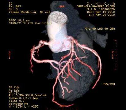
7 Coronary CT Angiography
8 Coronary CT Angiography (CCTA) Coronary CT angiography is currently regarded as the diagnostic imaging method of choice for evaluating coronary arteries.
 Coronary CT angiography (CTCA) Invasive methods (ICA) Expensive Superior spatial & temporal resolution Non")
9 Conventional vs. Modern Angiography Conventional coronary angiography (conventional cardiac catheterization) Coronary CT angiography (CTCA) Invasive methods (ICA) Expensive Superior spatial & temporal resolution Non invasive Improved spatial & temporal resolution Higher dose to patients Studies have shown that coronary CT angiography has a high diagnostic accuracy for the detection of significant CAD ( 50% lumen stenosis) when compared to ICA (Sun Z, et.al., 2011).


10

11 With latest MSCT ( 64 slice CT), CTCA has been reported to have high diagnostic value & can be used as reliable alternative to invasive coronary angiography in selected patients.
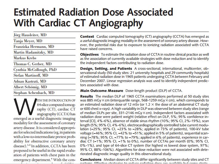
12 Mean effective dose for MSCT coronary angiography was significantly higher than that for conventional angiography MSCT angiography (14.7 msv) vs. conventional angiography (5.6 msv). (Duncan R. et.al, 2006)


13 Coronary CT Angiography 2015 Total = 278 Abd AP Cardiac Head HN Neck NT NTAP Spine TAP TA Thorax Pelvis 2016 Total = 356 In AMDI, only 3 cardiac CT examinations were performed since Jan 2015 CTA (1 patients) + CAScore (2 patients) 0.7% of total studies performed in 2015.
 depends on patient s height Pitch: 0.")
14 Coronary CT Angiography Study Protocols: Siemens SOMATOM Definition AS+ (128 slices/dect) Tube voltage: 120 kvp Tube current: AEC for mas Scan length: cm (as reported in literature) depends on patient s height Pitch: 0.2 Scan time: 0.33 s

15
16 CT scanner Scanning methods Slice thickness / collimation (mm) < 64 slices CTA (4/16/40- slices) 64-slices CTA > 64 slices CTA (128/256/320- slices) Retrospective/ Prospective/ High pitch/ ECG-controlled tube current modulation Retrospective/ Prospective/ High pitch/ low kvp/aec/ tube current modulation Retrospective/ Prospective/ High pitch/ tube current modulation Pitch (low pitch) (high pitch) 0.6, (low pitch) (high pitch) *most of the study used low pitch 0.5, 0.6, , (low pitch) Exposure settings kvp mas kvp mas kvp mas

 must be at least 20 mm & 30 mm for")
17 Min requirements: A 64-detector row (or above) is required. The detector width must be mm or less. Gantry rotation time should be <350 ms. The z-axis coverage (CC) must be at least 20 mm & 30 mm for DSCT mm for the best practice.
18 Contents: Introduction Radiation dosimetry in CT Radiation risk associated with coronary CT angiography Dose reduction strategies Coronary CTA for paediatric patients

19 Radiation dosimetry in CT
20 Dose Display (Post Study Data Page) The dose display is created upon completion of a study delivered CTDI vol, DLP for each series, and phantom size used to calculate these values. It is useful to check CTDI vol after a study is performed to ensure that the output of the scanner was as expected
21 Dose Display (Post Study Data Page) CTDI vol is calculated based on the technique factors used to acquire the data DLP is calculated based on the technique factors and scan length used
22 Radiation dosimetry in CT CT dose index (CTDI) is a standardized measure of radiation dose output of a CT scanner which allows the user to compare radiation output of different CT scanners. CTDI = 1 NT D z dz D(z) = the radiation dose profile along the z-axis, N = the number of tomographic sections imaged in a single axial scan. T = scan width AAPM Report 96
 PMMA cylindrical phantom.")
 is for adult head CT, and pediatric")
23 Radiation dosimetry in CT Measured in large (32 cm diameter) or small (16 cm or 10 cm diameter) PMMA cylindrical phantom. The CTDI measured in large phantom is for adult CT (chest, abdomen, and pelvis). The CTDI measured in small phantom (16 cm) is for adult head CT, and pediatric body CT and 10 cm phantom is for pediatric head.


24 CTDI 100 is a measure of radiation on a 100cm long pencil ionization chamber. CTDI weighted, CTDI w : Radiation dosimetry in CT CTDI w = 2 3 CTDI peripheral CTDI center AAPM Report 96
25 Radiation dosimetry in CT CTDI volume, CTDI vol is the approximate average radiation dose over x,y, and z axis of the patient. CTDI vol = NT I CTDI w CTDI vol = 1 CTDI pitch w CTDI vol estimates the average radiation dose within the irradiated volume for an object of similar attenuation to the CTDI phantom. AAPM Report 96
26 Radiation dosimetry in CT CTDI vol estimation of patient dose (in mgy) a standardized parameter to measure Scanner Radiation Output. CTDI vol is NOT patient dose! CTDI vol is slice-specific dose measurement CTDI vol is based on measurements made by the manufacturer in a factory setting.

27 CTDI vol does not represent the average dose for objects of substantially different size, shape, or attenuation!
28 Summary of Acquisition Parameter Settings Parameter Scan Mode Table Feed/Increment Detector Configuration Pitch Exposure Time Per Rotation Relationship to CTDI vol Changes in the scan mode may affect CTDI vol Table feed affects CTDI vol through its inclusion in pitch Decreasing the beam collimation typically, but not always, increases the CTDI vol CTDI vol relationship to pitch is vendor dependent CTDI vol relationship to exposure time per rotation is vendor dependent Tube Current CTDI vol µ tube current Tube Potential CTDI vol µ (kvp 1 /kvp 2 ) n n ~ 2 to 3 Tube Current Time Product Effective mas Field of Measurement Beam Shaping Filter CTDI vol µ tube current time product CTDI vol µ effective tube current time product Changes in the field of measurement may affect CTDI vol Changes in the beam shaping filter may affect CTDI vol
29 Radiation dosimetry in CT Dose-length product (DLP) product of the length of the irradiated scan volume and the average CTDI vol over that distance Unit: mgy.cm DLP = CTDI vol x scan length

30 CTDI vs. DLP
31 Radiation dosimetry in CT Effective dose, E is a measure of radiation and organ system specific damage in humans (in unit sievert, Sv) E is the DLP multiplied by conversion factor, k (takes into account organ size and radiosensitivity)

32 Radiation dosimetry in CT H T are the tissue-specific equivalent doses in tissues T w T are committee-defined dimensionless tissue-specific weighting factors
33 Table shows tissue weighting factor (as published by ICRP) Tissue or organ Weighting Factor, w T ICRP 26 (1977) ICRP 60 (1990) ICRP 103 (2007) Bone marrow (red) Breast Lung Stomach Colon Gonads Thyroid Bladder Liver Oesophagus Bone surface Skin Salivary glands Brain Remainder
34 Contents: Introduction Radiation dosimetry in CT Radiation risk associated with coronary CT angiography Dose reduction strategies Coronary CTA for paediatric patients
35 Cell Radiosensitivity Radiosensitivity relative susceptibility of cells, tissues & organs to the injurious action of radiation. Cell radiosensitivity is directly proportional to cell division rate and inversely proportional to cell differentiation degree. The most radiosensitive (most at risk from radiation) cells are: actively dividing cells (high division rate) not fully mature or non-specialized type cells that have a high metabolic rate well nourished cells
 Moderate Radiosensitivity Optic lens, stomach, growing cartilage, fine vasculature, growing")
36 Tissues radiosensitivity Increasing radiosensitivity High Radiosensitivity Lymphoid organs, bone marrow, blood, testes, ovaries, intestines Fairly High Radiosensitivity Skin and other organs with epithelial cell lining (cornea, oral cavity, esophagus, rectum, bladder, vagina, uterine cervix, ureters) Moderate Radiosensitivity Optic lens, stomach, growing cartilage, fine vasculature, growing bone Fairly Low Radiosensitivity Mature cartilage or bones, salivary glands, respiratory organs, kidneys, liver, pancreas, thyroid, adrenal and pituitary glands Low Radiosensitivity Muscle, brain, spinal cord
37 CT has become the single largest contributor to man-made radiation exposure. Increased patient radiation dose from CT!

38 CT scans delivers ~500 times the radiation of standard X-ray!!!
39 Radiation dose from common imaging tests Test Echocardiogram MRI Chest x-ray Mammogram Calcium scoring test Cardiac catheterization Chest CT Coronary CT angiography Radionuclide sestamibi stress test Radionuclide dual isotope myocardial perfusion imaging Radiation 0 msv 0 msv 0.05 msv 0.7 msv 1-2 msv 7 msv 10 msv 3-14 msv msv 25 msv
40
41
42 Radiation risk associated with CCTA Although we only scan cm of the chest for most cardiac CT exams, particularly CCTA, the doses are relatively higher because we re looking at very fine structures with overlapping data acquisition and matching the data with the cardiac cycle or the motion of the heart. Higher dose due to higher image quality higher resolution and thinner slices is needed. (Cristen C. B., 2008)
43
44

45 Radiation risk associated with CCTA Comparisons of effective radiation dose in adults for various CT procedures. FA. Mettler et.al., Radiology: 2008
46
47
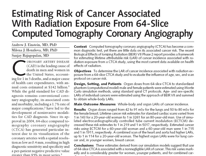
48 Radiation risk associated with CCTA One study estimated that one in every 270 women aged 40 years who undergo a CT coronary angiogram will develop cancer from the procedure. (Smith-Bindman R, et al., 2009)
49 Comparisons of effective radiation dose in adults with background radiation exposure for several radiological procedures. CT PROCEDURES Radiation risk associated with CCTA * An adult s approximate effective radiation dose is: Comparable to natural background radiation for: ABDOMINAL REGION: CT Abdomen + Pelvis 10 msv 3 years CT Abdomen + Pelvis, repeated with and without contrast 20 msv 7 years CT Colonography 6 msv 2 years CENTRAL NERVOUS SYSTEM: CT Head 2 msv 8 months CT Head, repeated with and without contrast 4 msv 16 months CT Spine 6 msv 2 years CHEST: CT Chest 7 msv 2 years CT-Lung Cancer Screening 1.5 msv 6 months DENTAL: Intraoral X-ray msv 1 day HEART: Coronary CTA 12 msv 4 years Cardiac CT for Calcium Scoring 3 msv 1 year Note: Paediatric patients vary in size. Doses given to paediatric patients will vary significantly from those given to adults. * The effective doses are typical values for an average-sized adult. The actual dose can vary substantially, depending on a person's size as well as on differences in imaging practices.
50 Contents: Introduction Radiation dosimetry in CT Radiation risk associated with coronary CT angiography Dose reduction strategies Coronary CTA for paediatric patients
51 The big challenges! Risks (radiation dose) Benefits (image quality)


52 Principles of radiation protection Justification JUSTIFICATION radiation exposures should not be performed unless it demonstrates significant benefit benefit exceeds the risk Optimisation OPTIMISATION the dose should be As Low As Reasonably Practicable (ALARP) Limitation LIMITATION dose limits apply to those who work with radiations and members of the public (ICRP 1999)
53 Dose reduction strategies: 3 immediate ways to reduce the radiation burden of CT: Review your CT imaging protocols radiation dose per examination is optimized, and minimized where appropriate. Avoid delivering higher radiation dose than what is necessary for optimal image quality. Ensure proper utilization of CT avoid inappropriate ordering of imaging procedures (variability between referring physicians). By using standards-based ordering decision support, referring clinicians can enter a patient s symptoms and qualify the effectiveness of their exam choice (e.g. Nuance s Radport is a decision support system for diagnostic image ordering). There are always alternative to CT go for nonionizing radiation based modalities (e.g. MRI and ultrasound). (Cristen C. Bolan, 2008)

54 What we already have? MDCT scanner Dual-energy CT Helical /Spiral CT Iterative image reconstruction Automatic exposure control (AEC)
55 Are we FULLY UTILIZE the technology advancements that we have? Are these technologies utilized appropriately so that their benefits ultimately reach our patients?


56 CCTA dose reduction strategies Adapt to Clinical Indications Implementing iterative reconstruction software Automatic exposure control (AEC) Dose reduction strategies Limiting the scan length & scan phase ECG pulsing Automatic pitch adaption (Cristen C. Bolan, 2008)


57 CCTA dose reduction strategies Patient Preparation Appropriate imaging indications Shield non-imaged organs Scanner Technology Iterative recostruction Dual energy Tube current modulation software Cardiac ECG-gating Acquisition Parameters Appropriate coverage Limit no. of acquisition Lowest possible kvp & mas High pitch, fast gantry rotation time Thick detector width Iodine dose optimization
58
59 CCTA dose reduction strategies
60 1) ECG-Based Tube Current Modulation Electrocardiogram (ECG) pulsing (or ECG controlled tube current modulation) for minimizing dose. If you are looking for coronary arteries and the heart is constantly moving, what we do is segment the heart in order to obtain a measure of the coronary arteries when they are least moving. We only want to acquire coronary arteries when the heart is relatively resting, which is the diastolic phase. The tube current is adjusted based on the ECG signal that we obtain. The tube current is increased when at resting phase or diastolic phase and then is reduced during the systolic phase, or when the heart is moving very rapidly. If the systolic phase is longer than the diastolic phase (occurs when patient s heart rate is too high) results in little dose reduction.
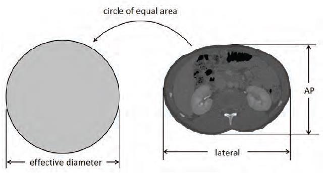
61 ECG-Based Tube Current Modulation AEC feature used with prospectively gated cardiac imaging that adjusts the tube current based on the phase within the cardiac cycle It is important for heart rate considerations when using prospective gating.
62 ECG-Based Tube Current Modulation (TCM) Radiation On Multiple heart beats and table positions may be required to collect all of the data required to reconstruct the FOV including the heart
63 The use of ECG-Based Tube Current Modulation with prospective gating will decrease CTDI vol compared to retrospective gating

64 Traditional way retrospective ECG-gating with tube modulation
65 Prospective ECG-Based TCM More dose reduction through prospective ECG triggering up to 80% of dose reduction. What s the difference between prospective ECG triggering and the ECG tube current modulation? In prospective ECG triggering instead of dropping the tube current during the phase when the heart is rapidly moving, we turn off the tube current to obtain much higher dose reduction. This technique is commonly employed for coronary CaScore and also coronary CTA, if the patient s heart rate is stable and slow. To avoid the tube to be off when the heart is relaxing.
66 Prospective ECG-Based TCM
67 2) Automatic Pitch Adaptation Most scanners have this technique. When the heart is moving fast (higher heart rate), the scanner will go faster (higher pitch), less overlapping, and higher pitch during scanning dose is reduced. For dual source MDCT - the extent to change the pitch is much greater. For SSCT (64-slice scanner), pitch change ranged from but for DSCT pith change ranged from 0.2 to With dual source you don t need the beta blockers because the scanner is faster, and the dose can be reduced using the pitch adaptation.
68 Pitch < 1 implies overlapping Pitch = 1 same as contiguous axial scanning Pitch > 1 implies extended imaging (preferable for dose reduction)

69 How pitch affect the average dose?
70 The drawbacks: Higher pitch gives lower dose, but poor image quality (less resolution)!
71 3) Automatic Exposure Control (AEC) This AEC technique, if used appropriately, will allow the radiologist/ cardiologist to reduce does in children by 30-50%. The clinician selects an appropriate level of image quality. The system then calculates the size of the patient (child/adult) and automatically uses the lowest possible dose to obtain the optimal image quality. AEC controls the tube current to adapt to the patient s size based on what image quality the clinician has specified patient dose can be optimized. With AEC, the radiologists need to decide based on the clinical indications and level of comfort to assess low radiation dose images. Inappropriate selection of superior image quality can actually increase the dose with AEC technique.

")


72 Redesigning CT acquisition protocols Decreased kvp Low kvp can reduce the radiation dose reduction, improve soft tissue contrast. However, mas likely have to be increased to compensate the image quality. Auto mas ma can be adjusted automatically (automatic dose modulation by AEC) based on the patient s size and shape (auto-ma). Decrease ma will reduce patient dose (but must maintain the diagnostic image quality). Pitch > 1 Dose inversely proportional to dose. Pitch < 1, beam overlapping, dose increased. Faster pitch (pitch > 1), will reduce radiation dose. But, reducing image quality. Nelson TR,
73 Lower tube potential (kvp) The use of 100 kv instead of 120 kv (conventionally used) can reduces the dose by 30-35%. Reducing the dose, will increase the image contrast and therefore you need higher volumes of contrasts. But it depends on the patient s size! 100 kv may produce optimal quality for average size but may produce inappropriate image quality for larger patient or else higher dose to smaller patients. So, the regional size of the body or large region of interest is very important and avoid doing lower kv with large size patients or patients with a large region of interest. Adapt the scanning protocol to the clinical indication and the size of the patient. Vendors don t know about the specific clinical indications, and the radiation awareness is a major concern for radiologists and team.
74 Adjusting CT dose based on size Adjust exposure based on individual size and shape. Standard kvp for adult is 120, but adults vary in size. By using a patient s BMI to determine patient size kvp can be accurately adjusted and optimized for dose reduction. 10 cm 16 cm 32 cm Measured CTDI vol = 47 mgy Measured CTDI vol = 37 mgy Measured CTDI vol = 18 mgy Displayed CTDI vol16 = 37 mgy CTDI vol10 = 47 mgy Displayed CTDI vol16 = 37 mgy CTDI vol32 = 18 mgy Displayed CTDI vol32 = 18 mgy Adapted from: Nelson TR, 2014

75
76 4) Limiting: Scan length & Scan phase Decreased length of scan coverage: The scan length is directly proportional to CT dose. Limits the length (z-axis) so that only the anatomy of clinical interest is included in the scan. Limitations on double scans & multi-phase studies: Whenever possible, eliminates non-contrast scans provide little additional diagnostic information and increase patient dose. Limits no. of phases (pre- & post-contrast monitoring) in multi-phase examinations.

77 Limiting the scan phase!

78 How many times you scan the same body part? By reducing the number of passes that you take of the same part of the body, you will reduce the dose. Reducing the number of phases in CT can help you reduce the dose.
79 How you position your patients? It is also important to center the patient right in the center of the gantry. If you don t center the patient right in the center of the gantry, you can increase the dose by 11-15%. This is a very common error.
80 Iterative reconstruction (IR) method Iterative Reconstruction Software / SafeCT: IR is the newest & effective method for CT image reconstruction. Advantages reducing image noise, reduce patient dose by 40% to 50%. Faster technique, improves image quality & SNR. Nelson TR,
81 Contents: Introduction Radiation dosimetry in CT Radiation risk associated with coronary CT angiography Dose reduction strategies Coronary CTA for paediatric patients
82 The Critical Group! Paediatric patients inherently more radiosensitive and because they have more remaining years of life during which a radiationinduced cancer could develop.
83 The Critical Group! There is a latent period following radiation exposure and the time it takes for the radiation effects to develop. The latent period for development of cancer following low level radiation dose is variable and can be as long as years. If a person getting a CT scan at age 60, he is unlikely to develop cancer in his remaining lifetime, but children have much longer to live. Children s cells are more susceptible to radiation and they are also likely to live longer than adults.
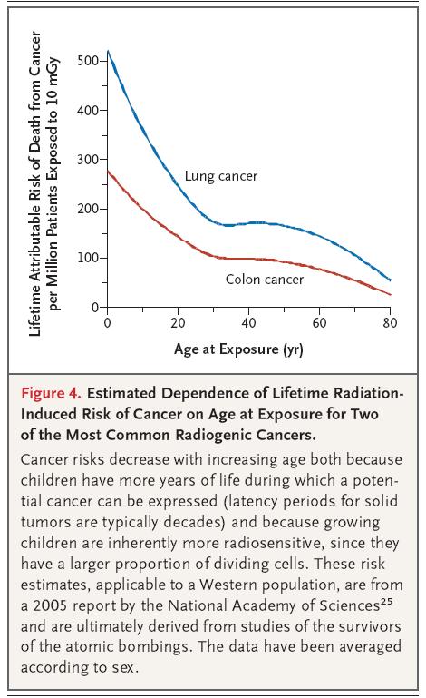
84 Estimated age-dependent CT doses to various organs Estimated age-dependent doses to various organs for typical single CT scan of head (assuming the same exposure techniques for all ages) Risk depends strongly on the age at the time of irradiation the younger the child, the higher the potential risk (Brenner DJ et.al., 2007)
 (Brenner DJ et.al., 2007)")
85 Estimated risk of death by cancer attributable to a CT scan at different ages Estimated lifetime CT-attributable cancer mortality risks as a function of age (for different gender) (Brenner DJ et.al., 2007)
86 CCTA for Paediatric patient Different imaging needs for paediatric patients! Child s cells dividing in different ways & more sensitive & susceptible to radiation risks! (Nelson TR, 2014)


87 Dose reduction for paediatric patient One size does not fit all...when imaging paediatric patients, radiation dose matters!
88 Dose reduction strategies for paediatric patient Children are more sensitive to radiation. So when we image, let's image gently. The right things to do: Scan only the indicated area. Selection of the kvp and ma child size. One scan (single phase) is often enough.
. Appropriate radiation levels for their age and size!")
89 Dose reduction strategies for paediatric patient A well-known strategy for reducing radiation exposure for paediatric is to decrease CT dose based on the patient s weight (Johnson et.al., 2012). Appropriate radiation levels for their age and size!

90 Establishment of Local Diagnostic Reference Levels (DRLs) Regularly analyse local CT dose values! Monthly/Annual dose audit.
")
91 National DRLs Recommended Malaysian DRLs for CT examination (MOH, 2013)
.")
92 Establishment of Local Diagnostic Reference Levels (DRLs) DRLs are proposed to help manage radiation dose to patients so that the dose is commensurate with the clinical purpose (ICRP Committee 3). As recommended by MOH, the respective medical institutions are advised to obtain individual local data in their setup in order to compare with the national DRLs (MOH, 2013).
93
94
 Benefits")
95 Take home notes! Risks (radiation dose) Benefits (image quality)
96 Thank you
Managing Radiation Risk in Pediatric CT Imaging
 Managing Radiation Risk in Pediatric CT Imaging Mahadevappa Mahesh, MS, PhD, FAAPM, FACR, FACMP, FSCCT. Professor of Radiology and Cardiology Johns Hopkins University School of Medicine Chief Physicist
CT Dose Estimation. John M. Boone, Ph.D., FAAPM, FSBI, FACR Professor and Vice Chair of Radiology. University of California Davis Medical Center
 CT Dose Estimation John M. Boone, Ph.D., FAAPM, FSBI, FACR Professor and Vice Chair of Radiology 1 University of California Davis Medical Center CT Dose Estimation Introduction The CTDI Family of Metrics
Low Dose Era in Cardiac CT
 Low Dose Era in Cardiac CT DIANA E. LITMANOVICH, MD Department of Radiology Beth Israel Deaconess Medical Center Harvard Medical School Disclosures Neither I nor my immediate family members have a financial
Why is CT Dose of Interest?
 Why is CT Dose of Interest? CT usage has increased rapidly in the past decade Compared to other medical imaging CT produces a larger radiation dose. There is direct epidemiological evidence for a an increase
ESTABLISHING DRLs in PEDIATRIC CT. Keith Strauss, MSc, FAAPM, FACR Cincinnati Children s Hospital University of Cincinnati College of Medicine
 ESTABLISHING DRLs in PEDIATRIC CT Keith Strauss, MSc, FAAPM, FACR Cincinnati Children s Hospital University of Cincinnati College of Medicine CT Dose Indices CTDI INTRODUCTION CTDI 100, CTDI w, CTDI vol
Measurement of organ dose in abdomen-pelvis CT exam as a function of ma, KV and scanner type by Monte Carlo method
 Iran. J. Radiat. Res., 2004; 1(4): 187-194 Measurement of organ dose in abdomen-pelvis CT exam as a function of ma, KV and scanner type by Monte Carlo method M.R. Ay 1, M. Shahriari 2, S. Sarkar 3, P.
CT Radiation Risks and Dose Reduction
 CT Radiation Risks and Dose Reduction Walter L. Robinson, M.S. D.A.B.S.N.M., D.A.B.M.P., D.A.B.R. Consultant Certified Medical Radiation Health & Diagnostic Imaging Physicist Medical Radiation and Children
Assessment of effective dose in paediatric CT examinations
 Assessment of effective dose in paediatric CT examinations E. Dougeni 1,2 CL. Chapple 1, J. Willis 1, G. Panayiotakis 2 1 Regional Medical Physics Department, Freeman Hospital, Freeman Road, Newcastle
Doses from pediatric CT examinations in Norway Are pediatric scan protocols developed and in daily use?
 Doses from pediatric CT examinations in Norway Are pediatric scan protocols developed and in daily use? Eva Godske Friberg * Norwegian Radiation Protection Authority, P.O. Box, Østerås, Norway Abstract.
SOMATOM Drive System Owner Manual Dosimetry and imaging performance report
 www.siemens.com/healthcare SOMATOM Drive System Owner Manual Dosimetry and imaging performance report Table of contents 1 Dosimetry and imaging performance report 5 1.1 Dose information 5 1.1.1 General
Accounting for Imaging Dose
 Accounting for Imaging Dose High Profile Over-exposures Lead to Growing Concern FDA issues warning in October 2009-209 patients exposed to 8 times typical dose for CT brain perfusion scan (3-4 Gy) - Some
Computed tomography Acceptance testing and dose measurements
 Computed tomography Acceptance testing and dose measurements Jonas Andersson Medical Physicist, Ph.D. Department of Radiation Sciences University Hospital of Norrland, Umeå Sweden Contents The Computed
CURRENT CT DOSE METRICS: MAKING CTDI SIZE-SPECIFIC
 CURRENT CT DOSE METRICS: MAKING CTDI SIZE-SPECIFIC Keith Strauss, MSc, FAAPM, FACR Cincinnati Children s Hospital University of Cincinnati College of Medicine Acknowledgments John Boone, PhD Michael McNitt-Grey,
Fundamentals, Techniques, Pitfalls, and Limitations of MDCT Interpretation and Measurement
 Fundamentals, Techniques, Pitfalls, and Limitations of MDCT Interpretation and Measurement 3 rd Annual Imaging & Physiology Summit November 20-21, 21, 2009 Seoul, Korea Wm. Guy Weigold, MD, FACC Cardiovascular
Doses from Cervical Spine Computed Tomography (CT) examinations in the UK. John Holroyd and Sue Edyvean
 examinations in the UK. John Holroyd and Sue Edyvean") Doses from Cervical Spine Computed Tomography (CT) examinations in the UK John Holroyd and Sue Edyvean Why a new dose survey? Number of enquires received concerning the current NDRL Concern that could
Doses from Cervical Spine Computed Tomography (CT) examinations in the UK John Holroyd and Sue Edyvean Why a new dose survey? Number of enquires received concerning the current NDRL Concern that could
Managing Patient Dose in Computed Tomography (CT) INTERNATIONAL COMMISSION ON RADIOLOGICAL PROTECTION
 INTERNATIONAL COMMISSION ON RADIOLOGICAL PROTECTION") Managing Patient Dose in Computed Tomography (CT) International Commission on Radiological Protection Information abstracted from ICRP Publication 87 Available at www.icrp.org Task Group: M.M. Rehani,
Managing Patient Dose in Computed Tomography (CT) International Commission on Radiological Protection Information abstracted from ICRP Publication 87 Available at www.icrp.org Task Group: M.M. Rehani,
CT Dosimetry in the Clinical Environment: Methods and Analysis
 CT Dosimetry in the Clinical Environment: Methods and Analysis Manuel Arreola, Ph.D. DABR Associate Chair of Radiology Director, Medical Physics Graduate Program Department of Radiology University of Florida
Skyscan 1076 in vivo scanning: X-ray dosimetry
 Skyscan 1076 in vivo scanning: X-ray dosimetry DOSIMETRY OF HIGH RESOLUTION IN VIVO RODENT MICRO-CT IMAGING WITH THE SKYSCAN 1076 An important distinction is drawn between local tissue absorbed dose in
Pushing the limits of cardiac CT. Steven Dymarkowski Radiology / Medical Imaging Research Centre
 Pushing the limits of cardiac CT Steven Dymarkowski Radiology / Medical Imaging Research Centre 5 X 2013 Introduction Rapid technological advances and new clinical applications in cardiovascular imaging
SPECIFIC PRINCIPLES FOR DOSE REDUCTION IN HEAD CT IMAGING. Rajiv Gupta, MD, PhD Neuroradiology, Massachusetts General Hospital Harvard Medical School
 SPECIFIC PRINCIPLES FOR DOSE REDUCTION IN HEAD CT IMAGING Rajiv Gupta, MD, PhD Neuroradiology, Massachusetts General Hospital Harvard Medical School OUTLINE 1 st Presentation: Dose optimization strategies
Cardiac CT - Coronary Calcium Basics Workshop II (Basic)
") Cardiac CT - Coronary Calcium Basics Workshop II (Basic) J. Jeffrey Carr, MD, MSCE Dept. of Radiology & Public Health Sciences Wake Forest University School of Medicine Winston-Salem, NC USA No significant
Cardiac CT - Coronary Calcium Basics Workshop II (Basic) J. Jeffrey Carr, MD, MSCE Dept. of Radiology & Public Health Sciences Wake Forest University School of Medicine Winston-Salem, NC USA No significant
Radiation Dosimetry for CT Protocols
 Radiation Dosimetry for CT Protocols This document contains radiation dosimetry information from CT scans and can be used by investigators to estimate the dosimetry information required by the JRSC or
A comparison of radiation doses from modern multi-slice Computed Tomography angiography and conventional diagnostic Angiography:
 A comparison of radiation doses from modern multi-slice Computed Tomography angiography and conventional diagnostic Angiography: Rob Loader Oliver Gosling Introduction Approached by Dr Oliver Gosling (Research
The radiation dose in retrospective
 The radiation dose in retrospective gated tdcoronary computed td tomography (CCT) Saeed AL Ahmari, Ghormallah AL Zahrani, Sumiah AL Helali, Samir AL Dulikan, Abdullah Bafagih, HibaKhashojji Prince Sultan
CT Optimisation for Paediatric SPECT/CT Examinations. Sarah Bell
 CT Optimisation for Paediatric SPECT/CT Examinations Sarah Bell Sarah.bell14@nhs.net Outline 1. Introduction 2. Aims and Objectives 3. Methods 4. Results 5. Discussion 6. Conclusions 7. References Introduction
Combined Anatomical and Functional Imaging with Revolution * CT
 GE Healthcare Case studies Combined Anatomical and Functional Imaging with Revolution * CT Jean-Louis Sablayrolles, M.D. Centre Cardiologique du Nord, Saint-Denis, France Case 1 Whole Brain Perfusion and
Debra Pennington, MD Director of Imaging Dell Children s Medical Center
 Debra Pennington, MD Director of Imaging Dell Children s Medical Center 1 Gray (Gy) is 1 J of radiation energy/ 1 kg matter (physical quantity absorbed dose) Diagnostic imaging doses in mgy (.001 Gy)
8/18/2011. Acknowledgements. Managing Pediatric CT Patient Doses INTRODUCTION
 Managing Pediatric CT Patient Doses Keith J. Strauss, MSc, FAAPM, FACR President X-Ray Computations, Inc. Boston, Massachusetts Acknowledgements Marilyn Goske, MD John Boone, PhD Cynthia McCollough, PhD
Thoracic examinations with 16, 64, 128 and 256 slices CT: comparison of exposure doses measured with an anthropomorphic phantom and TLD dosimeters
 Thoracic examinations with 16, 64, 128 and 256 slices CT: comparison of exposure doses measured with an anthropomorphic phantom and TLD dosimeters Poster No.: C-2584 Congress: ECR 2015 Type: Scientific
A more accurate method to estimate patient dose during body CT examinations with tube current modulation
 A more accurate method to estimate patient dose during body CT examinations with tube current modulation Poster No.: C-0738 Congress: ECR 2014 Type: Scientific Exhibit Authors: A. Kawaguchi 1, Y. Matsunaga
Chief Radiographer TEI Clinical Associate 2016
 MDCT Principles i and Applications Ε ΑGADAKOS MSc Ε. ΑGADAKOS MSc Chief Radiographer TEI Clinical Associate 2016 Aim To understand d recent technological advances in MSCT and how they can be effectively
Ultrasound. Computed tomography. Case studies. Utility of IQon Spectral CT in. cardiac imaging
 Ultrasound Computed tomography Case studies Utility of IQon Spectral CT in cardiac imaging Cardiac imaging is a challenging procedure where it is necessary to image a motion-free heart. This requires a
3 rd International Symposium on the System of Radiological Protection Seoul, October John Harrison
 3 rd International Symposium on the System of Radiological Protection Seoul, October 2015 John Harrison UK Task Group 79 : Use of Effective Dose as a Risk-related Radiological Protection Quantity John
How do the Parameters affect Image Quality and Dose for Abdominal CT? Image Review
 How do the Parameters affect Image Quality and Dose for Abdominal CT? Image Review Mannudeep K. Kalra, MD, DNB Massachusetts General Hospital Harvard Medical School Financial Disclosure This presentation
Calculation of Effective Doses for Radiotherapy Cone-Beam CT and Nuclear Medicine Hawkeye CT Laura Sawyer
 Calculation of Effective Doses for Radiotherapy Cone-Beam CT and Nuclear Medicine Hawkeye CT Laura Sawyer Department of Medical Physics and Bioengineering, Royal United Hospital, Bath Overview Varian Acuity
Managing Patient Dose in Computed Tomography (CT)
") Managing Patient Dose in Computed Tomography (CT) International Commission on Radiological Protection Information abstracted from ICRP Publication 87 Available at www.icrp.org Task Group: M.M. Rehani,
Managing Patient Dose in Computed Tomography (CT) International Commission on Radiological Protection Information abstracted from ICRP Publication 87 Available at www.icrp.org Task Group: M.M. Rehani,
Estimating Patient Radiation Dose from Computed Tomography
 Estimating Patient Radiation Dose from Computed Tomography C. Cagnon, J. DeMarco, E. Angel, M. McNitt-Gray UCLA David Geffen School of Medicine 1 Patient Dose from CT Advances in Technology... Helical
Recent Progress in Radiation Dosimetry for Epidemiology and Radiological Protection. John Harrison ICRP Committee 2
 Recent Progress in Radiation Dosimetry for Epidemiology and Radiological Protection John Harrison ICRP Committee 2 Joint ICRP-RERF-JHPS Workshop: Tokyo, December 2017 Task Group 79 : Use of Effective Dose
Translating Protocols Across Patient Size: Babies to Bariatric
 Translating Protocols Across Patient Size: Babies to Bariatric Cynthia H. McCollough, PhD, FACR, FAAPM Professor of Radiologic Physics Director, CT Clinical Innovation Center Department of Radiology Mayo
Cone Beam CT Protocol Optimisation for Prostate Imaging with the Varian Radiotherapy OBI imaging system. Dr Craig Moore & Dr Tim Wood
 Cone Beam CT Protocol Optimisation for Prostate Imaging with the Varian Radiotherapy OBI imaging system Dr Craig Moore & Dr Tim Wood Background With the increasing use of CBCT imaging alongside complex
X-Ray & CT Physics / Clinical CT
 Computed Tomography-Basic Principles and Good Practice X-Ray & CT Physics / Clinical CT INSTRUCTORS: Dane Franklin, MBA, RT (R) (CT) Office hours will be Tuesdays from 5pm to 6pm CLASSROOM: TIME: REQUIRED
Low-dose and High-resolution Cardiac Imaging with Revolution CT
 GE Healthcare Case study Low-dose and High-resolution Cardiac Imaging with Revolution CT Prof. Philipp A. Kaufmann, M.D. Ronny R. Buechel, M.D. Fran Mikulicic, M.D. Dominik C. Benz, M.D. University of
Disclosure Information
 Coronary CTA Pearls and Pitfalls Ricardo C. Cury, MD, FSCCT, FAHA, FACC Chairman of Radiology Radiology Associates of South Florida Director of Cardiac Imaging Miami Cardiac and Vascular Institute Past-President
Patient / Organ Dose in CT
 Patient / Organ Dose in CT Patient specific and organ dose estimation H.D. Nagel Dr. HD Nagel, Science & Technology for Radiology Buchholz / Germany www.sascrad.com 1 Topics CTDI & patient dose SSDE Organ
Dosimetric Consideration in Diagnostic Radiology
 Dosimetric Consideration in Diagnostic Radiology Prof. Ng Kwan-Hoong Department of Biomedical Imaging University of Malaya ngkh@um.edu.my Radiation Dosimetry Workshop, 28-29 March 2014 2 Why do we measure
Alessandro Albonico Philips
 Alessandro Albonico Philips Alessandro.albonico@philips.com Noise (Standard Deviation in HU) Virtually noise-free Characteristic of a true knowledge-based IR 80 70 Standard Recon idose4 Level6 1 mm Slice
Cardiac Computed Tomography
 Cardiac Computed Tomography Authored and approved by Koen Nieman Stephan Achenbach Francesca Pugliese Bernard Cosyns Patrizio Lancellotti Anastasia Kitsiou Contents CARDIAC COMPUTED TOMOGRAPHY Page 1.
Toshiba Aquillion 64 CT Scanner. Phantom Center Periphery Center Periphery Center Periphery
 Comparison of radiation dose and imaging performance for the standard Varian x-ray tube and the Richardson Healthcare ALTA750 replacement tube for the Toshiba Aquillion CT scanners. by Robert L. Dixon,
Cardiac CT Lowering the Dose Dramatically
 Cardiac CT Lowering the Dose Dramatically U. Joseph Schoepf, MD, FAHA, FSCBT MR, FSCCT Professor of Radiology, Medicine, and Pediatrics Director of Cardiovascular Imaging Disclosures Consultant for / research
Radiation Burden in Cardiology. A quick tour! David Sutton
 Radiation Burden in Cardiology A quick tour! David Sutton Contents Radiation Doses to Staff Doses to the eye Radiation Doses to Patients Nuclear Cardiology CCTA Cardiac x-ray procedures Guiding Principles
FDG-18 PET/CT - radiation dose and dose-reduction strategy
 FDG-18 PET/CT - radiation dose and dose-reduction strategy Poster No.: C-1856 Congress: ECR 2014 Type: Authors: Keywords: DOI: Scientific Exhibit P. Nicholson, S. McSweeney, K. O'Regan; Cork/IE Radiation
Regional diagnostic reference levels and collective effective doses from CT scanners in India
 Regional diagnostic reference levels and collective effective doses from CT scanners in India Roshan S Livingstone and Paul M Dinakaran Department of Radiology Christian Medical College, Vellore, S India
Survey of patients CT radiation dose in Jiangsu Province
 Original Article Page 1 of 6 Survey of patients CT radiation dose in Jiangsu Province Yuanyuan Zhou 1, Chunyong Yang 1, Xingjiang Cao 1, Xiang Du 1, Ningle Yu 1, Xianfeng Zhou 2, Baoli Zhu 1, Jin Wang
Introduction Pediatric malignancies Changing trends & Radiation burden Radiation exposure from PET/CT Image gently PET & CT modification - PET/CT
 Introduction Pediatric malignancies Changing trends & Radiation burden Radiation exposure from PET/CT Image gently PET & CT modification - PET/CT protocols Tips Leukaemia / lymphoma: ~ 35% acute lymphoblastic
Clinical Image Gallery Next Generation Volume 1
 Clinical Image Gallery Next Generation Volume 1 Dr. Russell Bull Royal Bournemouth Hospital, Bournemouth, United Kingdom After long experience with the first generation, a next generation Aquilion ONE
Fellows on this rotation are expected to attend nuclear conferences and multimodality imaging conference.
 Rotation: Imaging 1 Imaging 1 provides COCATS Level 1 experience for nuclear cardiology (including SPECT and PET) and cardiac CT. Fellows will administer, process, and read cardiac nuclear studies with
Managing the imaging dose during Image-guided Radiotherapy. Martin J Murphy PhD Department of Radiation Oncology Virginia Commonwealth University
 Managing the imaging dose during Image-guided Radiotherapy Martin J Murphy PhD Department of Radiation Oncology Virginia Commonwealth University Radiographic image guidance has emerged as the new paradigm
Machine Learning Powered Automatic Organ Classification for Patient Specific Organ Dose Estimation
 Machine Learning Powered Automatic Organ Classification for Patient Specific Organ Dose Estimation Junghwan Cho, Eunmi Lee, Hyunkwang Lee, Bob Liu, Xinhua Li, Shahein Tajmir, Dushyant Sahani, and Synho
A Snapshot on Nuclear Cardiac Imaging
 Editorial A Snapshot on Nuclear Cardiac Imaging Khalil, M. Department of Physics, Faculty of Science, Helwan University. There is no doubt that nuclear medicine scanning devices are essential tool in the
Cardiac CT Angiography
 Cardiac CT Angiography Dr James Chafey, Radiologist Why do we need a better test for C.A.D? 1. CAD is the leading cause of death in the US CAD 31% Cancer 23% Stroke 7% 2. The prevalence of atherosclerosis
Triple Rule-out using 320-row-detector volume MDCT: A comparison of the wide volume and helical modes
 Triple Rule-out using 320-row-detector volume MDCT: A comparison of the wide volume and helical modes Poster No.: C-0488 Congress: ECR 2012 Type: Authors: Keywords: DOI: Scientific Exhibit E.-J. Kang,
IMAGE GENTLY HOW CAN YOU HELP?
 IMAGE GENTLY HOW CAN YOU HELP? Keith J. Strauss, MSc, FAAPM, FACR Director, Radiology Physics & Engineering Children s s Hospital Boston Harvard Medical School Acknowledgment Marilyn J. Goske,, MD Robert
Cardiac CT Techniques in Neonates (and infants)
") Cardiac CT Techniques in Neonates (and infants) Siddharth P. Jadhav, MD Director, Body CT and MRI Edward B. Singleton Department of Pediatric Radiology Texas Children s Hospital Disclosures None Objectives
Cardiac CT Techniques in Neonates (and infants) Siddharth P. Jadhav, MD Director, Body CT and MRI Edward B. Singleton Department of Pediatric Radiology Texas Children s Hospital Disclosures None Objectives
Gender differences in CT calcium scoring: A phantom study
 Gender differences in CT calcium scoring: A phantom study Nicholas Petrick, Qin Li, Benjamin Berman, Marios A Gavrielides, Rongping Zeng, Berkman Sahiner CDRH/OSEL/DIDSR U.S. Food and Drug Administration
Simon Nepveu 1, Irina Boldeanu 1, Yves Provost 1, Jean Chalaoui 1, Louis-Mathieu Stevens 2,3, Nicolas Noiseux 2,3, Carl Chartrand-Lefebvre 1,3
 Coronary Artery Bypass Graft Imaging with CT Angiography and Iterative Reconstruction: Quantitave Evaluation of Radiation Dose Reduction and Image Quality Simon Nepveu 1, Irina Boldeanu 1, Yves Provost
Horizon Scanning Technology Summary. Magnetic resonance angiography (MRA) imaging for the detection of coronary artery disease
 imaging for the detection of coronary artery disease") Horizon Scanning Technology Summary National Horizon Scanning Centre Magnetic resonance angiography (MRA) imaging for the detection of coronary artery disease April 2007 This technology summary is based
Horizon Scanning Technology Summary National Horizon Scanning Centre Magnetic resonance angiography (MRA) imaging for the detection of coronary artery disease April 2007 This technology summary is based
Cardiac Imaging Tests
 Cardiac Imaging Tests http://www.medpagetoday.com/upload/2010/11/15/23347.jpg Standard imaging tests include echocardiography, chest x-ray, CT, MRI, and various radionuclide techniques. Standard CT and
AAPM Task Group 180 Image Guidance Doses Delivered During Radiotherapy: Quantification, Management, and Reduction
 AAPM Task Group 180 Image Guidance Doses Delivered During Radiotherapy: Quantification, Management, and Reduction Parham Alaei, Ph.D. Department of Radiation Oncology University of Minnesota NCCAAPM Fall
Computed Tomography of the Coronary Arteries
 Cardiology Update DAVOS 2011 Computed Tomography of the Coronary Arteries Anders Persson M.D., Ph.D Director, Assoc. Professor Center for Medical Image Science and Visualization Linköping University SWEDEN
Radiography/Radiology
 Radiography/Radiology Activity for 2017 Activity No: A1(17) Topic CT radiation Article CT radiation: key concepts for gentle and wise use Approved for (3) Clinical Continuing Educational Units (CEU s)
Acknowledgments. A Specific Diagnostic Task: Lung Nodule Detection. A Specific Diagnostic Task: Chest CT Protocols. Chest CT Protocols
 Personalization of Pediatric Imaging in Terms of Needed Indication-Based Quality Per Dose Acknowledgments Duke University Medical Center Ehsan Samei, PhD Donald Frush, MD Xiang Li PhD DABR Cleveland Clinic
Dose Reduction Options in Cardiac CT
 Dose Reduction Options in Cardiac CT Doyle P, Ball P*, Donnelly P # Radiological Sciences & Imaging, Forster Green Hospital *Department of Radiology, Ulster Hospital # Department of Cardiology, Ulster
Coronary Artery Imaging. Suvipaporn Siripornpitak, MD Inter-hospital Conference : Rajavithi Hospital
 Coronary Artery Imaging Suvipaporn Siripornpitak, MD Inter-hospital Conference : Rajavithi Hospital Larger array : cover scan area Detector size : spatial resolution Rotation speed : scan time Retrospective
3/5/2015. Don t Electrocute Me!: Common Misconceptions in Imaging and Radiation Safety (and What to Do About Them)
") Don t Electrocute Me!: Common Misconceptions in Imaging and Radiation Safety (and What to Do About Them) Rebecca Milman Marsh, Ph.D. University of Colorado Department of Radiology Who in the Facility Works
Don t Electrocute Me!: Common Misconceptions in Imaging and Radiation Safety (and What to Do About Them) Rebecca Milman Marsh, Ph.D. University of Colorado Department of Radiology Who in the Facility Works
Quality Control and Patient Dosimetry on line for Computed Tomography
 Quality Control and Patient Dosimetry on line for Computed Tomography Jose I. Ten 1,2, Eliseo Vano 2,3, Jose M. Fernandez-Soto 2,3, Roberto Sanchez 3, Juan Arrazola 1,2 1 Diagnostic Radiology Service and
Εξελίξεις και νέες προοπτικές στην καρδιαγγειακή απεικόνιση CT. Σταμάτης Κυρζόπουλος Ωνάσειο Καρδιοχειρουργικό Κέντρο
 Εξελίξεις και νέες προοπτικές στην καρδιαγγειακή απεικόνιση CT Σταμάτης Κυρζόπουλος Ωνάσειο Καρδιοχειρουργικό Κέντρο No conflict of interest to disclose Noninvasive Cardiac Imaging Unresolved Issues-Future
Dianna Cody, PhD, DABR, FAAPM Professor & Clinical Operations Director Imaging Physics U.T. M.D. Anderson Cancer Center Houston, TX
 Dianna Cody, PhD, DABR, FAAPM Professor & Clinical Operations Director Imaging Physics U.T. M.D. Anderson Cancer Center Houston, TX Learning Objectives: Limitations for estimating patient dose for CT Methods
Ask EuroSafe Imaging Tips & Tricks. CT Working Group
 Ask EuroSafe Imaging Tips & Tricks CT Working Group Organ Based Tube Current Modulation to Reduce Radiation Dose to Superficial Radiosensitive Organs Eileen Kelly (Galway University Hospitals, IE) Matthias
Is computed tomography angiography really useful in. of coronary artery disease?
 Is computed tomography angiography really useful in screening patients with high risk of coronary artery disease? Myeong-Ki Hong, M.D. Ph D Professor of Medicine Division of Cardiology, Severance Cardiovascular
Dual Energy CT of the Heart: Perfusion and Beyond
 Dual Energy CT of the Heart: Perfusion and Beyond U. Joseph Schoepf, MD, FAHA, FSCBT MR, FSCCT Professor of Radiology, Medicine, and Pediatrics Director of Cardiovascular Imaging Disclosures Consultant
CT Perfusion. U. Joseph Schoepf, MD, FAHA, FSCBT MR, FSCCT Professor of Radiology, Medicine, and Pediatrics Director of Cardiovascular Imaging
 CT Perfusion U. Joseph Schoepf, MD, FAHA, FSCBT MR, FSCCT Professor of Radiology, Medicine, and Pediatrics Director of Cardiovascular Imaging Disclosures Consultant for / research support from Bayer Bracco
CT Radiation Dosimetry Study Using Monte Carlo Simulation and. Computational Anthropomorphic Phantoms
 CT Radiation Dosimetry Study Using Monte Carlo Simulation and Computational Anthropomorphic Phantoms by Yakun Zhang Graduate Program in Medical Physics Duke University Date: Approved: Ehsan Samei, Supervisor
Anthem Blue Cross and Blue Shield Virginia Advanced Imaging Procedures Requiring Precertification Revised 02/13/2013
 Anthem Blue Cross and Blue Shield Virginia Advanced Imaging Procedures Requiring Precertification Revised 02/13/2013 Modality and CT Head CTA Head: Cerebrovascular MRI Head MRA Head: Cerebrovascular Functional
Technology Assessment Institute: Summit on CT Dose Cardiac CT - Optimal Use of Evolving Scanner Technologies
 Cardiac CT - Optimal Use of Evolving Scanner Technologies P. Rogalla, M.D. Dept. of Medical Imaging University of Toronto Special thanks to Dr. Lembcke, Dr. Hein Charité, Berlin Disclosures No salaries
CT Imaging of Atherosclerotic Plaque. William Stanford MD Professor-Emeritus Radiology University of Iowa College of Medicine Iowa City, IA
 CT Imaging of Atherosclerotic Plaque William Stanford MD Professor-Emeritus Radiology University of Iowa College of Medicine Iowa City, IA PREVALENCE OF CARDIOVASCULAR DISEASE In 2006 there were 80 million
Les Outils Cliniques de Demain en Scanner Cardiaque. Cardiaque Status en ECR 2018 From Diagnosis to Prognosis
 ECR 2018 From Diagnosis to Prognosis ECR 2018 From Diagnosis to Prognosis Thursday, March 1, 2018/08:30-10:00/Room N Les Outils Cliniques de Demain en Scanner Cardiaque Cardiaque Status en 2018 Rodrigo
To Shield or Not to Shield? Lincoln L. Berland, M.D.
 To Shield or Not to Shield? Lincoln L. Berland, M.D. Disclosures Consultant to: Nuance, Inc. Page 2 Breast Radiation on CT Use of chest CT has increased in women vulnerable to cancer induction by radiation.
Radiation dose reduction in computed tomography: techniques and future perspective
 REVIEW Radiation dose reduction in computed tomography: techniques and future perspective Despite universal consensus that computed tomography (CT) overwhelmingly benefits patients when used for appropriate
Radiation Dose Optimization in Cardiac CT: A Technical Review. CHENG Wai Kwong. 17 May Contents
 Radiation Dose Optimization in Cardiac CT: A Technical Review CHENG Wai Kwong 17 May 2014 Contents 1. Introduction and background 2. Cardiac CT synchronization techniques 3. 4. Conclusion 1 Introduction
A new method for radiation dose reduction at cardiac CT with multi-phase data-averaging and non-rigid image registration: preliminary clinical trial
 A new method for radiation dose reduction at cardiac CT with multi-phase data-averaging and non-rigid image registration: preliminary clinical trial Poster No.: C-0595 Congress: ECR 2013 Type: Authors:
Aquilion ONE: Pediatric Imaging. Richard Mather, PhD. Senior Manager, CT Clinical Science Toshiba America Medical Systems, Inc.
 Aquilion ONE: Pediatric Imaging Richard Mather, PhD Senior Manager, CT Clinical Science Toshiba America Medical Systems, Inc. The use of CT in pediatric diagnostic procedures has increased significantly
The Computed Tomography Examination
 CONTENT SPECIFICATIONS The Computed Tomography Examination The purpose of The American Registry of Radiologic Technologists (ARRT ) Computed Tomography Examination is to assess the knowledge and cognitive
Estimated Radiation Dose Associated With Low-Dose Chest CT of Average-Size Participants in the National Lung Screening Trial
 Medical Physics and Informatics Original Research Larke et al. Estimated Radiation Dose for Low-Dose Chest CT Medical Physics and Informatics Original Research Frederick J. Larke 1 Randell L. Kruger 2
HEALTHFIRST 2011 RADIOLOGY PROGRAM CODE LIST
 HEALTHFIRST 2011 RADIOLOGY PROGRAM CODE LIST Outpatient Radiology utilization call Carecore at 1-877-773-6964 Modality CPT CODE Description CT SCANS 70450 CT HEAD/BRAIN W/O CONTRAST CT SCANS 70460 CT HEAD/BRAIN
Recommended Standards in ECG Gated Cardiac CT Training: British Society of Cardiac Imaging (BSCI)
") President: Dr Roger Bury Secretary: Dr Mark Hamilton Treasurer: Dr Giles Roditi www.bsci.org.uk Recommended Standards in ECG Gated Cardiac CT Training: British Society of Cardiac Imaging (BSCI) Background
President: Dr Roger Bury Secretary: Dr Mark Hamilton Treasurer: Dr Giles Roditi www.bsci.org.uk Recommended Standards in ECG Gated Cardiac CT Training: British Society of Cardiac Imaging (BSCI) Background
Justification of the use of CT for individual health assessment of asymptomatic people: the Chinese experience
 Justification of the use of CT for individual health assessment of asymptomatic people: the Chinese experience Liang Wang, MD, PhD Professor, attending radiologist wang6@tjh.tjmu.edu.cn Dept. of Radiology
Exploration the Method of Low Dose Coronary Artery Imaging with Dual-Source CT
 Journal of Biosciences and Medicines, 2013, 1, 6-10 doi:10.4236/jbm.2013.11002 Published Online February 2013 (http://www.scirp.org/journal/jbm/) Exploration the Method of Low Dose Coronary Artery Imaging
ADI Procedure Codes. August 2016 Revised April 2017 Page 1 of 7 ADI Procedure Codes
 Code Description 70450 CT Head without contrast 70460 CT Head with contrast 70470 CT Head with & without contrast 70480 CT Orbit, et al without contrast 70481 CT Orbit, et al with contrast 70482 CT Orbit,
 Page 1 of 5 Patient Safety: Radiation Dose in X-Ray and CT Exams What are x-rays and what do they do? X-rays are forms of radiant energy, like light or radio waves. Unlike light, x-rays can penetrate the
State of the art and future development for standardized estimation of organ doses in CT
 State of the art and future development for standardized estimation of organ doses in CT March 2015 William J. O Connel, Dr. Ph, Senior Medical Physicist Imagination at work. Agenda Introduction Duke Florida