Human Anatomy, First Edition
|
|
|
- Alexander Armstrong
- 5 years ago
- Views:
Transcription

1 Human Anatomy, First Edition McKinley & O'Loughlin Chapter 22 : Heart 1
2 Functions of the Heart Center of the cardiovascular system, the heart. Connects to blood vessels that transport blood between the heart and other body tissues. arteries carry blood away from the heart veins carry blood back to the heart Arteries carry blood high in oxygen. (except for the pulmonary arteries) Veins carry blood low in oxygen. (except for the pulmonary veins) Arteries and veins entering and leaving the heart are called the great vessels. 22-2
3 Characteristics and Functions of the Heart Ensures the unidirectional flow of blood through both the heart and the blood vessels. Backflow of blood is prevented by valves within the heart. Acts like two independent, side-by-side pumps that work independently but at the same rate. (double circuit) one directs blood to the lungs for gas exchange the other directs blood to body tissues for nutrient delivery 22-3
4 Characteristics and Functions of the Heart Develops blood pressure through alternate cycles of heart wall contraction and relaxation. Minimum blood pressure is essential to push blood through blood vessels to the body tissues for nutrient and waste exchange. 22-4
5 5
6 6
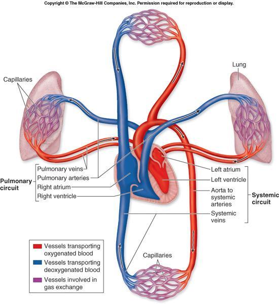
7 Pulmonary and Systemic Circuits The pulmonary circuit consists of the chambers on the right side of the heart (right atrium and ventricle) as well as the pulmonary arteries and veins. conveys blood to the lungs via pulmonary arteries to reduce carbon dioxide and replenish oxygen levels in the blood Blood returns to the heart in pulmonary veins 22-7
8 Pulmonary and Systemic Circuits Blood returns to the left side of the heart, where it then enters the systemic circuit. The systemic circuit consists of the chambers on the left side of the heart (left atrium and ventricle), along with all the other named blood vessels. carries blood to all the peripheral organs and tissues of the body 22-8
9 Pulmonary and Systemic Circuits Oxygenated blood from the left side of the heart is pumped into the aorta the largest systemic artery in the body then into smaller systemic arteries. Gas exchange in tissues occurs from capillaries. Systemic veins then carry deoxygenated blood (high in carbon dioxide) and waste products. Most veins merge and drain into the superior and inferior venae cavae drain blood into the right atrium. There, the blood enters the pulmonary circuit, and the cycle repeats. 22-9

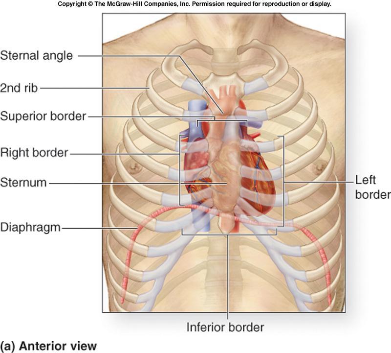
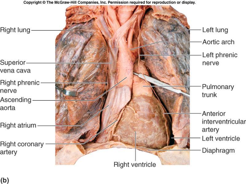
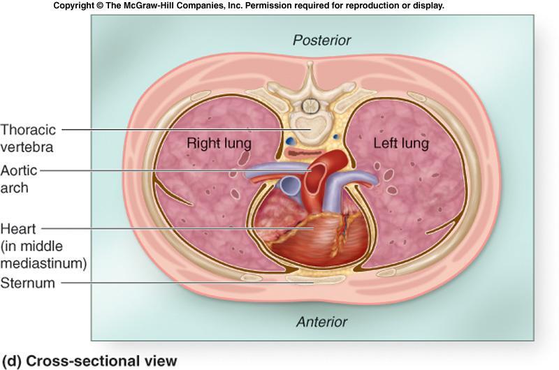
10 Anatomy of the Heart Relatively small, conical organ approximately the size of a person s clenched fist. it weighs about 250 to 350 grams Located left of the body midline posterior to the sternum in the middle mediastinum. Rotated such that its right side or border (right atrium and ventricle) is located more anteriorly, while its left side or border (left atrium and ventricle) is located more posteriorly.


11 Mediastinum is abroad central partition that separates the two laterally placed plural cavities. It extends: 1-from the sternum to the bodies of the vertebrae. 2-from Superior thoracic apparatus to the diaphragm. It contains the thymus gland, the pericardial sac, the heart, the trachea, major arteries and veins. 11

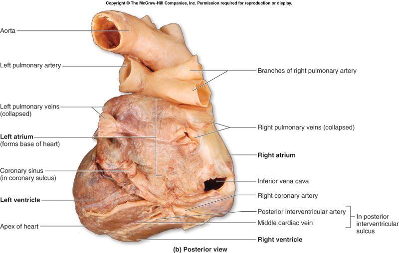
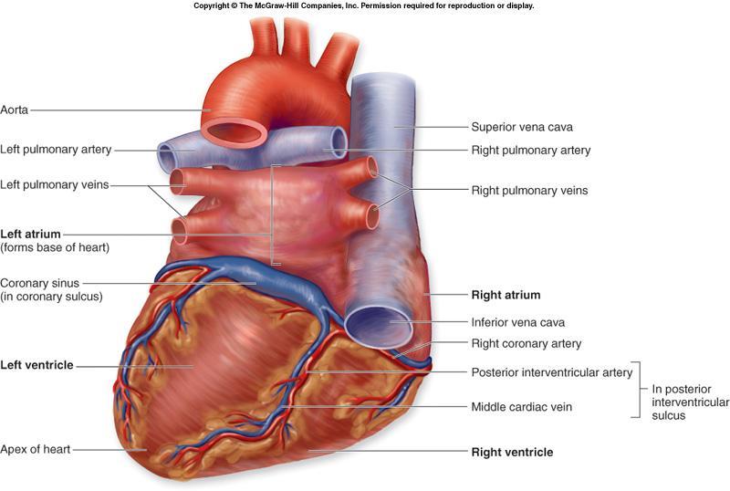
12 Anatomy of the Heart The posterosuperior surface of the heart, formed primarily by the left atrium, is called the base. The pulmonary veins that enter the left atrium border this base. The inferior, conical end is called the apex. It projects slightly anteroinferiorly toward the left side of the body
13 13
14 14
15 15
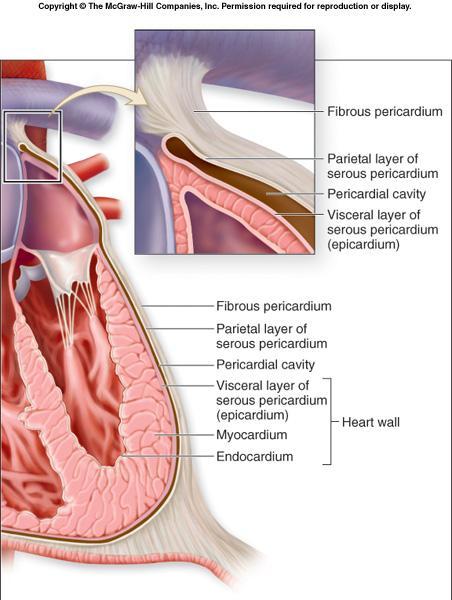
16 Pericardium Fibrous, serous sac Contains the heart In the mediastinum Held in place by connective tissue The external wall of the great vessels superior to the heart diaphragm inferior. Restricts heart movements Prevents the heart from overfilling with blood
17 17
18 Pericardium Outer portion tough, dense connective tissue called the fibrous pericardium. attached to both the sternum and the diaphragm Inner portion thin, double-layered serous membrane called the serous pericardium. parietal layer visceral layer 22-18
19 19
20 20
21 21
22 Heart Wall Structure Three distinctive layers: external epicardium middle myocardium internal endocardium Epicardium outermost heart layer also known as the visceral layer of serous pericardium. Simple squamous epithelium underlined by fat As we age, more fat is deposited in the epicardium this layer becomes thicker and more fatty
23 23

24 Heart Wall Structure Myocardium middle layer of the heart wall composed chiefly of cardiac muscle tissue. thickest of the three heart wall layers. lies deep to the epicardium and superficial to the endocardium Endocardium covers internal surface of the heart and the external surfaces of the heart valves thin endothelium areolar CT under the endothelium 22-24
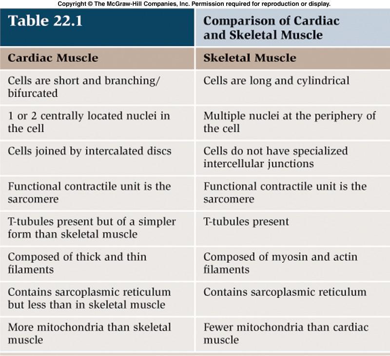
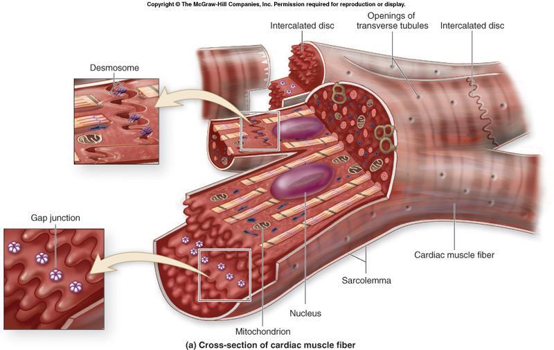
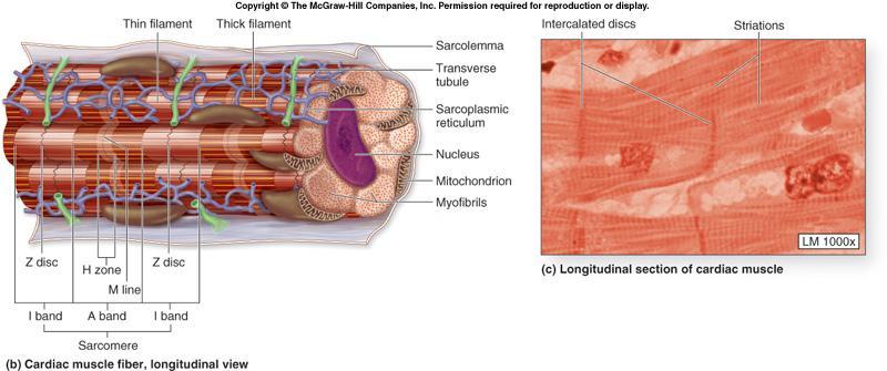
25 Cardiac Muscle Tissue Fiber Characteristics short, branched fibers one or two central nuclei numerous mitochondria for ATP supply. striated, with extensive capillary networks 22-25
26 22-26
27 Cardiac Muscle Tissue Fibers contract as a single unit Intercalated discs Specialized cell cell contacts Contain gap junctions contain desmosomes Muscle impulses are distributed immediately and simultaneously throughout all fibers either of the atria or of the ventricles
28 28
29 29
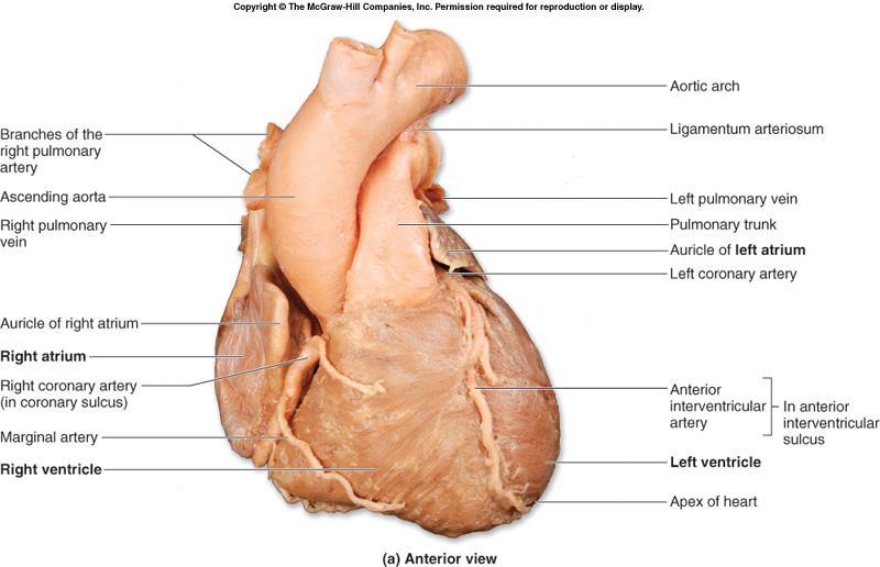
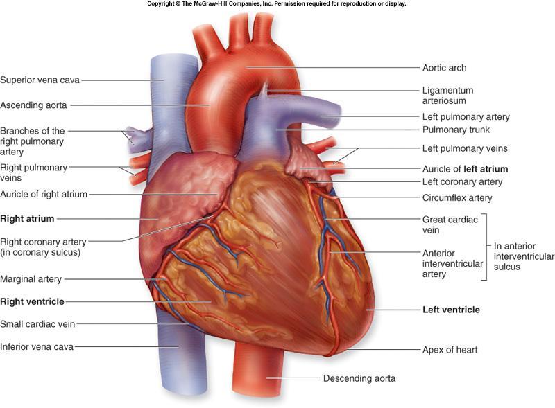
30 External Anatomy of the Heart Chambers: four hollow chambers: Atria two smaller atria two larger ventricles. thin-walled, located superiorly. anterior part of each atrium is a wrinkled, flaplike extension called an auricle Atria receive blood through both circulatory circuits. right atrium receives blood from the systemic circuit left atrium receives blood from the pulmonary circuit 22-30
31 External Anatomy of the Heart Blood that enters an atrium is passed to the ventricle on the same side of the heart. Ventricles the inferior chambers. Two large arteries, the pulmonary trunk and the aorta exit the heart at the basal surface. The pulmonary trunk carries blood from the right ventricle into the pulmonary circuit. The aorta conducts blood from the left ventricle into the systemic circuit 22-31


32 External Anatomy of the Heart Atria are separated from the ventricles externally by coronary sulcus (or atrioventricular sulcus) extends around the circumference of the heart

33 External Anatomy of the Heart On both the anterior and posterior surfaces of the heart, the anterior interventricular sulcus and the posterior interventricular sulcus are located between the left and right ventricles. These sulci extend inferiorly from the coronary sulcus toward the heart apex
34 34
35 35
36 36
37 37

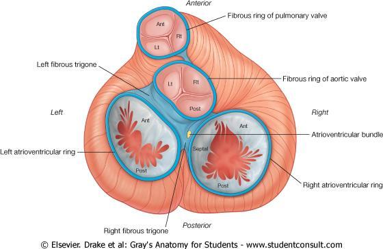
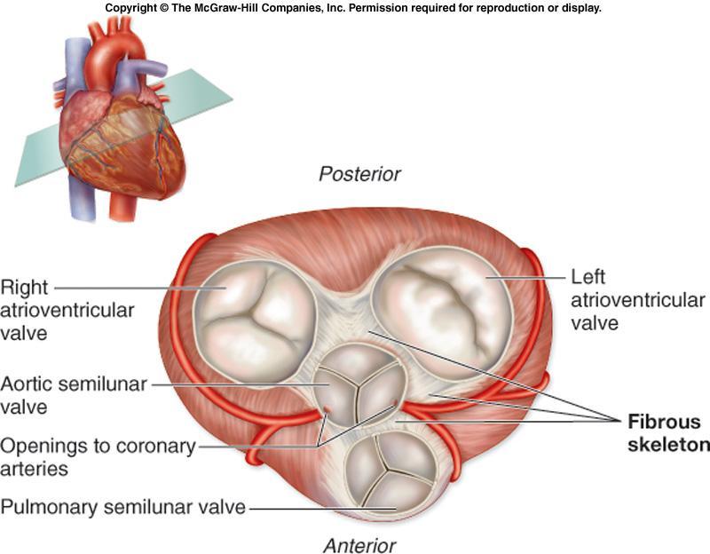
38 The Fibrous Skeleton of the Heart 38
39 39
40 Functions of the Fibrous Skeleton of the Heart Located between the atria and the ventricles Formed from dense irregular connective tissue. separates the atria and ventricles anchors heart valves by forming supportive rings at their attachment points provides electrical insulation between atria and ventricles ensures that muscle impulses are not spread randomly throughout the heart prevents all of the heart chambers from beating at the same time Provides a rigid framework for the attachment of cardiac muscle tissue
41 41
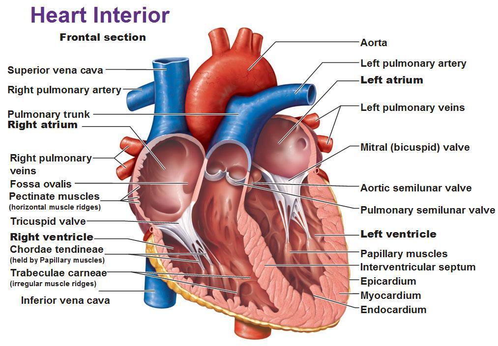
42 Internal Anatomy of the Heart There are four heart chambers: right atrium right ventricle left atrium left ventricle Each plays a role in the continuous process of blood circulation. Valves permit the passage of blood in one direction and prevent its backflow
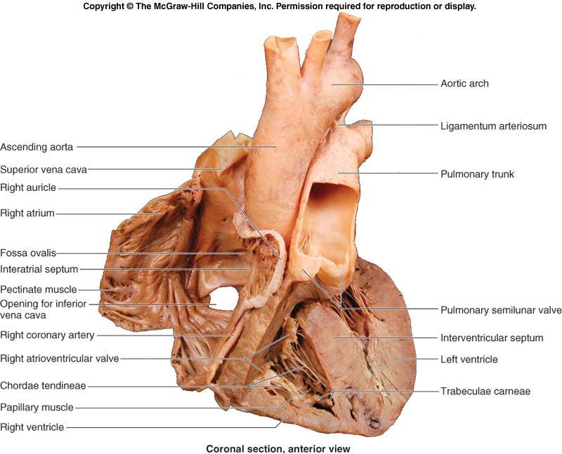
43 Right Atrium Receives venous blood from the systemic circuit from the heart muscle itself. Three major vessels empty into the right atrium: superior vena cava (SVC) drains blood from the head, upper limbs, and superior regions of the trunk inferior vena cava (IVC) drains blood from the lower limbs and trunk coronary sinus drains blood from the heart wall The interatrial septum forms a wall between the right and left atria
44 Right Atrioventricular (AV) Valve Separates the right atrium from the right ventricle. Also called the tricuspid valve. has three triangular flaps Venous blood flows from the right atrium, through the valve into the right ventricle. Is forced closed when the right ventricle begins to contract preventing blood backflow into the right atrium 22-44
45 Aortic valve 45
46 Right Ventricle Receives deoxygenated venous blood from the right atrium. An interventricular septum forms a wall between the right and left ventricles. Papillary muscles on the internal wall surface cone-shaped, muscular projections anchor chordae tendineae attach to the cusp of the right AV valve and prevent everting and flipping into the atrium when contracting 22-46
47 Pulmonary Trunk At its superior end it narrows into a smooth-walled, conical region called the conus arteriosus. The pulmonary semilunar valve marks the end of the right ventricle and the entrance into the pulmonary trunk. Pulmonary trunk divides shortly into right and left pulmonary arteries. carry deoxygenated blood to the lungs 22-47
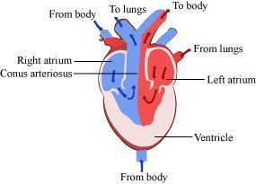
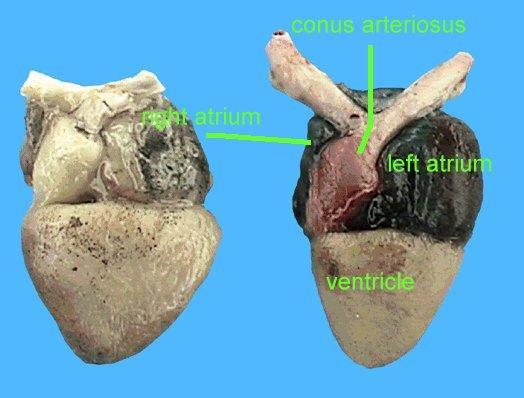
48 conus arteriosus 22-48
49 Semilunar Valves Located within the walls of both ventricles immediately before the connection of the ventricle to the pulmonary trunk and aorta. Composed of three thin, pocketlike semilunar cusps. As blood is pumped into the arterial trunks, it pushes against the cusps forcing the valves open. When ventricular contraction ceases blood is prevented from flowing back into the ventricles. causes the cusps to inflate and meet at the artery center, effectively blocking blood backflow 22-49
50 50
51 51
52 Left Atrium Once gas exchange occurs in the lungs, the oxygenated blood travels through the pulmonary veins to the left atrium. Smooth posterior wall of the left atrium contains openings for approximately four pulmonary veins. two left pulmonary veins two right pulmonary veins 22-52

53 Left Atrioventricular (AV) Valve Separates the left atrium from the left ventricle. Also called the bicuspid valve or the mitral valve. Left AV valve has chordae tendineae similar to those of the right AV valve. Oxygenated blood flows from the left atrium into the left ventricle. Is forced closed when the left ventricle begins to contract prevents blood backflow into the left atrium 22-53
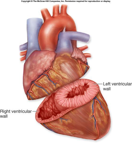
54 Left Ventricle Largest of the four heart chambers. Wall is typically three times thicker than the right ventricular wall. Requires thick walls in order to generate enough pressure to force the oxygenated blood from the lungs into the aorta and then through the entire systemic circuit. right ventricle only has to pump blood to the nearby lungs 22-54
55 Left Ventricle Two large papillary muscles attach to the chordae tendineae that help support the left AV valve. At the superior end of the ventricular cavity, the aortic semilunar valve marks the end of the left ventricle and the entrance into the aorta
56 22-56
57 57
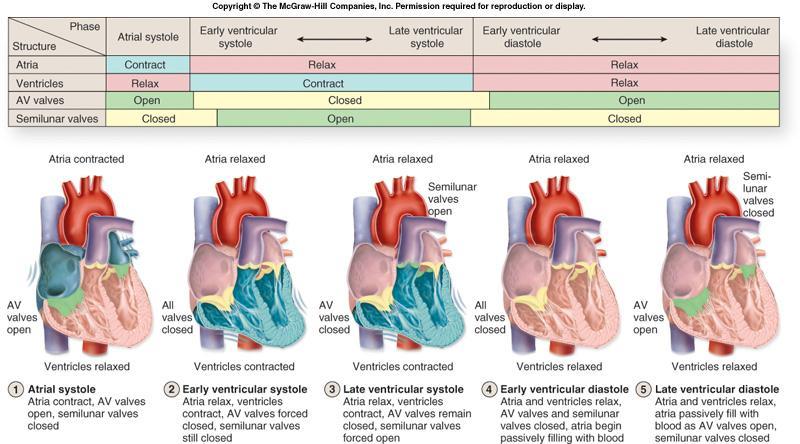
58 Cardiac Cycle The inclusive period of time from the start of one heartbeat to the initiation of the next. All chambers within the heart experience alternate periods of contraction and relaxation. Contraction of a heart chamber is called systole. forces blood into another chamber (from atrium to ventricle) forces blood into a blood vessel (from a ventricle into the attached large artery) Relaxation phase of a heart chamber is termed diastole. myocardium of each chamber relaxes between contraction phases and the chamber fills with blood
59 59
60 Conduction System of the Heart Exhibits autorhythmicity the heart itself (not external nerves) is responsible for initiating the heartbeat. Certain cardiac muscle fibers are specialized to conduct muscle impulses to the contractile muscle cells of the myocardium. Specialized cells are part of the heart s conduction system

61 The Conductive System of the Heart
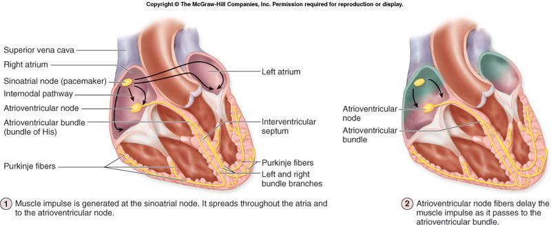
62 CONDUCTION SYSTEM Sequence of excitation 1. sinoatrial (SA) node - spreads to both atria 2. atrioventricular (AV) node 3. atrioventricular (AV) bundle (bundle of His) 4. right & left bundle branches in the interventricular septum 5. Purkinje fibers conduction myofibers
63 Conduction System of the Heart Sinoatrial (SA) Node Heartbeat is initiated by the cardiac muscle fibers of the sinoatrial (SA) node. located in the posterior wall of the right atrium, adjacent to the entrance of the superior vena cava Act as the pacemaker. rhythmic center that establishes the pace for cardiac activity Initiates impulses times per minute
64 Conduction System of the Heart Atrioventricular (AV) Node Impulse travels to both atria, stimulating atrial systole. And via an internodal conduction pathway through an opening in the fibrous skeleton to the atrioventricular (AV) node. located in the floor of the right atrium between the right AV valve and the coronary sinus 22-64
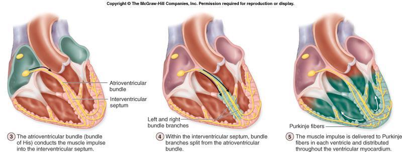
65 Conduction System of the Heart Atrioventricular (AV) Bundle Cardiac impulse then travels from the AV node to the atrioventricular (AV) bundle (bundle of His). extends into the interventricular septum and then divides into one right and two left bundle branches. Conduct the impulse to conduction fibers called Purkinje fibers in the heart apex. Purkinje fibers are larger than other cardiac muscle fibers. Muscle impulse conduction along the Purkinje fibers is extremely rapid. The impulse spreads immediately throughout the ventricular myocardium
66 66
67 67
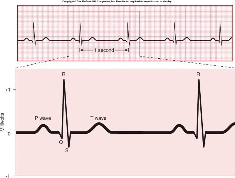
68 The Electrocardiogram 68
 ECG AV node")
69 Figure SA node (pacemaker) ECG AV node Bundle branches Heart apex Purkinje fibers

70 Heart Sounds 70
71 Innervation of the Heart Innervated by the autonomic nervous system. Consists of both sympathetic and parasympathetic components. referred to as the coronary plexus Autonomic innervation by autonomic centers in the hindbrain doesn t initiate heartbeat, but it can increase or decrease the heartbeat. Rich innervation to SA and AV nodes, but also to myocardial cells


72 Sympathetic Innervation Sympathetic innervation increases the rate and the force of heart contractions arises from T1-T5 segments of spinal cord enter sympathetic trunk (sympathetic chain), ascend and pass through ganglia travel through heart via cardiac nerves 22-72

73 Innerva tion 22-73
")
74 Parasympathetic Innervation Parasympathetic innervation decreases heart rate, but tends to have no effect on the force of contractions, except in special circumstances comes off of the medulla oblongata via right and left vagus nerves (CN X) 22-74
75 Coronary Circulation Left and right coronary arteries travel in the coronary sulcus (atrioventricular groove) of the heart to supply the heart wall. the only branches of the ascending aorta Located immediately superior to the aortic semilunar valve. The right coronary artery typically branches into the marginal artery supplies the right border of the heart posterior interventricular artery supplies both the left and right ventricles 22-75
76 Coronary Circulation Left coronary artery typically branches into the anterior interventricular artery. also called the left anterior descending artery supplies the anterior surface of both ventricles and most of the interventricular septum Circumflex artery. supplies the left atrium and ventricle Arterial pattern can vary greatly among individuals
77 77
78 78
2. right heart = pulmonary pump takes blood to lungs to pick up oxygen and get rid of carbon dioxide
 A. location in thorax, in inferior mediastinum posterior to sternum medial to lungs superior to diaphragm anterior to vertebrae orientation - oblique apex points down and to the left 2/3 of mass on left
The Heart. The Heart A muscular double pump. The Pulmonary and Systemic Circuits
 C H A P T E R 19 The Heart The Heart A muscular double pump circuit takes blood to and from the lungs Systemic circuit vessels transport blood to and from body tissues Atria receive blood from the pulmonary
Chapter 20 (1) The Heart
 The Heart") Chapter 20 (1) The Heart Learning Objectives Describe the location and structure of the heart Describe the path of a drop of blood from the superior vena cava or inferior vena cava through the heart out
Chapter 20 (1) The Heart Learning Objectives Describe the location and structure of the heart Describe the path of a drop of blood from the superior vena cava or inferior vena cava through the heart out
Chapter 14. The Cardiovascular System
 Chapter 14 The Cardiovascular System Introduction Cardiovascular system - heart, blood and blood vessels Cardiac muscle makes up bulk of heart provides force to pump blood Function - transports blood 2
Ch 19: Cardiovascular System - The Heart -
 Ch 19: Cardiovascular System - The Heart - Give a detailed description of the superficial and internal anatomy of the heart, including the pericardium, the myocardium, and the cardiac muscle. Trace the
The Heart. Size, Form, and Location of the Heart. 1. Blunt, rounded point; most inferior part of the heart.
 12 The Heart FOCUS: The heart is composed of cardiac muscle cells, which are elongated, branching cells that appear striated. Cardiac muscle cells behave as a single electrical unit, and the highly coordinated
the Cardiovascular System I
 the Cardiovascular System I By: Dr. Nabil A Khouri MD, MsC, Ph.D MEDIASTINUM 1. Superior Mediastinum 2. inferior Mediastinum Anterior mediastinum. Middle mediastinum. Posterior mediastinum Anatomy of
Anatomy of the Heart. Figure 20 2c
 Anatomy of the Heart Figure 20 2c Pericardium & Myocardium Remember, the heart sits in it s own cavity, known as the mediastinum. The heart is surrounded by the Pericardium, a double lining of the pericardial
CV Anatomy Quiz. Dr Ella Kim Dr Pip Green
 CV Anatomy Quiz Dr Ella Kim Dr Pip Green Q1 The location of the heart is correctly described as A) lateral to the lungs. B) medial to the sternum. C) superior to the diaphragm. D) posterior to the spinal
THE CARDIOVASCULAR SYSTEM. Part 1
 THE CARDIOVASCULAR SYSTEM Part 1 CARDIOVASCULAR SYSTEM Blood Heart Blood vessels What is the function of this system? What other systems does it affect? CARDIOVASCULAR SYSTEM Functions Transport gases,
The HEART. What is it???? Pericardium. Heart Facts. This muscle never stops working It works when you are asleep
 This muscle never stops working It works when you are asleep The HEART It works when you eat It really works when you exercise. What is it???? Located between the lungs in the mid thoracic region Apex
Chapter 18 - Heart. I. Heart Anatomy: size of your fist; located in mediastinum (medial cavity)
") Chapter 18 - Heart I. Heart Anatomy: size of your fist; located in mediastinum (medial cavity) A. Coverings: heart enclosed in double walled sac called the pericardium 1. Fibrous pericardium: dense connective
Chapter 18 - Heart I. Heart Anatomy: size of your fist; located in mediastinum (medial cavity) A. Coverings: heart enclosed in double walled sac called the pericardium 1. Fibrous pericardium: dense connective
THE HEART. A. The Pericardium - a double sac of serous membrane surrounding the heart
 THE HEART I. Size and Location: A. Fist-size weighing less than a pound (250 to 350 grams). B. Located in the mediastinum between the 2 nd rib and the 5 th intercostal space. 1. Tipped to the left, resting
THE HEART OBJECTIVES: LOCATION OF THE HEART IN THE THORACIC CAVITY CARDIOVASCULAR SYSTEM
 BIOLOGY II CARDIOVASCULAR SYSTEM ACTIVITY #3 NAME DATE HOUR THE HEART OBJECTIVES: Describe the anatomy of the heart and identify and give the functions of all parts. (pp. 356 363) Trace the flow of blood
The Heart. Happy Friday! #takeoutyournotes #testnotgradedyet
 The Heart Happy Friday! #takeoutyournotes #testnotgradedyet Introduction Cardiovascular system distributes blood Pump (heart) Distribution areas (capillaries) Heart has 4 compartments 2 receive blood (atria)
The Cardiovascular System (Heart)
") The Cardiovascular System The Cardiovascular System (Heart) A closed system of the heart and blood vessels The heart pumps blood Blood vessels allow blood to circulate to all parts of the body The function
The Cardiovascular System The Cardiovascular System (Heart) A closed system of the heart and blood vessels The heart pumps blood Blood vessels allow blood to circulate to all parts of the body The function
Chapter 20: Cardiovascular System: The Heart
 Chapter 20: Cardiovascular System: The Heart I. Functions of the Heart A. List and describe the four functions of the heart: 1. 2. 3. 4. II. Size, Shape, and Location of the Heart A. Size and Shape 1.
Anatomy of the Heart
 Biology 212: Anatomy and Physiology II Anatomy of the Heart References: Saladin, KS: Anatomy and Physiology, The Unity of Form and Function 8 th (2018). Required reading before beginning this lab: Chapter
Cardiovascular System
 Cardiovascular System The Heart Cardiovascular System The Heart Overview What does the heart do? By timed muscular contractions creates pressure gradients blood moves then from high pressure to low pressure
- what other structures, besides the heart, does the mediastinum contain?
 Basic A & P II Dr. L. Bacha Chapter Outline (Martini & Nath 2010) An Introduction to the Cardiovascular System - read the paragraphs under this heading on page 580 The Heart is a Four Chambered Organ describe
The Cardiovascular System
 The Cardiovascular System The Manila Times College of Subic Prepared by: Stevens B. Badar, RN, MANc THE HEART Anatomy of the Heart Location and Size approx. the size of a person s fist, hollow and cone-shaped,
The Cardiovascular System
 11 PART A The Cardiovascular System PowerPoint Lecture Slide Presentation by Jerry L. Cook, Sam Houston University ESSENTIALS OF HUMAN ANATOMY & PHYSIOLOGY EIGHTH EDITION ELAINE N. MARIEB The Cardiovascular
Heart Anatomy. 7/5/02 Stephen G Davenport 1
 Heart Anatomy Copyright 1999, Stephen G. Davenport, No part of this publication may be reproduced, stored in a retrieval system, or transmitted, in any form without prior written permission. 7/5/02 Stephen
11/10/2014. Muscular pump Two atria Two ventricles. In mediastinum of thoracic cavity 2/3 of heart's mass lies left of midline of sternum
 It beats over 100,000 times a day to pump over 1,800 gallons of blood per day through over 60,000 miles of blood vessels. During the average lifetime, the heart pumps nearly 3 billion times, delivering
LECTURE 5. Anatomy of the heart
 LECTURE 5. Anatomy of the heart Main components of the CVS: Heart Blood circulatory system arterial compartment haemomicrocirculatory (=microvascular) compartment venous compartment Lymphatic circulatory
Cardiovascular System
 Cardiovascular System Purpose Transport oxygen and nutrients Take waste products away from tissues & organs Things we learned Blood pressure: the force of blood pushing against the walls of blood vessels
The Cardiovascular System
 Essentials of Human Anatomy & Physiology Elaine N. Marieb Seventh Edition Chapter 11 The Cardiovascular System Slides 11.1 11.19 Lecture Slides in PowerPoint by Jerry L. Cook The Cardiovascular System
Figure ) The specific chamber of the heart that is indicated by letter A is called the. Diff: 1 Page Ref: 364
 The specific chamber of the heart that is indicated by letter A is called the. Diff: 1 Page Ref: 364") Essentials of Anatomy and Physiology, 9e (Marieb) Chapter 11 The Cardiovascular System Short Answer Figure 11.1 Using Figure 11.1, identify the following: 1) The Purkinje fibers are indicated by label.
Essentials of Anatomy and Physiology, 9e (Marieb) Chapter 11 The Cardiovascular System Short Answer Figure 11.1 Using Figure 11.1, identify the following: 1) The Purkinje fibers are indicated by label.
This lab activity is aligned with Visible Body s A&P app. Learn more at visiblebody.com/professors
 1 This lab activity is aligned with Visible Body s A&P app. Learn more at visiblebody.com/professors 2 PRE-LAB EXERCISES: A. Watch the video 29.1 Heart Overview and make the following observations: 1.
human anatomy 2016 lecture thirteen Dr meethak ali ahmed neurosurgeon
 Heart The heart is a hollow muscular organ that is somewhat pyramid shaped and lies within the pericardium in the mediastinum. It is connected at its base to the great blood vessels but otherwise lies
The Cardiovascular System. Chapter 15. Cardiovascular System FYI. Cardiology Closed systemof the heart & blood vessels. Functions
 Chapter 15 Cardiovascular System FYI The heart pumps 7,000 liters (4000 gallons) of blood through the body each day The heart contracts 2.5 billion times in an avg. lifetime The heart & all blood vessels
Lab Activity 23. Cardiac Anatomy. Portland Community College BI 232
 Lab Activity 23 Cardiac Anatomy Portland Community College BI 232 Cardiac Muscle Histology Branching cells Intercalated disc: contains many gap junctions connecting the adjacent cell cytoplasm, creates
Cardiovascular System
 Cardiovascular System I. Structure of the Heart A. Average adult heart is 14 cm long and 9 cm wide. B. Lies in the mediastinum. C. Enclosed in the pericardium. 1. Fibrous pericardium- Outer, tough connective
CIRCULATORY SYSTEM BLOOD VESSELS
 Name: Block: CIRCULATORY SYSTEM Multicellular organisms (above the level of roundworms) rely on a circulatory system to bring nutrients to, and take wastes away from, cells. In higher organisms such as
The Cardiovascular System
 Essentials of Human Anatomy & Physiology Elaine N. Marieb Slides 11.1 11.19 Seventh Edition Chapter 11 The Cardiovascular System Functions of the Cardiovascular system Function of the heart: to pump blood
LAB 12-1 HEART DISSECTION GROSS ANATOMY OF THE HEART
 LAB 12-1 HEART DISSECTION GROSS ANATOMY OF THE HEART Because mammals are warm-blooded and generally very active animals, they require high metabolic rates. One major requirement of a high metabolism is
The Heart. C h a p t e r. PowerPoint Lecture Slides prepared by Jason LaPres Lone Star College - North Harris
 C h a p t e r 20 The Heart PowerPoint Lecture Slides prepared by Jason LaPres Lone Star College - North Harris Copyright 2009 Pearson Education, Inc., publishing as Pearson Benjamin Cummings Introduction
The Cardiovascular System: The Heart
 PowerPoint Lecture Slides prepared by Meg Flemming Austin Community College C H A P T E R 12 The Cardiovascular System: The Heart Chapter 12 Learning Outcomes 12-1 12-2 Describe the anatomy of the heart,
4. The two inferior chambers of the heart are known as the atria. the superior and inferior vena cava, which empty into the left atrium.
 Answer each statement true or false. If the statement is false, change the underlined word to make it true. 1. The heart is located approximately between the second and fifth ribs and posterior to the
Circulation. Circulation = is a process used for the transport of oxygen, carbon! dioxide, nutrients and wastes through-out the body
 Circulation Circulation = is a process used for the transport of oxygen, carbon! dioxide, nutrients and wastes through-out the body Heart = muscular organ about the size of your fist which pumps blood.
Cardiovascular Anatomy Dr. Gary Mumaugh
 Cardiovascular Anatomy Dr. Gary Mumaugh Location of Heart Approximately the size of your fist Location o Superior surface of diaphragm o Left of the midline in mediastinum o Anterior to the vertebral column,
Pearson's Comprehensive Medical Assisting Administrative and Clinical Competencies
 Pearson's Comprehensive Medical Assisting Administrative and Clinical Competencies THIRD EDITION CHAPTER 27 The Cardiovascular System Lesson 1: Overview of the Cardiovascular System Lesson Objectives Upon
The Cardiovascular System
 The Cardiovascular System The Cardiovascular System A closed system of the heart and blood vessels The heart pumps blood Blood vessels allow blood to circulate to all parts of the body The function of
The cardiovascular system is composed of the heart and blood vessels that carry blood to and from the body s organs. There are 2 major circuits:
 1 The cardiovascular system is composed of the heart and blood vessels that carry blood to and from the body s organs. There are 2 major circuits: pulmonary and systemic. The pulmonary goes out to the
Introduction to Anatomy. Dr. Maher Hadidi. Bayan Yanes. April/9 th /2013
 Introduction to Anatomy Dr. Maher Hadidi Bayan Yanes 27 April/9 th /2013 KEY POINTS: 1) Right side of the heart 2) Papillary muscles 3) Left side of the heart 4) Comparison between right and left sides
Read Me. covering the Heart Anatomy. Labs. textbook. use. car: you
 Heart Anatomy Lab Pre-Lab Exercises Read Me These exercises should be done before coming to lab, after watching the videos covering the Heart Anatomy Labs. Answer the questions in this guide using the
10. Thick deposits of lipids on the walls of blood vessels, called, can lead to serious circulatory issues. A. aneurysm B. atherosclerosis C.
 Heart Student: 1. carry blood away from the heart. A. Arteries B. Veins C. Capillaries 2. What is the leading cause of heart attack and stroke in North America? A. alcohol B. smoking C. arteriosclerosis
10/23/2017. Muscular pump Two atria Two ventricles. In mediastinum of thoracic cavity 2/3 of heart's mass lies left of midline of sternum
 It beats over 100,000 times a day to pump over 1,800 gallons of blood per day through over 60,000 miles of blood vessels. During the average lifetime, the heart pumps nearly 3 billion times, delivering
Ch.15 Cardiovascular System Pgs {15-12} {15-13}
 Ch.15 Cardiovascular System Pgs {15-12} {15-13} E. Skeleton of the Heart 1. The skeleton of the heart is composed of rings of dense connective tissue and other masses of connective tissue in the interventricular
The Cardiovascular System: The Heart: Part A
 PowerPoint Lecture Slides prepared by Janice Meeking, Mount Royal College CHAPTER 18 The Cardiovascular System: The Heart: Part A Heart Anatomy Approximately the size of a fist Location In the mediastinum
Approximately the size of your fist Location. Pericardial physiology
 Heart Anatomy Approximately the size of your fist Location Superior surface of diaphragm Left of the midline Anterior to the vertebral column, posterior to the sternum Wednesday, March 28, 2012 Muscle
1. What kind of blood is found in the rt. atrium? (oxygenated or deoxygenated)
") Carl Christennsen, PhD Chap. 19, 20, & 21 - Circulatory System Bio. 2304 Human Anatomy HEART 1. What kind of blood is found in the rt. atrium? (oxygenated or deoxygenated) Where does this blood come from?
Carl Christennsen, PhD Chap. 19, 20, & 21 - Circulatory System Bio. 2304 Human Anatomy HEART 1. What kind of blood is found in the rt. atrium? (oxygenated or deoxygenated) Where does this blood come from?
Cardiovascular System. Heart Anatomy
 Cardiovascular System Heart Anatomy 1 The Heart Location & general description: Atria vs. ventricles Pulmonary vs. systemic circulation Coverings Walls The heart is found in the mediastinum, the medial
The Heart and Cardiovascular System
 The Heart and Cardiovascular System What you will learn The location of the heart 3 layers and covering of the heart Explain the function of the heart as 2 separate pumps Identify the 4 chambers of the
BIOLOGY 2060 LECTURE NOTES ANATOMY & PHYSIOLOGY II (A. IMHOLTZ) HEART P1 OF 5
 HEART P1 OF 5") BIOLOGY 2060 LECTURE NOTES ANATOMY & PHYSIOLOGY II (A. IMHOLTZ) HEART P1 OF 5 1. Heart Functions a. Generates pressure that propels blood thru blood vessels. (Tissue perfusion.) b. Separates oxygenated
BIOLOGY 2060 LECTURE NOTES ANATOMY & PHYSIOLOGY II (A. IMHOLTZ) HEART P1 OF 5 1. Heart Functions a. Generates pressure that propels blood thru blood vessels. (Tissue perfusion.) b. Separates oxygenated
The Cardiovascular System
 PowerPoint Lecture Slide Presentation by Patty Bostwick-Taylor, Florence-Darlington Technical College The Cardiovascular System 11 PART A The Cardiovascular System A closed system of the heart and blood
Heart Dissection. 5. Locate the tip of the heart or the apex. Only the left ventricle extends all the way to the apex.
 Heart Dissection Page 1 of 6 Background: The heart is a four-chambered, hollow organ composed primarily of cardiac muscle tissue. It is located in the center of the chest in between the lungs. It is the
Collin County Community College. ! BIOL Anatomy & Physiology! WEEK 5. The Heart
 Collin County Community College! BIOL. 2402 Anatomy & Physiology! WEEK 5 The Heart 1 (1578-1657) A groundbreaking work in the history of medicine, English physician William Harvey s Anatomical Essay on
Approximately the size of your fist Location Superior surface of diaphragm Left of the midline in mediastinum Anterior to the vertebral column,
 Dr. Gary Mumaugh Approximately the size of your fist Location Superior surface of diaphragm Left of the midline in mediastinum Anterior to the vertebral column, posterior to the sternum Posteriorly the
BIOL 4350 Cardiovascular Physiology Dr. Hamilton. Using the figure above, match the following: 1. Purkinje fibers. 2. SA node. 3. AV node.
 BIOL 4350 Cardiovascular Physiology Dr. Hamilton Using the figure above, match the following: 1. Purkinje fibers. 2. SA node. 3. AV node. 1 Using the figure above, match the following: 4. Atrial depolarization.
Part 1. Copyright 2011 Pearson Education, Inc. Figure Copyright 2011 Pearson Education, Inc.
 PowerPoint Lecture Slides prepared by Leslie Hendon University of Alabama, Birmingham C H A P T E R The Heart 19 Part 1 The Heart A muscular double pump circuit vessels transport blood to and from the
CARDIOVASCULAR SYSTEM
 CARDIOVASCULAR SYSTEM Overview Heart and Vessels 2 Major Divisions Pulmonary Circuit Systemic Circuit Closed and Continuous Loop Location Aorta Superior vena cava Right lung Pulmonary trunk Base of heart
Human Anatomy and Physiology Chapter 19 Worksheet 1- The Heart
 Human Anatomy and Physiology Chapter 19 Worksheet 1- The Heart Name Date Period 1. The "double pump" function of the heart includes the right side, which serves as the circuit pump, while the left side
Chapter 13. Cardiovascular System
 Chapter 13 Cardiovascular System 1 Introduction A. The cardiovascular system consists of the heart and vessels (arteries, capillaries and veins.) B. A functional cardiovascular system is vital for supplying
Major Function of the Cardiovascular System. Transportation. Structures of the Cardiovascular System. Heart - muscular pump
 Structures of the Cardiovascular System Heart - muscular pump Blood vessels - network of tubes Blood - liquid transport vehicle brachiocephalic trunk superior vena cava right pulmonary arteries right pulmonary
Read Chapters 21 & 22, McKinley et al
 ACTIVITY 9: BLOOD AND HEART OBJECTIVES: 1) How to get ready: Read Chapters 21 & 22, McKinley et al., Human Anatomy, 5e. All text references are for this textbook. Read dissection instructions BEFORE YOU
STRUCTURES OF THE CARDIOVASCULAR SYSTEM
 STRUCTURES OF THE CARDIOVASCULAR SYSTEM CARDIOVASCULAR SYSTEM Also called the circulatory system Consists of the heart, arteries, veins, and capillaries Main function is to pump/circulate oxygenated blood
Cardiovascular System. I. Structures of the heart A. : Pericardium sack that surrounds the heart
 Cardiovascular System I. Structures of the heart A. : Pericardium sack that surrounds the heart 1. : Pericardial Cavity serous fluid filled space between the heart and the pericardium B. Heart Wall 1.
Principles of Anatomy and Physiology
 Principles of Anatomy and Physiology 14 th Edition CHAPTER 20 The Cardiovascular System: The Heart Introduction The purpose of the chapter is to: 1. Learn about the components of the cardiovascular system
Cardiovascular System Module 3: Heart Anatomy *
 OpenStax-CNX module: m49683 1 Cardiovascular System Module 3: Heart Anatomy * Donna Browne Based on Heart Anatomy by OpenStax This work is produced by OpenStax-CNX and licensed under the Creative Commons
Cardiovascular System Notes: Physiology of the Heart
 Cardiovascular System Notes: Physiology of the Heart Interesting Heart Fact Capillaries are so small it takes ten of them to equal the thickness of a human hair. Review What are the 3 parts of the cardiovascular
INTRODUCTORY REMARKS:
 INTRODUCTORY REMARKS: The circulatory system provides a way for the blood to be transported throughout the body. This provides nutrients to the cells and allows wastes to be removed. Open vs. Closed Circulatory
37 1 The Circulatory System
 H T H E E A R T 37 1 The Circulatory System The circulatory system and respiratory system work together to supply cells with the nutrients and oxygen they need to stay alive. a) The respiratory system:
The Cardiovascular System. anatom.ua 1
 The Cardiovascular System anatom.ua 1 The Closed Circulatory System Humans have a closed circulatory system, typical of all vertebrates, in which blood is confined to vessels and is distinct from the interstitial
Anatomy Review: The Heart Graphics are used with permission of A.D.A.M. Software, Inc. and Benjamin/Cummings Publishing Co.
 Anatomy Review: The Heart Graphics are used with permission of A.D.A.M. Software, Inc. and Benjamin/Cummings Publishing Co. Anatomy Views Label the diagrams of the heart below: Interactive Physiology Study
BIOLOGY 2060 LECTURE NOTES ANATOMY & PHYSIOLOGY II (A. IMHOLTZ) HEART P1 OF 7
 HEART P1 OF 7") BIOLOGY 2060 LECTURE NOTES ANATOMY & PHYSIOLOGY II (A. IMHOLTZ) HEART P1 OF 7 1. Heart a. Generates the pressure that propels blood thru blood vessels. b. Separates oxygenated and deoxygenated blood separate.
BIOLOGY 2060 LECTURE NOTES ANATOMY & PHYSIOLOGY II (A. IMHOLTZ) HEART P1 OF 7 1. Heart a. Generates the pressure that propels blood thru blood vessels. b. Separates oxygenated and deoxygenated blood separate.
ACTIVITY 9: BLOOD AND HEART BLOOD
 ACTIVITY 9: BLOOD AND HEART OBJECTIVES: 1) How to get ready: Read Chapters 21 & 22, McKinley et al., Human Anatomy, 4e. All text references are for this textbook. Read dissection instructions BEFORE YOU
Cardiovascular System
 Hole s Essentials of Human Anatomy & Physiology David Shier Jackie Butler Ricki Lewis Created by Dr. Melissa Eisenhauer Head Athletic Trainer/Assistant Professor Trevecca Nazarene University Chapter 13
Heart. Heart 2-Tunica media: middle layer (media ='middle') muscle fibers (smooth or cardiac).
 muscle fibers (smooth or cardiac).") t. innermost lumenal General Circulatory system heart and blood vessels walls have 3 layers (inside to outside) 1-Tunica interna: aka tunica intima layer--lumenal layer epithelium--endothelium simple squamous
t. innermost lumenal General Circulatory system heart and blood vessels walls have 3 layers (inside to outside) 1-Tunica interna: aka tunica intima layer--lumenal layer epithelium--endothelium simple squamous
The Cardiovascular and Lymphatic Systems Cardiovascular System Blood Vessels Blood Vessels Arteries Arteries Arteries
 CH 12 The Cardiovascular and s The Cardiovascular and s OUTLINE: Cardiovascular System Blood Vessels Blood Pressure Cardiovascular System The cardiovascular system is composed of Blood vessels This system
Health Science 20 Circulatory System Notes
 Health Science 20 Circulatory System Notes Functions of the Circulatory System The circulatory system functions mainly as the body s transport system. It transports: o Oxygen o Nutrients o Cell waste o
CIRCULATION & GAS EXCHANGE
 AP BIOLOGY ACTIVITY2.13 Text:Campbell,v.8,chapter42 NAME DATE HOUR CIRCULATION & GAS EXCHANGE 1. In general, what is the function of transport systems? 2. What method/structure do most invertebrates use
THE HEART. Unit 3: Transportation and Respiration
 THE HEART Unit 3: Transportation and Respiration The Circulatory System Also called the Cardiovascular System Circulates blood in the body Transports nutrients, oxygen, carbon dioxide, hormones, and blood
HUMAN HEART. Learn the following structures on the heart models.
 HUMAN HEART Learn the following structures on the heart models. The human heart has four chambers that consist of the right atrium, left atrium, right ventricle, and left ventricle. The atria are smaller
The Heart. PowerPoint Lecture Presentations prepared by Jason LaPres. Lone Star College North Harris Pearson Education, Inc.
 20 The Heart PowerPoint Lecture Presentations prepared by Jason LaPres Lone Star College North Harris An Introduction to the Cardiovascular System Learning Outcomes Describe the superficial anatomy of
Cardiovascular system
 BIO 301 Human Physiology Cardiovascular system The Cardiovascular System: consists of the heart plus all the blood vessels transports blood to all parts of the body in two 'circulations': pulmonary (lungs)
Middle mediastinum---- heart & pericardium. Dep. of Human Anatomy Zhou Hongying
 Middle mediastinum---- heart & pericardium Dep. of Human Anatomy Zhou Hongying eaglezhyxzy@163.com Subdivisions of the mediastinum Contents of Middle mediastinum Heart Pericardium: a serous sac enclosing
MODULE 2: CARDIOVASCULAR SYSTEM ANTOMY An Introduction to the Anatomy of the Heart and Blood vessels
 MODULE 2: CARDIOVASCULAR SYSTEM ANTOMY An Introduction to the Anatomy of the Heart and Blood vessels The cardiovascular system includes a pump (the heart) and the vessels that carry blood from the heart
The Cardiovascular System Part I: Heart Outline of class lecture After studying part I of this chapter you should be able to:
 The Cardiovascular System Part I: Heart Outline of class lecture After studying part I of this chapter you should be able to: 1. Describe the functions of the heart 2. Describe the location of the heart,
The Cardiovascular and Lymphatic Systems
 BIOLOGY OF HUMANS Concepts, Applications, and Issues Fifth Edition Judith Goodenough Betty McGuire 12 The Cardiovascular and Lymphatic Systems Lecture Presentation Anne Gasc Hawaii Pacific University and
Heart & Pericardium. December, 2015
 Heart & Pericardium December, 2015 2 Pericardium Definition Fibro-serous sac that encloses the heart and the roots of great vessels Function Restrict excessive movements of the heart as a whole Serve as
Ch. 12 The Circulatory System. The heart. The heart is a double pump. A quick note on arteries vs. veins. = the muscular pump of the CV system
 Ch. 12 The Circulatory System The heart A.k.a. the cardiovascular system Blood was discussed in Ch. 11 Focus of Ch. 12: heart and blood vessels = the muscular pump of the CV system ~ 100,000 heartbeats/day!
Chapter 14. Circulatory System Images. VT-122 Anatomy & Physiology II
 Chapter 14 Circulatory System Images VT-122 Anatomy & Physiology II The mediastinum Dog heart Dog heart Cat heart Dog heart ultrasound Can see pericardium as distinct bright line Pericardial effusion Fluid
CJ Shuster A&P2 Lab Addenum Beef Heart Dissection 1. Heart Dissection. (taken from Johnson, Weipz and Savage Lab Book)
") CJ Shuster A&P2 Lab Addenum Beef Heart Dissection 1 Heart Dissection. (taken from Johnson, Weipz and Savage Lab Book) Introduction When you have finished examining the model, you are ready to begin your
CJ Shuster A&P2 Lab Addenum Beef Heart Dissection 1 Heart Dissection. (taken from Johnson, Weipz and Savage Lab Book) Introduction When you have finished examining the model, you are ready to begin your
Large Arteries of Heart
 Cardiovascular System (Part A-2) Module 5 -Chapter 8 Overview Arteries Capillaries Veins Heart Anatomy Conduction System Blood pressure Fetal circulation Susie Turner, M.D. 1/5/13 Large Arteries of Heart
Anatomy & Physiology of Cardiovascular System. Chapter 18 & 19
 Anatomy & Physiology of Cardiovascular System Chapter 18 & 19 Objectives..cont 1. Discuss the physiological stages of cardiac muscle contraction. 2. Trace a typical ECG and label each wave or complex 3.
Chapter 4: The thoracic cavity and heart. The Heart
 Chapter 4: The thoracic cavity and heart The thoracic cavity is divided into right and left pleural cavities by a central partition, the mediastinum. The mediastinum is bounded behind by the vertebral
The Circulatory System. The Heart, Blood Vessels, Blood Types
 The Circulatory System The Heart, Blood Vessels, Blood Types The Closed Circulatory System Humans have a closed circulatory system, typical of all vertebrates, in which blood is confined to vessels and
PART ONE General Characteristics of the Cardiovascular System
 HUMAN ANATOMY & PHYSIOLOGY CARDIOVASCULAR SYSTEM Chapter 15 Notes OBJECTIVES HOLE S HA&P CHAPTER FIFTEEN 1. Discuss the functions of the organs of the cardiovascular system. 2. Distinguish between the
Biology Unit 3 The Human Heart P
 Biology 2201 Unit 3 The Human Heart P 314-321 Structure and Function of the Human Heart Structure of the Human Heart Has four Chambers (2 Atria and 2 Ventricles) Made of Cardiac Muscle Found in Chest Cavity