HEMOPTYSIS. Prof. G. Zuliani
|
|
|
- Gerald Hardy
- 5 years ago
- Views:
Transcription

1 HEMOPTYSIS Prof. G. Zuliani
2 HEMOPTYSIS Hemoptysis is the expectoration of blood, that can range from blood-streaking of sputum (Hemoptoe) to the presence of gross blood in the absence of any accompanying sputum. The term massive hemoptysis is reserved for bleeding that is potentially life-threatening; it has been defined by a number of different criteria, more often as more than 600 ml of blood over a 24 hour period.
3 HEMOPTYSIS 1000 ml of blood in the alveolar space is sufficient to cause significant hindrance to the oxygen transfer. Massive hemoptysis accounts for only 1.5% of all hemoptysis. The majority of hemoptysis originates from the bronchial arteries (about 90%)
4 VASCULAR ORIGIN OF HEMOPTYSIS Blood traversing the lungs can arrive from: pulmonary arteries bronchial arteries Virtually, the entire cardiac output courses through the low-pressure pulmonary arteries and arterioles to be oxygenated in the pulmonary capillary bed In contrast, the bronchial arteries carry only a small portion of the cardiac output, but are under much higher systemic pressure
5 VASCULAR ORIGIN OF HEMOPTYSIS Despite the quantitatively smaller contribution of the bronchial circulation to pulmonary blood flow, the bronchial arteries are generally a more important source of hemoptysis than the pulmonary circulation In addition to being perfused at a higher pressure, they also supply blood to the airways and to lesions within the airways
6 HEMOPTYSIS The total mortality rate related to hemoptysis is influenced by: 1. malignant etiologies 2. bleeding rate The coexistence of hemoptysis over 1000 ml/24hr and malignancy in the same patient brought the mortality rate to about 80%.
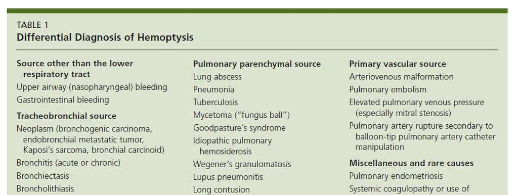
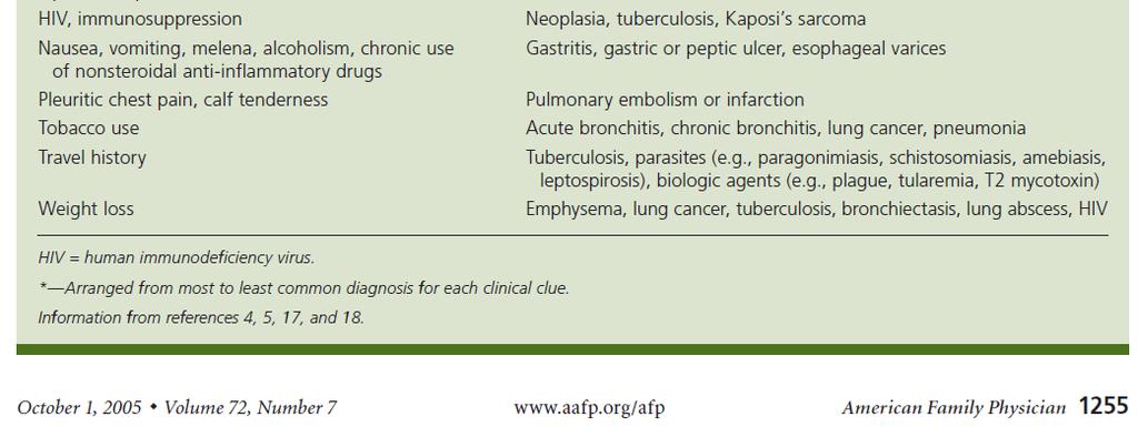
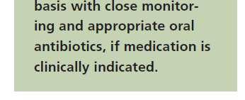
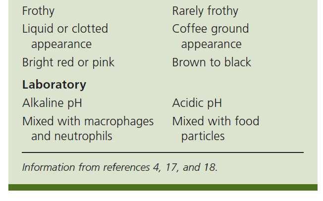
7 DIFFERENTIAL DIAGNOSIS OF HEMOPTYSIS Before assuming a lower respiratory source of the bleeding, it is always important to consider whether the blood may be coming from a non-pulmonary source, such as the upper airways or the gastrointestinal tract Alkaline ph, foaminess, or the presence of pus may sometimes suggest the lungs as the primary source of bleeding rather than the stomach.
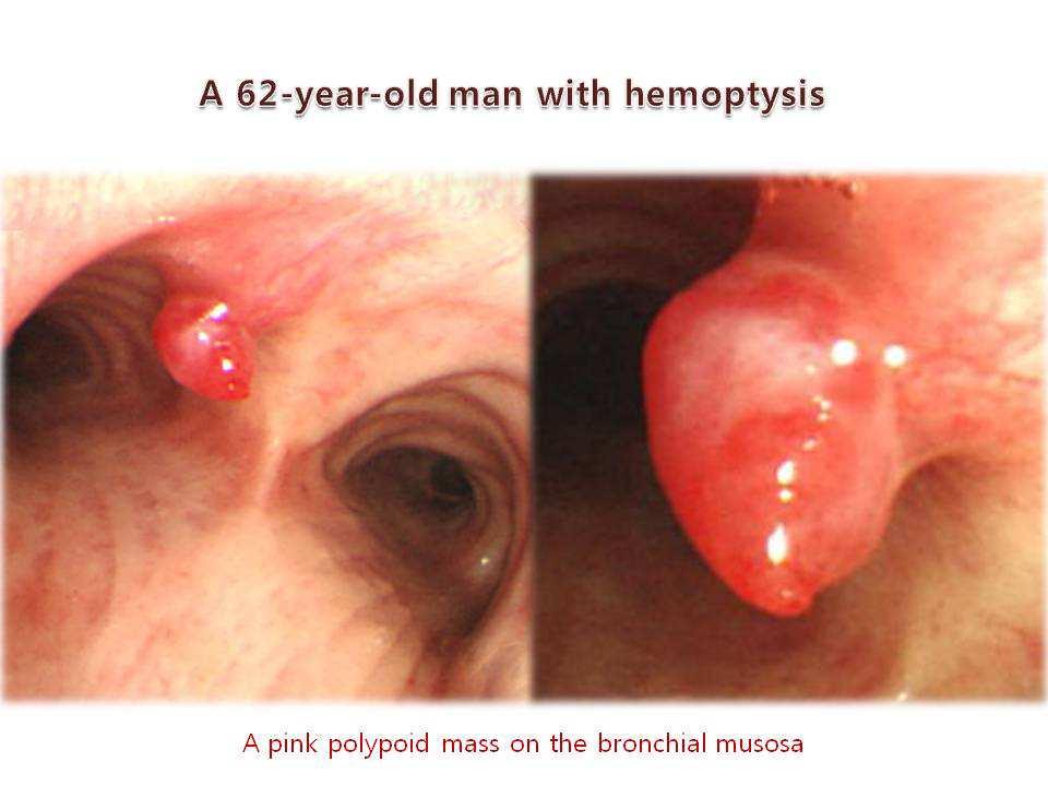
8 1. Airways diseases The most common source of hemoptysis is airways disease: Inflammatory diseases: such as acute bronchitis or bronchiectasis Neoplasms: including primary bronchogenic carcinoma, endobronchial metastatic carcinoma or bronchial carcinoid Foreign body & Airway trauma Kaposi's sarcoma: in patients with AIDS, involving the airways and/or the pulmonary parenchyma Fistula between a vessel and the tracheo-bronchial tree: fistulas between the aorta and the airway are associated with aneurysms of the thoracic aorta; are fatal if not diagnosed and surgically treated

9 1. Airways diseases - Bronchitis - Bronchiectasis - COPD - Bronchogenic carcinoma are common causes of hemoptysis.


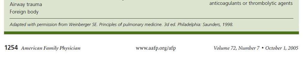
10 2. Pulmonary parenchymal diseases Infections: especially tuberculosis, pneumonia, aspergilloma, and lung abscess Hemoptysis, which can be life-threatening, complicates the course of percent of patients with an Aspergilloma Tuberculosis can cause massive hemoptysis through multiple mechanisms Active cavitary or non-cavitary lung disease can cause small or large amounts of bleeding Active disease can cause sudden rupture of a Rasmussen's aneurysm (aneurysm of the pulmonary artery that slowly expands into an adjacent cavity because of inflammatory erosion of the external vessel wall until it bursts)
11 2. Pulmonary parenchymal diseases Inflammatory or immune disorders Goodpasture's syndrome, idiopathic pulmonary hemosiderosis, lupus pneumonitis, Wegener's granulomatosis Coagulopaties thrombocytopenia or use of anticoagulants Iatrogenic: especially due to either percutaneous or transbronchial lung biopsy. Hemoptysis, which is usually minor and transient, occurs in five to 10% of percutaneous lung biopsies (but massive hemorrhage and death have also been reported)

12 2. Miscellaneous causes of pulmonary parenchymal hemorrhage Cocaine-induced pulmonary hemorrhage Hemoptysis has been described in about 6% of habitual smokers of free-base cocaine ("crack") and has been associated with diffuse alveolar hemorrhage Catamenial hemoptysis hemoptysis that in women is recurrent and coincident with menses. The cause is intrathoracic endometriosis, usually involving the pulmonary parenchyma but occasionally affecting the airways

13 3. Pulmonary vascular disorders Pulmonary embolism (infarction) Pulmonary A-V malformation, either with or without underlying Rendu-Osler-Weber syndrome Elevated pulmonary capillary pressure mitral valve stenosis significant left ventricular failure congenital heart diseases severe pulmonary hypertension Iatrogenic pulmonary artery perforation from a Swan-Ganz catheter
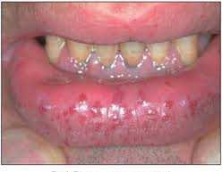
14 Hereditary hemorrhagic telangiectasia: ROW
15 4. Cryptogenic hemoptysis Depending upon the study, up to 30% of patients with hemoptysis have no cause identified even after careful evaluation In a series of patients with cryptogenic hemoptysis, the prognosis was generally good, and most patients had resolution of bleeding within six months of evaluation Adelman M et al. Cryptogenic hemoptysis. Clinical features, bronchoscopic findings, and natural history in 67 patients. Ann Intern Med
16
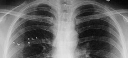
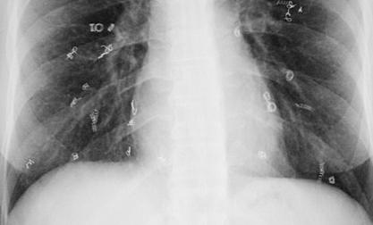
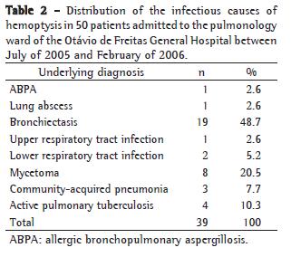
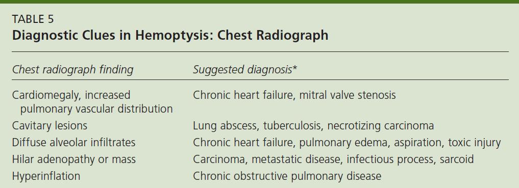
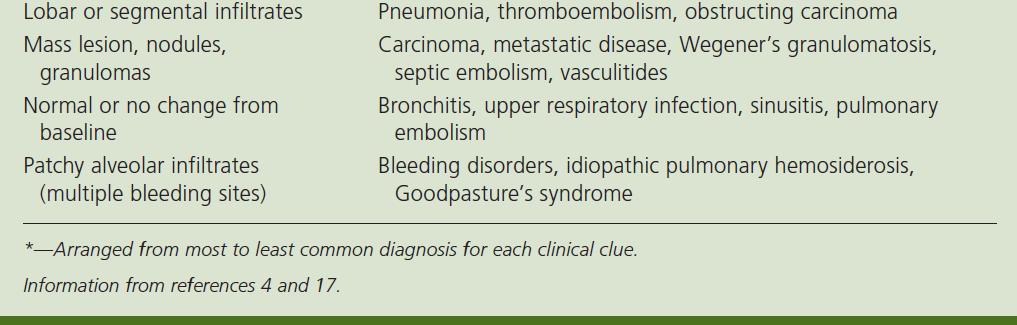
17 EVALUATION OF HEMOPTYSIS The evaluation should begin with the initial history and physical examination supplemented by chest radiograph Important features of the history include: age, smoking history, duration of hemoptysis, and association with symptoms of acute bronchitis or an acute exacerbation of chronic bronchitis
18
19 Physical examination The presence of many telangiectasias suggests R-O-W disease A skin rash may be suggestive of vasculitis Splinter hemorrhages suggest endocarditis or vasculitis Clubbing is non-specific, since it can occur in many chronic lung diseases Pulmonary hypertension may be suggested by an augmented P2, murmurs of tricuspid regurgitation or pulmonary insufficiency, or a right ventricular lift Cardiac murmurs also raise the question of congenital heart disease, endocarditis with septic emboli, or, when a diastolic rumble or opening snap is present, mitral stenosis The legs should be examined carefully for possible deep venous thrombi (eco-doppler)
20 EVALUATION OF HEMOPTYSIS No immediate further work-up is indicated if the clinical picture is not suggestive of a lung carcinoma: negative chest radiograph age <40 years no smoking history hemoptysis < 1 week duration suggestive of acute bronchitis (blood streaking superimposed upon purulent sputum) The patient should be treated for bronchitis and observed for recurrence of hemoptysis following improvement in purulent sputum production
21 EVALUATION OF HEMOPTYSIS
22
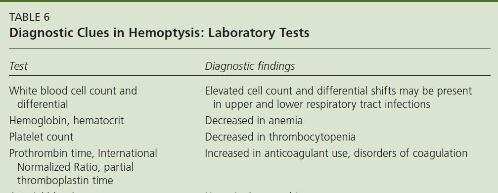
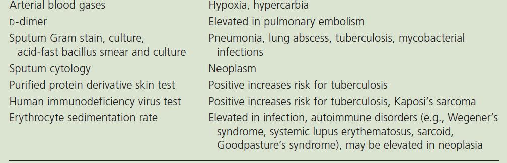
23 Diagnostic Studies 1. Sputum Examination 2. Chest Radiography 3. Bronchoscopy 4. CT - HRCT 5. Angiography
24
25 EVALUATION OF HEMOPTYSIS Further evaluation is indicated if the patient has risk factors for carcinoma or if the hemoptysis does not occur in the setting of acute bronchitis Bronchoscopy is the preferred next procedure in those patients with risk factors for tumor or chronic bronchitis On the other hand, HRCT is the preferred next procedure in patients at lower risk for tumor or chronic bronchitis, but with a history or radiograph suggestive of bronchiectasis or an arteriovenous malformation.
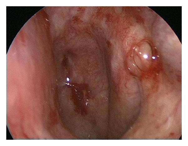
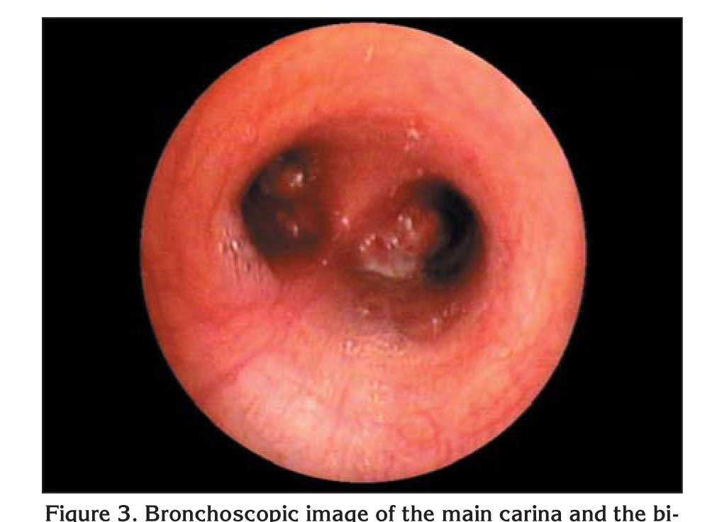
26 DIAGNOSTIC PROCEDURES Bronchoscopy often considered in patients with hemoptysis and a normal or non-localizing CXR to rule out endobronchial malignancy performed early in the evaluation, while the patient is actively bleeding, provides the highest yield for localizing the bleeding site Risk factors predicting those individuals most likely to have tumor found on bronchoscopy include: Male sex Older age, greater than 50 years Smoking history greater than 30 pack/year Duration of hemoptysis greater than one week
27
28 Arteriography If the patient continues to bleed and the source is still unknown, then Arteriography should next be performed, since it may be useful diagnosis as well as for therapy Since the majority of massive bleeds arise from the bronchial circulation, bronchial arteriography has a higher yield than arteriography of the pulmonary or systemic arterial beds When the pulmonary arterial circulation is the source, the most common underlying conditions are pulmonary A-V malformations, Rasmussen's aneurysms or iatrogenic pulmonary artery tears
29 CT scan of the chest Use of early chest CT has been advocated to help localize the bleeding site and diagnose the cause of hemoptysis The advantage of CT is that it may suggest one of several diagnoses such as: bronchiectasis, lung abscess, and mass lesions including cancer, mycetomas, and A-V lesions It may also help in the acute setting to guide arteriography or bronchoscopy to the regions of highest yield The disadvantage of chest CT is that it may require temporary movement of an unstable patient
30 Bronchoscopy versus HRCT Bronchoscopy and HRCT are, in many ways, complementary studies, each with specific advantages in certain clinical situations In one study of 91 patients with hemoptysis, HRCT demonstrated all tumors seen by bronchoscopy as well as, several which were beyond bronchoscopic range. On the other hand, HRCT could not detect bronchitis or subtle mucosal abnormalities which could be seen by bronchoscopy In another report of 57 patients, HRCT was particularly useful in diagnosing bronchiectasis and aspergillomas, while bronchoscopy was diagnostic of bronchitis and mucosal lesions such as Kaposi's sarcoma
31
32 Acute management of hemoptysis Initial priorities are insuring adequate airway protection, ventilation, and cardiovascular function (ABC) Possible coagulation disorders should be rapidly reversed (PT, aptt) Patients with poor gas exchange, rapid ongoing hemoptysis, hemodynamic instability, or severe shortness of breath should be orally intubated with a large bore endotracheal tube (size 8.0 or greater)
33 Use of bronchoscopy in acute management of hemoptysis A bronchoscopic option for protecting the nonbleeding lung is Balloon Tamponade of the bleeding site, involving placement of a Fogarty balloon catheter in the segmental or subsegmental bronchus leading to the bleeding site The balloon is left inflated for 24 to 48 hours, and the patient is then observed for rebleeding with the balloon deflated for several hours There is a potential risk of ischemic mucosal injury and post-obstructive pneumonia, but these complications have not been reported
34
35 Use of bronchoscopy in acute management of hemoptysis Bronchoscopic techniques used to slow or stop bleeding have included lavage with saline and application of topical epinephrine (1:20.000), vasopressin, thrombin or a fibrinogenthrombin combination None of these methods has been tested in controlled trials (!) If bronchoscopy visualizes a localized bleeding mucosal lesion, laser therapy or electrocautery may be considered, if available
36 Protection of the non-bleeding lung If the location or side of bleeding is known, placing the bleeding lung in a dependent position may prevent blood spillage into the non-bleeding lung: place the patient on lateral decubitus toward the site of bleeding Selective intubation: an alternative strategy involves placement of a typical, single lumen endotracheal tube into either the right or left mainstem bronchus. This approach is less practical when the right lung is bleeding, because selective intubation of the left mainstem bronchus may be quite difficult A third alternative is the placement of a double lumen endotracheal tube specially designed for selective intubation of the right or left mainstem bronchi
37
38 Arteriographic embolization The other option for the patient who continues to bleed is Arteriographic Embolization, either as "semi-definitive" treatment or as a bridge to elective surgery In the hands of experienced angiographers, embolization successfully stops bleeding more than 85% of the time Unfortunately, embolization is only "semi-definitive," because re-bleeding occurs in 10-20% of patients over the next 6-12 months Late re-bleeding may be due to incomplete embolization, revascularization, or recanalization.
39
40 Surgery Patients with lateralized uncontrollable bleeding should be assessed early for possible surgery Relative contraindications to surgery include: severe underlying pulmonary disease, active TBC, diffuse underlying lung disease (cystic fibrosis, multiple A-V Ms, multifocal bronchiectasis), and diffuse alveolar hemorrhage Morbidity and mortality are significantly greater with emergent surgery for persistent massive bleeding compared with elective surgery In most series of emergent therapy, surgical mortality for treatment of massive hemoptysis is approximately 20 %
41 RECOMMENDATIONS First, stabilize the patient and then perform early Bronchoscopy along with other appropriate diagnostic studies If the patient continues to bleed aggressively, Arteriography is most reasonable for localization and therapy If bleeding persists despite embolization or if the patient is too ill to go to angiography, then Blockade therapy or a double lumen tube should be considered While Surgery remains the only truly definitive therapy, it should not be used in the acute emergent setting unless it cannot be avoided
42
COUGH Dr. A m A it i e t sh A g A garwa w l Le L ctu t rer Departm t ent t o f f M e M dic i in i e
 COUGH Dr. Amitesh Aggarwal Lecturer Department of Medicine Cough is an explosive expiration that provides a normal protective mechanism for clearing the tracheobronchial tree of secretions and foreign
Lung Cancer - Suspected
 Lung Cancer - Suspected Shared Decision Making Lung Cancer: http://www.enhertsccg.nhs.uk/ Patient presents with abnormal CXR Lung cancer - clinical presentation History and Examination Incidental finding
Anatomy. The respiratory system starts from the nose, mouth, larynx, trachea, and the two lungs.
 Respiratory System Anatomy The respiratory system starts from the nose, mouth, larynx, trachea, and the two lungs. Within the lungs, the bronchi transport air with oxygen to the alveoli on inspiration
Extracorporeal Life Support Organization (ELSO) Guidelines for Pediatric Respiratory Failure
 Guidelines for Pediatric Respiratory Failure") Extracorporeal Life Support Organization (ELSO) Guidelines for Pediatric Respiratory Failure Introduction This pediatric respiratory failure guideline is a supplement to ELSO s General Guidelines for all
Extracorporeal Life Support Organization (ELSO) Guidelines for Pediatric Respiratory Failure Introduction This pediatric respiratory failure guideline is a supplement to ELSO s General Guidelines for all
Pathology of pulmonary vascular disease. Dr.Ashraf Abdelfatah Deyab. Assistant Professor of Pathology Faculty of Medicine Almajma ah University
 Pathology of pulmonary vascular disease Dr.Ashraf Abdelfatah Deyab Assistant Professor of Pathology Faculty of Medicine Almajma ah University Pulmonary vascular disease Type of pulmonary circulation: Types
Lung diseases of Vascular Origin. By: Shefaa Qa qqa
 Lung diseases of Vascular Origin By: Shefaa Qa qqa Pulmonary Hypertension Pulmonary hypertension is defined as a mean pulmonary artery pressure greater than or equal to 25 mm Hg at rest. Based on underlying
Specific Basic Standards for Osteopathic Fellowship Training in Pulmonary / Critical Care Medicine
 Specific Basic Standards for Osteopathic Fellowship Training in Pulmonary / Critical Care Medicine American Osteopathic Association and American College of Osteopathic Internists BOT Rev. 2/2011 These
Surgical indications: Non-malignant pulmonary diseases. Punnarerk Thongcharoen
 Surgical indications: Non-malignant pulmonary diseases Punnarerk Thongcharoen Non-malignant Malignant as a pathological term: Cancer Non-malignant = not cancer Malignant as an adjective: Disposed to cause
INDEPENDENT LUNG VENTILATION
 INDEPENDENT LUNG VENTILATION Giuseppe A. Marraro, MD Director Anaesthesia and Intensive Care Department Paediatric Intensive Care Unit Fatebenefratelli and Ophthalmiatric Hospital Milan, Italy gmarraro@picu.it
Vascular Lung Diseases
 Vascular Lung Diseases SESSION SPECIFIC OBJECTIVES List the major types of vascular lung disease Recognize and describe the pathology of vascular lung disease: Pulmonary embolism, thrombosis, hypertension,
Subject Index. Bacterial infection, see Suppurative lung disease, Tuberculosis
 Subject Index Abscess, virtual 107 Adenoidal hypertrophy, features 123 Airway bleeding, technique 49, 50 Airway stenosis, see Stenosis, airway Anaesthesia biopsy 47 complications 27, 28 flexible 23 26
Atlas of the Vasculitic Syndromes
 CHAPTER e40 Atlas of the Vasculitic Syndromes Carol A. Langford Anthony S. Fauci Diagnosis of the vasculitic syndromes is usually based upon characteristic histologic or arteriographic findings in a patient
Cystic Fibrosis Complications ANDRES ZIRLINGER, MD STANFORD UNIVERSITY MEDICAL CENTER MARCH 3, 2012
 Cystic Fibrosis Complications ANDRES ZIRLINGER, MD STANFORD UNIVERSITY MEDICAL CENTER MARCH 3, 2012 INTRODUCTION PNEUMOTHORAX HEMOPTYSIS RESPIRATORY FAILURE Cystic Fibrosis Autosomal Recessive Genetically
Discussing feline tracheal disease
 Vet Times The website for the veterinary profession https://www.vettimes.co.uk Discussing feline tracheal disease Author : ANDREW SPARKES Categories : Vets Date : March 24, 2008 ANDREW SPARKES aims to
Epidermiology Early pulmonary embolism
 Epidermiology Early pulmonary embolism Sitang Nirattisaikul Faculty of Medicine, Prince of Songkla University 3 rd most common cause of cardiovascular death in the United States, following ischemic heart
Tests Your Pulmonologist Might Order. Center For Cardiac Fitness Pulmonary Rehab Program The Miriam Hospital
 Tests Your Pulmonologist Might Order Center For Cardiac Fitness Pulmonary Rehab Program The Miriam Hospital BASIC ANATOMY OF THE LUNGS Lobes of Lung 3 lobes on the Right lung 2 lobes on the Left Blood
Pulmonary Pathophysiology
 Pulmonary Pathophysiology 1 Reduction of Pulmonary Function 1. Inadequate blood flow to the lungs hypoperfusion 2. Inadequate air flow to the alveoli - hypoventilation 2 Signs and Symptoms of Pulmonary
L. Freitag*, E. Tekolf*, G. Stamatis*, M. Montag**, D. Greschuchna*
 Eur Respir J, 1994, 7, 0 07 DOI: 10.118/090196.94.07110 Printed in UK - all rights reserved Copyright ERS Journals Ltd 1994 European Respiratory Journal ISSN 090-196 Three years experience with a new balloon
Indications of Coronary Angiography Dr. Shaheer K. George, M.D Faculty of Medicine, Mansoura University 2014
 Indications of Coronary Angiography Dr. Shaheer K. George, M.D Faculty of Medicine, Mansoura University 2014 Indications for cardiac catheterization Before a decision to perform an invasive procedure such
Localized Pulmonary Hemorrhage Associated With Massive Hemoptysis: Report of a Case
 113 Localized Pulmonary Hemorrhage Associated With Massive Hemoptysis: Report of a Case Yang-Hao Yu, Te-Chun Hsia, Liang-Wen Hang, Tze-Yi Lin 1, Nan-Yung Hsu 2, Der-Yuan Wang 3 Devision of Pulmonary and
Hospital-acquired Pneumonia
 Hospital-acquired Pneumonia Hospital-acquired pneumonia (HAP) Pneumonia that occurs at least 2 days after hospital admission. The second most common and the leading cause of death due to hospital-acquired
Exam 2 Respiratory Disorders
 Exam 2 Respiratory Disorders Common Cold Common Cold Pathology Common Cold Consequences Rhinosinusitis Rhinosinusitis Pathology Rhinosinusitis ostia can close due to Influenza (Flu) Influenza Pathology
Introduction to Chest Radiography
 Introduction to Chest Radiography RSTH 366: DIAGNOSTIC TECHNIQUES Alan Alipoon BS, RCP, RRT Instructor Department of Cardiopulmonary Sciences 1 Introduction Discovered in 1895 by Wilhelm Roentgen Terminology
Index. Note: Page numbers of article titles are in boldface type
 Index Note: Page numbers of article titles are in boldface type A Acute coronary syndrome, perioperative oxygen in, 599 600 Acute lung injury (ALI). See Lung injury and Acute respiratory distress syndrome.
Cor pulmonale. Dr hamid reza javadi
 1 Cor pulmonale Dr hamid reza javadi 2 Definition Cor pulmonale ;pulmonary heart disease; is defined as dilation and hypertrophy of the right ventricle (RV) in response to diseases of the pulmonary vasculature
INDICATIONS AND COMPLICATIONS OF BRONCHOSCOPY: AN EXPERIENCE OF 100 CASES IN A TERTIARY CARE HOSPITAL
 ORIGINAL ARTICLE INDICATIONS AND COMPLICATIONS OF BRONCHOSCOPY: AN EXPERIENCE OF 00 CASES IN A TERTIARY CARE HOSPITAL Amir Suleman, Qazi Ikramullah, Farooq Ahmed, M Yousaf Khan Department of Medicine and
DEPARTMENT OF SURGERY CARDIOVASCULAR-THORACIC SECTION
 DEPARTMENT OF SURGERY CARDIOVASCULAR-THORACIC SECTION DIRECTIONS: This must accompany all initial applications for appointment to the Cardiovascular-Thoracic Section, Department of Surgery. Please indicate
Beyond Pulmonary Emboli: Acquired Abnormalities of the Pulmonary Arteries on CTA
 Beyond Pulmonary Emboli: Acquired Abnormalities of the Pulmonary Arteries on CTA Sean Cleary, MD and Katherine Kaproth-Joslin, MD/PhD University of Rochester Medical Center Rochester, NY Financial Disclosures
Adult Echocardiography Examination Content Outline
 Adult Echocardiography Examination Content Outline (Outline Summary) # Domain Subdomain Percentage 1 2 3 4 5 Anatomy and Physiology Pathology Clinical Care and Safety Measurement Techniques, Maneuvers,
Therapeutic Bronchoscopy Etiology - Benign Stenosis Post - intubation Trauma Post - operative Inflammatory Idiopathic
 Endobronchial Palliation of Airway Disease Douglas E. Wood, MD Professor and Chief Division of Cardiothoracic Surgery Vice-Chair, Department of Surgery Endowed Chair in Lung Cancer Research University
Endoscopy. Pulmonary Endoscopy
 Pulmonary 1 Direct visualization of TB tree Developed in 1890 s to remove foreign bodies - rigid metal tube Advances added light system, Sx Flexible fiberoptic scopes introduced in early 1960 s 2 Used
TB Intensive Houston, Texas
 TB Intensive Houston, Texas October 15-17, 17 2013 Diagnosis of TB: Radiology Rosa M Estrada-Y-Martin, MD MSc FCCP October 16, 2013 Rosa M Estrada-Y-Martin, MD MSc FCCP, has the following disclosures to
Clinical Indications for Echocardiography
 Clinical Indications for Echocardiography Echocardiography is widely utilised and potential applications are increasing with advances in technology. The aim of this document is two-fold: 1) To define clinical
MRSA pneumonia mucus plug burden and the difficult airway
 Case report Crit Care Shock (2016) 19:54-58 MRSA pneumonia mucus plug burden and the difficult airway Ann Tsung, Brian T. Wessman An 80-year-old female with a past medical history of chronic obstructive
HEMODYNAMIC DISORDERS
 HEMODYNAMIC DISORDERS Normal fluid homeostasis requires vessel wall integrity as well as maintenance of intravascular pressure and osmolarity within certain physiologic ranges. Increases in vascular volume
Respiratory system. Applied Anatomy &Physiology
 Respiratory system Applied Anatomy &Physiology Anatomy The respiratory system consists of 1)The Upper airway : Nose, mouth and larynx 2)The Lower airways Trachea and the two lungs. Within the lungs,
Patient Management Code Blue in the CT Suite
 Patient Management Code Blue in the CT Suite David Stultz, MD November 28, 2001 Case Presentation A 53-year-old woman experienced acute respiratory distress during an IV contrast enhanced CT scan of the
Index. Note: Page numbers of article titles are in boldface type.
 Index Note: Page numbers of article titles are in boldface type. A Acute coronary syndrome(s), anticoagulant therapy in, 706, 707 antiplatelet therapy in, 702 ß-blockers in, 703 cardiac biomarkers in,
October 2017 Pulmonary Embolism
 October 2017 Pulmonary Embolism Prof. Ahmed BaHammam, FRCP, FCCP Professor of Medicine College of Medicine King Saud University 1 Objectives Epidemiology Pathophysiology Diagnosis Massive PE Treatment
Unconscious exchange of air between lungs and the external environment Breathing
 Respiration Unconscious exchange of air between lungs and the external environment Breathing Two types External Exchange of carbon dioxide and oxygen between the environment and the organism Internal Exchange
B-I-2 CARDIAC AND VASCULAR RADIOLOGY
 (YEARS 1 3) CURRICULUM FOR RADIOLOGY 13 B-I-2 CARDIAC AND VASCULAR RADIOLOGY KNOWLEDGE To describe the normal anatomy of the heart and vessels including the lymphatic system as demonstrated by radiographs,
Pneumothorax. Defined as air in the pleural space which can occur through a number of mechanisms
 Pneumothorax Defined as air in the pleural space which can occur through a number of mechanisms Traumatic pneumothorax Penetrating chest trauma Common secondary to bullet or knife penetration Chest tube
ENDOBRONCHIAL ABLATIVE THERAPIES. Christopher Cortes, MD, FPCCP
 ENDOBRONCHIAL ABLATIVE THERAPIES Christopher Cortes, MD, FPCCP Choice of Ablative Therapy Size of the lesion Location of the lesion Characteristics of the lesion Availability of the different therapies
CLINICAL FEATURES IN PULMONARY TUBERCULOSIS
 CLINICAL FEATURES IN PULMONARY TUBERCULOSIS Dr. Amitesh Aggarwal Department of Medicine Tuberculosis Captain of all the Men of Death Great White Plague devastating effect on society 100 years ago one in
Aneurysms & a Brief Discussion on Embolism
 Aneurysms & a Brief Discussion on Embolism Aneurysms, overview = congenital or acquired dilations of blood vessels or the heart True aneurysms -involve all three layers of the artery (intima, media, and
Echocardiography as a diagnostic and management tool in medical emergencies
 Echocardiography as a diagnostic and management tool in medical emergencies Frank van der Heusen MD Department of Anesthesia and perioperative Care UCSF Medical Center Objective of this presentation Indications
CHEST INJURY PULMONARY CONTUSION
 CHEST INJURY PULMONARY CONTUSION Introduction Pulmonary contusion refers to blunt traumatic lung parenchymal injury which results in oedema and haemorrhaging into alveolar spaces. It may also result in
L angiografia ha ancora un ruolo?
 Aggiornamenti di Radiologia Interventistica L angiografia ha ancora un ruolo? Roberto Iezzi Dipartimento di Bioimmagini e Scienze Radiologiche Istituto di Radiologia, Policlinico A. Gemelli Università
Diffuse Alveolar Hemorrhage: Initial and Follow-up HRCT Features
 Diffuse Alveolar Hemorrhage: Initial and Follow-up HRCT Features Poster No.: E-0037 Congress: ESTI 2012 Type: Authors: Keywords: Scientific Exhibit M. Y. Kim; Seoul/KR Lung, CT-High Resolution, CT, Computer
SIR 2008 Annual Meeting Film Panel Case: Radiation-induced Angiosarcoma
 Special Communications SIR 2008 Annual Meeting Film Panel Case: Radiation-induced Angiosarcoma Daniel Y. Sze, MD, PhD, and Judy H. Huang, MD J Vasc Interv Radiol 2008; 19:1133 1137 HISTORY From the Division
Dr. Rami M. Adil Al-Hayali Assistant Professor in Medicine
 Dr. Rami M. Adil Al-Hayali Assistant Professor in Medicine Venous thromboembolism: pulmonary embolism (PE) deep vein thrombosis (DVT) 1% of all patients admitted to hospital 5% of in-hospital mortality
The Surgical Treatment of Tracheobronchial Tuberculosis. The Thoracic Department of Beijing Chest Hospital, Capital Medical University
 The Surgical Treatment of Tracheobronchial Tuberculosis ) The Thoracic Department of Beijing Chest Hospital, Capital Medical University Named also: endobronchial tuberculosis,ebtb defined as tuberculous
Case Report Stenting as a Rescue Treatment of a Pulmonary Artery False Aneurysm Caused by Swan-Ganz Catheterization
 Case Reports in Pulmonology, Article ID 893647, 4 pages http://dx.doi.org/10.1155/2014/893647 Case Report Stenting as a Rescue Treatment of a Pulmonary Artery False Aneurysm Caused by Swan-Ganz Catheterization
Role of multi-detector Computed Tomographic (CT) angiography, in patients with hemoptysis.
 angiography, in patients with hemoptysis.") Role of multi-detector Computed Tomographic (CT) angiography, in patients with hemoptysis. Poster No.: C-2094 Congress: ECR 2014 Type: Scientific Exhibit Authors: V. Bizimi, V. Papalouka, C. Chrona, N.
Role of multi-detector Computed Tomographic (CT) angiography, in patients with hemoptysis. Poster No.: C-2094 Congress: ECR 2014 Type: Scientific Exhibit Authors: V. Bizimi, V. Papalouka, C. Chrona, N.
University of Florida Department of Surgery. CardioThoracic Surgery VA Learning Objectives
 University of Florida Department of Surgery CardioThoracic Surgery VA Learning Objectives This service performs coronary revascularization, valve replacement and lung cancer resections. There are 2 faculty
Annual High Claims Survey. Year Ending 31 December 2016
 Annual High Claims Survey Year Ending 31 December 2016 Released July 2017 Summary The Private Healthcare Australia Annual High Claims Survey Report analyses the nature and magnitude of high claims met
Pulmonary Aspergillosis
 May 2005 Pulmonary Aspergillosis Nancy Wei, Harvard Medical School, Year III Overview Pulmonary aspergillosis background information Patient presentations Common radiographic findings for each type of
Bronchial syndrome. Atelectasis Draining bronchus Bronchiectasis
 Bronchial syndrome Atelectasis Draining bronchus Bronchiectasis Etienne Leroy Terquem Pierre L Her SPI / ISP Soutien Pneumologique International / International Support for Pulmonology Atelectasis Consequence
Pulmonary hypertension
 Zurich Open Repository and Archive University of Zurich Main Library Strickhofstrasse 39 CH-8057 Zurich www.zora.uzh.ch Year: 2012 Pulmonary hypertension Glaus, T M Posted at the Zurich Open Repository
Valvular Heart Disease Mitral Stenosis
 Valvular Heart Disease Mitral Stenosis A 75 year old woman with loud first heart sound and mid-diastolic murmur Chronic dyspnea Class 2/4 Fatigue Recent orthopnea/pnd Nocturnal palpitation Pedal edema
An aneurysm is a localized abnormal dilation of a blood vessel or the heart Types: 1-"true" aneurysm it involves all three layers of the arterial
 An aneurysm is a localized abnormal dilation of a blood vessel or the heart Types: 1-"true" aneurysm it involves all three layers of the arterial wall (intima, media, and adventitia) or the attenuated
Respiratory Disease. Dr Amal Damrah consultant Neonatologist and Paediatrician
 Respiratory Disease Dr Amal Damrah consultant Neonatologist and Paediatrician Signs and Symptoms of Respiratory Diseases Cardinal Symptoms Cough Sputum Hemoptysis Dyspnea Wheezes Chest pain Signs and Symptoms
Acute Respiratory Distress Syndrome (ARDS) An Update
 An Update") Acute Respiratory Distress Syndrome (ARDS) An Update Prof. A.S.M. Areef Ahsan FCPS(Medicine) MD(Critical Care Medicine) MD ( Chest) Head, Dept. of Critical Care Medicine BIRDEM General Hospital INTRODUCTION
Acute Respiratory Distress Syndrome (ARDS) An Update Prof. A.S.M. Areef Ahsan FCPS(Medicine) MD(Critical Care Medicine) MD ( Chest) Head, Dept. of Critical Care Medicine BIRDEM General Hospital INTRODUCTION
DISEASES OF THE RESPIRATORY SYSTEM 2018 DR HEYAM AWAD LECTURE 3: CHRONIC BRNCHITIS AND BRONCHIECTASIS
 DISEASES OF THE RESPIRATORY SYSTEM 2018 DR HEYAM AWAD LECTURE 3: CHRONIC BRNCHITIS AND BRONCHIECTASIS INTRDUCTION In the last lecture we discussed the difference between restrictive and obstructive lung
100 Chest X Rays for Study Group. by Dr. Suneet Khurana
 100 Chest X Rays for Study Group by Dr. Suneet Khurana Approach to - Chest X Ray (shadow of the viscera on a photographic plate) Gas appears Black Fat appears Dark Grey Water Appears as Light Grey Bone
Ughmm Let Me Clear My Throat
 Ughmm Let Me Clear My Throat The Initial Evaluation and Management of Hemoptysis 41 st Annual Refresher Course for the Family Physician Christopher Hogrefe, MD Clinical Assistant Professor Departments
GUIDELINES FOR THE EARLY MANAGEMENT OF PATIENTS WITH ACUTE ISCHEMIC STROKE
 2018 UPDATE QUICK SHEET 2018 American Heart Association GUIDELINES FOR THE EARLY MANAGEMENT OF PATIENTS WITH ACUTE ISCHEMIC STROKE A Summary for Healthcare Professionals from the American Heart Association/American
Unit II Problem 2 Pathology: Pneumonia
 Unit II Problem 2 Pathology: Pneumonia - Definition: pneumonia is the infection of lung parenchyma which occurs especially when normal defenses are impaired such as: Cough reflex. Damage of cilia in respiratory
Intended Learning Outcomes
 2011 Acute Limb Ischemia Definition, Etiology & Pathophysiology Clinical Evaluation Management Ali SABBOUR Prof. of Vascular Surgery, Ain Shams University Acute Limb Ischemia Intended Learning Outcomes
October 2012 Imaging Case of the Month. Michael B. Gotway, MD Associate Editor Imaging. Department of Radiology Mayo Clinic Arizona Scottsdale, AZ
 October 2012 Imaging Case of the Month Michael B. Gotway, MD Associate Editor Imaging Department of Radiology Mayo Clinic Arizona Scottsdale, AZ Clinical History: A 65-year-old non-smoking woman presented
SUPPLEMENTAL MATERIAL. Supplemental digital content 1, Appendix 1. Ischemic symptoms and electrocardiography
 SUPPLEMENTAL MATERIAL Supplemental digital content 1, Appendix 1. Ischemic symptoms and electrocardiography findings 1. Ischemic symptoms included any of the following: chest discomfort, arm discomfort,
Interventional procedures guidance Published: 20 December 2017 nice.org.uk/guidance/ipg600
 Endobronchial valve insertion to reduce lung volume in emphysema Interventional procedures guidance Published: 20 December 2017 nice.org.uk/guidance/ipg600 Your responsibility This guidance represents
The ABC s of Chest Trauma
 The ABC s of Chest Trauma J Bradley Pickhardt MD, FACS Providence St Patrick Hospital What s the Problem? 2/3 of trauma patients have chest trauma Responsible for 25% of all trauma deaths Most injuries
AORTIC DISSECTION. DISSECTING ANEURYSMS OF THE AORTA or CLASSIFICATION
 DISSECTING ANEURYSMS OF THE AORTA or AORTIC DISSECTION CLASSIFICATION DeBakey classified aortic dissections into types I, II, and III :- Type I dissection the tear site originates in the ascending aorta,
Emergency Surgery Course Graz, March UPPER GI BLEEDING. Carlos Mesquita Coimbra
 UPPER GI BLEEDING Carlos Mesquita Coimbra Aim Causes Management Problem Above angle of Treitz Common emergency 1-2/1000 pts 10% rebleeed 1% angioembolization 20% over 60
Episodes of Care Risk Adjustment
 Episodes of Care Risk Adjustment Episode Types Wave 1 Asthma Acute Exacerbation Perinatal Total Joint Replacement Wave 2 Acute Percutaneous Coronary Intervention COPD Acute Exacerbation Non-acute Percutaneous
WF RESPIRATORY SYSTEM. RESPIRATORY MEDICINE
 WF RESPIRATORY SYSTEM. RESPIRATORY MEDICINE 1 Societies 11 History 13 Dictionaries. Encyclopaedias. Bibliographies Use for general works only. Classify with specific aspect where possible 15 Classification.
Ischemic heart disease
 Ischemic heart disease Introduction In > 90% of cases: the cause is: reduced coronary blood flow secondary to: obstructive atherosclerotic vascular disease so most of the time it is called: coronary artery
Cardiovascular Nursing Practice: A Comprehensive Resource Manual and Study Guide for Clinical Nurses 2 nd Edition
 Cardiovascular Nursing Practice: A Comprehensive Resource Manual and Study Guide for Clinical Nurses 2 nd Edition Table of Contents Volume 1 Chapter 1: Cardiovascular Anatomy and Physiology Basic Cardiac
an inflammation of the bronchial tubes
 BRONCHITIS DEFINITION Bronchitis is an inflammation of the bronchial tubes (or bronchi), which are the air passages that extend from the trachea into the small airways and alveoli. Triggers may be infectious
Respiratory Diseases and Disorders
 Chapter 9 Respiratory Diseases and Disorders Anatomy and Physiology Chest, lungs, and conducting airways Two parts: Upper respiratory system consists of nose, mouth, sinuses, pharynx, and larynx Lower
10/16/2014. CCRN Review - Cardiovascular. CCRN Review - Cardiovascular. CCRN Review - Cardiovascular
 Hypertrophic (IHSS) Diagnosis Chest x ray cardiomegaly Electrocardiography LV hypertrophy, ST segment T was changes, Q waves in inferior & precordial leads Atrial & ventricular dysrhythmias Hypertrophic
Excavated pulmonary nodule: steps to diagnosis?
 Excavated pulmonary nodule: steps to diagnosis? Poster No.: C-1044 Congress: ECR 2014 Type: Authors: Keywords: DOI: Educational Exhibit W. Mnari, M. MAATOUK, A. Zrig, B. Hmida, M. GOLLI; Monastir/ TN Metastases,
Bronchogenic Carcinoma
 A 55-year-old construction worker has smoked 2 packs of ciggarettes daily for the past 25 years. He notes swelling in his upper extremity & face, along with dilated veins in this region. What is the most
ERDHEIM-CHESTER DISEASE LUNG & HEART ISSUES
 ERDHEIM-CHESTER DISEASE LUNG & HEART ISSUES GIULIO CAVALLI, M.D. INTERNAL MEDICINE AND CLINICAL IMMUNOLOGY IRCCS SAN RAFFAELE HOSPITAL VITA-SALUTE SAN RAFFAELE UNIVERSITY MILAN, ITALY cavalli.giulio@hsr.it
4/17/2010 C ini n ca c l a Ev E a v l a ua u t a ion o n of o ILD U dat a e t e i n I LDs
 Update in ILDs Diagnosis 101: Clinical Evaluation April 17, 2010 Jay H. Ryu, MD Mayo Clinic, Rochester MN Clinical Evaluation of ILD Outline General aspects of ILDs Classification of ILDs Clinical evaluation
NURSING DEPARTMENT CRITICAL CARE POLICY MANUAL CRITICAL CARE PROTOCOLS. ACTIVASE (t-pa) INFUSION PROTOCOL FOR ACUTE MYOCARDIAL INFARCTION
 INFUSION PROTOCOL FOR ACUTE MYOCARDIAL INFARCTION") NURSING DEPARTMENT CRITICAL CARE POLICY MANUAL CRITICAL CARE PROTOCOLS ACTIVASE (t-pa) FOR ACUTE MYOCARDIAL INFARCTION I. PURPOSE: A. To reduce the extent of myocardial infarction by lysing the clot in
NURSING DEPARTMENT CRITICAL CARE POLICY MANUAL CRITICAL CARE PROTOCOLS ACTIVASE (t-pa) FOR ACUTE MYOCARDIAL INFARCTION I. PURPOSE: A. To reduce the extent of myocardial infarction by lysing the clot in
Audra Fuller MD, Mark Sigler MD, Shrinivas Kambali MD, Raed Alalawi MD
 Clinical Series Successful treatment of post-intubation tracheal stenosis with balloon dilation, argon plasma coagulation, electrocautery and application of mitomycin C Audra Fuller MD, Mark Sigler MD,
Diagnosing Idiopathic Pulmonary Fibrosis on Evidence-Based Guidelines
 Diagnosing Idiopathic Pulmonary Fibrosis on Evidence-Based Guidelines Rebecca Keith, MD Assistant Professor, Division of Pulmonary and Critical Care Medicine National Jewish Health, Denver, CO Objectives
University of Wisconsin - Madison Cardiovascular Medicine Fellowship Program UW CICU Rotation Goals and Objectives
 Background: The field of critical care cardiology has evolved considerably over the past 2 decades. Contemporary critical care cardiology is increasingly focused on the management of patients with advanced
Group B: Directed self-study Group C: Anatomy lab. Lecture: Structure and function of larynx. Lecture: Dead space & compliance of lungs
 Timetable Week 1 (1 st January 2018) Theme: Structure and functions of the lungs Group A: Anatomy lab Group C: Histology lab Upper Group B: Anatomy lab Group C: Anatomy lab Group A: Histology lab Upper
CVICU EXAM. Mrs. Jennings is a 71-year-old post-op CABG x5 with an IABP in her left femoral artery
 CVICU EXAM 1111 North 3rd Street Mrs. Jennings is a 71-year-old post-op CABG x5 with an IABP in her left femoral artery 1. Nursing standards for a patient on an IABP device include: a. Know results of
Detailed Order Request Checklists for Cardiology
 Next Generation Solutions Detailed Order Request Checklists for Cardiology 8600 West Bryn Mawr Avenue South Tower Suite 800 Chicago, IL 60631 www.aimspecialtyhealth.com Appropriate.Safe.Affordable 2018
A Repeat Case of Idiopathic Spontaneous Hemothorax
 Case Report A Repeat Case of Idiopathic Spontaneous Hemothorax Felix R. Gaw, MD Jack H. Bloch, MD, PhD, FACS Nolan J. Anderson, MD, FACS Spontaneous hemothorax, a collection of blood in the pleural cavity
A 44-year-old man with hemoptysis: A review of pertinent imaging studies and radiographic interventions
 IMAGING IN PRACTICE ARLENE SIRAJUDDIN, MD Section of Thoracic Imaging, Imaging Institute, Cleveland Clinic TAN-LUCIEN H. MOHAMMED, MD Section of Thoracic Imaging, Imaging Institute, Cleveland Clinic A
TAVR : Caring for your patients before and after TAVR
 TAVR : Caring for your patients before and after TAVR Zubair Ahmed MD FSCAI Interventional Cardiologist Washington Regional Medical Center / Walker Heart Institute What is Aortic Valve Stenosis? AVA ~4
Thoracic Surgery; An Overview
 Thoracic Surgery What we see Thoracic Surgery; An Overview James P. Locher, Jr, MD Methodist Cardiovascular and Thoracic Surgery Lung cancer Mets Fungus and TB Lung abcess and empyema Pleural based disease
Ultrasound-enhanced, catheter-directed thrombolysis for pulmonary embolism
 NATIONAL INSTITUTE FOR HEALTH AND CARE EXCELLENCE Interventional procedure consultation document Ultrasound-enhanced, catheter-directed thrombolysis for pulmonary embolism A pulmonary embolism (PE) is
Operative Treatment of Massive Hemoptysis
 Operative Treatment of Massive Hemoptysis Anatole Gourin, M.D., and Antonio A. Garzon, M.D. ABSTRACT Fifty-five pulmonary resections have been performed at our institution for hemoptysis of 600 ml. or
Pulmonary vascular anatomy & anatomical variants
 Review Article Pulmonary vascular anatomy & anatomical variants Asha Kandathil, Murthy Chamarthy Department of Radiology, University of Texas Southwestern Medical Center, Dallas, TX, USA Contributions: