SCPA602 Cardiovascular System
|
|
|
- Lindsey Chapman
- 5 years ago
- Views:
Transcription


1 SCPA602 Cardiovascular System Associate Professor Dr. Wannee Jiraungkoorskul Department of Pathobiology, Faculty of Science, Mahidol University Tel: , 1
2 Objectives Describe the anatomy and histology of cardiovascular system Describe the interrelationships and functions of the different parts of the cardiovascular system. 2
3 HEART:- FUNCTION Distribution of O 2 & nutrients (glucose, amino acids) to tissue Transportation of CO 2 and waste products from tissue Distribution of water, electrolytes and hormones through the body Supporting the host defense system Thermoregulation 3
4 4
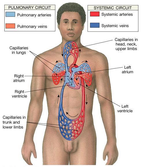
5 HEART:- CIRCUIT Pulmonary Circulation The right ventricle contracts forcing blood through the pulmonary valve into the pulmonary artery Blood traverses the lung capillaries and enters the pulmonary vein and finally the left atrium The heart itself receives its blood via the coronary circulation Systemic Circulation Blood from left ventricle, enters aorta Blood flows to arterioles to capillaries then to venules and converge at superior and inferior vena cava enters right atrium and flows through the tricuspid valve into the right ventricle 5

6 6
7 7

8 HEART 8

9 HEART:- PERICARDIUM Pericardium composed of two layers separated by a space called the pericardial cavity Outer layer - the parietal pericardium Inner layer - the visceral pericardium 9
10 HEART:- PERICARDIUM Pericardial cavity between visceral and parietal pericardium filled with pericardial fluid about 25 to 35 ml lubricates and reduces friction between heart and sac Pericarditis inflammation of pericardium; causes dryness of membrane and painful friction of the heart as it beats 10

11 HEART:- PERICARDIUM The parietal pericardium consists of Outer layer - thick, fibrous connective tissue, attached to the diaphragm forms a strong protective sac for the heart Inner layer -serous, consisting largely of mesothelium, small amount of connective tissue, forms a simple squamous epithelium and secretes a small amount of fluid. 11

12 HEART:- PERICARDIUM The visceral pericardium (epicardium) forms the outer covering of the heart and has an external layer of flat mesothelial cells M. These cells lie on a stroma of fibrocollagenous F support tissue, which contains elastic fibres. A = adipose tissue 12
13 Epicardium or visceral pericardium The external tunic of the heart, the epicardium, is the site of the coronary vessels and contains considerable adipose tissue. This section of atrium shows part of the myocardium (M) and epicardium (Ep). The epicardium consists of loose connective tissue (CT) containing both autonomic nerves (N) and fat (F). The epicardium is the visceral layer of the pericardium and is covered by the simple squamous-to-cuboidal epithelium (arrows) that also lines the pericardial space. These mesothelial cells secrete a lubricate fluid that prevents friction as the beating heart contacts the parietal pericardium on the other side of the pericardial cavity. X100. H&E. 13

14 Epicardium or visceral pericardium 14
15 HEART:- MYOCARDIUM Myocardium - contractile element composed of specialised striated muscle fibres called cardiac muscle. The amount of myocardium and the diameter of muscle fibres in the chambers of the heart varies according to the workload of the chamber. The left and right atria push blood into empty ventricles against minimal resistance during diastole and therefore have a thin wall composed of cells of small diameter. 15
16 HEART:- MYOCARDIUM The right ventricle pushes blood through the pulmonary valve and through the lungs, to enter the left atrium. It therefore has a moderately thick muscle layer composed of fibres intermediate in diameter between atrial and left ventricular muscle cells. The left ventricle pumps blood through the highpressure systemic arterial system and therefore has the thickest myocardium with the largest diameter muscle fibres. 16

17 HEART:- MYOCARDIUM A section through the left and right ventricles showed the difference in thickness between the wall and their different shapes. 17
18 HEART:- ENDOCARDIUM Endocardium - composed of three layers 1. Outmost layer - irregularly arranged collagen fibres, Purkinje fibres (conducting system) 2. Middle layer - thickest endocardial layer, regularly arranged collagen fibres, variable numbers of elastic fibres 3. Inner layer - flat endothelial cells, which are continuous with the endothelial cells lining the vessels entering and emerging from the heart. 18
 containing small nerves and in the ventricles the conducting (Purkinje) fibers (P) of the")
19 Endocardium & subendocardial conducting network The endocardium (En) is a thin layer of connective tissue lined by simple squamous endothelium. Between the endocardium and myocardium is a layer of variable thickness called the subendocardial layer (SEn) containing small nerves and in the ventricles the conducting (Purkinje) fibers (P) of the subendocardial conducting network. These fibers are cardiac muscle cells joined by intercalated disks but specialized for impulse conduction rather than contraction. Purkinje fibers are usually larger than contractile cardiac muscle fibers with large amounts of lightly stained glycogen filling most of the cytoplasm and displacing sparse myofibrils to the periphery. (a): Purkinje fibers running separately within the subendocardial layer. 19

20 Endocardium & subendocardial conducting network (b): Purkinje fibers intermingling with contractile fibers within the myocardium (M). Along with the nodes of specialized cardiac muscle in the right atrium which generate the electrical impulse, the network of conducting fibers comprises the conducting system of the heart. Both X200. H&E. 20
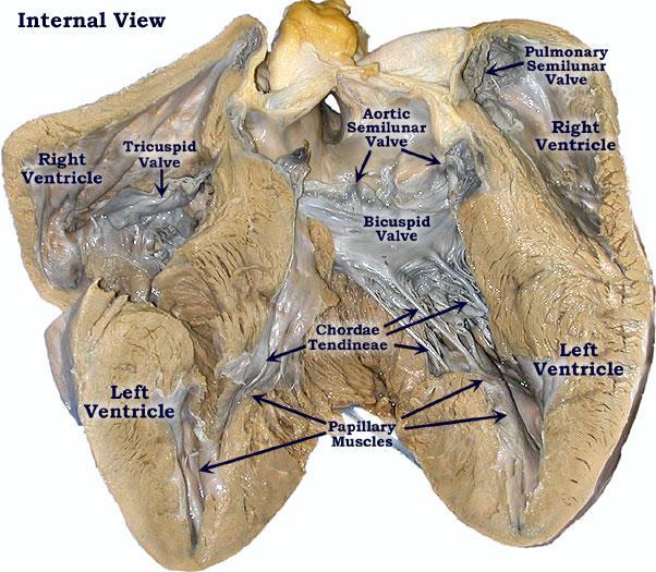
21 HEART:- VALVES Ensure one-way blood flow Atrioventricular (AV) valves (2) right AV valve has 3 cusps (tricuspid valve) left AV valve has 2 cusps (mitral, bicuspid valve) chordae tendineae - cords connect AV valves to papillary muscles (on floor of ventricles) Semilunar valves - control flow into great arteries Pulmonary semilunar valve: from right ventricle into pulmonary trunk Aortic semilunar valve: from left ventricle into aorta 21


22 aortic valves mitral valves pulmonary valves tricuspid valves 22

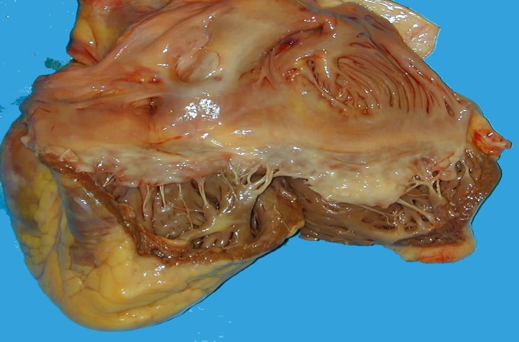
23 HEART:- VALVES Consist of leaflets of collagenous tissue, the surfaces being invested with a thin endothelial layer continuous with that of the heart chambers and great vessels. The base of a valve arising from the myocardium M. The tough central fibrous sheet, the lamina fibrosa F, represents a merging of the fibro-elastic supporting layers S beneath the endothelium E. Burkitt, Young, Heath (1993) Wheater's Functional Histology 3rd Ed. Fig. 8.5, p
24 24
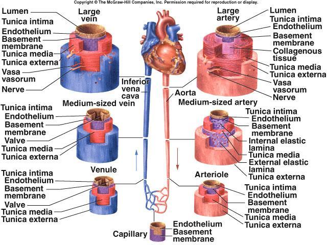
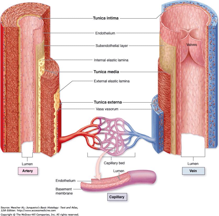
25 ARTERIES 3 types: large/elastic medium/muscular small arteries/arterioles All 3 have same layers in wall: tunica interna (intima) tunica media tunica externa (adventitia) 25
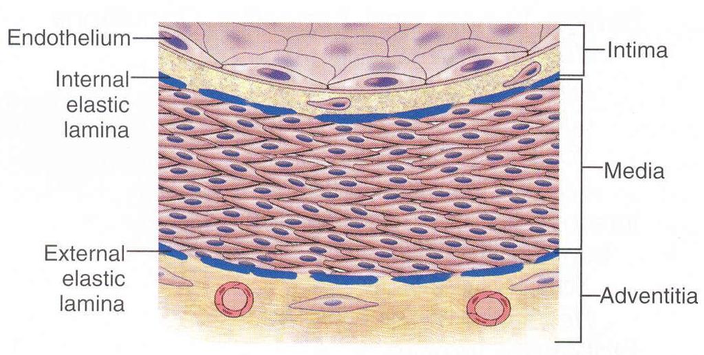
26 Tunica intima The tunica intima has one layer of endothelial cells supported by a thin subendothelial layer of loose connective tissue with occasional smooth muscle cells. In arteries, the intima is separated from the media by an internal elastic lamina, the most external component of the intima. This lamina, composed of elastin, has holes (fenestrae) that allow the diffusion of substances to nourish cells deep in the vessel wall. As a result of the loss of blood pressure and contraction of the vessel at death, the tunica intima of arteries may have a slightly folded appearance in tissue sections. 26
27 Tunica media The tunica media, the middle layer, consists chiefly of concentric layers of helically arranged smooth muscle cells. Interposed among the smooth muscle cells are variable amounts of elastic fibers and lamellae, reticular fibers of collagen type III, proteoglycans, and glycoproteins, all of which is produced by these cells. In arteries, the media has a thinner external elastic lamina, which separates it from the tunica adventitia. 27
28 Tunica adventitia The tunica adventitia or tunica externa consists principally of type I collagen and elastic fibers. This adventitial layer is gradually continuous with the stromal connective tissue of the organ through which the blood vessel runs. 28
29 29
30 ARTERIES:- WALL 30
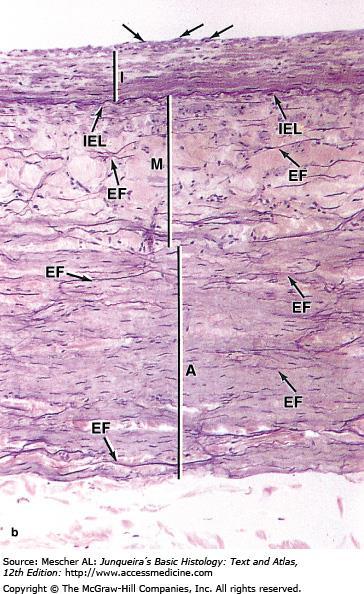
31 Tunics of the vascular wall Comparison of the three major layers or tunics in the largest artery and vein. (a): aorta (b): vena cava. Simple squamous endothelial cells (arrows) line the tunica intima (I) which has subendothelial loose connective tissue and is separated from the tunica media by the internal elastic lamina (IEL), a prominent sheet of elastin. The media (M) contains elastic lamellae and fibers (EF) and multiple layers of smooth muscle not seen well here. The tunica media is much thicker in large arteries than veins, with relatively more elastin. Elastic fibers are also present in the outer tunica adventitia (A), which is relatively thicker in large veins. Vasa vasorum (V) are seen in the adventitia of the aorta. The connective tissue of the adventitia always merges with the less dense connective tissue around it. Both X122. Elastic. 31

32 Tunics of the vascular wall 32
 with only one or two layers of smooth muscle, and usually thin, inconspicuous adventitia (Ad). X350. Masson trichrome. http://histonano.")
33 Arterioles (a): Arterioles are microvessels with a tunica intima (I) that consists only of the endothelium (E), in which the cells may have rounded nuclei. They have tunica media (M) with only one or two layers of smooth muscle, and usually thin, inconspicuous adventitia (Ad). X350. Masson trichrome. 33

34 Arterioles (b): Three arterioles of various sizes are shown here and a capillary. X400. H&E. 34

35 Arterioles (c): A large mesenteric arteriole is cut obliquely and longitudinally and clearly shows the endothelial cells (arrow heads) and one or two layers of smooth muscle cells (M) cut transversely. Adventitia merges imperceptibly with neighboring connective tissue. X300. PT. 35

36 VEINS Classified as large, medium and small Large and medium veins usually accompany large and medium arteries Veins have lower blood pressure: avg. 10mmHg with little fluctuation thinner walls, less muscular and elastic tissue expand easily, have high capacitance one-way venous valves aid skeletal muscles in upward blood flow 36
: Micrograph of small vein (V) shows a relatively large lumen compared to the small muscular artery (A) with its thick media (M) and adventitia (Ad).")
37 Vein Veins usually travel near arteries and are classified as small, medium, or large based on size and development of the tunics. (a): Micrograph of small vein (V) shows a relatively large lumen compared to the small muscular artery (A) with its thick media (M) and adventitia (Ad). The wall of a small vein is very thin, containing only two or three layers of smooth muscle. X200. H&E. 37

38 Vein (b): Micrograph of a convergence between two small veins showing valves (arrow). Valves are thin folds of tunica intima projecting well into the lumen which act to prevent backflow of blood. X200. H&E. 38

39 Vein (c): Micrograph of a medium vein (MV) showing a thicker wall, but still less prominent than that of the accompanying muscular artery (MA). Both the media and adventitia are better developed, but the wall is often folded around the relatively large lumen. X100. H&E. 39

40 (d): Micrograph of a medium vein containing blood and showing valve folds (arrows). X200. Masson trichrome. Vein 40
, composed of the subendothelial connective tissue with endothelium on both sides. X100, PT.")
41 Wall of large vein with valve. Large veins have a muscular tunica media (TM) that is very thin compared to the tunica adventitia (TA) composed of dense irregular connective tissue. The wall is often folded as shown here. The tunica intima here projects into the lumen as a valve (V), composed of the subendothelial connective tissue with endothelium on both sides. X100, PT. 41
, small capillaries (C) and venules (V) make up the microvasculature where, in almost every organ, exchange takes place between blood and the interstitial fluid of the")
42 Vessels of the microvasculature. Arterioles (A), small capillaries (C) and venules (V) make up the microvasculature where, in almost every organ, exchange takes place between blood and the interstitial fluid of the tissues. X200. Masson trichrome. 42


43 References Kierszenbaum AL, Tres L. Histology and Cell Biology: An Introduction to Pathology. 4 th ed., Sounders. 2015, 752pp. Moore KL, Agur AMR and Dalley AF Essential Clinical Anatomy. 5 nd Edition. Wolters Kluwer. 686pp. 43
Practical Histology. Cardiovascular System. Dr Narmeen S. Ahmad
 Practical Histology Cardiovascular System Dr Narmeen S. Ahmad The Cardiovascular System A closed system of the heart and blood vessels Functions of cardiovascular system: Transport nutrients, hormones
The cardiovascular system
 The cardiovascular system Components of the Cardiovascular system Heart Vessels: Arteries Capillaries Veins Functions of CVS: Transportation system where blood is the transporting vehicle Carries oxygen,
Chapter 14. The Cardiovascular System
 Chapter 14 The Cardiovascular System Introduction Cardiovascular system - heart, blood and blood vessels Cardiac muscle makes up bulk of heart provides force to pump blood Function - transports blood 2
Cardiovascular System
 Cardiovascular System Purpose Transport oxygen and nutrients Take waste products away from tissues & organs Things we learned Blood pressure: the force of blood pushing against the walls of blood vessels
Anatomy of the Heart. Figure 20 2c
 Anatomy of the Heart Figure 20 2c Pericardium & Myocardium Remember, the heart sits in it s own cavity, known as the mediastinum. The heart is surrounded by the Pericardium, a double lining of the pericardial
Chapter 20 (1) The Heart
 The Heart") Chapter 20 (1) The Heart Learning Objectives Describe the location and structure of the heart Describe the path of a drop of blood from the superior vena cava or inferior vena cava through the heart out
Chapter 20 (1) The Heart Learning Objectives Describe the location and structure of the heart Describe the path of a drop of blood from the superior vena cava or inferior vena cava through the heart out
Human Anatomy, First Edition
 Human Anatomy, First Edition McKinley & O'Loughlin Chapter 22 : Heart 1 Functions of the Heart Center of the cardiovascular system, the heart. Connects to blood vessels that transport blood between the
the Cardiovascular System I
 the Cardiovascular System I By: Dr. Nabil A Khouri MD, MsC, Ph.D MEDIASTINUM 1. Superior Mediastinum 2. inferior Mediastinum Anterior mediastinum. Middle mediastinum. Posterior mediastinum Anatomy of
Histology of the Cardiac System. Dr. Nabil Khoury Anatomy Department
 Histology of the Cardiac System Dr. Nabil Khoury Anatomy Department Objectives 1. Identify the 3 layers of the heart endocardium, myocardium, epicardium 2. Differentiate cardiacmuscle 3. Define intercalated
Heart. Heart 2-Tunica media: middle layer (media ='middle') muscle fibers (smooth or cardiac).
 muscle fibers (smooth or cardiac).") t. innermost lumenal General Circulatory system heart and blood vessels walls have 3 layers (inside to outside) 1-Tunica interna: aka tunica intima layer--lumenal layer epithelium--endothelium simple squamous
t. innermost lumenal General Circulatory system heart and blood vessels walls have 3 layers (inside to outside) 1-Tunica interna: aka tunica intima layer--lumenal layer epithelium--endothelium simple squamous
STRUCTURES OF THE CARDIOVASCULAR SYSTEM
 STRUCTURES OF THE CARDIOVASCULAR SYSTEM CARDIOVASCULAR SYSTEM Also called the circulatory system Consists of the heart, arteries, veins, and capillaries Main function is to pump/circulate oxygenated blood
Cardiovascular Anatomy Dr. Gary Mumaugh
 Cardiovascular Anatomy Dr. Gary Mumaugh Location of Heart Approximately the size of your fist Location o Superior surface of diaphragm o Left of the midline in mediastinum o Anterior to the vertebral column,
2. right heart = pulmonary pump takes blood to lungs to pick up oxygen and get rid of carbon dioxide
 A. location in thorax, in inferior mediastinum posterior to sternum medial to lungs superior to diaphragm anterior to vertebrae orientation - oblique apex points down and to the left 2/3 of mass on left
Cardiovascular System. I. Structures of the heart A. : Pericardium sack that surrounds the heart
 Cardiovascular System I. Structures of the heart A. : Pericardium sack that surrounds the heart 1. : Pericardial Cavity serous fluid filled space between the heart and the pericardium B. Heart Wall 1.
THE HEART OBJECTIVES: LOCATION OF THE HEART IN THE THORACIC CAVITY CARDIOVASCULAR SYSTEM
 BIOLOGY II CARDIOVASCULAR SYSTEM ACTIVITY #3 NAME DATE HOUR THE HEART OBJECTIVES: Describe the anatomy of the heart and identify and give the functions of all parts. (pp. 356 363) Trace the flow of blood
CIRCULATORY SYSTEM BLOOD VESSELS
 Name: Block: CIRCULATORY SYSTEM Multicellular organisms (above the level of roundworms) rely on a circulatory system to bring nutrients to, and take wastes away from, cells. In higher organisms such as
The Heart. Size, Form, and Location of the Heart. 1. Blunt, rounded point; most inferior part of the heart.
 12 The Heart FOCUS: The heart is composed of cardiac muscle cells, which are elongated, branching cells that appear striated. Cardiac muscle cells behave as a single electrical unit, and the highly coordinated
CVS HISTOLOGY. Dr. Nabil Khouri.
 CVS HISTOLOGY Dr. Nabil Khouri http://anatomy.kmu.edu.tw/blockhis/block3/slides/block4_24.html The Heart Wall Contract as a single unit Cardiac Muscle Simultaneous contraction due to depolarizing at the
Heart Anatomy. 7/5/02 Stephen G Davenport 1
 Heart Anatomy Copyright 1999, Stephen G. Davenport, No part of this publication may be reproduced, stored in a retrieval system, or transmitted, in any form without prior written permission. 7/5/02 Stephen
Major Function of the Cardiovascular System. Transportation. Structures of the Cardiovascular System. Heart - muscular pump
 Structures of the Cardiovascular System Heart - muscular pump Blood vessels - network of tubes Blood - liquid transport vehicle brachiocephalic trunk superior vena cava right pulmonary arteries right pulmonary
Approximately the size of your fist Location Superior surface of diaphragm Left of the midline in mediastinum Anterior to the vertebral column,
 Dr. Gary Mumaugh Approximately the size of your fist Location Superior surface of diaphragm Left of the midline in mediastinum Anterior to the vertebral column, posterior to the sternum Posteriorly the
The Cardiovascular System
 The Cardiovascular System The Cardiovascular System A closed system of the heart and blood vessels The heart pumps blood Blood vessels allow blood to circulate to all parts of the body The function of
CV Anatomy Quiz. Dr Ella Kim Dr Pip Green
 CV Anatomy Quiz Dr Ella Kim Dr Pip Green Q1 The location of the heart is correctly described as A) lateral to the lungs. B) medial to the sternum. C) superior to the diaphragm. D) posterior to the spinal
Ch 19: Cardiovascular System - The Heart -
 Ch 19: Cardiovascular System - The Heart - Give a detailed description of the superficial and internal anatomy of the heart, including the pericardium, the myocardium, and the cardiac muscle. Trace the
Figure ) The specific chamber of the heart that is indicated by letter A is called the. Diff: 1 Page Ref: 364
 The specific chamber of the heart that is indicated by letter A is called the. Diff: 1 Page Ref: 364") Essentials of Anatomy and Physiology, 9e (Marieb) Chapter 11 The Cardiovascular System Short Answer Figure 11.1 Using Figure 11.1, identify the following: 1) The Purkinje fibers are indicated by label.
Essentials of Anatomy and Physiology, 9e (Marieb) Chapter 11 The Cardiovascular System Short Answer Figure 11.1 Using Figure 11.1, identify the following: 1) The Purkinje fibers are indicated by label.
The Cardiovascular System
 11 PART A The Cardiovascular System PowerPoint Lecture Slide Presentation by Jerry L. Cook, Sam Houston University ESSENTIALS OF HUMAN ANATOMY & PHYSIOLOGY EIGHTH EDITION ELAINE N. MARIEB The Cardiovascular
Cardiac Conduction System
 Cardiac Conduction System What causes the Heart to Beat? Heart contracts by electrical signals! Cardiac muscle tissue contracts on its own an electrical signal is sent out by the heart so that all cells
THE HEART. A. The Pericardium - a double sac of serous membrane surrounding the heart
 THE HEART I. Size and Location: A. Fist-size weighing less than a pound (250 to 350 grams). B. Located in the mediastinum between the 2 nd rib and the 5 th intercostal space. 1. Tipped to the left, resting
10. Thick deposits of lipids on the walls of blood vessels, called, can lead to serious circulatory issues. A. aneurysm B. atherosclerosis C.
 Heart Student: 1. carry blood away from the heart. A. Arteries B. Veins C. Capillaries 2. What is the leading cause of heart attack and stroke in North America? A. alcohol B. smoking C. arteriosclerosis
The Circulatory System
 The Circulatory System Dr. Sami Zaqout The circulatory system Circulatory system Blood vascular systems Lymphatic vascular systems Blood vascular systems Blood vascular systems The circulatory system Circulatory
The Heart and Cardiovascular System
 The Heart and Cardiovascular System What you will learn The location of the heart 3 layers and covering of the heart Explain the function of the heart as 2 separate pumps Identify the 4 chambers of the
Heart Dissection. 5. Locate the tip of the heart or the apex. Only the left ventricle extends all the way to the apex.
 Heart Dissection Page 1 of 6 Background: The heart is a four-chambered, hollow organ composed primarily of cardiac muscle tissue. It is located in the center of the chest in between the lungs. It is the
The Cardiovascular System (Heart)
") The Cardiovascular System The Cardiovascular System (Heart) A closed system of the heart and blood vessels The heart pumps blood Blood vessels allow blood to circulate to all parts of the body The function
The Cardiovascular System The Cardiovascular System (Heart) A closed system of the heart and blood vessels The heart pumps blood Blood vessels allow blood to circulate to all parts of the body The function
a) Endocardium The endocardium is the innermost layer of the heart wall. It forms the internal lining of the atria and ventricles.
 Endocardium The endocardium is the innermost layer of the heart wall. It forms the internal lining of the atria and ventricles.") Chapter 11 Circulatory System The circulatory system consists of a muscular, pulsing heart and a system of blood vessels.the blood vessels include: Arteries which will carry the blood and it's dissolved
Chapter 11 Circulatory System The circulatory system consists of a muscular, pulsing heart and a system of blood vessels.the blood vessels include: Arteries which will carry the blood and it's dissolved
Microscopic Anatomy CARDIOVASCULAR SYSTEM
 Microscopic Anatomy CARDIOVASCULAR SYSTEM I. Introduction The cardiovascular system is a closed system consisting of a pump, the heart, and a series of tubular blood vessels that interconnect all body
LECTURE 5. Anatomy of the heart
 LECTURE 5. Anatomy of the heart Main components of the CVS: Heart Blood circulatory system arterial compartment haemomicrocirculatory (=microvascular) compartment venous compartment Lymphatic circulatory
The Cardiovascular and Lymphatic Systems Cardiovascular System Blood Vessels Blood Vessels Arteries Arteries Arteries
 CH 12 The Cardiovascular and s The Cardiovascular and s OUTLINE: Cardiovascular System Blood Vessels Blood Pressure Cardiovascular System The cardiovascular system is composed of Blood vessels This system
Cardiovascular (Circulatory) System
 System") Cardiovascular (Circulatory) System Piryaei May 2011 Circulatory System Heart Blood Vessels Macrovasculature (More than 0.1mm) Elastic Artery Muscular (Distributing) Artery Large Arteriol Small Vein Muscular
Cardiovascular (Circulatory) System Piryaei May 2011 Circulatory System Heart Blood Vessels Macrovasculature (More than 0.1mm) Elastic Artery Muscular (Distributing) Artery Large Arteriol Small Vein Muscular
Cardiovascular System
 Cardiovascular System The Heart Cardiovascular System The Heart Overview What does the heart do? By timed muscular contractions creates pressure gradients blood moves then from high pressure to low pressure
Histology of the myocardium and blood vessels. Prof. Abdulameer Al-Nuaimi
 Histology of the myocardium and blood vessels Prof. Abdulameer Al-Nuaimi E-mail: a.al-nuaimi@sheffield.ac.uk E-mail: abdulameerh@yahoo.com Histology of blood vessels The walls of arteries and veins are
Health Science 20 Circulatory System Notes
 Health Science 20 Circulatory System Notes Functions of the Circulatory System The circulatory system functions mainly as the body s transport system. It transports: o Oxygen o Nutrients o Cell waste o
Cardiovascular System
 Cardiovascular System I. Structure of the Heart A. Average adult heart is 14 cm long and 9 cm wide. B. Lies in the mediastinum. C. Enclosed in the pericardium. 1. Fibrous pericardium- Outer, tough connective
The Heart. Happy Friday! #takeoutyournotes #testnotgradedyet
 The Heart Happy Friday! #takeoutyournotes #testnotgradedyet Introduction Cardiovascular system distributes blood Pump (heart) Distribution areas (capillaries) Heart has 4 compartments 2 receive blood (atria)
THE CARDIOVASCULAR SYSTEM. Part 1
 THE CARDIOVASCULAR SYSTEM Part 1 CARDIOVASCULAR SYSTEM Blood Heart Blood vessels What is the function of this system? What other systems does it affect? CARDIOVASCULAR SYSTEM Functions Transport gases,
The Cardiovascular System
 Essentials of Human Anatomy & Physiology Elaine N. Marieb Seventh Edition Chapter 11 The Cardiovascular System Slides 11.1 11.19 Lecture Slides in PowerPoint by Jerry L. Cook The Cardiovascular System
1. What kind of blood is found in the rt. atrium? (oxygenated or deoxygenated)
") Carl Christennsen, PhD Chap. 19, 20, & 21 - Circulatory System Bio. 2304 Human Anatomy HEART 1. What kind of blood is found in the rt. atrium? (oxygenated or deoxygenated) Where does this blood come from?
Carl Christennsen, PhD Chap. 19, 20, & 21 - Circulatory System Bio. 2304 Human Anatomy HEART 1. What kind of blood is found in the rt. atrium? (oxygenated or deoxygenated) Where does this blood come from?
2. capillaries - allow exchange of materials between blood and tissue fluid
 Chapter 19 - Vascular System A. categories and general functions: 1. arteries - carry blood away from heart 2. capillaries - allow exchange of materials between blood and tissue fluid 3. veins - return
Cardiovascular System Notes: Physiology of the Heart
 Cardiovascular System Notes: Physiology of the Heart Interesting Heart Fact Capillaries are so small it takes ten of them to equal the thickness of a human hair. Review What are the 3 parts of the cardiovascular
The cardiovascular system is composed of the heart and blood vessels that carry blood to and from the body s organs. There are 2 major circuits:
 1 The cardiovascular system is composed of the heart and blood vessels that carry blood to and from the body s organs. There are 2 major circuits: pulmonary and systemic. The pulmonary goes out to the
The Heart. The Heart A muscular double pump. The Pulmonary and Systemic Circuits
 C H A P T E R 19 The Heart The Heart A muscular double pump circuit takes blood to and from the lungs Systemic circuit vessels transport blood to and from body tissues Atria receive blood from the pulmonary
The Cardiovascular and Lymphatic Systems
 BIOLOGY OF HUMANS Concepts, Applications, and Issues Fifth Edition Judith Goodenough Betty McGuire 12 The Cardiovascular and Lymphatic Systems Lecture Presentation Anne Gasc Hawaii Pacific University and
Pearson's Comprehensive Medical Assisting Administrative and Clinical Competencies
 Pearson's Comprehensive Medical Assisting Administrative and Clinical Competencies THIRD EDITION CHAPTER 27 The Cardiovascular System Lesson 1: Overview of the Cardiovascular System Lesson Objectives Upon
The HEART. What is it???? Pericardium. Heart Facts. This muscle never stops working It works when you are asleep
 This muscle never stops working It works when you are asleep The HEART It works when you eat It really works when you exercise. What is it???? Located between the lungs in the mid thoracic region Apex
4. The two inferior chambers of the heart are known as the atria. the superior and inferior vena cava, which empty into the left atrium.
 Answer each statement true or false. If the statement is false, change the underlined word to make it true. 1. The heart is located approximately between the second and fifth ribs and posterior to the
The Circulatory System. The Heart, Blood Vessels, Blood Types
 The Circulatory System The Heart, Blood Vessels, Blood Types The Closed Circulatory System Humans have a closed circulatory system, typical of all vertebrates, in which blood is confined to vessels and
11/10/2014. Muscular pump Two atria Two ventricles. In mediastinum of thoracic cavity 2/3 of heart's mass lies left of midline of sternum
 It beats over 100,000 times a day to pump over 1,800 gallons of blood per day through over 60,000 miles of blood vessels. During the average lifetime, the heart pumps nearly 3 billion times, delivering
The Cardiovascular System. Chapter 15. Cardiovascular System FYI. Cardiology Closed systemof the heart & blood vessels. Functions
 Chapter 15 Cardiovascular System FYI The heart pumps 7,000 liters (4000 gallons) of blood through the body each day The heart contracts 2.5 billion times in an avg. lifetime The heart & all blood vessels
Human Anatomy and Physiology Chapter 19 Worksheet 1- The Heart
 Human Anatomy and Physiology Chapter 19 Worksheet 1- The Heart Name Date Period 1. The "double pump" function of the heart includes the right side, which serves as the circuit pump, while the left side
The Cardiovascular System
 The Cardiovascular System The Manila Times College of Subic Prepared by: Stevens B. Badar, RN, MANc THE HEART Anatomy of the Heart Location and Size approx. the size of a person s fist, hollow and cone-shaped,
Extra notes for lab- 1 histology. Slide 1 : cross section in the elastic artery ( aortic arch, ascending aorta, descending aorta )
") Extra notes for lab- 1 histology Slide 1 : cross section in the elastic artery ( aortic arch, ascending aorta, descending aorta ) - twin of ascending aorta is the pulmonary trunk. Ascending aorta represents
Extra notes for lab- 1 histology Slide 1 : cross section in the elastic artery ( aortic arch, ascending aorta, descending aorta ) - twin of ascending aorta is the pulmonary trunk. Ascending aorta represents
Cardiovascular System. Heart Anatomy
 Cardiovascular System Heart Anatomy 1 The Heart Location & general description: Atria vs. ventricles Pulmonary vs. systemic circulation Coverings Walls The heart is found in the mediastinum, the medial
組織學 Historlogy 台北醫學大學 / 解剖學科教授 : 邱瑞珍分機號碼 :3261. 電子郵件信箱
 組織學 Historlogy 台北醫學大學 / 解剖學科教授 : 邱瑞珍分機號碼 :3261 電子郵件信箱 :rueijen@tmu.edu.tw 1 The Circulatory System 台北醫學大學 / 解剖學科教授 : 邱瑞珍分機號碼 :3261 電子郵件信箱 :rueijen@tmu.edu.tw 2 學習目的 The structure of the arteries The structure
Unit 10 Cardiovascular System
 Unit 10 Cardiovascular System I. Functions Deliver nutrients to cells > O 2, sugars, amino acids, lipids, ions, H 2 O... Remove waste from cells > CO 2, pathogens, toxins, lactic acid... Fight off infection
The Cardiovascular System Part I: Heart Outline of class lecture After studying part I of this chapter you should be able to:
 The Cardiovascular System Part I: Heart Outline of class lecture After studying part I of this chapter you should be able to: 1. Describe the functions of the heart 2. Describe the location of the heart,
Chapter 12 Lecture Outline
 Chapter 12 Lecture Outline See separate PowerPoint slides for all figures and tables preinserted into PowerPoint without notes. Copyright The McGraw-Hill Companies, Inc. Permission required for reproduction
Blood Vessels. Types of Blood Vessels Arteries carry blood away from the heart Capillaries smallest blood vessels. Veins carry blood toward the heart
 C H A P T E R Blood Vessels 20 Types of Blood Vessels Arteries carry blood away from the heart Capillaries smallest blood vessels The site of exchange of molecules between blood and tissue fluid Veins
- what other structures, besides the heart, does the mediastinum contain?
 Basic A & P II Dr. L. Bacha Chapter Outline (Martini & Nath 2010) An Introduction to the Cardiovascular System - read the paragraphs under this heading on page 580 The Heart is a Four Chambered Organ describe
Anatomy: Cardiovascular System
 In the name of God Anatomy: Cardiovascular System Moradian MD, MPH, PhD candidate Tehran University of Medical Sciences drmoradian@sums.ac.ir 2015 The Cardiovascular System A closed system of the heart
1. Which of the following blood vessels has a thin elastic layer? A. Aorta. B. Pulmonary artery. C. Posterior vena cava. D. Mesenteric capillary.
 CIRCULATORY SYSTEM 1. Which of the following blood vessels has a thin elastic layer? A. Aorta. B. Pulmonary artery. C. Posterior vena cava. D. Mesenteric capillary. 2. Capillary beds are equipped with
Function: Transportation of. Oxygen Nutrients Waste Hormones gases
 Function: Transportation of Oxygen Nutrients Waste Hormones gases Pericardium: double sac of serous membrane filled with fluid (pericardial fluid to be exact) that surrounds the heart. Parietal pericardium:
Lab Activity 25. Blood Vessels & Circulation. Portland Community College BI 232
 Lab Activity 25 Blood Vessels & Circulation Portland Community College BI 232 Artery and Vein Histology Walls have 3 layers: Tunica intima Tunica media Tunica externa 2 Tunica Intima Is the innermost layer
Anatomy of the Heart
 Biology 212: Anatomy and Physiology II Anatomy of the Heart References: Saladin, KS: Anatomy and Physiology, The Unity of Form and Function 8 th (2018). Required reading before beginning this lab: Chapter
Test Review Circulatory System Chapters
 Test Review Circulatory System Chapters 13-2010 1. The tissue that forms the tight fitting sac around the heart is the a. parietal pericardium c. myocardium b. visceral pericardium d. endocardium 2. Which
Mr. Epithelium s Anatomy and Physiology Test SSSS
 Mr. Epithelium s Anatomy and Physiology Test SSSS You have 50 minutes to complete this test packet. One 8.5 x 11 cheat sheet is allowed, along with 1 non-programmable calculator dedicated to computation.
Chapter 20: Cardiovascular System: The Heart
 Chapter 20: Cardiovascular System: The Heart I. Functions of the Heart A. List and describe the four functions of the heart: 1. 2. 3. 4. II. Size, Shape, and Location of the Heart A. Size and Shape 1.
Cardiovascular Physiology
 Cardiovascular Physiology Lecture 1 objectives Explain the basic anatomy of the heart and its arrangement into 4 chambers. Appreciate that blood flows in series through the systemic and pulmonary circulations.
AN ATOMY OF THE CARDIOVASCULAR SYSTEM
 Student Name CHAPTER 18 AN ATOMY OF THE CARDIOVASCULAR SYSTEM T he heart is actually two pumps one moves blood to the lungs, the other pushes it out into the body. These two functions seem rather elementary
Remember: the CVS system is deriving from the mesenchyma and is lined by simple squamous epithelium called the endothelium
 Lecture 10 General med_2nd semester Microscopic anatomy and embryology of cardiovascular system Microscopic structure of the heart, excitomotoric system - its structural peculiarities Blood vessels - arteries
Figure 10.1A Transparency Master 79
 Brain Carotid arteries Jugular vein Right front leg Lungs (inflated) Cranial Right atrium To left front leg Left subclavian Bronchus capillaries Brachiocephalic vein Left atrium Dorsal aorta Right ventricle
Circulation. Circulation = is a process used for the transport of oxygen, carbon! dioxide, nutrients and wastes through-out the body
 Circulation Circulation = is a process used for the transport of oxygen, carbon! dioxide, nutrients and wastes through-out the body Heart = muscular organ about the size of your fist which pumps blood.
Anatomy & Physiology of Cardiovascular System. Chapter 18 & 19
 Anatomy & Physiology of Cardiovascular System Chapter 18 & 19 Objectives..cont 1. Discuss the physiological stages of cardiac muscle contraction. 2. Trace a typical ECG and label each wave or complex 3.
10/23/2017. Muscular pump Two atria Two ventricles. In mediastinum of thoracic cavity 2/3 of heart's mass lies left of midline of sternum
 It beats over 100,000 times a day to pump over 1,800 gallons of blood per day through over 60,000 miles of blood vessels. During the average lifetime, the heart pumps nearly 3 billion times, delivering
Chapter 18 - Heart. I. Heart Anatomy: size of your fist; located in mediastinum (medial cavity)
") Chapter 18 - Heart I. Heart Anatomy: size of your fist; located in mediastinum (medial cavity) A. Coverings: heart enclosed in double walled sac called the pericardium 1. Fibrous pericardium: dense connective
Chapter 18 - Heart I. Heart Anatomy: size of your fist; located in mediastinum (medial cavity) A. Coverings: heart enclosed in double walled sac called the pericardium 1. Fibrous pericardium: dense connective
Anatomy Review: The Heart Graphics are used with permission of A.D.A.M. Software, Inc. and Benjamin/Cummings Publishing Co.
 Anatomy Review: The Heart Graphics are used with permission of A.D.A.M. Software, Inc. and Benjamin/Cummings Publishing Co. Anatomy Views Label the diagrams of the heart below: Interactive Physiology Study
IB TOPIC 6.2 THE BLOOD SYSTEM
 IB TOPIC 6.2 THE BLOOD SYSTEM TERMS TO KNOW circulation ventricle artery vein THE BLOOD SYSTEM 6.2.U1 - Arteries convey blood at high pressure from the ventricles to the tissues of the body Circulation
Ch. 12 The Circulatory System. The heart. The heart is a double pump. A quick note on arteries vs. veins. = the muscular pump of the CV system
 Ch. 12 The Circulatory System The heart A.k.a. the cardiovascular system Blood was discussed in Ch. 11 Focus of Ch. 12: heart and blood vessels = the muscular pump of the CV system ~ 100,000 heartbeats/day!
Scrub In: Red blood cells are called: Which component of blood is necessary for the initiation of the blood clotting process:
 Scrub In: Red blood cells are called: a. erythrocytes b. leukocytes c. melanocytes d. thrombocytes Which component of blood is necessary for the initiation of the blood clotting process: a. erythrocytes
The Cardiovascular System
 Essentials of Human Anatomy & Physiology Elaine N. Marieb Slides 11.1 11.19 Seventh Edition Chapter 11 The Cardiovascular System Functions of the Cardiovascular system Function of the heart: to pump blood
Blood and Heart. Student Learning Objectives:
 Blood and Heart Student Learning Objectives: Identify the major components of the blood. Identify the primary structures associated with the heart Follow the blood through the path of the circulation.
Cardiovascular System Notes: Heart Disease & Disorders
 Cardiovascular System Notes: Heart Disease & Disorders Interesting Heart Facts The Electrocardiograph (ECG) was invented in 1902 by Willem Einthoven Dutch Physiologist. This test is still used to evaluate
Circulation. Sinoatrial (SA) Node. Atrioventricular (AV) Node. Cardiac Conduction System. Cardiac Conduction System. Linked to the nervous system
 Node. Atrioventricular (AV) Node. Cardiac Conduction System. Cardiac Conduction System. Linked to the nervous system") Circulation Cardiac Conduction System AHS A H S Your body resembles a large roadmap. There are routes or arteries that take you downtown to the heart of the city and veins that take you to the outskirts
Circulation Cardiac Conduction System AHS A H S Your body resembles a large roadmap. There are routes or arteries that take you downtown to the heart of the city and veins that take you to the outskirts
Circulatory System 10.1
 1 Circulatory System 10.1 2 ARTERIES Arteries-blood vessels that carry blood away from the heart Thick walls Inner & Outer layers: connective tissue Middle layers are muscle and elastic connective tissue
Cardiovascular System Blood Vessels
 Cardiovascular System Blood Vessels Structure of Blood Vessels The three layers (tunics) Tunica intima composed of simple squamous epithelium Tunica media sheets of smooth muscle Contraction vasoconstriction
Happy One Week before Spring Break!
 Happy One Week before Spring Break! Essential Question Today How does the structure of the heart ensure its functions? Objectives Identify the major parts of the heart. Describe the pathway of circulation,
Lecture name: blood 2 & The Circulatory System Edited by: Buthainah Al masaeed & Yousef Qandeel
 Lecture name: blood 2 & The Circulatory System Edited by: Buthainah Al masaeed & Yousef Qandeel Now we will take about A granulocytes : Lymphocyte Monocytes 1- Lymphocyte - The second major type of presence
Circulatory system. Lecture #2
 Circulatory system Lecture #2 The essential components of the human cardiovascular system: Heart Blood Blood vessels Arteries - blood vessels that conduct arterial blood from heart ventricle to organs
2/28/18. Cardiovascular System. Introduction. Anatomy. Chapter 26. Body is 60% to 80% fluid (by volume) Systems responsible for fluid movement are:
 Systems responsible for fluid movement are:") Cardiovascular System Chapter 26 1 Introduction Body is 60% to 80% fluid (by volume) Systems responsible for fluid movement are: - Cardiovascular helps move fluid - Lymphatic Both called pick-up and delivery
Cardiovascular System Chapter 26 1 Introduction Body is 60% to 80% fluid (by volume) Systems responsible for fluid movement are: - Cardiovascular helps move fluid - Lymphatic Both called pick-up and delivery
BIOL 4350 Cardiovascular Physiology Dr. Hamilton. Using the figure above, match the following: 1. Purkinje fibers. 2. SA node. 3. AV node.
 BIOL 4350 Cardiovascular Physiology Dr. Hamilton Using the figure above, match the following: 1. Purkinje fibers. 2. SA node. 3. AV node. 1 Using the figure above, match the following: 4. Atrial depolarization.
Circulatory System: Introduction. Dr. Carmen E. Rexach Anatomy 35 Mt. San Antonio College
 Circulatory System: Introduction Dr. Carmen E. Rexach Anatomy 35 Mt. San Antonio College Components Cardiovascular system Lymphatic system Cardiovascular system Heart, blood vessels, blood Functions: transport
Section 5.1 The heart and heart disease
 Section 5.1 The heart and heart disease Mammals are too large to rely on diffusion. They need a circulatory system to move substances around the body. Blood moves down pressure gradients, from high to