Dr. Zahiri. In the name of God
|
|
|
- Meagan Norman
- 5 years ago
- Views:
Transcription
1 Dr. Zahiri In the name of God
2 small intestine = small bowel is the part of the gastrointestinal tract Boundaries: Pylorus Ileosecal junction Function: digestion and absorption of food It receives bile juice and pancreatic juice through the hepatopancreatic duct, controlled by Sphincter of oddi Has 3 part: Duodenum, jejunum and ileum
3 Duodenum
4 duodenum is the first part of small intestine joints the stomach at the pylorus It is a C-shaped tube, about 12 inches long It lies between the stomach and jejunum receives the openings of the bile and pancreatic ducts
5 Peritoneal relation of duodenum: The first inch of duodenum is similar to stomach in structure. It isintraperitoneal for the first 2-3cm only The remainder of the duodenum is retroperitoneal
6 Parts of duodenum: For descriptive purposes, the duodenum is divided into four parts, as explained below: the superior, descending, horizontal, and ascending duodenum. Pylorus Duodenojejunal flecture
7 First part or superior (D1) begins as a continuation of the duodenal end of the pylorus It passes laterally to the right, superiorly and posteriorly, for approximately 5cm It is Ant. & Lat. To L1 It is intraperitoneal for the first 2-3cm only(duodenal cap) Sup: Hepatoduodenal lig. Inf: Greater Omentum

8 Relations : anteriorly- gallbladder, liver posteriorly-common bile duct, portal vein, gastroduodneal artery superiorly- epiploic foramen inferiorly- pancreatic head

9 Second part of duodenum(d2) is about 3 inches long It runs vertically downward on the right side of L2, L3 major duodenal papilla: the bile duct and the main pancreatic duct a little higher up minor duodenal papilla : accessory pancreatic duct Sup. Duodenal Flecture

10 The pancreatic duct and common bile duct enter the descending duodenum through the major duodenal papilla (ampulla of Vater or Hepatopancratico ampul). minor duodenal papilla: the entrance for the accessory pancreatic duct. The junction between the foregut and midgut: lies just below the major duodenal papilla.
 inferiorly- loops of jejunum laterally- ascending colon, hepatic flexure, right kidney medially -")
11 Relations : anteriorly- transverse mesocolon posteriorly- right kidney, right ureter, right adrenal gland superiorly- liver, gallbladder (variable) inferiorly- loops of jejunum laterally- ascending colon, hepatic flexure, right kidney medially - pancreatic head
12 Third part (D3) The third (inferior/horizontal) part of the duodenum begins at the inferior duodenal flexure and passes transverse 3 inches long It passes in front of the vertebral column, IVC, Aorta Sup. Duodenal Flecture Inf. Duodenal Flecture
13 Relations : anteriorly-small bowel mesentery Artery & vein & root posteriorly- right psoas muscle, right ureter, gonadal vessels, aorta and IVC superiorly- pancreatic head inferiorly- loops of jejunum

14 Fourth part or ascending(d4) superiorly, either anterior to, or to the left of, the aorta, until it reaches the inferior border of the body of the pancreas. terminates at the duodenojejunal flexure The duodenojejunal flexure is surrounded by a peritoneal fold containing muscle fibres: the ligament of Treitz. Duodenojejun al Flecture
15 Relations : superiorly- stomach inferiorly- loops of jejunum posteriorly- left psoas muscle, aorta
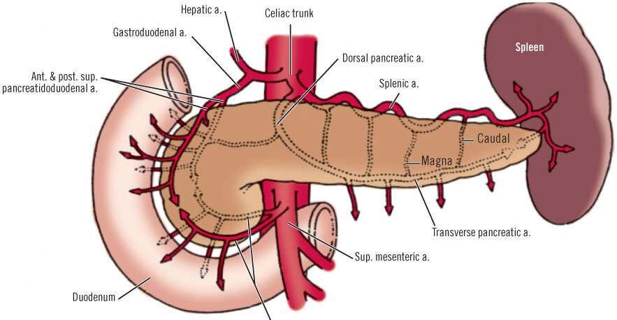
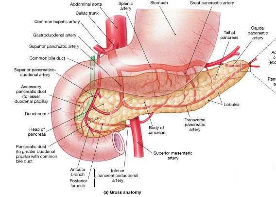
16 Blood supply arterial supply : Duodenal cap (first 2.5cm) -right gastric artery, right gastroepiploic artery Remaining D1 to mid-d2 -superior pancreaticodudenal artery (branch of gastroduodenal artery) Mid-D2 to ligament of Trietz: Inferior pancreaticoduodenal arteries (branch of SMA)

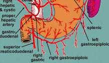
17 Common hepatic artery Supradeodenal artery
18
19 Venous drainage : The veins correspond to the arterie splenic vein & SMV portal vein Lymphatic supply Ant. Part pancreaticoduodenal& pyloric nodes(gastroduodenal) Post. Part SMV nods Finally they drain coeliac nodes
20 Nerve supply Duodenum receives both sympathetic and parasympathetic nerves from the celiac and superior mesenteric plexuses
21 The jejunum and ileum form the second and third part of the small intestine They collectively measure about 20 feet long jejunum : 8 feet, ileum: 12 feet long Each of jejunum and ileum has distinctive features, but there is a gradual change from one to the other and that is why it is better to describe both simultaneously.

22 jejunum begins at the duodenojejunal flexure jejunum gets coiled and gradually changes its features to become ileum, which ends at the ileocecal junction. The coils of jejunum and ileum are freely mobile They are attached to the posterior abdominal wall by the mesentery of small intestine, which is a fan-shaped fold of peritoneum.
 to the region of sacroiliac joint")
23 The root of this fold is along a line that extend from left side of L3 (downward from left to right ) to the region of sacroiliac joint

24 The branches of the superior mesenteric artery and vein, lymph vessels and nerves, all reach the intestine through the mesentery.

25 Differences between jejunum and ileum: Location: The jejunum lies coiled in the upper part of peritoneal cavity below the left of the transverse mesocolon, while the ileum is found in the lower part of the peritoneal cavity and in the pelvis.

 are larger, more numerous and more")

26 Structural features: The jejunum has thicker walls, and is redder in color. The infoldings of the mucous membrane (plicae circulars) are larger, more numerous and more closely set in the jejunum ileum, where they are smaller and widely spread in the upper part and totally absent in the lower part.

27 Attachment of mesentery: The mesentery of jejunum is attached to the left of aorta while the mesentery of ileum is attached to the right.

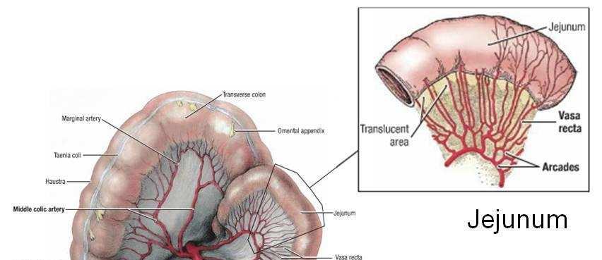
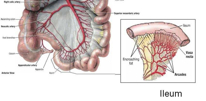
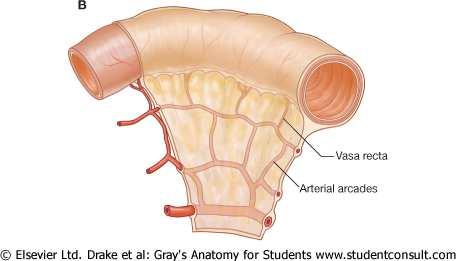
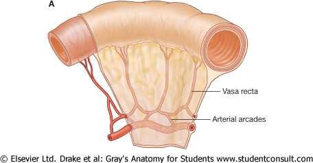
28 Vascular arcades: The mesenteric vessels of jejunum form only one or two arcades, which supply the jejunalwall through long and infrequent branches. the ileum receives numerous short terminal vessels that arise from a series of three, four or even more arcades.
29 Deposition of fat: At the jejunalend of mesentery, the fat is deposited near the root and is scanty near the jejunalwall. On the other hand, at the ilealend, the fat is deposited throughout so that it extends from the root to the intestinal wall.
30 Aggregations of lymphoid tissue: In the mucous membrane of lower ileum, there are aggregations of lymphoid tissue, known as Peyer s patches. Such aggregations are not found in the jejunum
31 Blood supply of jejunum and ileum: Arteries: branches of superior mesenteric artery(sma) These branches arise from the left side of the artery (from where no other branches arise) and run through the mesentery to reach the wall of intestine. The branches anastomosewith one another to form a series of arcades. The lowest part of ileum, near the ileocecal junction, is supplied by the ileocolicartery in addition to the usual blood supply.
32
33
34
35 Veins: The veins correspond to the arteries and eventually drain into the superior mesenteric vein.
36 Lymph drainage of jejunum and ileum: superior mesenteric nodes Ileocolic
37 Nerve supply to jejunum lymph node and ileum: Both the sympathetic and parasympathetic fibers come from the superior mesenteric plexus.


38 و سربلند باشيد پيروز
Anatomy of the SMALL INTESTINE. Dr. Noman Ullah Wazir PMC
 Anatomy of the SMALL INTESTINE Dr. Noman Ullah Wazir PMC SMALL INTESTINE The small intestine, consists of the duodenum, jejunum, and illium. It extends from the pylorus to the ileocecal junction were the
د. عصام طارق. Objectives:
 GI anatomy Lecture: 5 د. عصام طارق Objectives: To describe anatomy of stomach, duodenum & pancreas. To list their main relations. To define their blood & nerve supply. To list their lymph drainage. To
The abdominal Esophagus, Stomach and the Duodenum. Prof. Oluwadiya KS
 The abdominal Esophagus, Stomach and the Duodenum Prof. Oluwadiya KS www.oluwadiya.com Viscera of the abdomen Abdominal esophagus: Terminal part of the esophagus The stomach Intestines: Small and Large
The jejunum and the Ileum. Prof. Oluwadiya KS
 The jejunum and the Ileum Prof. Oluwadiya KS www.oluwadiya.siteled.com Introduction Introduction The small intestine (SI) comprises of the duodenum, jejunum and the ileum The jejunum is the second part
-Ensherah Mokheemer. -Shatha Al-Jaberi محمد المحتسب- 1 P a g e
 9-9 -Ensherah Mokheemer -Shatha Al-Jaberi محمد المحتسب- 1 P a g e Small intestine has three regions: ( االثني عشر( The duodenum The jejunum The ileum Small intestine Duodenum: -c-shaped -The concavity
ANATOMY OF THE SMALL & LARGE INTESTINES. Semester 1, 2011 A. Mwakikunga
 ANATOMY OF THE SMALL & LARGE INTESTINES Semester 1, 2011 A. Mwakikunga LEARNING OBJECTIVES 1. List the parts and anatomical regions of the small and large intestines 2. State anatomical relations of the
ABDOMEN - GI. Duodenum
 TALA SALEH ABDOMEN - GI Duodenum - Notice the shape of the duodenum, it looks like capital G shape tube which extends from the pyloroduodenal junction to the duodenojejunal junction. - It is 10 inches
Pancreas & Biliary System. Dr. Vohra & Dr. Jamila
 Pancreas & Biliary System Dr. Vohra & Dr. Jamila 1 Objectives At the end of the lecture, the student should be able to describe the: Location, surface anatomy, parts, relations & peritoneal reflection
Small Plicae Circularis. Short Closely packed together. Sparse, completely absent at distal part Lymphoid Nodule
 Intestines Differences Between Jejunum and Ileum Types Jejunum Ileum Color Deeper red Paler pink Calibre Bigger Smaller Thickness of wall Thick and Heavy Thin and Lighter Vascularity Highly vascularised
Duodenum retroperitoneal
 Duodenum retroperitoneal C shaped Initial region out of stomach into small intestine RETROperitoneal viscus Superior 1 st part duodenal cap ; moves upwards and backwards to lie on the R crura medial to
Preview from Notesale.co.uk Page 1 of 34
 Abdominal viscera and digestive tract Digestive tract Abdominal viscera comprise majority of the alimentary system o Terminal oesophagus, stomach, pancreas, spleen, liver, gallbladder, kidneys, suprarenal
Block 3: DISSECTION 2 CELIAC TRUNK, JEJUNUM/ILEUM, LARGE INTESTINE, DUODENUM, PANCREAS, PORTAL VEIN; MOBILIZATION OF THE LIVER
 1 Block 3: DISSECTION 2 CELIAC TRUNK, JEJUNUM/ILEUM, LARGE INTESTINE, DUODENUM, PANCREAS, PORTAL VEIN; MOBILIZATION OF THE LIVER Attempt to complete as much as you can of the dissection explained in the
Anatomy of the Large Intestine
 Large intestine Anatomy of the Large Intestine 2 Large Intestine Extends from ileocecal valve to anus Length = 1.5-2.5m = 5 feet Regions Cecum = 2.5-3 inch Appendix= 3-5 inch Colon Ascending= 5 inch Transverse=
THE ORAL CAVITY
 THE ORAL CAVITY WALL OF ABDOMEN (ANTERIOR) The paraumbilical vein drains into the portal vein and then through the liver. This is an important clinical connection. THE ABDOMINAL VISCERA The small
BLOCK IV: OFFICIAL BODY PARTS LIST FOR ANTERIOR ABDOMINAL WALL AND ABDOMINAL CONTENTS
 BLOCK IV: OFFICIAL BODY PARTS LIST FOR ANTERIOR ABDOMINAL WALL AND ABDOMINAL CONTENTS External oblique muscle Muscular portion Aponeurotic portion Superficial inguinal ring Lateral (inferior) crus Medial
The peritoneum. Prof. Oluwadiya KS, MBBS, FMCS(Orthop) Website:
 Website:") The peritoneum Prof. Oluwadiya KS, MBBS, FMCS(Orthop) Website: http://oluwadiya.com The peritoneum Serous membrane that lines the abdominopelvic cavity and invests the viscera The largest serous membrane
The peritoneum Prof. Oluwadiya KS, MBBS, FMCS(Orthop) Website: http://oluwadiya.com The peritoneum Serous membrane that lines the abdominopelvic cavity and invests the viscera The largest serous membrane
Pancreas and Biliary System
 Pancreas and Biliary System Please view our Editing File before studying this lecture to check for any changes. Color Code Important Doctors Notes Notes/Extra explanation Objectives At the end of the lecture,
-12. -Renad Habahbeh. -Dr Mohammad mohtasib
 -12 -Renad Habahbeh - -Dr Mohammad mohtasib The Gallbladder -The gallbladder has a body, a fundus (a rounded end), a neck, Hartmann s pouch before the neck and a cystic duct that meets the common hepatic
Peritoneum: Def. : It is a thin serous membrane that lines the walls of the abdominal and pelvic cavities and clothes the viscera.
 Peritoneum: Def. : It is a thin serous membrane that lines the walls of the abdominal and pelvic cavities and clothes the viscera. Layers of the peritoneum: 1. Outer Layer ( Parietal Peritoneum) : lines
Surface Anatomy. Location Shape Weight Role of Five Surfaces Borders Fissures Lobes Peritoneal Lig
 The Liver Functions Bile production and secretion Detoxification Storage of glycogen Protein synthesis Production of heparin and bile pigments Erythropoiesis (in fetus) Surface Anatomy Location Shape Weight
Nasogastric tube. Stomach. Pylorus. Duodenum 1. Duodenum 2. Duodenum 3. Duodenum 4
 Esophagus Barium Swallow Stomach and Duodenum 4 year old Upper GI Nasogastric tube Stomach and Duodenum 4 year old Upper GI Nasogastric tube Stomach Pylorus Duodenum 1 Duodenum 2 Duodenum 3 Duodenum 4
Done by: Dina Sawadha & Mohammad Abukabeer
 Done by: Dina Sawadha & Mohammad Abukabeer The stomach *the stomach is a dilated part of the gastro intestinal tract, it's "J" shape. *the lower surface of the stomach ( the greater curvature ) reaches
Lab Monitor Images Dissection of the Abdominal Vasculature + Lower Digestive System
 Lab Monitor Images Dissection of the Abdominal Vasculature + Lower Digestive System Stomach & Duodenum Frontal (AP) View Nasogastric tube 2 1 3 4 Stomach Pylorus Duodenum 1 Duodenum 2 Duodenum 3 Duodenum
ANATOMY OF THE DIGESTIVE SYSTEM PART II
 ANATOMY OF THE DIGESTIVE SYSTEM PART II 9.12.2014 Kaan Yücel M.D., Ph.D. http://fhs121.org Dr.Kaan Yücel http://fhs121.org Digestive system Part II 1. LIVER The liver is the largest gland in the body and,
Anatomy: Know Your Abdomen
 Anatomy: Know Your Abdomen Glossary Abdomen - part of the body below the thorax (chest cavity); separated by the diaphragm. Anterior - towards the front of the body. For example, the umbilicus is anterior
1 Right & left Hepatic ducts Gastric Impression of spleen
 Pancreatic Model 1 Right & left Hepatic ducts 14 Gastric Impression of spleen 2 Common hepatic duct 15 Renal Impression of spleen 3 Cystic Duct 16 Colic Impression of spleen 4 Common Bile Duct 17 Splenic
Accessory Glands of Digestive System
 Accessory Glands of Digestive System The liver The liver is soft and pliable and occupies the upper part of the abdominal cavity just beneath the diaphragm. The greater part of the liver is situated under
Exploring Anatomy: the Human Abdomen
 Exploring Anatomy: the Human Abdomen PERITONEUM AND PERITONEAL CAVITY PERITONEUM The peritoneum is a thin serous membrane that lines the abdominal cavity and covers, in variable amounts, the viscera within
GI module Lecture: 9 د. عصام طارق. Objectives:
 GI module Lecture: 9 د. عصام طارق Objectives: To list structures forming posterior abdominal wall. To follow aorta & its main branches. To describe IVC & its main tributaries. To list nerves of posterior
Al-Mohtaseb. Saba Alfayoumi. Mo Alfarra
 8 Al-Mohtaseb Saba Alfayoumi Mo Alfarra For the comparison purposes refer to the last page where you can find a table that summarizes them. Enjoy Jejunum and Ileum -They're intraperitoneal and freely mobile
Jhia Anjela D. Rivera 1 1. BS Biology, Department of Biology, College of Science, Polytechnic University of the Philippines
 DIGESTIVE SYSTEM Jhia Anjela D. Rivera 1 1 BS Biology, Department of Biology, College of Science, Polytechnic University of the Philippines DIGESTIVE SYSTEM Consists of the digestive tract (gastrointestinal
Mousa Salah. Dr. Mohammad Al. Mohtasib. 1 P a g e
 8 Mousa Salah Dr. Mohammad Al. Mohtasib 1 P a g e In the previous lecture we talked about the peritoneum, and we said that the peritonium is a serous sac, and it consists of two layers, visceral and parietal.
To describe the liver. To list main structures in porta hepatis.
 GI anatomy Lecture: 6 د. عصام طارق Objectives: To describe the liver. To list main structures in porta hepatis. To define portal system & portosystemic anastomosis. To list parts of biliary system. To
Embryology of the Midgut and Hind gut
 Embryology of the Midgut and Hind gut Prof. Abdulameer Al-Nuaimi E-mail: a.al-nuaimi@sheffield.ac.uk E-mail: abdulameerh@yahoo.com Abdominal organs www.google.co.uk/search? Development of Duodenum The
The posterior abdominal wall. Prof. Oluwadiya KS
 The posterior abdominal wall Prof. Oluwadiya KS www.oluwadiya.sitesled.com Posterior Abdominal Wall Lumbar vertebrae and discs. Muscles opsoas, quadratus lumborum, iliacus, transverse, abdominal wall
The Whipple Operation Illustrations
 The Whipple Operation Illustrations Fig. 1. Illustration of the sixstep pancreaticoduodenectomy (Whipple operation) as described in a number of recent text books by Dr. Evans. The operation is divided
It passes through the diaphragm at the level of the 10th thoracic vertebra to join the stomach
 The esophagus is a tubular structure (muscular, collapsible tube ) about 10 in. (25 cm) long that is continuous above with the laryngeal part of the pharynx opposite the sixth cervical vertebra The esophagus
Bushra Arafa Zayed & Hanan Jamal. - Dana AF
 - 10 - Bushra Arafa Zayed & Hanan Jamal - Dana AF - Mohammad Al Muhtaseb Notes: This sheet was written in the same order as the slides, and everything in the slides is mentioned in this sheet. Pictures
LECTURE 11 & 12: ABDOMINAL VISCERA ABDOMINAL CONTENTS DIVISION. The location of abdominal viscera is divided into 4 quadrants:
 LECTURE 11 & 12: ABDOMINAL VISCERA ABDOMINAL CONTENTS DIVISION The location of abdominal viscera is divided into 4 quadrants: - horizontal line across the umbilicus divides the upper quadrants from the
-the stones will obstruct the common bile duct and it might also be precancerous. -so the best treatment is chlolycyctoctomy.
 At the beginning this sheet includes the rest of last lecture s slides liver +gallbladder and the new lecture posterior abdominal wall and its vessels. We will start talking about Cholelithiasis -it means
GASTROINTESTINAL SYSTEM
 GASTROINTESTINAL SYSTEM Topographic Anatomy of the Abdomen Surface Landmarks Xiphoid process T9/T10 Inferior costal margin L2/L3 Iliac Crest L4 level ASIS L5/S1 level Pubic symphysis level of greater trochanter
Biology Human Anatomy Abdominal and Pelvic Cavities
 Biology 351 - Human Anatomy Abdominal and Pelvic Cavities Please place your name and I.D. number on the back of the last page of this exam. You must answer all questions on this exam. Because statistics
BY DR NOMAN ULLAH WAZIR
 BY DR NOMAN ULLAH WAZIR The stomach (from ancient Greek word stomachos, stoma means mouth) is a muscular, hollow and the most dilated part of the GIT. It starts from the point where esophagus ends. It
Peritoneal cavity. Infracolic compartment. Assoc. prof. dr. S. Delchev, MD, PhD
 Peritoneal cavity. Infracolic compartment Assoc. prof. dr. S. Delchev, MD, PhD Infracolic compartment The infracolic compartment lies inferior to the transverse mesocolon and posterior to the greater omentum
In the name ofgod. Abdomen 3. Dr. Zahiri
 In the name ofgod Abdomen 3 Dr. Zahiri Peritoneum Peritoneum It is the serous membrane(a type of loose connective tissue and is covered by mesothelium) that lines the abdominal cavity. Extensions of the
Common Bile Duct (CBD)
") Liver Last time we talked about the liver and the doctor started by revising some information about it: It has five surfaces. It reaches the 5 th intercostal space ; some books write that it reaches the
Liver Last time we talked about the liver and the doctor started by revising some information about it: It has five surfaces. It reaches the 5 th intercostal space ; some books write that it reaches the
ANATOMY OF PELVICAYCEAL SYSTEM -DR. RAHUL BEVARA
 1 ANATOMY OF PELVICAYCEAL SYSTEM -DR. RAHUL BEVARA 2 KIDNEY:ANATOMY OVERVIEW Kidneys are retroperitoneal, in posterior abdominal region, extending from T12 L3 Bean-shaped Right kidney is lower than left
SUBJECTS 2nd year, 1st semester I. 1. Primitive gut - limits, derivatives 2. Foregut -limits, evolution, derivatives 3. Midgut -limits, evolution,
 SUBJECTS 2nd year, 1st semester I. 1. Primitive gut - limits, derivatives 2. Foregut -limits, evolution, derivatives 3. Midgut -limits, evolution, derivatives 4. Hindgut- limits, evolution, derivatives
Netter's Anatomy Flash Cards Section 4 List 4 th Edition
 Netter's Anatomy Flash Cards Section 4 List 4 th Edition https://www.memrise.com/course/1577335/ Section 4 Abdomen (31 cards) Plate 4-1 Bony Framework of Abdomen 1.1 Costal cartilages 1.2 Iliac crest 1.3
Gastrointestinal Tract. Anatomy of GI Tract. Anatomy of GI Tract. (Effective February 2007) (1%-5%)
 (1%-5%)") Gastrointestinal Tract (Effective February 2007) (1%-5%) Anatomy of GI Tract Esophagus bulls-eye or target EG junction seen on sagittal scan posterior to left lobe of liver and anterior to aorta Anatomy
Gastrointestinal Tract (Effective February 2007) (1%-5%) Anatomy of GI Tract Esophagus bulls-eye or target EG junction seen on sagittal scan posterior to left lobe of liver and anterior to aorta Anatomy
Lab 5 Digestion and Hormones of Digestion. 7/16/2015 MDufilho 1
 Lab 5 Digestion and Hormones of Digestion 1 Figure 23.1 Alimentary canal and related accessory digestive organs. Mouth (oral cavity) Tongue* Parotid gland Sublingual gland Submandibular gland Salivary
- Digestion occurs during periods of low activity - Produces more energy than it uses. - Mucosa
 Introduction Digestive System Chapter 29 Provides processes to break down molecules into a state easily used by cells - A disassembly line: Starts at the mouth and ends at the anus Digestive functions
Anatomical Considerations for Lab Practical II
 Anatomical Considerations for Lab Practical II For each of the following please be prepared to provide: Identification System Organ(s) or ducts to Function(s) location which it is attached Use your lecture
The Spleen. Dr Fahad Ullah
 The Spleen BY Dr Fahad Ullah Spleen The spleen is an largest lymphoid organ shaped like a shoe that lies relative to the 9th and 11th ribs and is located in the left hypochondrium. Thus, the spleen is
- Digestion occurs during periods of low activity - Produces more energy than it uses. 3 Copyright 2016 by Elsevier Inc. All rights reserved.
 Introduction Digestive System Chapter 29 Provides processes to break down molecules into a state easily used by cells - A disassembly line: Starts at the mouth and ends at the anus Digestive functions
Biology Human Anatomy Abdominal and Pelvic Cavities
 Biology 351 - Human Anatomy Abdominal and Pelvic Cavities You must answer all questions on this exam. Because statistics demonstrate that, on average, between 2-5 questions on every 100-point exam are
Anatomy of the renal system. Professor Nawfal K. Al-Hadithi
 Anatomy of the renal system Professor Nawfal K. Al-Hadithi Objectives To describe the posterior abdominal wall To identify the main anatomical landmarks of the kidneys & ureters To describe the suprarenal
Midterm 2 is Tuesday 5/28/13
 Business Reminder: No class Monday (Memorial Day) Midterm 2 is Tuesday 5/28/13 Optional review session tomorrow @ 5pm Homework due in Lab 1. PreLab 8 (1pt) 2. Replace a Missing Assignment (4 pts) Homework
Dissection Lab Manuals: Required Content
 Dissection Lab Manuals: Required Content 1. Introduction a. Basic terminology (directions) b. External features of the cat c. Adaptations to predatory niche d. How to skin a cat e. How to make the incisions
458 Essentials of Human Anatomy and Physiology
 458 Essentials of Human Anatomy and Physiology Visceral peritoneum Intrinsic nerve plexuses: Myenteric nerve plexus Submucosal nerve plexus Submucosal glands Mucosa: Surface epithelium Lamina propria Muscle
Done by: nisreen obeidat
 Sheet: liver and pancreas Done by: nisreen obeidat Embryology of the liver The liver develops in the ventral mesentery of the foregut and divides the ventral mesentery :into 1)lesser omentum (between the
ABDOMEN. 2. The highest branch of the abdominal aorta is: (a) R suprarenal a (b) Coeliac trunk (c) L renal a (d) L gonadal a (e) SMA
 R suprarenal a (b) Coeliac trunk (c) L renal a (d) L gonadal a (e) SMA") ABDOMEN 1. The duodenum: (a) is a retroperitoneal structure (b) is 25cm long (c) lies between the levels of L2-L4 (d) in its fourth part lies to the R of the aorta (e) all of the above 2. The highest branch
ABDOMEN 1. The duodenum: (a) is a retroperitoneal structure (b) is 25cm long (c) lies between the levels of L2-L4 (d) in its fourth part lies to the R of the aorta (e) all of the above 2. The highest branch
Omar Sami --- Muhammad Al-Muhatasib
 8 Omar Sami --- Muhammad Al-Muhatasib This sheet is a remake from 2015 s sheet for the same lecture; I have checked the record, added, omitted, edited & illustrated all what the Professor mentioned in
The Digestive System and Body Metabolism
 14 PART B The Digestive System and Body Metabolism PowerPoint Lecture Slide Presentation by Jerry L. Cook, Sam Houston University ESSENTIALS OF HUMAN ANATOMY & PHYSIOLOGY EIGHTH EDITION ELAINE N. MARIEB
This lab activity is aligned with Visible Body s Human Anatomy Atlas app. Learn more at visiblebody.com/professors
 1 This lab activity is aligned with Visible Body s Human Anatomy Atlas app. Learn more at visiblebody.com/professors 2 A. Digestive System Overview To Start: Go to the Views menu and scroll down to the
SMALL INTESTINE LARGE INTESTINE RECTUM ABDOMINAL AORTA INFERIOR VENA CAVA THE ABDOMEN
 SMALL INTESTINE LARGE INTESTINE RECTUM ABDOMINAL AORTA INFERIOR VENA CAVA THE ABDOMEN THE SMALL INTESTINE THE SMALL INTESTINE: Is the longest part of the gastrointesanal tract - length of 5 metres (3 7
THE ABDOMEN SUPRARENAL GLANDS KIDNEY URETERS URINARY BLADDER
 THE ABDOMEN SUPRARENAL GLANDS KIDNEY URETERS URINARY BLADDER THE SUPRARENAL GLANDS The suprarenal (adrenal) glands lie immediately superior and slightly anterior to the upper pole of either kidney. Golden
Duodenum: -General features:
 Duodenum: -General features: -The duodenum pursues a C shaped course, it measure 10(25 cm) Inches in length. Concave retroperitoneal organ, the concavity tilts backward and to the right. -The head of pancreas
Exercise. Digestive System. Digestive system function. 1. Define the following terms: a. Chemical digestionb. Mechanical digestionc.
 Exercise 7 The Digestive System NAME: DATE: INSTRUCTOR: SECTION: Digestive system function 1. Define the following terms: a. Chemical digestionb. Mechanical digestionc. Ingestiond. Digestione. Absorptionf.
Liver o The liver is the largest gland in the body and has a wide variety of functions. - It s an accessory organ of GIT
 بسم رلاهللا You don t need to refer to the slides, we included everything here In this lecture we will talk about Liver & Gallbladder Liver o The liver is the largest gland in the body and has a wide variety
Development of pancreas and Small Intestine. ANATOMY DEPARTMENT DR.SANAA AL-AlSHAARAWY DR.ESSAM Eldin Salama
 Development of pancreas and Small Intestine ANATOMY DEPARTMENT DR.SANAA AL-AlSHAARAWY DR.ESSAM Eldin Salama OBJECTIVES At the end of the lecture, the students should be able to : Describe the development
ORAL CAVITY, ESOPHAGUS AND STOMACH
 ORAL CAVITY, ESOPHAGUS AND STOMACH 1 OBJECTIVES By the end of the lecture you should be able to: Describe the anatomy the oral cavity, (boundaries, parts, nerve supply). Describe the anatomy of the palate,
Chapter 9. The digestive system. Glossary. Louise McErlean
 Chapter 9 The digestive system Louise McErlean Glossary Absorption Process whereby the products of digestion move into the blood or lymph fluid. Acini glands Produce pancreatic juice. Amylase Carbohydrate
The Digestive System Laboratory
 The Digestive System Laboratory 1 The Digestive Tract The alimentary canal is a continuous tube stretching from the mouth to the anus. Liver Gallbladder Small intestine Anus Parotid, sublingual, and submaxillary
Ureters, Urinary Bladder & Urethra
 Ureters, Urinary Bladder & Urethra Please check our Editing File هذا العمل ال يغني عن المصدر األساسي للمذاكرة Lecture 2 } و م ن ي ت و ك ع ل ا لل ه ف ه و ح س ب ه { Objectives o Describe the course of ureter
Abdomen. Retroperitoneal space
 Abdomen. Retroperitoneal space Abdominal cavity The space bounded by: Anterolateral abdominal wall Posterior abdominal wall Diaphragm Pelvic walls and pelvic floor. Subdivided into: True abdominal cavity
DIGESTIVE SYSTEM. Chapter 25
 DIGESTIVE SYSTEM Chapter 25 DIGESTIVE SYSTEM Digestive Tract Mouth Pharynx Esophagus Stomach Small intestines Large intestines Anus Accessory Organs Teeth Tongue Salivary glands Pancreas Liver Gallbladder
Lab 9 Abdomen MUSCLES
 Lab 9 Abdomen MUSCLES External abdominal oblique continuous with the external intercostal muscle; its fibers point in a caudal direction as it moves anteriorly until it inserts on the linea alba via its
- Tamara Wahbeh. - Fareed Khdair. 0 P a g e
 -1 - Tamara Wahbeh - - Fareed Khdair 0 P a g e GI Embryology Note: I included everything in the records and slides; anything in the slide not included in this sheet was not mentioned by the doctor during
بسم االه الرحمن الرحيم
 MAY 3, 2012 [POSTERIOR ABDOMINAL WALL] LECTURE 26 ANATOMY Quick Revision: بسم االه الرحمن الرحيم Last time we started with the anterior abdominal wall and said that: 1. Diaphragm is the root of the abdomen.
The Digestive System PowerPoint Lecture Presentations prepared by Steven Bassett Southeast Community College Lincoln, Nebraska
 25 The Digestive System PowerPoint Lecture Presentations prepared by Steven Bassett Southeast Community College Lincoln, Nebraska Introduction The digestive system consists of: The digestive tract Accessory
ACTIVITY 11: RESPIRATORY AND DIGESTIVE SYSTEMS RESPIRATORY SYSTEM
 ACTIVITY 11: RESPIRATORY AND DIGESTIVE SYSTEMS OBJECTIVES: 1) How to get ready: Read Chapters 25 and 26, McKinley et al., Human Anatomy, 4e. All text references are for this textbook. 2) Identify structures
Misc Anatomy. Upper Limb! 2. Lower Limb! 5. Venous Drainage! Head & neck! 8
 Misc Anatomy Upper Limb! 2 Arteries!... 2 Veins!... 2 Spaces!... 4 Lower Limb! 5 Arteries!... 5 Venous Drainage!... 6 Spaces!... 7 Head & neck! 8 Artery!... 8 Ultrasound View for IJ CVL!... 8 Arteries
Anatomy of the spleen. Oluwadiya KS
 Anatomy of the spleen Oluwadiya KS www.oluwadiya.com Introduction The spleen is an ovoid, usually purplish, pulpy mass about the size and shape of one's fist. It is the largest lymphoid tissue in the body
RESPIRATORY SYSTEM. described: pp. 744,746 fig. 25.1, described: p. 746 fig described: p. 776 fig. 26.3
 ACTIVITY 11: RESPIRATORY AND DIGESTIVE SYSTEMS OBJECTIVES: 1) How to get ready: Read Chapters 25 and 26, McKinley et al., Human Anatomy, 5e. All text references are for this textbook. 2) Identify structures
Fareed Khdair, MD Assistant Professor Chief, Section of Pediatric Gastroenterology, Hepatology, and Nutrition University of Jordan School of Medicine
 Fareed Khdair, MD Assistant Professor Chief, Section of Pediatric Gastroenterology, Hepatology, and Nutrition University of Jordan School of Medicine Outline Lecture one : Gut formation Foregut: esophagus,
Pathways of Regional Spread in Pancreatic Cancer
 Pathways of Regional Spread in Pancreatic Cancer 12 Chusilp Charnsangavej, M.D. Regional spread of pancreatic ductal adenocarcinoma is common at the time of diagnosis, and it is often associated with poor
Overview of the Digestive
 Overview of the Digestive System Bởi: OpenStaxCollege The function of the digestive system is to break down the foods you eat, release their nutrients, and absorb those nutrients into the body. Although
Omran Saeed. Mohammad Al-muhtaseb. 1 P a g e
 13 Omran Saeed Mohammad Al-muhtaseb 1 P a g e Posterior abdominal wall - The diaphragm separates between thoracic cavity and abdominal cavity. Structures of posterior abdominal wall: (below diaphragm)
Lecture 02 Anatomy of the LIVER
 Lecture 02 Anatomy of the LIVER BY Dr Farooq Khan Aurakzai Dated: 02.01.2018 Introduction to Liver Largest gland in the body. 2 nd largest organ of the body. Weight approximately 1500 gm, and is roughly
GI Tract Lynn Ta Jennifer Zhang July 6, 2006 GI TRACT. 1) Other Names: Gastrointestinal tract Digestive tract Alimentary tract
 Other Names: Gastrointestinal tract Digestive tract Alimentary tract") GI Tract Lynn Ta Jennifer Zhang July 6, 2006 GI TRACT 1) Other Names: Gastrointestinal tract Digestive tract Alimentary tract 2) Definition/Location: Digestion and absorption are the primary functions
GI Tract Lynn Ta Jennifer Zhang July 6, 2006 GI TRACT 1) Other Names: Gastrointestinal tract Digestive tract Alimentary tract 2) Definition/Location: Digestion and absorption are the primary functions
DIGESTIVE. CHAPTER 17 Lecture: Part 1 Part 2 BIO 212: ANATOMY & PHYSIOLOGY II
 BIO 212: ANATOMY & PHYSIOLOGY II CHAPTER 17 Lecture: DIGESTIVE Part 1 Part 2 Dr. Lawrence G. Altman www.lawrencegaltman.com Some illustrations are courtesy of McGraw-Hill. SMALL INTESTINE DUODENUM > JEJUNUM
Midgut. Over its entire length the midgut is supplied by the superior mesenteric artery
 Gi Embryology 3 Midgut the midgut is suspended from the dorsal abdominal wall by a short mesentery and communicates with the yolk sac by way of the vitelline duct or yolk stalk Over its entire length the
Summarise the major structures of the oral cavity, teeth, tongue and salivary glands oral cavity teeth surface anatomy of the teeth tongue
 Anatomy Anatomy of the upper GI tract 3.2.16 Summarise the major structures of the oral cavity, teeth, tongue and salivary glands The oral cavity is anteriorly bordered by the oral orifice, which is surrounded
1. A stab wound into the abdomen transected the hepatoduodenal ligament. Each of the following structures would have been cut EXCEPT the:
 YR 1 GROSS ANATOMY UNIT EXAM 3 -- November 07, 1997. CHOOSE THE SINGLE BEST ANSWER FOR QUESTION 1-42. 1. A stab wound into the abdomen transected the hepatoduodenal ligament. Each of the following structures
Digestive Anatomy Lab
 Digestive Anatomy Lab In-Lab Exercises I have included the word list in this document. Any descrepencies between this document and the wordlist, you should default to this document. There is a lot of repetition
Posterior Abdominal wall-
 Structures of posterior abdominal wall: o Bony boundaries: 5 lumber vertebra and their intervertebral disc, iliac fossa and iliac crest. o Muscles: psoas major, quadrates lumborum, transversus abdominis,
Dr. Weyrich G07: Superior and Posterior Mediastina. Reading: 1. Gray s Anatomy for Students, chapter 3
 Dr. Weyrich G07: Superior and Posterior Mediastina Reading: 1. Gray s Anatomy for Students, chapter 3 Objectives: 1. Subdivisions of mediastinum 2. Structures in Superior mediastinum 3. Structures in Posterior
Liver. Function of the liver
 Liver & Gallbladder Liver The liver is the largest gland in the body and has a wide variety of functions Weight: 1/50 of body weight in adult & 1/20 of body weight in infant It is exocrine(bile) & endocrine